Abstract
Introduction:
Elizabethkingia anophelis (E. anophelis) has emerged as a multidrug-resistant pathogen with limited therapeutic options. This study aimed to characterize antimicrobial resistance mechanisms and virulence determinants in six clinical isolates from Shanghai, China, to inform evidence-based treatment strategies.
Methods:
Six strains were isolated from hospitalized patients (five community-acquired, one healthcare-associated) between September–November 2023. Antimicrobial susceptibility testing followed CLSI M100 guidelines. Whole-genome sequencing employed hybrid Illumina/PacBio approaches. Phylogenetic relationships were determined through 16S rRNA gene sequence analysis using the Neighbor-Joining method. Antimicrobial resistance genes and virulence factors were annotated using the Comprehensive Antibiotic Resistance Database (CARD) and Virulence Factor Database (VFDB), with relative gene abundance quantified via a TPM-like (transcripts per million-like) method.
Results:
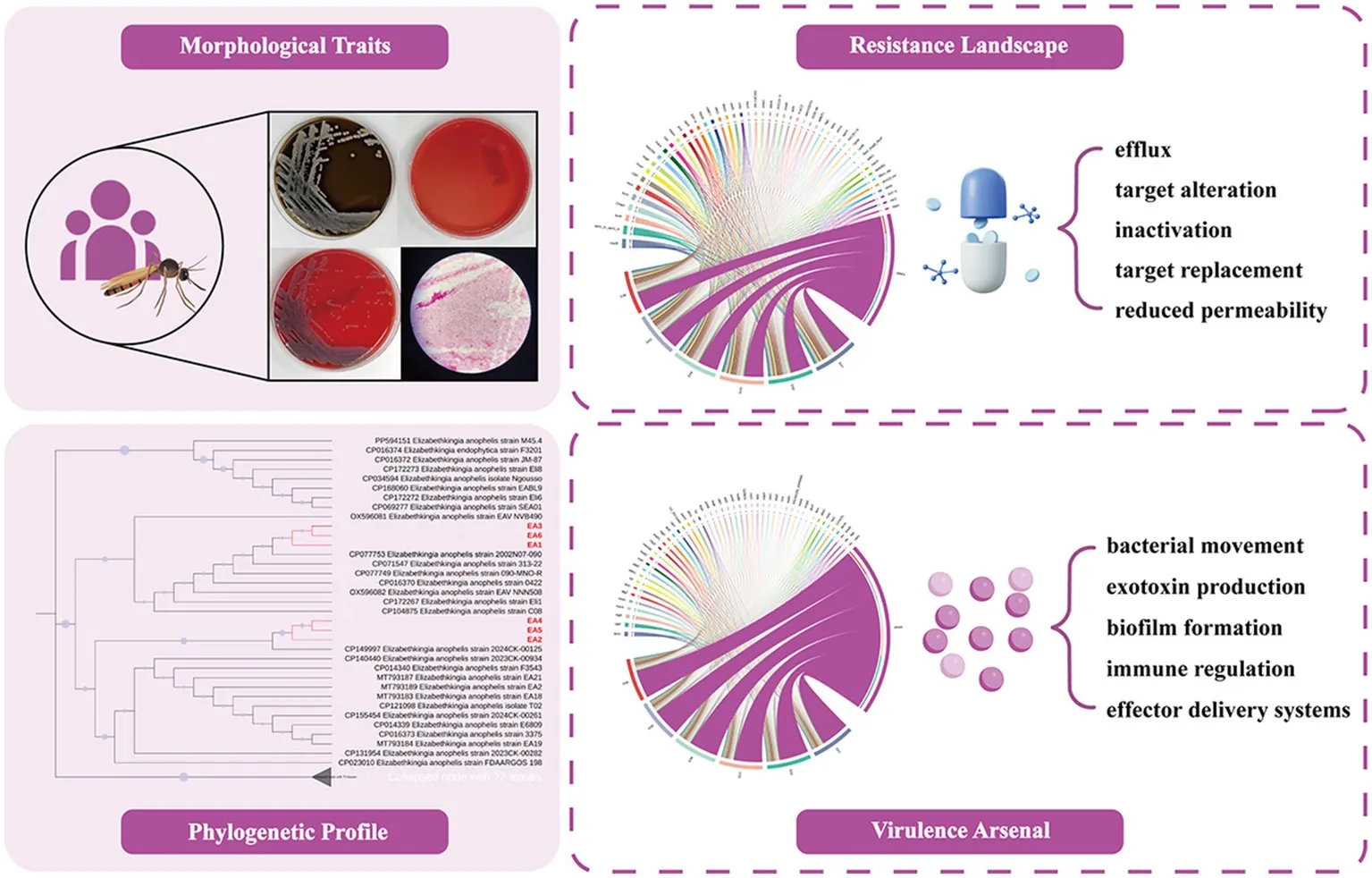
All isolates exhibited resistance to β-lactams, fluoroquinolones, carbapenems, and aminoglycosides, but retained minocycline susceptibility (MIC ≤1 μg/mL). Phylogenetic analysis revealed two distinct clusters: Cluster I (EA1, EA3, EA6) aligning with East/Southeast Asian isolates, and Cluster II (EA2, EA4, EA5) showing diverse geographic affinities. Five core resistance mechanisms were identified: antibiotic efflux, antibiotic target alteration, antibiotic inactivation, antibiotic target replacement, and reduced permeability to antibiotics. Virulence determinants included bacterial movement, exotoxin production, biofilm formation, immune regulation, and effector delivery systems. Strain EA5 exhibited unique signatures, including absence of cesH, unique sigE expression, elevated AAC(6′)-Iad/aadS and reduced qacL/OmpA.
Conclusion:
This study reveals phylogenetically divergent E. anophelis lineages in Shanghai with extensive multidrug resistance but preserved minocycline susceptibility. Findings support minocycline-based therapy, enhanced diagnostics, and regional surveillance networks for strain monitoring.
1 Introduction
The antimicrobial resistance (AMR) crisis has emerged as one of the most formidable global public health challenges of the 21st century. According to a recent forecast from GBD 2021 Antimicrobial Resistance Collaborators, AMR is projected to directly cause 1.91 million (1.56–2.26 million) deaths and contribute to 8.22 million deaths (6.85–9.65 million) indirectly by 2050 (). The World Health Organization has issued grave warnings that the proliferation of multidrug-resistant pathogens threatens to reverse decades of medical progress, potentially returning clinical practice to a “pre-antibiotic era” (). Concurrently, advances in molecular diagnostics—particularly whole-genome sequencing and high-throughput identification systems—have revolutionized pathogen identification, revealing a previously underappreciated diversity of opportunistic and emerging bacterial pathogens that possess inherent or acquired resistance mechanisms that substantially complicate clinical management (; ).
Elizabethkingia anophelis exemplifies this paradigm of emerging AMR pathogens initially overlooked by conventional diagnostics. First described by Kämpfer et al. from laboratory-reared Anopheles gambiae mosquitoes (Ifakara strain maintained in Stockholm, Sweden) (; ; ), this species has rapidly evolved into a clinically significant pathogen with transcontinental spread. Genomic surveillance in China have confirmed its stealthy infiltration, with recent studies documenting multidrug-resistant E. anophelis outbreaks in regions like Taizhou City – a sentinel warning of underrecognized transmission networks (). This organism demonstrates extremely ecological adaptability (), capable of both nosocomial transmission via contaminated medical devices or vertical transmission, and community-acquired infections manifesting as bacteremia, meningitis and pneumonia (; ). Critically, conventional microbial identification systems frequently misclassify E. anophelis as its phenotypically similar relative E. meningoseptica, leading to delayed recognition, inappropriate empirical therapies and potentially adverse clinical outcomes ().
This study investigates six E. anophelis infection cases at Tongji Hospital of Tongji University (Shanghai, China), employing a multi-approaches that combines phenotypic and molecular characterization: (1) standardized isolation and culture protocols to obtain purified strains; (2) broth microdilution susceptibility testing according to CLSI M100 guidelines (34th edition) to evaluate responses to 13 clinically relevant antimicrobial agents; and (3) whole-genome sequencing to elucidate phylogenetic relationships with closely related strains and comparatively inter-strain heterogeneity in resistome and virulence factor repertoires. While recent genomic studies have confirmed intrinsic antimicrobial resistance patterns in E. anophelis—including universal minocycline susceptibility (; )—most investigations focused on nosocomial outbreaks or single-center case series. The present study provided genomic characterization of predominantly community-acquired E. anophelis infections (5/6 cases) in Shanghai, China, and employed quantitative gene abundance profiling to reveal genomic heterogeneity underlying phenotypically uniform antimicrobial resistance. Our findings underscore the urgent need for enhanced molecular surveillance, targeted antimicrobial stewardship, molecular surveillance of emerging lineages, and tailored therapies to address the evolving resistance and virulence of E. anophelis.
2 Materials and methods
2.1 Study population and clinical data collection
Six patients hospitalized between September and November 2023 with confirmed E. anophelis infections were enrolled. Clinical samples, including blood and other secretions, were collected from each patient for bacterial isolation and culture.
Clinical and demographic data were retrieved exclusively from the hospital’s electronic medical records system, including age, sex, underlying comorbidities, primary diagnoses, antibiotic treatment regimens, and clinical outcomes. All specimens were processed according to standard operating procedures to ensure data accuracy and experimental reproducibility.
2.2 Pathogen identification and antimicrobial susceptibility testing
Suspected bacterial isolates from clinical samples were cultured on blood agar, chocolate agar, and MacConkey agar plates under aerobic conditions at 35 °C for 24–48 h. After incubation, morphologically typical colonies were selected for Gram staining and microscopic examination. Species identification was confirmed using MALDI-TOF MS (VITEK MS system) with 99.9% confidence scores.
Antimicrobial susceptibility testing was performed by the Broth Microdilution Method to determine minimum inhibitory concentrations (MICs) for E. anophelis, in accordance with the Clinical and Laboratory Standards Institute (CLSI) M100 document (34th edition) and non-Enterobacterales breakpoints. Plates were incubated aerobically at 35 °C for 16–20 h. Escherichia coli ATCC 25922 and Pseudomonas aeruginosa ATCC 27853 were included as quality control strains.
The following 13 antimicrobial agents were tested: ciprofloxacin, levofloxacin, piperacillin/tazobactam, ticarcillin/clavulanate, cefoperazone/sulbactam, ceftazidime, cefepime, imipenem, meropenem, amikacin, tobramycin, minocycline, and trimethoprim/sulfamethoxazole.
2.3 Sequencing and phylogenetic analysis
Whole-genome sequencing employed combined draft (Illumina PE150) and complete (hybrid Illumina + PacBio) genome approaches. For draft genomes, Illumina PE150 paired-end sequencing with ~400 bp insert size generated ≥100 × coverage per sample. For complete genomes, PacBio long-read sequencing with 8-10 kb fragments provided ≥100 × coverage per sample. Raw reads were quality-filtered using fastp v0.20.0. Draft genomes were assembled using SOAPdenovo v2.04 with GapCloser v1.12 for gap filling, while complete genomes utilized Unicycler for hybrid assembly with Pilon polishing. Gene prediction was performed using Prodigal (chromosomal) and GeneMarkS (plasmid), with tRNA and rRNA identification using tRNAscan-SE v2.0 and Barrnap.
For phylogenetic analysis, 16S rRNA gene sequences were extracted from the six E. anophelis genomes. These sequences from the clinical isolates (EA1-EA6) have been deposited in the NCBI GenBank database under accession numbers PV715948–PV715953, respectively. The extracted sequences were used as queries for BLASTn searches against the NCBI GenBank database. For each isolate, the top 50 most similar reference strains and 5 phylogenetically diverse strains were selected based on 16S rRNA sequence similarity. The 16S rRNA sequences from all retrieved reference strains were combined with those of the six study isolates, deduplicated, and aligned using MAFFT v7.490. Phylogenetic trees were constructed using the Neighbor-Joining method in MEGA7.0.26 software.
2.4 Analysis of resistance and virulence genes
Antimicrobial resistance genes and virulence factors were identified by the Comprehensive Antibiotic Resistance Database (CARD) and Virulence Factor Database (VFDB), respectively. To enable quantitative comparison of gene relative abundance across isolates, quality-filtered reads were mapped to predicted genes using Bowtie2 v2.4.5. Gene abundance was normalized using a TPM-like (transcripts per million-like) method adapted from metagenomic analysis (). This normalization adjusts for both gene length and sequencing depth, enabling direct comparison of relative gene abundance between isolates. To identify genes with altered abundance, we calculated the coefficient of variation (CV) for antimicrobial resistance genes and virulence factors separately across the six isolates, yielding CV = 5.4 and 5.7%, respectively. Using the higher CV as a conservative estimate, genes with TPM values ≥ 1.20-fold or ≤ 0.83-fold (1/1.20) relative to the median TPM were classified as having elevated or reduced relative abundance, respectively.
3 Results
3.1 Clinical management and outcomes
This study enrolled six patients with E. anophelis infections admitted between September and November 2023, including one healthcare-associated infection (EA4, diagnosed ≥48 h post-admission per Chinese national criteria) and five community-acquired infections (EA1, EA2, EA3, EA5, EA6, presenting at or before admission). The cohort included four males and two females, with ages ranging from 54 to 90 years (median: 68 years). All patients had ≥2 underlying comorbidities, including predominantly hepatobiliary diseases (4/6), acute infectious complications (4/6), and malignancies (2/6). Specimens were primarily isolated from blood (n = 2), bile (n = 2), both (n = 1), and multiple (n = 1). Various comorbidities significantly limited antimicrobial therapeutic options (detailed clinical data see Table 1).
Table 1
| ID | Age | Sex | Specimen | Comorbidities/diagnoses | Antibiotic regimen | Clinical outcome |
|---|---|---|---|---|---|---|
| EA1 | 54 | Male | Bile | Acute suppurative cholecystitis; Gallstones with cholecystitis; Biliary stasis | Cefmetazole sodium (2 g IV Q12H); Cefoperazone-sulbactam (3 g IV Q8H); Ornidazole (1 g IV BID); Doxycycline (100 mg PO Q12H) | Improved |
| EA2 | 90 | Male | Blood | Severe community-acquired pneumonia; Coronary atherosclerosis; Hypertension grade 3 | Meropenem (1 g IV Q8H); Levofloxacin (0.5 g IV QD); Cefoperazone-sulbactam (3 g IV Q8H); Colistin (4 M IU IV Q12H); Vancomycin (0.5 g via tube Q6H) | Unresolved |
| EA3 | 65 | Male | Blood, Bile | Pancreatic tumor; Hypertension grade 1 | Ceftazidime (2 g IV BID); Ornidazole (1 g → 0.5 g IV BID); Cefoperazone-sulbactam (3 g IV Q8H) | Improved |
| EA4 | 70 | Female | Blood | Multiple fractures; Acute renal failure; Pulmonary infection; Severe anemia | Meropenem (0.5 g IV Q8H); Caspofungin (50 mg IV QD); Colistin (4 M IU IV Q12H); Cefotaxime (1 g IV QD) | Improved |
| EA5 | 70 | Female | Bile | Obstructive jaundice; Pancreatic malignancy | Cefotaxime (2 g IV BID); Cefmetazole (2 g IV BID; 2 g intraoperative) | Improved |
| EA6 | 66 | Male | Bile, Pus, Blood, Ascites | Acute biliary pancreatitis; Gallstones with acute cholecystitis; Hypertension grade 2 | Imipenem-cilastatin (0.5 g IV Q8H); Cefepime (2 g IV BID); Cefoperazone-sulbactam (3 g IV BID); Metronidazole (500 mg IV BID) | Death |
Clinical data of the six cases of E. anophelis infection.
Based on microbiological culture results within 14 days of E. anophelis isolation, three cases (EA1, EA3, EA6) were monomicrobial infections, with E. anophelis as the sole pathogen isolated from sterile sites (bile, blood, or ascitic fluid). The other three cases (EA2, EA4, EA5) were polymicrobial, where E. anophelis was concurrently or sequentially isolated with other pathogens. Notably, in these polymicrobial infections, E. anophelis was consistently recovered from sterile compartments (blood or bile), whereas other multidrug-resistant organisms were mainly detected in respiratory or urinary specimens.
Among the four patients with favorable outcomes: EA1 (54-year-old male): E. anophelis was isolated from bile. Infection resolved after treatment with doxycycline combined with multiple antibiotics. EA3 (65-year-old male) and EA5 (70-year-old female): Both had pancreatic malignancies with E. anophelis confirmed by both blood and bile culture (EA3), and bile (EA5). Clinical improvement was achieved through biliary drainage combined with antibiotics. EA4 (70-year-old female): Hospitalized for multiple fractures, acute renal failure, and pulmonary infection, this healthcare-associated catheter-related infection caused by E. anophelis resolved following meropenem and colistin combination therapy.
Two cases exhibited refractory clinical courses: EA2 (90-year-old male): Admitted with sepsis and multi-organ dysfunction (severe pneumonia, coronary heart disease NYHA class IV, lacunar infarction with severe malnutrition). E. anophelis was isolated from blood. Despite combined antibiotic regimens, persistent bacteremia led to transfer for palliative long-term care. EA6 (66-year-old male): Hospitalized for acute biliary pancreatitis, E. anophelis was detected in multiple clinical specimens. Antimicrobial therapy failed, leading in death due to multiple organ failure.
3.2 Pathogen isolation, culture, and antimicrobial susceptibility testing
Suspected bacterial strains were incubated under aerobic conditions at 35 °C for 48 h. On chocolate agar, colonies were translucent, circular, and gray-white with a glossy surface (1–2 mm in diameter). No growth was observed on MacConkey agar, consistent with the non-fermentative characteristics of E. anophelis species. Blood agar supported the development of non-hemolytic, circular, gray-white colonies with smooth edges (1–2 mm in diameter). Pure colonies from blood agar were Gram-stained, revealing Gram-negative bacilli under microscopy (Figure 1).
Figure 1
The six E. anophelis strains exhibited a high degree of consistency in their resistance to various antibiotics (Table 2). All strains showed resistance to fluoroquinolones (ciprofloxacin, levofloxacin), β-lactams (piperacillin/tazobactam, ticarcillin/clavulanic acid, cefoperazone/sulbactam, ceftazidime, cefpirome), carbapenems (imipenem, meropenem), and aminoglycosides (amikacin, tobramycin). Notably, minocycline was the only agent demonstrating universal susceptibility, with all strains exhibiting MIC values ≤1 μg/mL (6/6, 100% susceptible). For trimethoprim-sulfamethoxazole, five strains (EA1, EA2, EA3, EA4, EA6) were susceptible, while strain EA5 exhibited resistance, resulting in an 83% (5/6) susceptibility rate for this agent.
Table 2
| Antibiotic class | Antibiotic name | Interpretive categories and MIC breakpoints (μg/mL) | EA1 | EA2 | EA3 | EA4 | EA5 | EA6 | ||
|---|---|---|---|---|---|---|---|---|---|---|
| S | I | R | ||||||||
| Fluoroquinolones | Ciprofloxacin | ≤1 | 2 | ≥4 | ≥4 (R) | ≥4 (R) | ≥4 (R) | ≥4 (R) | ≥4 (R) | ≥4 (R) |
| Levofloxacin | ≤2 | 4 | 8 | ≥8 (R) | ≥8 (R) | ≥8 (R) | ≥8 (R) | ≥8 (R) | ≥8 (R) | |
| β-Lactams | Piperacillin/Tazobactam | ≤16/4 | 32/4–64/4 | ≥128/4 | ≥128/4 (R) | ≥128/4 (R) | ≥128/4 (R) | ≥128/4 (R) | ≥128/4 (R) | ≥128/4 (R) |
| Ticarcillin/Clavulanate | ≤16/2 | 32/2–64/2 | ≥128/2 | ≥128/2 (R) | ≥128/2 (R) | ≥128/2 (R) | ≥128/2 (R) | ≥128/2 (R) | ≥128/2 (R) | |
| Cefoperazone/Sulbactam | ≤16 | 32 | ≥64 | ≥64 (R) | ≥64 (R) | ≥64 (R) | ≥64 (R) | ≥64 (R) | ≥64 (R) | |
| Ceftazidime | ≤8 | 16 | ≥32 | ≥64 (R) | ≥64 (R) | ≥64 (R) | ≥64 (R) | ≥64 (R) | ≥64 (R) | |
| Cefepime | ≤8 | 16 | ≥32 | ≥32 (R) | ≥32 (R) | ≥32 (R) | ≥32 (R) | ≥32 (R) | ≥32 (R) | |
| Carbapenems | Imipenem | ≤4 | 8 | ≥16 | ≥16 (R) | ≥16 (R) | ≥16 (R) | ≥16 (R) | ≥16 (R) | ≥16 (R) |
| Meropenem | ≤4 | 8 | ≥16 | ≥16 (R) | ≥16 (R) | ≥16 (R) | ≥16 (R) | ≥16 (R) | ≥16 (R) | |
| Aminoglycosides | Amikacin | ≤16 | 32 | ≥64 | ≥64 (R) | ≥64 (R) | ≥64 (R) | ≥64 (R) | ≥64 (R) | ≥64 (R) |
| Tobramycin | ≤4 | 8 | ≥16 | ≥16 (R) | ≥16 (R) | ≥16 (R) | ≥16 (R) | ≥16 (R) | ≥16 (R) | |
| Tetracyclines | Minocycline | ≤4 | 8 | ≥16 | ≤1 (S) | ≤1 (S) | ≤1 (S) | ≤1 (S) | ≤1 (S) | ≤1 (S) |
| Sulfonamides | Trimethoprim-Sulfamethoxazole | ≤2/38 | - | ≥4/76 | ≤2/38 (S) | ≤2/38 (S) | ≤2/38 (S) | ≤2/38 (S) | ≥4/76 (R) | ≤2/38 (S) |
Antibiotic resistance testing results for six E. anophelis isolates.
3.3 Phylogenetic analysis of pathogen 16S rRNA
Phylogenetic analysis based on 16S rRNA gene sequences grouped the six strains into two clusters (Figure 2). Cluster I (EA1, EA3, EA6) and Cluster II (EA2, EA4, EA5) exhibited a single nucleotide polymorphism (SNP) at position 73 of the coding region: adenine (A) in Cluster I versus guanine (G) in Cluster II. This SNP likely represents a geographic characteristic that distinguishes the two Cluster.
Figure 2
Cluster I shares close homology with the reference strain 0422 (GenBank accession no. CP016370), first isolated from a bloodstream infection in the United States in 1950. Despite its origin in America, this lineage has become the prevalent strain in East and Southeast Asia over the past decade, with recent isolates from Vietnam (accession nos. OX596081, OX596082), Malaysia (accession no. CP172267), and Taiwan (accession nos. CP077753, CP071547, CP104875) suggesting its dominance outbreaks across these regions. In contrast, Cluster II aligns with strain 3,375 (accession no. CP016373), initially identified in the U. S. in 1957. This lineage, formerly prevalent in Europe, North America, and Australia, has recently emerged in mainland China, as evidenced by accessions nos. MT793183, MT793184, MT793187, and MT793189 isolated post-2020. Geographic differences between clusters suggest that there may be potential adaptive evolution driven by regional antimicrobial measures or host-pathogen interactions.
3.4 Analysis of pathogen antibiotic resistance genes
Comprehensive resistance gene profiling of six E. anophelis strains revealed that these strains carry multiple resistance genes (Figure 3; Supplementary Table 1). The identified resistance genes conferred protection against multiple antibiotic classes, including macrolide (e.g., macB, mtrA, CRP), peptide (e.g., OmpA, bcrA, tsnR), tetracycline (e.g., tetA(58), TxR, tet(W/32/O)), aminoglycoside (e.g., AAC(6′)-Iad, AAC(3)-Ic, aadS), fluoroquinolone (e.g., evgA, CRP, marA), and glycopeptide (e.g., vanU_in_vanG_cl, YajC). These strains also shared core resistance mechanisms included antibiotic efflux (e.g., qacL, tetA(58)), antibiotic target alteration (e.g., vanU_in_vanG_cl, PmrF), antibiotic inactivation (e.g., AAC(6′)-Iad, aadS), antibiotic target replacement (e.g., mecI, dfrA3), and reduced permeability to antibiotics (e.g., OmpA, marA).
Figure 3
Strain-specific resistance gene abundance patterns revealed inter-isolate heterogeneity despite shared core mechanisms: EA1 showed high relative abundance of vanU_in_vanG_cl and bcrA; EA2 showed high relative abundance of MecI, SAT-4, evgA, FosC2, and CRP; EA3 and EA4 displayed increased tsnR but reduced qacL relative abundance; EA5 strain had high relative abundance of AAC(6′)-Iad, AAC(3)-Ic, aadS, catB11, LnuH, and MYO-1, alongside low qacL, tetA(58), ykkD, evgA, OmpA, and marA; EA6 showed high relative abundance of mecI and FosC2.
3.5 Analysis of pathogen virulence genes
Virulence gene analysis of the six E. anophelis strains revealed conserved core pathogenicity determinants (Figure 4; Supplementary Table 2). Genes with high abundance included those associated with bacterial movement (e.g., flmH), exotoxin production (e.g., clbP, clbD), biofilm formation (e.g., algR, algZ), immune regulation (e.g., mprA, sigH), and effector delivery systems (e.g., exsA, btrS).
Figure 4
Although the core virulence mechanisms are consistently present across all strains, distinct genomic signatures were observed: EA5 exhibited a unique genetic signature, lacking the cesH gene while uniquely carrying the sigE gene. This strain showed elevated relative abundance of pagR-XO2, SAUSA300_RS00840, ROD_RS25695, ML_RS08565, and fotS, alongside reduced PMI_RS02630, hopJ1, mprA, sodB, and ddrA. EA1 showed reduced relative abundance of PMI_RS02630, ML_RS08565, ahpC, and M3Q_RS01450; EA2 showed elevated PMI_RS02630, algR, hopJ1, panD, ahpC, btrS, pchR, and msrA/B relative abundance; EA3 displayed high relative abundance of mprA and pagR-XO1. EA4 had high relative abundance of acpXL, adeG, fur, and clbP. EA6 exhibited high relative abundance of acpXL, PMI_RS02630, bopD, btrS, and papX, contrasting with low relative abundance of ROD_RS25695, ML_RS08565, Cj0883c, adeG, panD, adeF, and ddrA.
4 Discussion
In this study, six E. anophelis strains were isolated from clinical specimens of hospitalized patients. Phylogenetic analysis based on the 16S rRNA gene revealed two distinct clusters. Cluster I strains (EA1, EA3, EA6) carried adenine (A) at position 73 of the 16S rRNA gene and demonstrated high phylogenetic similarity with isolates prevalent in East and Southeast Asia, while Cluster II (EA2, EA4, EA5) strains harbored guanine (G) at this locus and aligned closely with strains from Europe, North America, and Australia.
In recent years, infections caused by Elizabethkingia species have increased significantly worldwide (), with E. anophelis emerging as a particularly concerning pathogen due to its intrinsic multidrug resistance and high mortality rates in immunocompromised patients, posing a serious public health threat (). Current diagnostic challenges stem from overlapping clinical presentations and biochemical similarities between E. anophelis and E. meningoseptica, and this diagnostic ambiguity may lead to a systemic underestimation of the true epidemiological burden (). Our phylogenetic data suggest transmission of these lineages since the mid-20th century (), with modern population mobility posing risks for cross-border spread.
To address these diagnostic and surveillance challenges, we recommend healthcare institutions to regularly update microbial identification platforms (e.g., VITEK® 2 and MALDI-TOF MS) to improve species-level resolution for Elizabethkingia (), and establish molecular surveillance networks to monitor strain distribution patterns, detect emerging lineages, and support infection control interventions (; ).
The six E.anophelis strains investigated in this study exhibited multidrug resistance to β-lactams, carbapenems, aminoglycosides, and fluoroquinolones. Genomic analysis via the CARD confirmed the prevalence of five core resistance mechanisms: antibiotic efflux, antibiotic target alteration, antibiotic inactivation, antibiotic target replacement and reduced permeability to antibiotics. Notably, strain EA5 displayed unique genomic signatures: elevated relative abundance of AAC (6′)-Iad, AAC (3)-Ic, and aadS (aminoglycoside inactivation), alongside high catB11 and LnuH abundance (chloramphenicol/lincosamide resistance) (; ). Concurrently, reduced OmpA gene relative abundance in EA5 may impair outer membrane permeability (), synergizing with marA-regulated efflux pump activation to further diminish intracellular antibiotic accumulation, thereby potentially contributing to its distinct resistance profile ().
Despite the genomic detection of tetracycline resistance genes (tetA(58), TxR, tet(W/32/O)), all strains retained phenotypic susceptibility to minocycline in broth microdilution assays. This discrepancy between genotypic prediction and phenotypic drug sensitivity emphasizes the multifactorial regulation of resistance gene expression, and thus the need to combine antimicrobial susceptibility testing with genomic data for clinical decision-making. Our findings align with prior studies reporting 60–100% minocycline susceptibility in Elizabethkingia spp. (), as its enhanced lipophilicity enables superior penetration of bacterial outer membranes and reduced susceptibility to efflux compared to other tetracyclines, potentially explaining its retained activity despite resistance gene carriage.
Virulence gene profiling confirmed that all six E. anophelis strains harbored critical virulence determinants associated with bacterial movement, exotoxin production, biofilm formation, immune regulation, and effector delivery systems. Elevated abundance of biofilm-related genes (algR/algZ) may enhance bacterial adherence to medical devices (e.g., catheters), aligning with biofilm formation to nosocomial outbreaks (; ). The activation of exotoxin genes clbP/clbD may induce DNA interstrand crosslinking, exacerbating host tissue damage ().
Strain EA5 exhibited a distinctive virulence gene expression profile compared to other strains (Supplementary Table 2). Notably, EA5 completely lacked expression of CT_473 (effector delivery system) and cesH (exotoxin), while uniquely expressing sigE, a gene encoding an alternative sigma factor that was absent in all other strains. Alternative sigma factors are known to regulate stress response pathways and virulence gene expression in diverse bacterial pathogens (; ), though their specific roles in Elizabethkingia remain to be characterized. Additionally, EA5 displayed elevated expression of regulatory genes (pagR-XO2: TPM = 587.35 vs. mean 375.10) and adherence-related genes (ROD_RS25695, ML_RS08565), alongside reduced expression of sodB (superoxide dismutase) and ddrA (immune modulation). Reduced sodB expression may compromise defense against oxidative stress, a critical factor in host-pathogen interactions (). These strain-specific variations underscore the importance of genome-level profiling to capture inter-strain heterogeneity that is not evident from phenotypic antimicrobial susceptibility testing alone.
The intrinsic multidrug resistance observed across all isolates—including both community-acquired and nosocomial strains—suggests this resistance mechanisms represent native properties of E. anophelis (; ). Despite this shared resistance foundation, the observed inter-strain heterogeneity in resistance and virulence gene abundance reflects diverse evolutionary pressures acting on E. anophelis across different ecological niches. For the predominant community-acquired infections in this cohort (5/6 cases), evolutionary drivers likely include environmental antimicrobials in water distribution systems, biofilm formation, and interactions with environmental hosts (). In contrast, the healthcare-associated strain (EA4) may experience direct selective pressure from prolonged antibiotic exposure within clinical settings. These findings necessitate integrated genomic surveillance encompassing clinical and environmental sources to comprehensively track E. anophelis transmission dynamics and evolutionary trajectories.
All enrolled patients presented with multiple comorbidities (e.g., hepatobiliary diseases, malignancies). The severity of underlying conditions, prolonged hospitalization, and extensive antibiotic exposure contributed to complicated infection management (; ). It is important to note that half of the infections (3/6) were polymicrobial; however, E. anophelis was isolated from sterile sites (blood or bile) in 6 enrolled patients, confirming its invasive pathogenic role. For patients with confirmed multidrug-resistant E. anophelis infections, treatment strategies should be guided by antimicrobial susceptibility testing. Based on our findings, combination therapy should focus on effective agents such as minocycline-based regimens, with treatment selection individualized based on susceptibility profiles and patient factors (). Source control measures (e.g., catheter removal, biliary drainage) are essential adjuncts to antimicrobial therapy in device-associated or obstructive infections.
This study has the following limitations: (1) the single-center study design with a small sample size (n = 6) limits generalizability of findings; (2) antibiotic exposure history depended on electronic records, which may not capture undocumented treatments (e.g., prior outpatient antibiotic use or self-medication); (3) resistance gene analysis (e.g., aac(6′)-Iad) and virulence gene detection (e.g., sigE) were based on whole-genome sequencing data, requiring functional validation to confirm gene expression and activity; (4) retrospective design limited our ability to trace infection sources for community-acquired cases, and prior healthcare exposures cannot be definitively excluded.
5 Conclusion
This study characterizes six E. anophelis strains from Shanghai hospitalized patients, revealing critical insights into this pathogen’s antimicrobial resistance, virulence, and phylogenetic diversity. Phylogenetic analysis identified two distinct clusters: Cluster I aligning with East/Southeast Asian isolates, and Cluster II showing affinity with strains from diverse origins, suggesting independent introduction events and cross-border transmission potential. All strains exhibited extensively drug-resistant phenotypes with five core mechanisms: antibiotic efflux, antibiotic target alteration, antibiotic inactivation, antibiotic target replacement, and reduced permeability to antibiotics. Virulence profiling revealed pathogenicity determinants for bacterial movement, exotoxin production, biofilm formation, immune regulation, and effector delivery systems. Despite tetracycline resistance genes, all strains retained minocycline susceptibility, supporting its therapeutic value. Strain EA5 displayed unique genomic signatures with distinct resistance and virulence gene profiles, underscoring the necessity of genome-level profiling beyond phenotypic antimicrobial susceptibility testing. These findings advocate for: (1) enhanced diagnostics through updated identification platform; (2) minocycline-based regimens guided by antimicrobial susceptibility testing; (3) regional molecular surveillance networks to monitor strain evolution and transmission; (4) larger studies to validate genotype–phenotype correlations using transcriptomic/proteomic approaches and high-resolution typing.
Statements
Data availability statement
The datasets presented in this study can be found in online repositories. The names of the repository/repositories and accession number(s) can be found at: https://www.ncbi.nlm.nih.gov/genbank/, PV715948, PV715949, PV715950, PV715951, PV715952, and PV715953.
Ethics statement
The studies involving humans were approved by the Ethics Committee of Tongji Hospital, School of Medicine, Tongji University (Approval No. K-W-2024-011). The studies were conducted in accordance with the local legislation and institutional requirements. The ethics committee/institutional review board waived the requirement of written informed consent for participation from the participants or the participants’ legal guardians/next of kin because this study involved only retrospective analysis of anonymized clinical data. Patient confidentiality was protected through a two-tier anonymization protocol: (1) removal of direct identifiers (e.g., names, ID numbers), and (2) masking of indirect identifiers (e.g., rare comorbidities, precise admission dates) to prevent re-identification. Data access was restricted to authorized investigators through the hospital’s encrypted intranet system in compliance with data security regulations.
Author contributions
JX: Conceptualization, Formal analysis, Visualization, Writing – original draft, Writing – review & editing. TH: Data curation, Investigation, Formal analysis, Writing – original draft, Writing – review & editing. JH: Formal analysis, Validation, Writing – review & editing. YW: Visualization, Writing – review & editing. XH: Writing – review & editing. DY: Conceptualization, Methodology, Supervision, Writing – original draft, Writing – review & editing. WH: Data curation, Investigation, Resources, Writing – review & editing. YL: Conceptualization, Funding acquisition, Project administration, Supervision, Writing – original draft, Writing – review & editing.
Funding
The author(s) declared that financial support was received for this work and/or its publication. This research was supported by the Shanghai Hospital Development Center Foundation (SHDC12022609).
Acknowledgments
We thank Majorbio Bio-Pharm Technology Co., Ltd. for conducting gene sequencing.
Conflict of interest
JH is affiliated with both Shanghai Institute of Infectious Disease and Biosecurity, Fudan University and Innostellar Biotherapeutics Co. Ltd.
The author(s)/remaining author(s) declared that this work was conducted in the absence of any commercial or financial relationships that could be construed as a potential conflict of interest.
Generative AI statement
The author(s) declared that Generative AI was not used in the creation of this manuscript.
Any alternative text (alt text) provided alongside figures in this article has been generated by Frontiers with the support of artificial intelligence and reasonable efforts have been made to ensure accuracy, including review by the authors wherever possible. If you identify any issues, please contact us.
Publisher’s note
All claims expressed in this article are solely those of the authors and do not necessarily represent those of their affiliated organizations, or those of the publisher, the editors and the reviewers. Any product that may be evaluated in this article, or claim that may be made by its manufacturer, is not guaranteed or endorsed by the publisher.
Supplementary material
The Supplementary material for this article can be found online at: https://www.frontiersin.org/articles/10.3389/fmicb.2025.1751256/full#supplementary-material
SUPPLEMENTARY Table 1Gene Abundance (TPM) of antibiotic resistance genes across six clinical Elizabethkingia anophelis isolates.
SUPPLEMENTARY Table 2Gene Abundance (TPM) of virulence genes across six clinical Elizabethkingia anophelis isolates.
References
1
AdnaniN.RajskiS. R.BugniT. S. (2017). Symbiosis-inspired approaches to antibiotic discovery. Nat. Prod. Rep.34, 784–814. doi: 10.1039/c7np00009j,
2
AndriyanovP.ZhurilovP.MenshikovaA.TutrinaA.YashinI.KashinaD. (2024). Large-scale genomic analysis of Elizabethkingia anophelis. BMC Genomics25:1015. doi: 10.1186/s12864-024-10921-y,
3
Ayala-MuñozD.BurgosW. D.Sánchez-EspañaJ.CouradeauE.FalagánC.MacaladyJ. L. (2020). Metagenomic and metatranscriptomic study of microbial metal resistance in an acidic pit Lake. Microorganisms8:1350. doi: 10.3390/microorganisms8091350,
4
BreurecS.CriscuoloA.DiancourtL.RenduelesO.VandenbogaertM.PassetV.et al. (2016). Genomic epidemiology and global diversity of the emerging bacterial pathogen Elizabethkingia anophelis. Sci. Rep.6:30379. doi: 10.1038/srep30379,
5
CaiY.ShiQ.YuS.LiH.YangY.WangD.et al. (2025). Epidemiological and genomic features of clinical isolates of the Elizabethkingia genus in Taizhou City, China. J. Global Antimicrob. Resist.43, 162–172. doi: 10.1016/j.jgar.2025.04.017,
6
ChengY. H.PerngC. L.JianM. J.ChengY. H.LeeS. Y.SunJ. R.et al. (2019). Multicentre study evaluating matrix-assisted laser desorption ionization-time of flight mass spectrometry for identification of clinically isolated Elizabethkingia species and analysis of antimicrobial susceptibility. Clin. Microbiol. Infect.25, 340–345. doi: 10.1016/j.cmi.2018.04.015,
7
ChiangM. H.ChangF. J.KesavanD. K.VasudevanA.XuH.LanK. L.et al. (2022). Proteomic network of antibiotic-induced outer membrane vesicles released by extensively drug-resistant Elizabethkingia anophelis. Microbiol. Spectr.10:e0026222. doi: 10.1128/spectrum.00262-22,
8
DarbyE. M.TrampariE.SiasatP.GayaM. S.AlavI.WebberM. A.et al. (2023). Molecular mechanisms of antibiotic resistance revisited. Nat. Rev. Microbiol.21, 280–295. doi: 10.1038/s41579-022-00820-y,
9
GBD 2021 Antimicrobial Resistance Collaborators (2024). Global burden of bacterial antimicrobial resistance 1990-2021: a systematic analysis with forecasts to 2050. Lancet404, 1199–1226. doi: 10.1016/s0140-6736(24)01867-1,
10
GhafooriS. M.RoblesA. M.AradaA. M.ShirmastP.DranowD. M.MayclinS. J.et al. (2021). Structural characterization of a type B chloramphenicol acetyltransferase from the emerging pathogen Elizabethkingia anophelis NUHP1. Sci. Rep.11:9453. doi: 10.1038/s41598-021-88672-z,
11
HanM. S.KimH.LeeY.KimM.KuN. S.ChoiJ. Y.et al. (2017). Relative prevalence and antimicrobial susceptibility of clinical isolates of Elizabethkingia species based on 16S rRNA gene sequencing. J. Clin. Microbiol.55, 274–280. doi: 10.1128/jcm.01637-16,
12
HenggeR. (2008). The two-component network and the general stress sigma factor RpoS (sigma S) in Escherichia coli. Adv. Exp. Med. Biol.631, 40–53. doi: 10.1007/978-0-387-78885-2_4,
13
HuS.LvY.XuH.ZhengB.XiaoY. (2022a). Biofilm formation and antibiotic sensitivity in Elizabethkingia anophelis. Front. Cell. Infect. Microbiol.12:953780. doi: 10.3389/fcimb.2022.953780,
14
HuS.XuH.MengX.BaiX.XuJ.JiJ.et al. (2022b). Population genomics of emerging Elizabethkingia anophelis pathogens reveals potential outbreak and rapid global dissemination. Emerg. Microbes. Infect.11, 2590–2599. doi: 10.1080/22221751.2022.2132880,
15
ImlayJ. A. (2013). The molecular mechanisms and physiological consequences of oxidative stress: lessons from a model bacterium. Nat. Rev. Microbiol.11, 443–454. doi: 10.1038/nrmicro3032,
16
KämpferP.MatthewsH.GlaeserS. P.MartinK.LoddersN.FayeI. (2011). Elizabethkingia anophelis sp. nov., isolated from the midgut of the mosquito Anopheles gambiae. Int. J. Syst. Evol. Microbiol.61, 2670–2675. doi: 10.1099/ijs.0.026393-0,
17
KazmierczakM. J.WiedmannM.BoorK. J. (2005). Alternative sigma factors and their roles in bacterial virulence. Microbiol. Mol. Biol. Rev.69, 527–543. doi: 10.1128/mmbr.69.4.527-543.2005,
18
KukutlaP.LindbergB. G.PeiD.RaylM.YuW.SteritzM.et al. (2014). Insights from the genome annotation of Elizabethkingia anophelis from the malaria vector Anopheles gambiae. PLoS One9:e97715. doi: 10.1371/journal.pone.0097715,
19
LauS. K.ChowW. N.FooC. H.CurreemS. O.LoG. C.TengJ. L.et al. (2016). Elizabethkingia anophelis bacteremia is associated with clinically significant infections and high mortality. Sci. Rep.6:26045. doi: 10.1038/srep26045,
20
LauS. K.WuA. K.TengJ. L.TseH.CurreemS. O.TsuiS. K.et al. (2015). Evidence for Elizabethkingia anophelis transmission from mother to infant, Hong Kong. Emerg. Infect. Dis.21, 232–241. doi: 10.3201/eid2102.140623,
21
LiN.CaiQ.MiaoQ.SongZ.FangY.HuB. (2021). High-throughput metagenomics for identification of pathogens in the clinical settings. Small Methods5:2000792. doi: 10.1002/smtd.202000792,
22
LinJ. N.LaiC. H.LinS. Y.LeeC. C.LeeN. Y.LiuP. Y.et al. (2022). Effect of intragenomic sequence heterogeneity among multiple 16S rRNA genes on species identification of Elizabethkingia. Microbiol. Spectr.10:e0133822. doi: 10.1128/spectrum.01338-22
23
LinJ. N.LaiC. H.YangC. H.HuangY. H. (2018). Comparison of clinical manifestations, antimicrobial susceptibility patterns, and mutations of fluoroquinolone target genes between Elizabethkingia meningoseptica and Elizabethkingia anophelis isolated in Taiwan. J. Clin. Med.7:538. doi: 10.3390/jcm7120538,
24
LindhJ. M.Borg-KarlsonA. K.FayeI. (2008). Transstadial and horizontal transfer of bacteria within a colony of Anopheles gambiae (Diptera: Culicidae) and oviposition response to bacteria-containing water. Acta Trop.107, 242–250. doi: 10.1016/j.actatropica.2008.06.008,
25
LuoH. Y.LiuM. F.WangM. S.ZhaoX. X.JiaR. Y.ChenS.et al. (2018). A novel resistance gene, lnu(H), conferring resistance to lincosamides in Riemerella anatipestifer CH-2. Int. J. Antimicrob. Agents51, 136–139. doi: 10.1016/j.ijantimicag.2017.08.022,
26
McTaggartL. R.StapletonP. J.EshaghiA.SoaresD.BrisseS.PatelS. N.et al. (2019). Application of whole genome sequencing to query a potential outbreak of Elizabethkingia anophelis in Ontario, Canada. Access Microbiol.1:e000017. doi: 10.1099/acmi.0.000017,
27
NafeaA. M.WangY.WangD.SalamaA. M.AzizM. A.XuS.et al. (2023). Application of next-generation sequencing to identify different pathogens. Front. Microbiol.14:1329330. doi: 10.3389/fmicb.2023.1329330,
28
Sánchez-JiménezA.LlamasM. A.Marcos-TorresF. J. (2023). Transcriptional regulators controlling virulence in Pseudomonas aeruginosa. Int. J. Mol. Sci.24:11895. doi: 10.3390/ijms241511895,
29
SeongH.KimJ. H.KimJ. H.LeeW. J.AhnJ. Y.MD. N.et al. (2020). Risk factors for mortality in patients with Elizabethkingia infection and the clinical impact of the antimicrobial susceptibility patterns of Elizabethkingia species. J. Clin. Med.9:1431. doi: 10.3390/jcm9051431
30
SpencerH. K.SpitznogleS. L.BorjanJ.AitkenS. L. (2020). An overview of the treatment of less common non-lactose-fermenting gram-negative Bacteria. Pharmacotherapy40, 936–951. doi: 10.1002/phar.2447,
31
WangL.ZhangX.LiD.HuF.WangM.GuoQ.et al. (2020). Molecular characteristics and antimicrobial susceptibility profiles of Elizabethkingia clinical isolates in Shanghai, China. Infect. Drug Resist.13, 247–256. doi: 10.2147/idr.S240963,
32
WHO (2024). WHO at UNGA: Call for urgent, high-level action to address global scourge of antimicrobial resistance [Press release]. Available online at: https://www.who.int/news/item/19-09-2024-who-at-unga-call-for-urgent--high-level-action-to-address-global-scourge-of-antimicrobial-resistance (Accessed November 19, 2025).
33
WuF.WuY.ZhangM.TianL.LiX.LuoX.et al. (2025). Comparative genomic analysis of ten Elizabethkingia anophelis isolated from clinical patients in China. Microbiol. Spectr.13:e0178024. doi: 10.1128/spectrum.01780-24,
34
WuC.XiongL.LiaoQ.ZhangW.XiaoY.XieY. (2024). Clinical manifestations, antimicrobial resistance and genomic feature analysis of multidrug-resistant Elizabethkingia strains. Ann. Clin. Microbiol. Antimicrob.23:32. doi: 10.1186/s12941-024-00691-6,
35
ZajmiA.TeoJ.YeoC. C. (2022). Epidemiology and characteristics of Elizabethkingia spp. infections in Southeast Asia. Microorganisms10:882. doi: 10.3390/microorganisms10050882,
Summary
Keywords
antimicrobial resistance, Elizabethkingia anophelis, emerging pathogen, phylogenetic analysis, virulence determinants, whole-genome sequencing
Citation
Xiong J, Han T, Hu J, Wu Y, Huang X, Yang D, Hou W and Lin Y (2026) Genomic profiling of Elizabethkingia anophelis clinical isolates from a Shanghai hospital: phylogenetic divergence coexists with heterogeneous antibiotic resistance and virulence determinants. Front. Microbiol. 16:1751256. doi: 10.3389/fmicb.2025.1751256
Received
21 November 2025
Revised
20 December 2025
Accepted
24 December 2025
Published
16 January 2026
Volume
16 - 2025
Edited by
Uelinton Manoel Pinto, University of São Paulo, Brazil
Reviewed by
Anna Kopf, Technical University Dresden, Germany
Edward D. Walker, Michigan State University, United States
Updates
Copyright
© 2026 Xiong, Han, Hu, Wu, Huang, Yang, Hou and Lin.
This is an open-access article distributed under the terms of the Creative Commons Attribution License (CC BY). The use, distribution or reproduction in other forums is permitted, provided the original author(s) and the copyright owner(s) are credited and that the original publication in this journal is cited, in accordance with accepted academic practice. No use, distribution or reproduction is permitted which does not comply with these terms.
*Correspondence: Dianyu Yang, ydy1988cs@163.com; Weiwei Hou, hou43993701@tongji.edu.cn; Yan Lin, linyan_summer@tongji.edu.cn
†These authors have contributed equally to this work and share first authorship
Disclaimer
All claims expressed in this article are solely those of the authors and do not necessarily represent those of their affiliated organizations, or those of the publisher, the editors and the reviewers. Any product that may be evaluated in this article or claim that may be made by its manufacturer is not guaranteed or endorsed by the publisher.