 Hannah Lyden1*
 Sarah I. Gimbel2Larissa Del Piero1A. Bryna Tsai1
 Matthew Sachs2
 Jonas T. Kaplan1,2Gayla Margolin1
 Darby Saxbe1
- 1Department of Psychology, University of Southern California, Los Angeles, CA, USA
- 2Department of Psychology, Brain and Creativity Institute, University of Southern California, Los Angeles, CA, USA
A corrigendum on
Associations between Family Adversity and Brain Volume in Adolescence: Manual vs. Automated Brain Segmentation Yields Different Results
by Lyden, H., Gimbel, S. I., Del Piero, L., Tsai, A. B., Sachs, M., Kaplan, J. T., et al. (2016). Front. Neurosci. 10:398. doi: 10.3389/fnins.2016.00398
Reason for Corrigendum:
In the original article there was an error in the beta value reporting the association between left amygdala volume calculated with manual segmentation and family aggression exposure in early life. The correct version of Table 3 appears below.
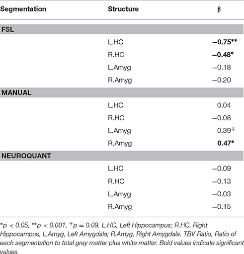
Table 3. Separate multivariate linear regression analyses of family aggression exposure manual and automated bilateral hippocampal and amygdala segmentations adjusting for age, gender, and total brain volume.
In the “Results” section, sub section “Manual Segmentation,” the second sentence has been added stating the following: A positive relationship between left amygdala volume and family aggression exposure using manual segmentation was approaching significance (b = 0.39, p = 0.09). The authors sincerely apologize for the error. This correlation was not previously mentioned or discussed in the manuscript. Therefore, this error does not change the scientific conclusions of the paper in any way.
Conflict of Interest Statement
The authors declare that the research was conducted in the absence of any commercial or financial relationships that could be construed as a potential conflict of interest.
Keywords: amygdala, hippocampus, methodology, family aggression, early life stress, adolescence
Citation: Lyden H, Gimbel SI, Del Piero L, Tsai AB, Sachs M, Kaplan JT, Margolin G and Saxbe D (2016) Corrigendum: Associations between Family Adversity and Brain Volume in Adolescence: Manual vs. Automated Brain Segmentation Yields Different Results. Front. Neurosci. 10:555. doi: 10.3389/fnins.2016.00555
Received: 01 November 2016; Accepted: 18 November 2016;
Published: 29 November 2016.
Edited and reviewed by: Yaroslav O. Halchenko, Dartmouth College, USA
Copyright © 2016 Lyden, Gimbel, Del Piero, Tsai, Sachs, Kaplan, Margolin and Saxbe. This is an open-access article distributed under the terms of the Creative Commons Attribution License (CC BY). The use, distribution or reproduction in other forums is permitted, provided the original author(s) or licensor are credited and that the original publication in this journal is cited, in accordance with accepted academic practice. No use, distribution or reproduction is permitted which does not comply with these terms.
*Correspondence: Hannah Lyden, bHlkZW5AdXNjLmVkdQ==