 Simon Burg1*
 Martin Gosau
 Audrey Laure Céline Grust
- 1Department of Oral and Maxillofacial Surgery, University Medical Center Hamburg-Eppendorf (UKE), Hamburg, Germany
- 2Department of Oral and Maxillofacial Surgery, Division of “Regenerative Orofacial Medicine”, University Medical Center Hamburg-Eppendorf (UKE), Hamburg, Germany
- 3Precura Center, Darmstadt, Germany
We present the case of a 53-year-old woman who was diagnosed with early-stage lung cancer by targeted cancer screening consisting of an immunological biopsy-based blood test followed by radiological imaging. The PanTum Detect blood test detects the biomarkers Apo10/DNaseX and Transketolase-like 1 (TKTL1) in circulating macrophage-like cells from peripheral blood samples to identify asymptomatic individuals with a high risk for malignancy. The elevated blood test values initiated an 18F-FDG PET/CT visualization for further clarification. In this case, imaging indicated a lung carcinoma in the right upper lobe. A biopsy confirmed the presence of a lung carcinoma, which was removed surgically. Histologic examination revealed a typical I A2 carcinoid, which was completely removed, making further therapy obsolete.
1 Introduction
Lung cancer is still one of the most common causes of cancer morbidity and mortality (1). It is generally well known that the success of a cancer therapy depends very decisively on the time of the diagnosis and on tumor characteristics such as invasiveness and therapy resistance. When detected at a localized stage, most entities can be cured by surgery alone and without further systemic therapy (2). Therefore, it is of great importance to establish diagnostic methods enabling early detection. Controlled trials with high-risk smokers and ex-smokers have shown that low-dose computed tomography (LDCT) can significantly reduce lung cancer mortality (3). Imaging modalities such as positron emission tomography using the glucose analogue 2-[18F]fluoro-2-deoxy-D-glucose (FDG-PET) in combination with computed tomography (FDG-PET/CT) have proven to be powerful tools for the localization and assessment of various tumor types (4–8). However, due to the cost and risk of radiation exposure, whole-body FDG-PET/CT does not yet appear suitable for general cancer screening in asymptomatic populations (4, 9). In contrast, a blood test that detects a variety of tumor types at early, asymptomatic stages can be used to pre-select subjects with a high likelihood of (pre)cancerous lesions on subsequent imaging (10). The PanTum Detect blood test is based on the unique technique of epitope detection in monocytes (EDIM) utilizing the fact that activated monocytes/macrophages (CD14+/CD16+), belonging to the group of circulating cancer-associated macrophage-like cells (CAMLs), phagocytose tumor cells and contain tumor proteins intracellularly (11–15). To prove internalization of tumor particles by CD14+/CD16+ monocytes, co-culture experiments were performed (16). These can be detected by flow cytometry from peripheral blood samples using specific antibodies. The PanTum Detect blood test screens for the biomarkers Apo10 and TKTL1, which identify basic biophysical mechanisms that are considerably altered in various forms of tumors (13, 17–19).
2 Case description
2.1 Patient information
The 53-year-old woman, 169 cm tall, weighing 77 kg (BMI 26.96) participated in the prospective non-interventional study “Pre-PanTum”, which was conducted between November 2020 and April 2021 at the Precura Center, Darmstadt, Germany (Ethics Committee of the Medical Association of Hessen, Frankfurt, Germany, approval number: 2020-1981-evBO). Anamnesis revealed a hot thyroid nodule, incipient osteoarthritis, cysts in the breast, neurodermatitis, arthritis of the left shoulder, hay fever, and various unspecified allergies. Since 01.07.2019, the subject has been medicated with 25 µg Levothyroxine (Thyronajod Henning, Sanofi-Aventis, 65926 Frankfurt/Main, Germany) per day. No other comorbidities, complaints, as well as current cancer diagnoses, or indications and symptoms suspecting a cancer disease were reported. She stated to have smoked about one cigarette per day for 7 years but being an ex-smoker for 24 years. Family history showed that her mother was diagnosed with ovarian cancer at age 75 and her uncle with colorectal cancer at age 67.
2.2 Clinical findings
The PanTum Detect blood test resulted in an elevated combined PanTum score of 270 (Apo10 score: 144, TKTL1 score: 126). All other tumor markers tested (CEA, CA 19-9, CA 125, CA 15-3, AFP, beta HCG, CRP) were not elevated. Further PET/CT imaging analysis was recommended for clarification.
2.3 Timeline
Figure 1. Timeline to demonstrate the diagnostic and therapeutic workflow from Pantum Detect blood test until required surgical resection and Follow-Up. The time from blood test sample receipt to complete surgical resection was 12 weeks.

Figure 1 Timeline showing the diagnostic and therapeutic schedule from November 2020 until May 2021.
2.4 Diagnostic assessments
2.4.1 Blood collection and blood test analysis
At least 2 ml of EDTA whole blood was collected per subject after a minimum of 60 minutes of fasting using a Sarstedt Monovette 2.7 ml EDTA. Shipping to the laboratory was undertaken by a transport service provider specialized in shipping blood samples, and the samples were stored at room temperature (15-25°C). Staining was performed with antibodies CD14 (OFC-14D), CD16 (Hi-16a), Apo10 (clone JFC 19X63) and TKTL1 (clone JF- C12T10), and flow cytometry analysis took place within 36 hours after blood collection at PreMed Labor GmbH, Pfungstadt, Germany with a BD FACSCantoTM II Flow Cytometry (Canto) operating software BD FACSDivaTM software version 8.0.3 and version 9.0.1. Test results were considered positive if the sum of the two individual scores for Apo10 and TKTL1 reached or exceeded the threshold of 260 and, in addition, the Apo10 individual score was ≥ 140 (hereafter total score). In case of elevated values, further imaging was recommended for clarification.
2.4.2 Imaging
For the assessment of the FDG-PET/CT, she was instructed to fast for a minimum of 6 hours and to avoid strenuous exercise 48 hours prior to the examination. She was injected with 97 MBq 18F-FDG and imaged on a Siemens Biograph Vision 600 PET/CT (Siemens Healthineers AG, 91052 Erlangen, Germany) in CT mode (153 mGy.cm). Following an uptake-phase (60 to 90 minutes) a low-dose CT was performed from the base of the skull to the thighs without iodinated contrast. Further details of the FDG-PET/CT imaging conditions can be found elsewhere (10).
The multiplanar reformate image (MPR) and lung CT revealed a 16 mm x 12 mm mass in the right upper lobe (Figures 2A, B, marked by arrows). In accordance with this, axial CT soft tissue window (Figure 2C) and transaxial fused PET-CT image (Figure 2D) showed an increased focal 18F-FDG uptake (SUVmax 6.3) in the right upper lobe adjacent to the hilus. In addition, a bilateral diffuse enhancement with mild focal emphasis cranially of the hilus (left SUVmax 5.8 > right SUVmax 4.8) was detected. The radiological findings revealed an urgent suspicion of lung carcinoma in the right upper lobe parahilar without pathologic lymph nodes. For bioptic/histological clarification, the patient was then referred for presentation to a pneumological/thoracic surgery center.
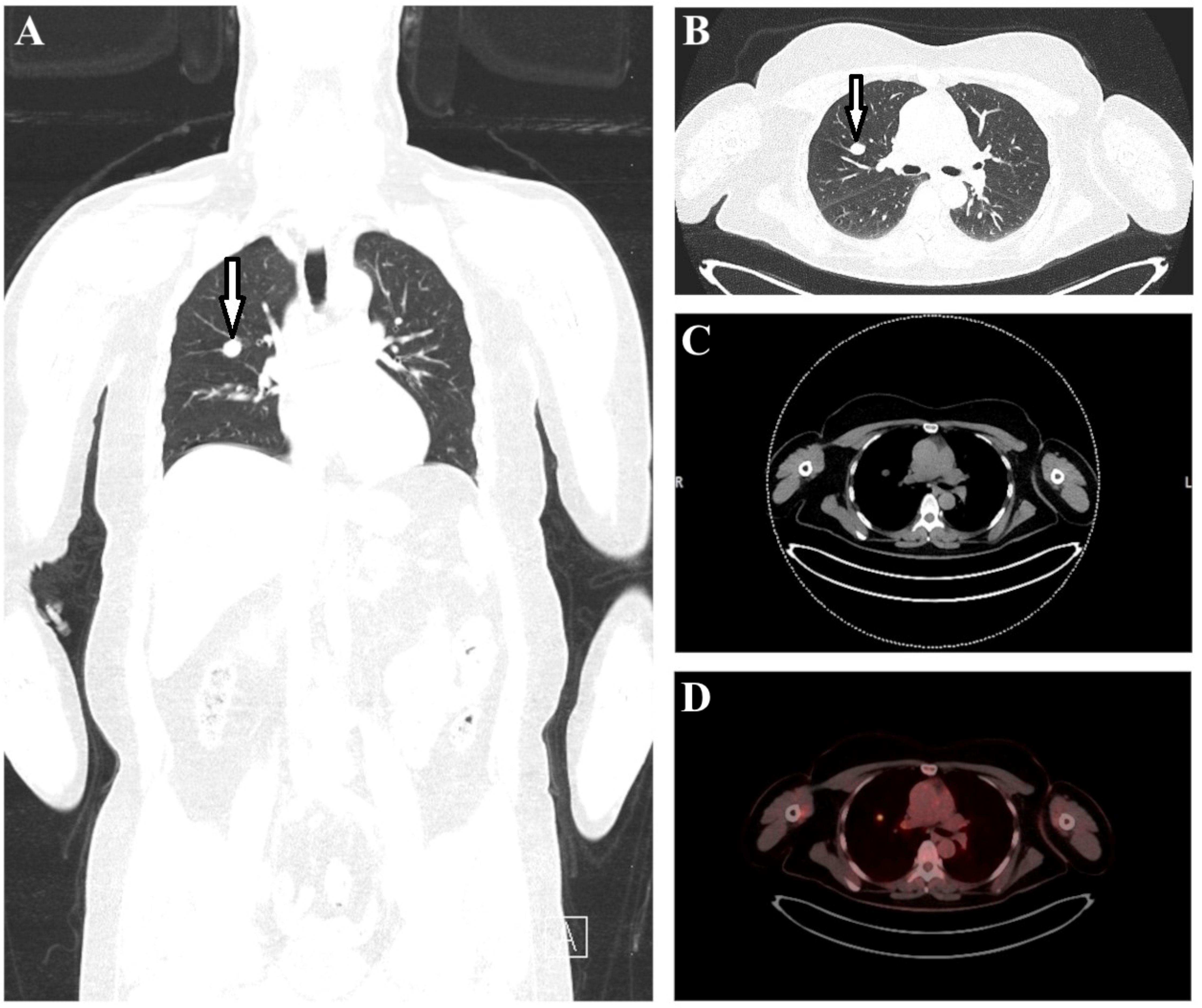
Figure 2 The multiplanar reformate image (A) and lung CT (B) revealed a 16 mm x 12 mm mass in the right upper lobe (marked with arrows). Axial CT (C) and transaxial fused PET-CT image (D) showed an increased focal 18F-FDG uptake (SUVmax 6.3) in the right upper lobe parahilar.
2.5 Therapeutic intervention
Biopsy confirmed malignant lung carcinoma. The surgical resection and subsequent histologic examination revealed a typical stage I A2 carcinoid without nodes or metastases (I A2 ED 01/21 pT1b (1.2 cm), pN0 (0/38), L0, V0, Pn0, R0 cM0) of the right upper lobe. As the tumor was completely removed, no further therapy was required.
2.6 Follow-up
At a follow-up three months after surgery, the subject reported about an occasional paresthesia in the thoracotomy area. The surgical scar appeared irritation-free and healed well. The thoracic CT showed postoperative changes, with no evidence of tumor recurrence. No adverse or unanticipated events were recorded.
3 Discussion
The field of early cancer detection is becoming increasingly important, which is also reflected in the growing number of publications in this area in recent years.
In a prospective, interventional study, called DETECT-A the feasibility and safety of multicancer blood testing coupled with PET/CT was evaluated in 10,006 asymptomatic women (6). On the one hand, screening of cell-free DNA (cfDNA) and specific protein biomarkers can provide first hints on the tumor entity, on the other hand, the concentration of cfDNA in the blood must be sufficiently high for this (20). The EDIM technology used for the PanTum Detect blood test however uses the body’s own immune system to detect disease-specific epitopes inside macrophages and this represents a very sensitive and specific biological mechanisms, which can achieve a very high diagnostic performance.
A study meanwhile conducted with more than 5,000 participants indicates that the upstream use of the PanTum Detect blood test allows for the pre-selection of individuals with a high tumor probability (PPV = 82.12%) and offers the possibility of using imaging procedures to screen these preselected asymptomatic individuals for possible tumors - also under suitable cost and radiation aspects (10). Since TKTL1 expression is linked to increased glucose uptake into the cell, an increased glucose uptake is a clear indication of transformation of a benign to a malignant tumor. PET/CT technology exploits the process of increased glucose uptake by using radiolabeled glucose to visualize increased glucose uptake in cells. The crucial conversion of a benign to a malignant tumor is thus detectable using the PanTum Detect blood® test by measuring elevated levels of TKTL1 and can be confirmed by PET/CT through increased glucose uptake in morphologically conspicuous tissues. For this reason, the combination of a blood test and imaging techniques can be used to precisely detect premalignant lesions that are on the verge of becoming dangerous in their development.
Like any diagnostic tool, the PanTum Detect blood test also has some limitations which are outlined below. For example, a manifest cancer diagnosis or suspected cancer as well as treatment with immunosuppressants (e.g., corticosteroids), amygdalin intake, acute febrile diseases, active herpes zoster, vaccinations, intake of contrast agents, surgeries or serious injuries may influence the test results and thus represent temporary exclusion criteria.
At present, the test is solely applied in early cancer screening. It is currently not used in monitoring, staging or follow-up. In case of elevated values, imaging techniques such as MRI or PET/CT must be performed for verification and localization. In addition, multiple small individual events could potentially add up to an elevated blood test level, although these are not yet detectable by current imaging techniques due to insufficient size. However, in case these small lesions continue to grow and the test is performed regularly, they will be detectable on imaging in a timely manner.
In clinical practice, the Pantum Detect blood test offers the advantage of non-invasive in vitro diagnostics without significant side effects or risks. Blood collection can be easily performed by a physician and subsequent analysis by trained laboratory personnel. Moreover, this blood test offers the advantage of exploiting the specific elimination of tumor cells by the innate immune system without dilution effects, as antigens are detected directly in macrophages. In addition, the two biomarkers Apo10 and TKTL1 allow the detection of many different tumor entities - including those for which tumor markers are not yet available (10).
The case report presented here conclusively shows that the PanTum Detect blood test in combination with subsequent radiological imaging offers the possibility to detect tumors at an early, localized stage, when prospects of cure are still favorable. In the presented case, the blood test indicated a high tumor probability in an asymptomatic individual, with subsequent radiological imaging confirming this suspicion and detecting early-stage lung cancer. Further customization and expansion of current screening programs to include such blood tests and imaging techniques should be considered in the future.
4 Patient perspective
The patient describes the entire process - from the Pantum Detect blood test to PET/CT imaging to surgical resection - as “life-saving.” At the time of the blood test, she had no complaints and felt healthy. She positively emphasized that after the abnormal blood test, all further measures were taken quickly and thus the period of uncertainty was short. She is particularly pleased that, thanks to the blood test and subsequent imaging, the tumor was detected at such an early stage and completely removed that no chemotherapy was required, and she is considered cured. The Patient approved the publication of this case report.
Data availability statement
The raw data supporting the conclusions of this article will be made available by the authors, without undue reservation.
Ethics statement
The studies involving human participants were reviewed and approved by Ethik Kommission der Ärztekammer Hamburg Weidestrasse 122b 22083 Hamburg Germany. The patients/participants provided their written informed consent to participate in this study.
Written informed consent was obtained from the individual(s) for the publication of any potentially identifiable images or data included in this article.
Author contributions
All authors contributed to the article and approved the submitted version. KF supervised the subject and performed the MRI examination. SB and AG drafted the final manuscript. RS performed the critical revision. RS and MG gave the final approval of the version to be published.
Conflict of interest
Authors SB, RS, KF, and AG declare financial funding from Zyagnum AG.
The remaining author declares that the research was conducted in the absence of any commercial or financial relationships that could be construed as a potential conflict of interest.
Publisher’s note
All claims expressed in this article are solely those of the authors and do not necessarily represent those of their affiliated organizations, or those of the publisher, the editors and the reviewers. Any product that may be evaluated in this article, or claim that may be made by its manufacturer, is not guaranteed or endorsed by the publisher.
References
1. Sung H, Ferlay J, Siegel RL, Laversanne M, Soerjomataram I, Jemal A, et al. Global cancer statistics 2020: GLOBOCAN estimates of incidence and mortality worldwide for 36 cancers in 185 countries. CA: A Cancer J Clin (2021) 71(3):209–49. doi: 10.3322/caac.21660
2. Siegel RL, Miller KD, Jemal A. Cancer statistics, 2017. CA: A Cancer J Clin (2017) 67(1):7–30. doi: 10.3322/caac.21387
3. Manser R, Lethaby A, Irving LB, Stone C, Byrnes G, Abramson MJ, et al. Screening for lung cancer. Cochrane Database Systematic Rev (2013) 6). doi: 10.1002/14651858.CD001991.pub3/full?highlightAbstract=standard%7Cgold%7Ccancer
4. Chan HP, Liu WS, Liou WS, Hu C, Chiu YL, Peng NJ. Comparison of FDG-PET/CT for cancer detection in populations with different risks of underlying malignancy. In Vivo. (2020) 34(1):469–78. doi: 10.21873/invivo.11797
5. Kwee TC, Kwee RM. Combined FDG-PET/CT for the detection of unknown primary tumors: systematic review and meta-analysis. Eur Radiol (2009) 19(3):731–44. doi: 10.1007/s00330-008-1194-4
6. Lennon AM, Buchanan AH, Kinde I, Warren A, Honushefsky A, Cohain AT, et al. Feasibility of blood testing combined with PET-CT to screen for cancer and guide intervention. Science (2020) 369(6499):eabb9601. doi: 10.1126/science.abb9601
7. Zhu A, Lee D, Shim H. Metabolic positron emission tomography imaging in cancer detection and therapy response. Semin Oncol (2011) 38(1):55–69. doi: 10.1053/j.seminoncol.2010.11.012
8. Cohen JD, Li L, Wang Y, Thoburn C, Afsari B, Danilova L, et al. Detection and localization of surgically resectable cancers with a multi-analyte blood test. Science (2018) 359(6378):926–30. doi: 10.1126/science.aar3247
9. Hatazawa J. Cancer screening by FDG-PET: benefit or risk? Ann Nucl Med (2011) 25(9):667–8. doi: 10.1007/s12149-011-0543-6
10. Burg S, Grust ALC, Feyen O, Failing K, Banat GA, Coy JF, et al. Blood-test based targeted visualization enables early detection of premalignant and malignant tumors in asymptomatic individuals. J Clin Med Images (2022) 6(9).
11. Herwig R, Pelzer A, Horninger W, Rehder P, Klocker H, Ramoner R, et al. Measurement of intracellular versus extracellular prostate-specific antigen levels in peripheral macrophages: a new approach to noninvasive diagnosis of prostate cancer. Clin Prostate Cancer. (2004) 3(3):184–8. doi: 10.3816/CGC.2004.n.029
12. Leers MPG, Nap M, Herwig R, Delaere K, Nauwelaers F. Circulating PSA-containing macrophages as a possible target for the detection of prostate cancer a three-Color/Five-Parameter flow cytometric study on peripheral blood samples. Am J Clin Pathol (2008) 129(4):649–56. doi: 10.1309/THWWRU8L42U5H9PB
13. Coy JF. EDIM-TKTL1/Apo10 blood test: an innate immune system based liquid biopsy for the early detection, characterization and targeted treatment of cancer. Int J Mol Sci (2017) 18(4):878–95. doi: 10.3390/ijms18040878
14. Stagno MJ, Schmidt A, Bochem J, Urla C, Handgretinger R, Cabanillas Stanchi KM, et al. Epitope detection in monocytes (EDIM) for liquid biopsy including identification of GD2 in childhood neuroblastoma–a pilot study. Br J Cancer. (2022) 127:1324–31. doi: 10.1038/s41416-022-01855-x
15. Sutton TL, Patel RK, Anderson AN, Bowden SG, Whalen R, Giske NR, et al. Circulating cells with macrophage-like characteristics in cancer: the importance of circulating neoplastic-immune hybrid cells in cancer. Cancer (Basel) (2022) 14(16):3871. doi: 10.3390/cancers14163871
16. Urla C, Stagno MJ, Schmidt A, Handgretinger R, Fuchs J, Warmann SW, et al. Epitope detection in monocytes (EDIM) as a new method of liquid biopsy in pediatric rhabdomyosarcoma. Cancers (2022) 10(8):1812. doi: 10.3390/biomedicines10081812
17. Feyen O, Coy JF, Prasad V, Schierl R, Saenger J, Baum RP. EDIM-TKTL1 blood test: a noninvasive method to detect upregulated glucose metabolism in patients with malignancies. Future Oncol (2012) 8(10):1349–59. doi: 10.2217/fon.12.98
18. Grimm M, Schmitt S, Teriete P, Biegner T, Stenzl A, Hennenlotter J, et al. A biomarker based detection and characterization of carcinomas exploiting two fundamental biophysical mechanisms in mammalian cells. BMC Cancer. (2013) 13(1):569–86. doi: 10.1186/1471-2407-13-569
19. Saman S, Stagno MJ, Warmann SW, Malek NP, Plentz RR, Schmid E. Biomarkers Apo10 and TKTL1: epitope-detection in monocytes (EDIM) as a new diagnostic approach for cholangiocellular, pancreatic and colorectal carcinoma. Cancer biomark (2020) 27(1):129–37. doi: 10.3233/CBM-190414
Keywords: early cancer screening, Apo10/DNaseX, TKTL1, immunological biopsy, blood test, FDG-PET/CT
Citation: Burg S, Smeets R, Gosau M, Failing K and Grust ALC (2023) Case Report: Early detection of lung carcinoid in an asymptomatic individual by blood-test initiated PET-CT imaging. Front. Oncol. 13:1177237. doi: 10.3389/fonc.2023.1177237
Received: 01 March 2023; Accepted: 19 May 2023;
Published: 06 June 2023.
Edited by:
Remy Guillevin, Centre Hospitalier Universitaire (CHU) de Poitiers, FranceReviewed by:
Dawid Sigorski, University of Warmia and Mazury in Olsztyn, PolandMatthew Pincus, Downstate Health Sciences University, United States
Copyright © 2023 Burg, Smeets, Gosau, Failing and Grust. This is an open-access article distributed under the terms of the Creative Commons Attribution License (CC BY). The use, distribution or reproduction in other forums is permitted, provided the original author(s) and the copyright owner(s) are credited and that the original publication in this journal is cited, in accordance with accepted academic practice. No use, distribution or reproduction is permitted which does not comply with these terms.
*Correspondence: Simon Burg, cy5idXJnQHVrZS5kZQ==