 Taiping Liao
 Guoxu Fu
- Department of Nuclear Medicine, The Third Hospital of Mianyang (Sichuan Mental Health Center), Mianyang, China
Metaplastic carcinoma of the breast is an extremely rare and highly aggressive malignancy associated with a poor prognosis. Breast spindle cell carcinoma is a subtype of metaplastic carcinoma. We present the case of a 48-year-old woman who was found to have a breast nodule during a routine examination two years ago. Pathological examination following surgical resection confirmed the diagnosis of breast spindle cell carcinoma. One year later, a follow-up CT scan detected a progressively enlarging mass in the left lower lobe of the lung, which was histologically confirmed as metastatic breast spindle cell carcinoma after surgical excision. Recently, the patient developed chest discomfort and severe left thigh pain, prompting an18F-FDG PET/CT scan, which revealed metastases to the lung, heart, pleura, and femur. Subsequently, the patient’s condition deteriorated rapidly within a short period. This report highlights the rare imaging findings of cardiac metastasis following surgery for breast spindle cell carcinoma, underscoring the highly aggressive nature of this tumor and the pivotal role of18F-FDG PET/CT in the post-operative monitoring of patients with breast spindle cell carcinoma.
Introduction
Metaplastic breast carcinoma is a rare subtype of breast cancer characterized by squamous or mesenchymal differentiation, which may exhibit spindle cell, chondroid, osseous, or rhabdomyoid differentiation patterns (1). Spindle cell carcinoma, a subtype of metaplastic breast carcinoma, is even rarer, and cardiac metastasis from this tumor is exceptionally uncommon. Here, we report the clinical course and management of a patient with breast spindle cell carcinoma, highlighting the highly aggressive nature of this malignancy and the critical role of 18F-FDG PET/CT in post-treatment surveillance.
Case presentation
A 48-year-old woman presented for a routine physical examination two years ago, during which an ultrasound detected a nodule in the left breast. Contrast-enhanced MRI suggested a high probability of malignancy (Figures 1A–C, long arrow). Fine-needle aspiration of the breast nodule was subsequently performed, and pathological analysis identified heterologous elements of bone and cartilage (Figures 1D–F), supporting a diagnosis of metaplastic breast carcinoma. The patient underwent a mastectomy, and postoperative pathological examination confirmed spindle cell carcinoma (Figures 1K–M). Immunohistochemical staining demonstrated negative expression of ER, PR, and HER2.
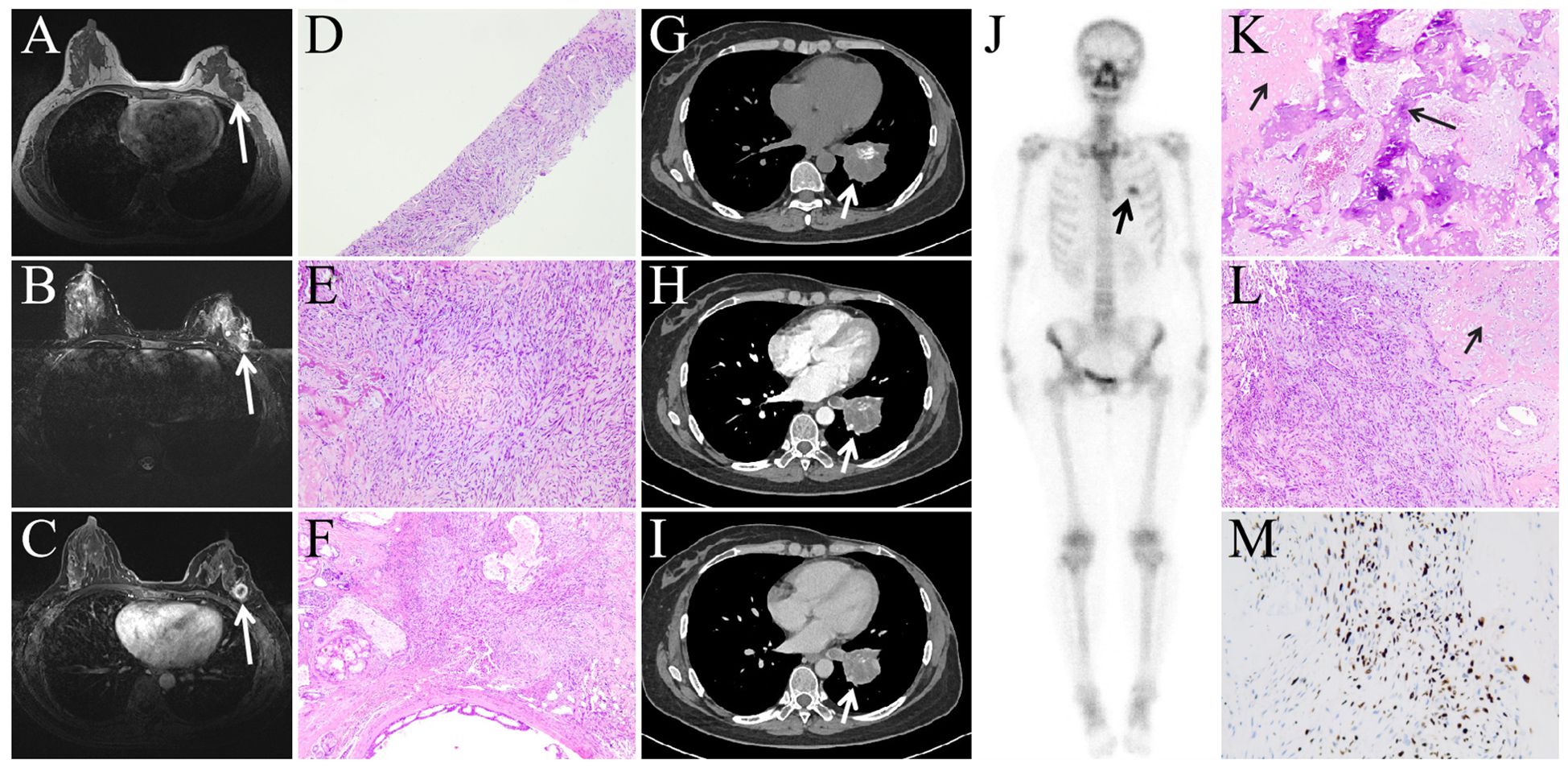
Figure 1. Breast MRI (A: T1WI, B: T2WI, C: contrast-enhanced scan) revealed a nodule in the left upper outer quadrant (long arrow), exhibiting long T1WI and long T2WI signals, with ring enhancement on contrast-enhanced scan. The biopsy result of the breast nodule (D: HE, 4×10, E: HE, 20×10, F: HE, 10×10) showed numerous spindle cells with heterologous components of bone and cartilage. Contrast-enhanced CT examination (G-I) revealed a mass in the left lower lobe (short arrow), with mild enhancement on contrast-enhanced scan, patchy high-density areas were also observed within the mass, which demonstrated intense radiotracer uptake on whole-body bone scintigraphy images (J, long arrow),suggesting active bone metabolism within the mass, consistent with the presence of bone and cartilage components found in the postoperative pathological images. Figures K-M (K: HE, 10×10, L: HE, 10×10, M: DAB, 10×10) represent postoperative pathological images of the left lower lobe lung lesion, which, similar to the breast lesion case results, also revealed heterologous components of bone (K, long arrow) and cartilage (K-L, short arrow).
The patient subsequently completed six cycles of chemotherapy with Taxol, doxorubicin, and cyclophosphamide. One year later, a follow-up CT scan revealed a mass in the left lower lobe of the lung (Figures 1G–I, short arrow), which progressively enlarged over two months, raising concerns for metastatic disease. A whole-body bone scan was performed, which showed no evidence of bone metastases; however, increased radiotracer uptake was observed in the left lower lobe lesion (Figure 1J, short arrow). The patient subsequently underwent surgical resection of the left lower lobe lesion, and postoperative pathological examination confirmed metastatic metaplastic breast carcinoma with negative expression of ER, PR, and HER2.
Subsequently, the patient was treated with five cycles of Utidelone plus Capecitabine. Utidelone is an analog of epothilone generated by genetically manipulating the polyketide biosynthetic gene cluster in S. cellulosum (2). It is reported that Utidelone demonstrated promising efficacy in the treatment of patients with advanced anthracycline/taxane-refractory metastatic breast cancer (3). One year later, she developed symptoms of fatigue and severe pain in her left thigh. To assess the treatment response and investigate the underlying cause, an 18F-FDG PET/CT scan was performed (Figures 2A–G). This scan identified nodular soft tissue lesions with abnormal FDG uptake in the right lung (Figures 2A, E–G, short arrow) and left pleura (Figure 2A, black arrowhead). Notably, an irregular soft tissue mass with markedly elevated FDG uptake was detected in the left atrium (Figures 2A–D, long arrow), along with an intramedullary soft tissue density shadow in the upper left femur (not shown in figure) that also demonstrated abnormal FDG uptake. In subsequent follow-up, the patient did not undergo pathological biopsy of the aforementioned lesions, but based on her clinical history and imaging findings, all these lesions were diagnosed as metastatic metaplastic breast carcinoma.
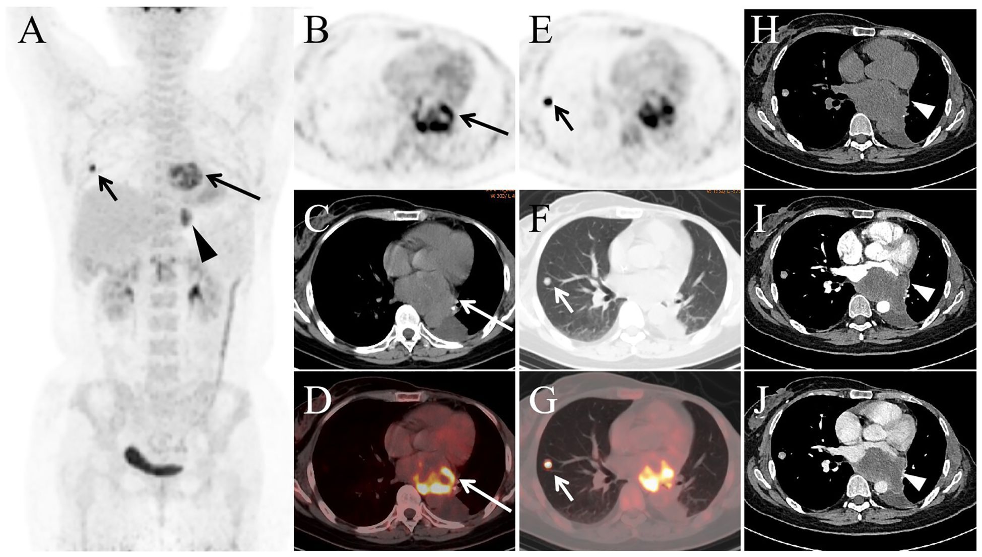
Figure 2. The MIP image (A) revealed increased FDG activity in lesions located in the heart (long arrow), right lung (short arrow), and left pleura (arrowhead). Transaxial images (B: PET, C: CT, D: PET/CT) displayed a large soft tissue mass in the left atrial region, exhibiting significantly elevated FDG activity with an SUVmax of 9.5. Additionally, a nodule was observed in the right upper lobe (E-G), showing abnormal FDG uptake with an SUVmax of 10.1. Contrast-enhanced CT (H-J) identified mild enhancement of the mass in the left atrium (arrowhead).
The patient subsequently underwent contrast-enhanced CT (Figures 2H–J), which showed mild enhancement of the left atrial mass. She has since received ten sessions of radiation therapy targeting the left femoral lesion, with each session delivering a dose of 3 Gy. Currently, the patient experiences occasional atrial fibrillation, reports poor overall well-being, and is undergoing treatment with Anlotinib and Toripalimab.Anlotinib is a new, orally administered tyrosine kinase inhibitor that targets vascular endothelial growth factor receptor (VEGFR), fibroblast growth factor receptor (FGFR), platelet-derived growth factor receptors (PDGFR), and c-kit (4). Studies have reported that anlotinib is effective in treating advanced metaplastic breast cancer (5, 6).
Discussion
Metaplastic carcinoma of the breast is an uncommon form of triple-negative carcinoma characterized by the transformation of either a portion or the entirety of its glandular carcinoma component into a non-glandular, or metaplastic, component (7–9). Metaplastic breast carcinoma has a high recurrence rate (10). Spindle cell carcinoma represents approximately <0.3% of invasive breast carcinomas (11). It has a more aggressive biological behavior with increased risk of recurrence and death due to disease compared to triple negative breast cancers (12, 13). The most common site of metastasis for metaplastic breast carcinoma is the lungs (14). Cardiac metastases are relatively rare. Nova-Camacho et al. performed autopsies on 1,294 adult cancer patients and identified 61 cases of cardiac metastases, including three cases of breast cancer (two cases of invasive ductal carcinoma and one case of lobular carcinoma) (15). Taguchi reviewed autopsy studies conducted by other researchers on cancer patients and reported that the incidence of cardiac metastases in malignant tumors ranges from 10% to 15% (16). However, none of these studies documented cases of cardiac metastases from spindle cell carcinoma of the breast. Cardiac metastases from spindle cell carcinoma are exceedingly rare, with reports limited to spindle cell carcinomas originating from the esophagus and larynx (17, 18). Literature has reported the use of 18F-FDG PET/CT in diagnosing metaplastic breast carcinoma (19, 20), but there are no documented cases of cardiac metastasis.
We analyzed cases of metastatic spindle cell carcinoma over the past 20 years, as summarized in Table 1 (17, 18, 21–25). The primary sites of spindle cell carcinoma were most frequently located in the breast. For the treatment of spindle cell carcinoma, monotherapy with pharmacotherapy or radiotherapy was rarely adopted. In cases with limited metastatic spread, a combination of surgical treatment and pharmacotherapy was more commonly selected as the treatment regimen, and the prognosis of most patients was relatively favorable following systematic therapy. However, for patients with widespread metastases before or after treatment, the prognosis was generally poor.
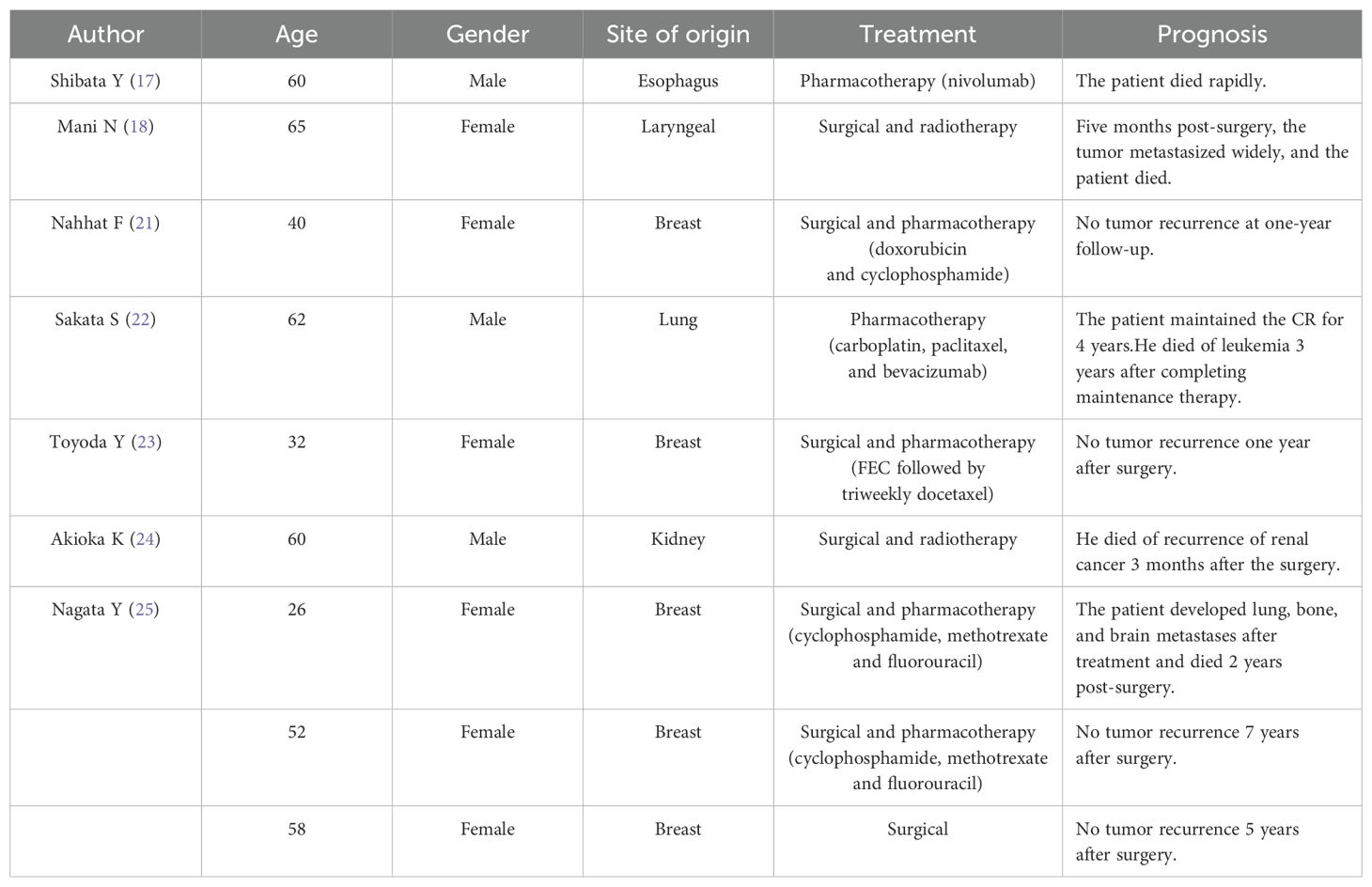
Table 1. Summary of the literature in the clinical treatment and prognosis of spindle cell carcinoma with metastases.
In our case, we report a rare occurrence of cardiac metastasis from spindle cell carcinoma of the breast, which is also the first documented case of cardiac metastasis in breast spindle cell carcinoma detected by 18F-FDG PET/CT, highlighting the significant role of 18F-FDG PET/CT in monitoring metastasis and recurrence in patients with breast spindle cell carcinoma. Unfortunately, despite aggressive treatment, recent enhanced CT scans show that the lung and cardiac lesions continue to enlarge, and the patient has recently developed atrial fibrillation, with new metastatic lesions detected in both kidneys, indicating a potentially poor prognosis.
Data availability statement
The original contributions presented in the study are included in the article/Supplementary Material. Further inquiries can be directed to the corresponding author.
Ethics statement
The studies involving humans were approved by The Third Hospital of Mianyang. The studies were conducted in accordance with the local legislation and institutional requirements. The participants provided their written informed consent to participate in this study. Written informed consent was obtained from the individual(s) for the publication of any potentially identifiable images or data included in this article.
Author contributions
TL: Writing – original draft, Writing – review & editing. GF: Writing – review & editing. LXL: Writing – review & editing. QQ: Writing – review & editing. LL: Writing – review & editing. YL: Writing – review & editing.
Funding
The author(s) declare that no financial support was received for the research and/or publication of this article.
Conflict of interest
The authors declare that the research was conducted in the absence of any commercial or financial relationships that could be construed as a potential conflict of interest.
Generative AI statement
The author(s) declare that no Generative AI was used in the creation of this manuscript.
Publisher’s note
All claims expressed in this article are solely those of the authors and do not necessarily represent those of their affiliated organizations, or those of the publisher, the editors and the reviewers. Any product that may be evaluated in this article, or claim that may be made by its manufacturer, is not guaranteed or endorsed by the publisher.
References
1. Narayan P, Kostrzewa CE, Zhang Z, O’Brien DAR, Mueller BA, Cuaron JJ, et al. Metaplastic carcinoma of the breast: matched cohort analysis of recurrence and survival. Breast Cancer Res Treat. (2023) 199:355–61. doi: 10.1007/s10549-023-06923-1
2. Zhang P, Sun M, Qiu R, Tang L, Dou G, Xu B. Phase I clinical and pharmacokinetic study of UTD1, a genetically engineered epothilone analog in patients with advanced solid tumors. Cancer Chemother Pharmacol. (2011) 68:971–8. doi: 10.1007/s00280-011-1571-6
3. Zhang P, Sun T, Zhang Q, Yuan Z, Jiang Z, Wang XJ, et al. Utidelone plus capecitabine versus capecitabine alone for heavily pretreated metastatic breast cancer refractory to anthracyclines and taxanes: a multicentre, open-label, superiority, phase 3, randomised controlled trial. Lancet Oncol. (2017) 18:371–83. doi: 10.1016/S1470-2045(17)30088-8
4. Shen G, Zheng F, Ren D, Du F, Dong Q, Wang Z, et al. Anlotinib: a novel multi-targeting tyrosine kinase inhibitor in clinical development. J Hematol Oncol. (2018) 11:120. doi: 10.1186/s13045-018-0664-7
5. Fu Y, Liu J, Jiang Y. Partial response after toripalimab plus anlotinib for advanced metaplastic breast carcinoma: A case report. Front Endocrinol (Lausanne). (2022) 13:810747. doi: 10.3389/fendo.2022.810747
6. Zou J, Yang X, Duan J, Wang J, Yang Z, Luo D, et al. A case report of targeted therapy with anlotinib in a patient with advanced breast metaplastic carcinoma. Onco Targets Ther. (2021) 14:4599–607. doi: 10.2147/OTT.S318645
7. McMullen ER, Zoumberos NA, Kleer CG. Metaplastic breast carcinoma: update on histopathology and molecular alterations. Arch Pathol Lab Med. (2019) 143:1492–6. doi: 10.5858/arpa.2019-0396-RA
8. Hasdemir OA, Tokgöz S, Köybaşıoğlu F, Karabacak H, Yücesoy C, İmamoğlu Gİ. Clinicopathological features of metaplastic breast carcinoma. Adv Clin Exp Med. (2018) 27:509–13. doi: 10.17219/acem/68293
9. Cooper CL, Karim RZ, Selinger C, Carmalt H, Lee CS, O’Toole SA. Molecular alterations in metaplastic breast carcinoma. J Clin Pathol. (2013) 66:522–8. doi: 10.1136/jclinpath-2012-201086
10. Haroon S, Zia S, Shirazi UA, Ahmed O, Asghar IA, Diwan MA, et al. Metaplastic breast carcinoma: clinicopathological parameters and prognostic profile. Cureus. (2021) 13:e14347. doi: 10.7759/cureus.14347
11. Sanmugasiva V, Hamid MTR, Fadzli F, Ab Mumin N, Rahmat K. Spindle cell metaplastic breast carcinoma. Curr Med Imaging. (2022) 18:684–8. doi: 10.2174/1573405617666211004114041
12. Niver HE, Foxhall E, Lahiry A. KEYNOTE-522 and male spindle cell carcinoma of the breast: A case report. Rare Tumors. (2023) 15:20363613231163730. doi: 10.1177/20363613231163730
13. El Zein D, Hughes M, Kumar S, Peng X, Oyasiji T, Jabbour H, et al. Metaplastic carcinoma of the breast is more aggressive than triple-negative breast cancer: A study from a single institution and review of literature. Clin Breast Cancer. (2017) 17:382–91. doi: 10.1016/j.clbc.2017.04.009
14. Takala S, Heikkilä P, Nevanlinna H, Blomqvist C, Mattson J. Metaplastic carcinoma of the breast: Prognosis and response to systemic treatment in metastatic disease. Breast J. (2019) 25:418–24. doi: 10.1111/tbj.13234
15. Nova-Camacho LM, Gomez-Dorronsoro M, Guarch R, Cordoba A, Cevallos MI, Panizo-Santos A. Cardiac metastasis from solid cancers: A 35-year single-center autopsy study. Arch Pathol Lab Med. (2023) 147:177–84. doi: 10.5858/arpa.2021-0418-OA
16. Taguchi S. Comprehensive review of the epidemiology and treatments for Malignant adult cardiac tumors. Gen Thorac Cardiovasc Surg. (2018) 66:257–62. doi: 10.1007/s11748-018-0912-3
17. Shibata Y, Ohmura H, Komatsu K, Sagara K, Matsuyama A, Nakano R, et al. Myocardial metastasis from ZEB1- and TWIST-positive spindle cell carcinoma of the esophagus: A case report. World J Gastroenterol. (2024) 30:1636–43. doi: 10.3748/wjg.v30.i11.1636
18. Mani N, Lowe D, Pope L, El-Daly H, Pfleiderer A. An unusual case of laryngeal spindle cell carcinoma metastasising to the orbit and heart. J Laryngol Otol. (2007) 121:e19. doi: 10.1017/S0022215107009498
19. Chen X, Chen G, Fu Z, Li Q. 18F-FDG PET/CT in a patient with matrix-producing carcinoma of the breast. Clin Nucl Med. (2022) 47:e340–3. doi: 10.1097/RLU.0000000000003916
20. Yin H, Xiu Y, Luo R, Zhang H, Shi H. Metaplastic squamous cell carcinoma of breast demonstrated on 18F-FDG PET/CT. Clin Nucl Med. (2018) 43:341–3. doi: 10.1097/RLU.0000000000001996
21. Nahhat F, Doyya M, Zabad K, Ksiri H. Metaplastic breast cancer with a unique presentation and complete response to chemotherapy: a case report. BMC Womens Health. (2024) 24:285. doi: 10.1186/s12905-024-03134-8
22. Sakata S, Saeki S, Sato R, Ishizuka S, Sasaki J, Fujii K. Long-term complete response to carboplatin plus paclitaxel combined with bevacizumab in a patient with metastatic spindle cell carcinoma. Respir Investig. (2017) 55:372–5. doi: 10.1016/j.resinv.2017.08.005
24. Akioka K, Masuda K, Harada S, Nakao T, Nakamura T, Okugawa K, et al. How long should we follow the post-transplantation patient after graft loss? A case report of renal cancer in the grafted kidney that occurred 16 years after graft loss. Transplant Proc. (2014) 46:626–9. doi: 10.1016/j.transproceed.2013.11.017
Keywords: 18F, FDG, PET/CT, metaplastic breast carcinoma, cardiac metastasis
Citation: Liao T, Fu G, Li L, Qi Q, Li L and Long Y (2025) The role of 18F-FDG PET/CT in detecting rare post-surgical cardiac metastasis of metaplastic breast carcinoma: a case report. Front. Oncol. 15:1521361. doi: 10.3389/fonc.2025.1521361
Received: 01 November 2024; Accepted: 28 March 2025;
Published: 11 April 2025.
Edited by:
Jing-Quan Wang, St. John’s University, United StatesReviewed by:
Abirami Sivapiragasam, MUSC Health, United StatesCherry Bansal, Tantia University, India
Copyright © 2025 Liao, Fu, Li, Qi, Li and Long. This is an open-access article distributed under the terms of the Creative Commons Attribution License (CC BY). The use, distribution or reproduction in other forums is permitted, provided the original author(s) and the copyright owner(s) are credited and that the original publication in this journal is cited, in accordance with accepted academic practice. No use, distribution or reproduction is permitted which does not comply with these terms.
*Correspondence: Yongjun Long, NTAzMjYwNzcwQHFxLmNvbQ==