 Roberta Lucianò1
 Maurizio Colecchia1,2
 Francesca Sanvito1Irene Locatelli3Chiara Venegoni3Alessia Di Coste3
 Davide Danilo Zani4
 Angelica Stranieri4
 Chiara Giudice4
 Antonella Rigillo5
 Matteo Gambini5
 Francesco Montorsi2,5
 Andrea Salonia2,3Marco Moschini3
 Massimo Alfano3*†
- 1Department of Pathology, Istituto di Ricovero e Cura a Caratterle Scientifico (IRCCS) San Raffaele Scientific Institute, Milan, Italy
- 2Università Vita-Salute San Raffaele, Milan, Italy
- 3Division of Experimental Oncology/Unit of Urology, Urological Research Institute (URI), Istituto di Ricovero e Cura a Caratterle Scientifico (IRCCS) San Raffaele Scientific Institute, Milan, Italy
- 4Department of Veterinary Medicine and Animal Science, University of Milan, Lodi, Italy
- 5I-Vet Diagnostica Veterinaria, Brescia, Italy
Objective: Urothelial carcinoma (UC) accounts for more than 90% of all bladder cancers both in humans and dogs. Human and canine UC share many genetic mutations and tumor markers and clinical and therapeutic interventions. The unmet clinical needs are similar such as the early detection and treatment of the high-grade residual disease responsible for tumor recurrence and progression. The aim of this study was to investigate the expression of the α5β1 integrin and its specificity in high-grade UC in humans and dogs, a marker recently reported in the human bladder in situ carcinoma and murine model of orthotopic bladder cancer.
Methods: Expression of integrin α5β1 was established by immunohistochemistry in 67 human bladder samples [four non-tumor tissues, 10 low-grade, 10 intermediate-grade, and 43 high-grade non-muscle invasive bladder cancer (NMIBC)] and 12 canine bladder tumor specimens.
Results: The α5β1 integrin was not expressed by urothelial cells in the conditions of inflammatory cystitis, actinic cystitis, benign hyperplasia, and low/intermediate grade NMIBC; it was identified as a specific marker expressed only by the malignant cells in the urothelium in 81% of human and all canine high-grade NMIBC.
Conclusions: The expression of α5β1 integrin is a specific marker of high-grade UC located in the urothelium of humans and dogs and might be tested for targeted delivery of contrast agents or drugs. Given the close similarity between high-grade UC in humans and dogs, basic research in the two species and comparative data analysis could strengthen the prospects for rapid development of an improved clinical strategy for the identification and treatment of the small neoplastic lesions responsible for residual high-grade in both species.
Introduction
One of the unmet clinical needs in the management of urothelial carcinoma (UC) is the identification and treatment of the residual disease after optimal treatment, responsible for disease relapse and progression. The bladder urothelium is uniformly exposed to the carcinogens with several areas of normal tissue affected by mutated clones present at the same time, named “field cancerization effect.” For patients with high-risk non-muscle-invasive bladder cancer (NMIBC), the “field cancerization effect” is associated with the development and recurrence of bladder cancer and may be associated with treatment outcomes (1), and in the preclinical model, the topographic modification of the extracellular matrix induced by radiotherapy sustains early onset of tumor growth induced by nitrosamine (2).
Most human and canine tumors share molecular genetics and clinical similarities (3). UC represents approximately 2% of all cancers, both in human (4) and dogs (5). The onset of UC is common in several canine breeds, with the highest odds ratio in the Scottish terrier (6) accounting for 11.7% of this breed (5).
In humans, 75% of UC are NMIBC at the first diagnosis (7), while in dogs, more than 90% are MIBC (81% papillary and 14% non-papillary UC) (5, 8) and high-grade in 76% (6, 8). For canine UC partial cystectomy and medical therapy are available. Indeed, disease progression due to recurrence occurs in the majority of animals treated with partial cystectomy (66%–100%) (8, 9), and the median progression-free survival after adjuvant therapy is 83–101 days (9, 10) and 119 days for medical therapy with intravesical instillation of Mitomycin C (11). Likewise, in humans, recurrence of bladder cancer at 2 and 5 years occurs in 36% and 50% of patients with high-grade NMIBC treated with Bacillus Calmette–Guerin (BCG) (12), and 10%–30% may progress at 5 years (13, 14).
Furthermore, human and canine UC share the same tumoral targets: among others, epithelial growth factor receptor (EGFR) that is overexpressed in 75% of high-grade human UC and 73% of canine muscle-invasive high-grade UC and cyclooxygenase-2 (Cox-2) that is overexpressed in 80% of both human and canine muscle-invasive high-grade UC (5). Likewise, dogs and humans share similar gut and urinary microbiomes (15–18), making dogs a more reliable model than rodents for studying UC.
We have recently characterized a murine model of orthotopic bladder cancer whose neoplastic cells express the integrin α5β1, an integrin that is also expressed in 81% of human high-grade NMIBC (19, 20). In the preclinical murine model, we also demonstrated that targeting the integrin α5β1 with engineered gold nanorods allows the detection and treatment of cancer lesions <1 mm (19, 20). Apart from developing the orthotopic bladder tumor, the preclinical model based on the intravesical instillation of MB49 bladder cancer cells does not show other signs of disease, for example, inflammatory reactions, as instead present in clinical specimens.
In this study, we deepen the integrin α5β1 expression pattern in the urothelium, by providing detailed pathological and therapeutical data of a large cohort of human high-grade NMIBC, low- and intermediate-grade NMIBC and non-oncological clinical specimens and included cases of canine spontaneous high-grade UC. We aimed to assess the feasibility of the integrin α5β1 to be a specific marker of malignant transformation of the urothelial cells in humans and dogs. This study demonstrates that the integrin α5β1 is a marker of high-grade non-invasive UC, both in humans and dogs, and its expression is not mediated neither by pro-inflammatory environment nor by the process of benign hyperplasia, nor previous intravesical immune-/chemotherapy.
Materials and methods
Human patient cohort
The collection of clinical specimens was through transurethral resection of bladder tumor (TURBT) of the following cohorts: i) four non-oncological bladder samples from the follow-up of four prostate cancer patients after radical prostatectomy and radiotherapy, with bladder area characterized by normal urothelium or signs of actinic cystitis; ii) 10 low-grade and 10 intermediate-grade NMIBC from 20 patients; and iii) 43 high-grade neoplastic tissues from 27 bladder cancer patients (Table 1).
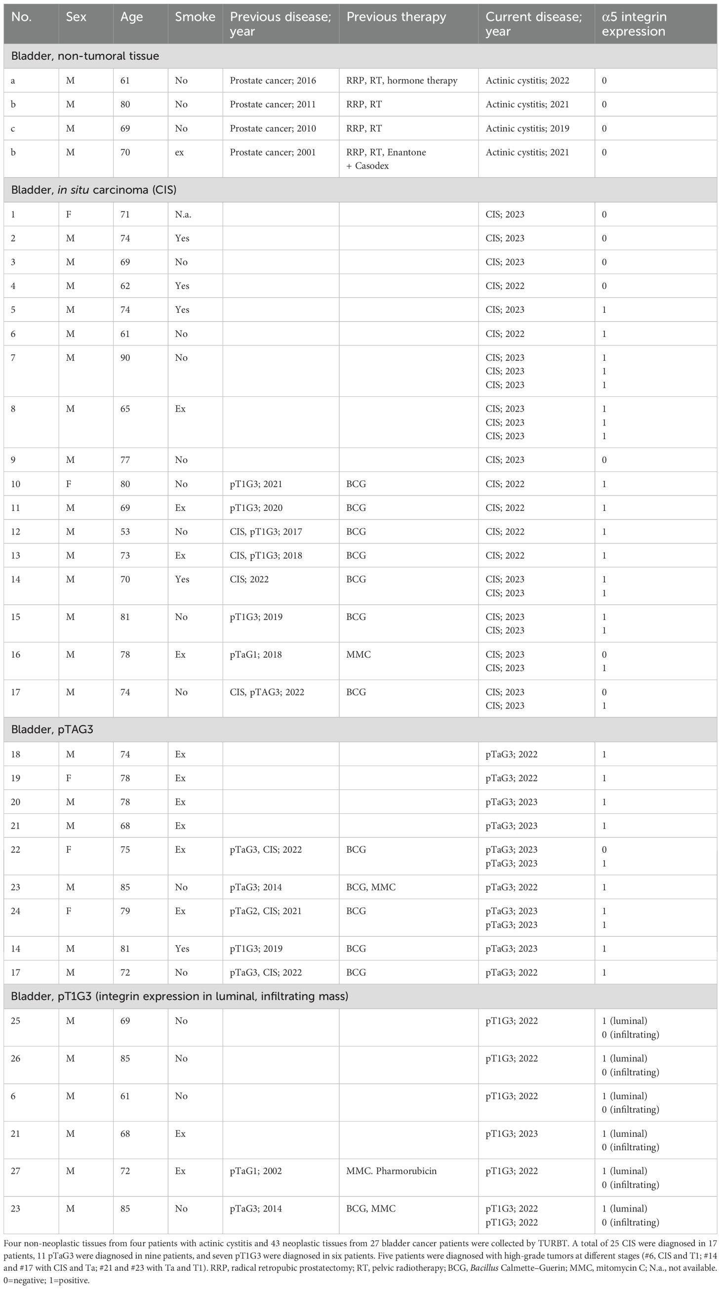
Table 1. Anamnestic data and tumor staging of human patients with high-grade urothelial carcinoma.
All surgical specimens span the entire thickness of the bladder wall and were staged by experienced pathologists (RL, MC, and FS) according to the TNM classification (21) and morpho-architectural criteria according to the WHO classification (22). Formalin-fixed paraffin-embedded blocks were retrieved and stained using an automatic hematoxylin/eosin (HE) slide stainer (HistoCore SPECTRA ST, Leica). An expert genitourinary pathologist (R.L.) reviewed the HE slides.
Canine patient cohort. Canine clinical specimens were collected through TURBT from 12 treatment-naive dogs (23) (Table 2). Canine specimens were classified according to the most recent diagnostic criteria, and malignant tumors were graded according to the latest published grading system for canine urothelial carcinoma (24). The diagnosis of canine specimens was further confirmed according to the TNM classification (21) and morpho-architectural criteria according to the WHO classification (25) used for human specimens.
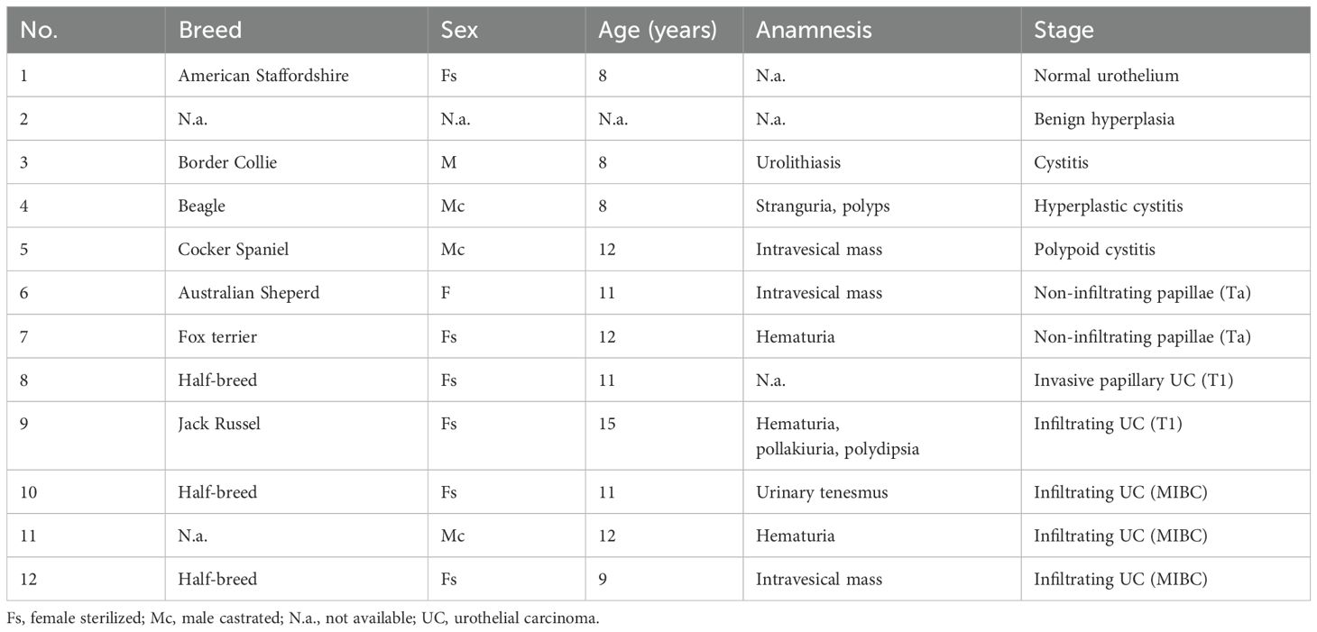
Table 2. Anamnestic data and tumor staging of dog patients with urothelial carcinoma.
Immunohistochemistry analysis
Immunohistochemistry (IHC) on human and canine formalin-fixed paraffin-embedded tissues was conducted on 2-µm tissue sections that were deparaffinized with xylol and decreasing scale of ethanol. Antigen retrieval was performed with heat with Tris-EDTA buffer for 40 min at 97°C. After washing with TBS and quenching the endogenous peroxidase (5 min incubation with 3% H2O2), tissue sections were incubated for 10 min with 3%BSA/TBS to perform non-specific antigen blocking.
IHC for the alpha5 integrin was carried out with 1/400 dilution of rabbit recombinant monoclonal integrin alpha 5 antibody (clone EPR7854, Abcam) in Da Vinci diluent (Biocare Medical, Pacheco, CA, USA) for 1 h at room temperature. After washing, the binding of rabbit primary antibodies was detected using a Universal HRP-Polymer Biotin-free detection system (MACH4, BioCare Medical, USA) and 3,3-diaminobenzidine free base (DAB) as a chromogen. Tissue samples were then counterstained with Harris hematoxylin.
IHC for the beta1 integrin was carried out as reported above by incubating tissue sections with ½,000 dilution of rabbit recombinant monoclonal antibody (clone EPR16895, Abcam, Cambridge, CB2 0AX, UK). Negative controls for IHC analysis on both human and canine specimens were by omission of the primary Ab.
Results
The α5β1 integrin is expressed by neoplastic cells in high-grade human NMIBC
We previously reported that the α5β1 integrin is expressed in six out of eight (75%) patient samples diagnosed with carcinoma in situ (CIS) and by the orthotopic tumor in the MB49-derived pre-clinical murine model (19). In this study, we implemented this information by carrying out the analysis of an extended number of CIS and included specimens from high-grade pTa and pT1, for a total of 43 specimens of high-grade NMIBC and four non-tumoral bladders (Table 1). We also extended the study to low- and intermediate-grade NMIBC, pTaG1 (n=10), and pTaG2 (n=10), respectively.
In the non-tumoral tissues, the α5 subunit was expressed by the stromal cells present in the lamina propria but not expressed by the urothelial and umbrella cells present in the urothelium (Figure 1A). The same expression pattern was observed in the clinical specimen of chronic reactive urothelium following radiotherapy, characterized by irregular polarization of the nuclei of urothelial cells, hyperplastic umbrella cells, and inflammation in the lamina propria. In the same non-neoplastic tissues, also the cells in the Von Brunn’s nest were negative for the integrin α5 (Figure 1B).
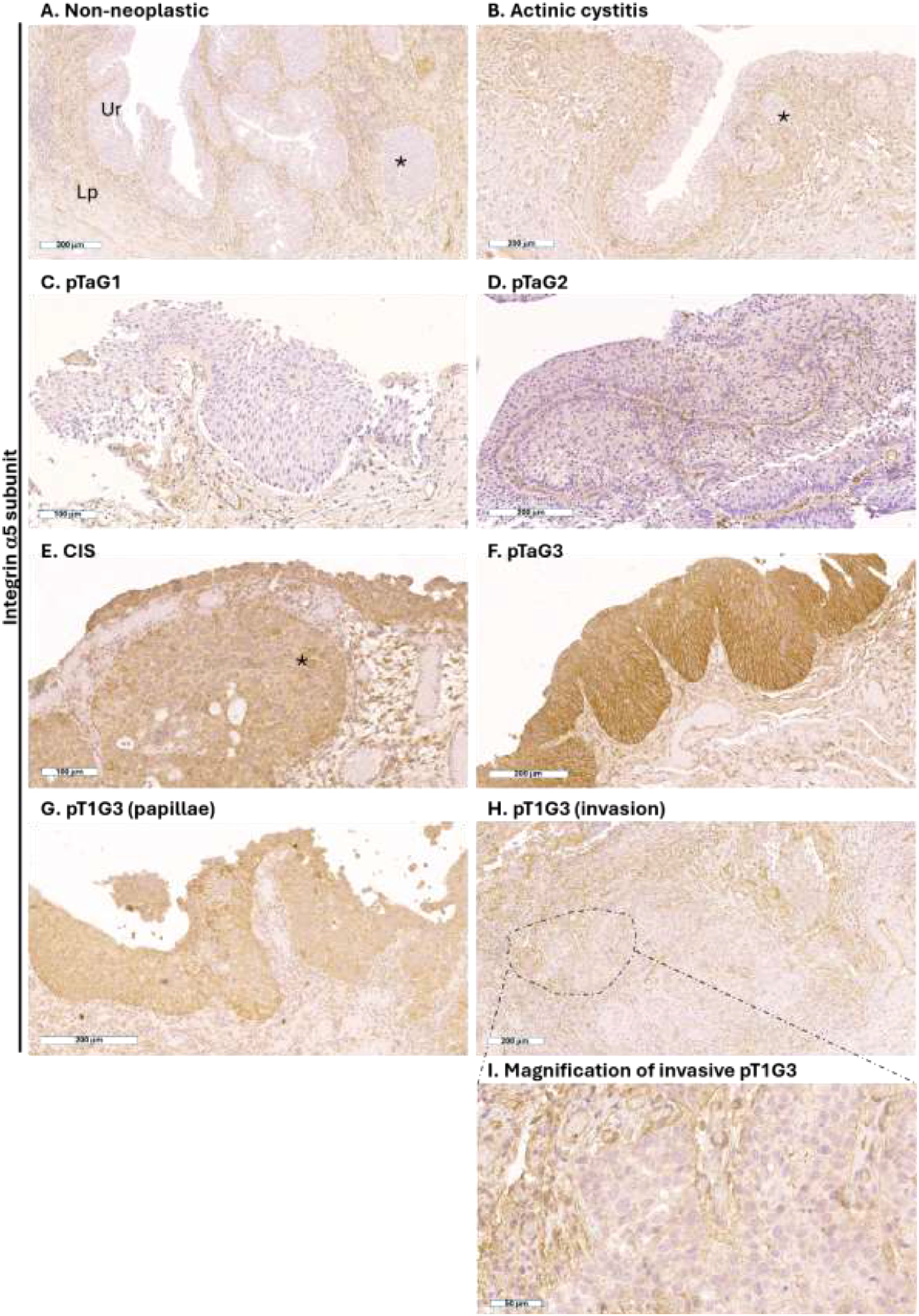
Figure 1. Expression of the α5 integrin subunit in human high-grade NMIBC. Representative immunohistochemistry photomicrographs of human bladder sections of non-neoplastic tissue (A, B), pTaG1 (C), pTaG2 (D), CIS (E), pTaG3 (F), and pT1G3 in the urothelial layer (G) and in the lamina propria (H); representative tumor area highlighted by dashed circle, and magnification in panel (I); all tissues were obtained by TURB. Scale bar is reported at the bottom left of each panel. Ur, urothelium; Lp, lamina propria. *Representative Von Brunn’s nest.
In low-grade NMIBC (10 pTAg1), urothelial cells were negative for α5 subunit expression, as in non-tumor tissues. (Figure 1C).
In intermediate-grade NMIBC, membranous expression of the α5 subunit was identified on a minority of urothelial cells in pTaG2 of 4 out of 10 patients (Figure 1D), and no expression was identified in the urothelial cells in pTaG2 of six patients.
In the high-grade NMIBC specimens, the membranous expression of the α5 subunit was identified in the cancer cells of CIS (n=18/25 cases, 72%), including CIS that colonized the Von Brunn’s nest (Figure 1E), and high-grade pTa (n=10/11 cases, 91%) (Figure 1F). In high-grade pT1, the α5 subunit was expressed only in the non-invasive region of the tumor (Figure 1G); when tumor cells invaded the lamina propria (n=7/7, 100%), the α5 subunit was not expressed (Figure 1H), while the stromal cells surrounding the tumor cells expressed the integrin (Figure 1I).
Integrin β1 subunit was always expressed by urothelial cells, such as in non-neoplastic condition (Figure 2A) that also includes chronic reaction as in the actinic cystitis (Figure 2B), low-grade UC (Figure 2C), intermediate-grade UC (Figure 2D), and high-grade UCs (Figures 2E–G). This result agrees with previous reports showing that integrin beta1 is expressed in all layers of the urothelium at intercellular junctions (26–28).
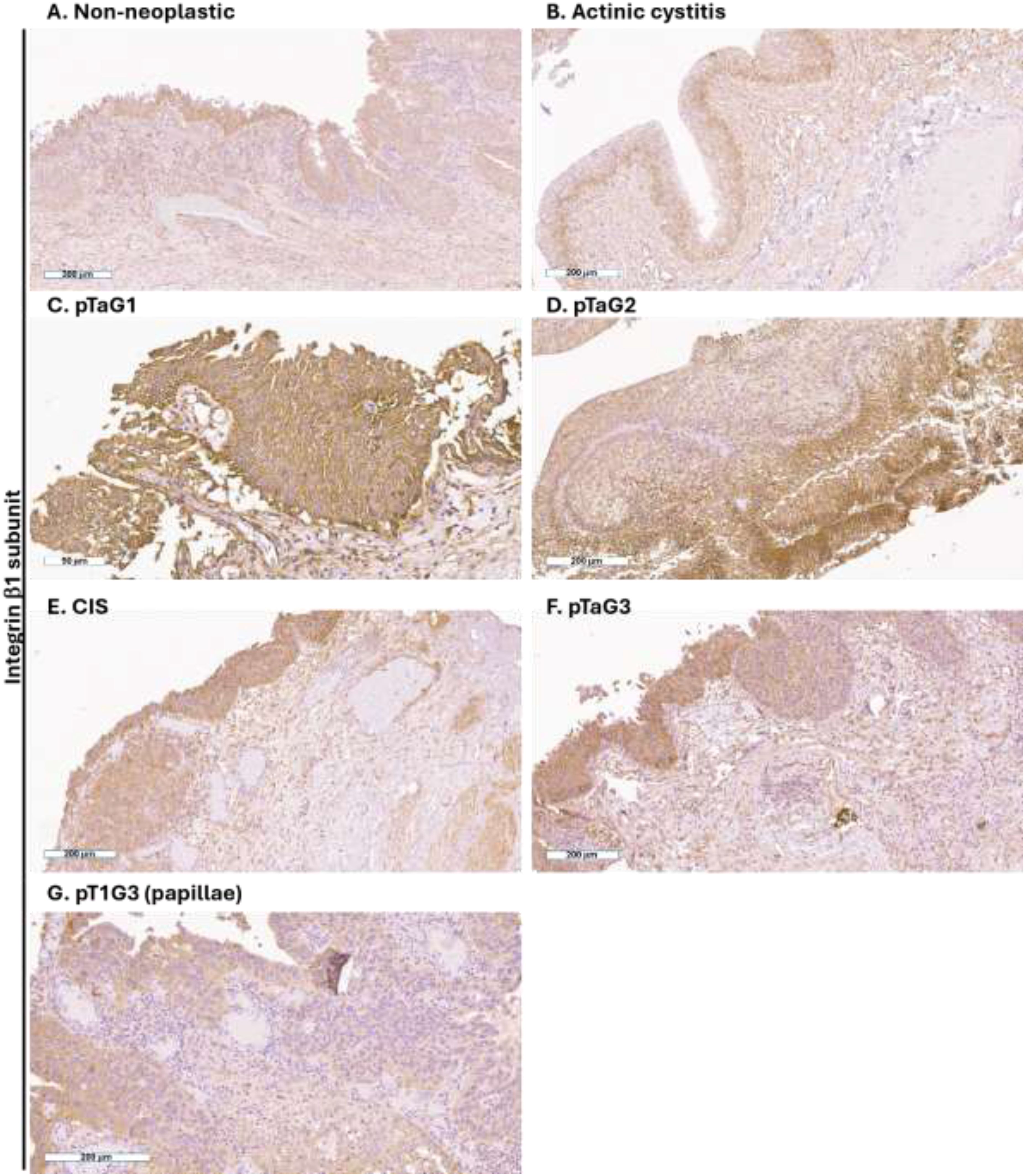
Figure 2. Expression of the β1 integrin subunit in human high-grade NMIBC. Representative immunohistochemistry photomicrographs of human bladder sections of non-neoplastic tissue (A, B), pTaG1 (C), pTaG2 (D), CIS (E), pTaG3 (F), and pT1G3 in the urothelial layer (G); all tissues were obtained by TURB. Scale bar is reported at the bottom left of each panel.
The outcome of this analysis shows that non-neoplastic urothelial cells and low/intermediate NMIBC were negative for the expression of the α5β1 integrin and that 81% (35 out of 43 cases) of high-grade NMIBC in the urothelial layer express the α5β1 integrin. Overall, these data suggest that the expression of the α5β1 integrin is specific to the neoplastic transformation of urothelial cells when high-grade neoplasia is in the urothelial layer but is not associated with chronic inflammation. Furthermore, the expression of α5β1 integrin by neoplastic urothelial cells was independent of previous intravesical treatment (Table 1), either immunotherapy with the BCG or chemotherapy with pharmorubicin or mitomycin-C.
Integrin α5b1 expressed by high-grade canine UC
Bladder specimens from dogs were validated by an experienced uro-pathologist (RL). Immunohistochemical analysis was carried out for the expression of the α5 and β1 chains of the integrin. In the non-tumoral tissues (specimens 1–5 in Table 2), the α5 subunit was expressed by the stromal cells present in the lamina propria but not expressed by the urothelial cells (Figure 3A). The same expression pattern was observed in the clinical specimen characterized by cystitis (Figures 3B,C), and benign hyperplasia (Figure 3D). In the canine UC, the α5 subunit was expressed also by the cancer cells in the non-invasive papillary carcinoma (Figure 3E) (specimens 6–8 in Table 2). Cancer cells that invaded the lamina propria were negative for the expression of the α5 subunit (Figures 3F,G) (specimens 9–12 in Table 2).
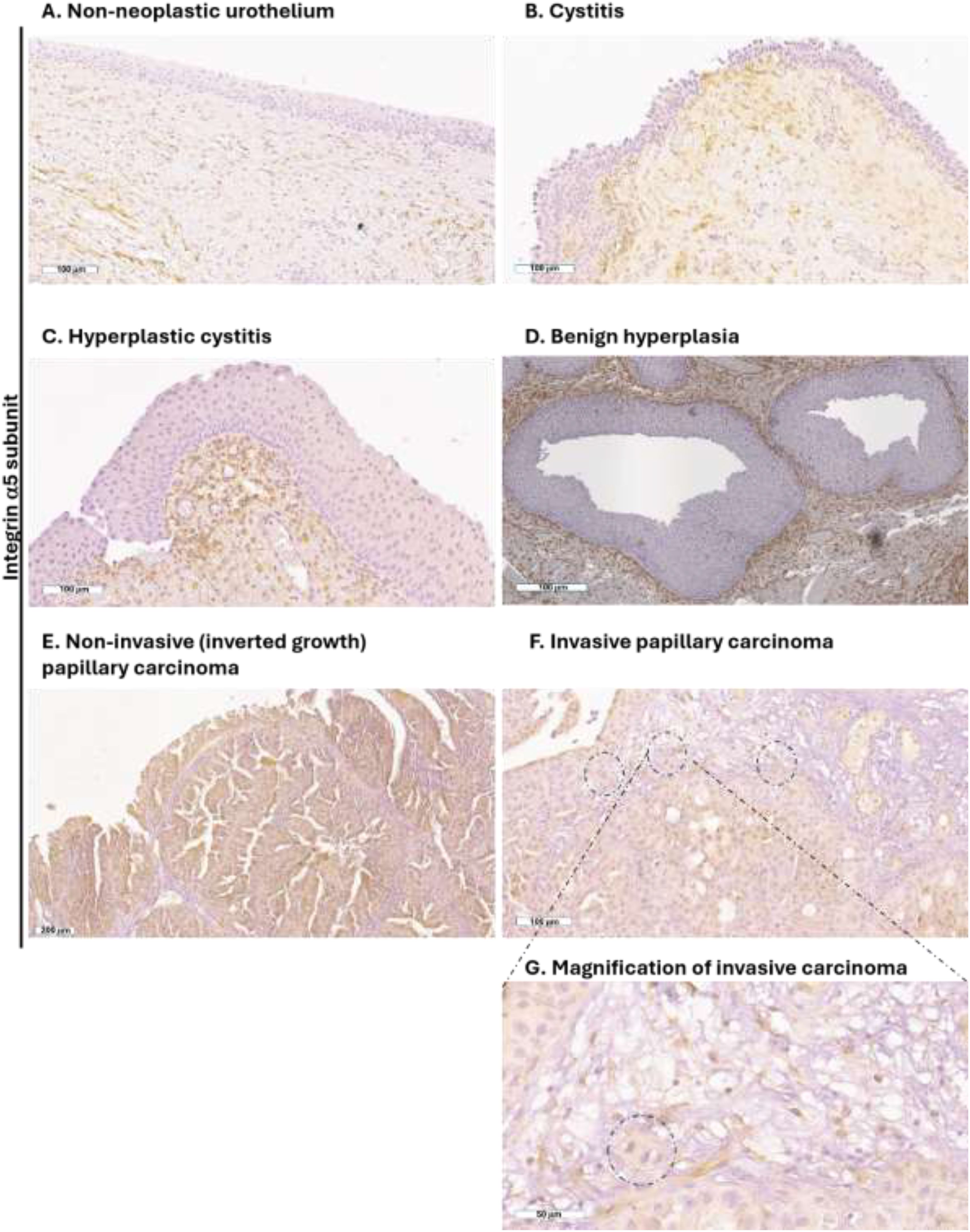
Figure 3. Expression of the α5 integrin subunit in canine UC. Representative immunohistochemistry photomicrographs of canine bladder sections of non-neoplastic tissue (A-D), non-invasive papillary UC (E), and invasive papillary UC spreading in the lamina propria (F); representative tumor area highlighted by dashed circle, and magnification in panel (G). All tissues were obtained by TURBT. Scale bar is reported at the bottom left of each panel.
As recently reported in human and murine bladder section (19), we here confirmed that the β1 subunit was expressed by normal urothelial, stromal, and tumor cells of the canine bladder. The β1 subunit was expressed by normal urothelial and stromal cells, and the same expression was observed for benign and malignant neoplasia (Figure 4).
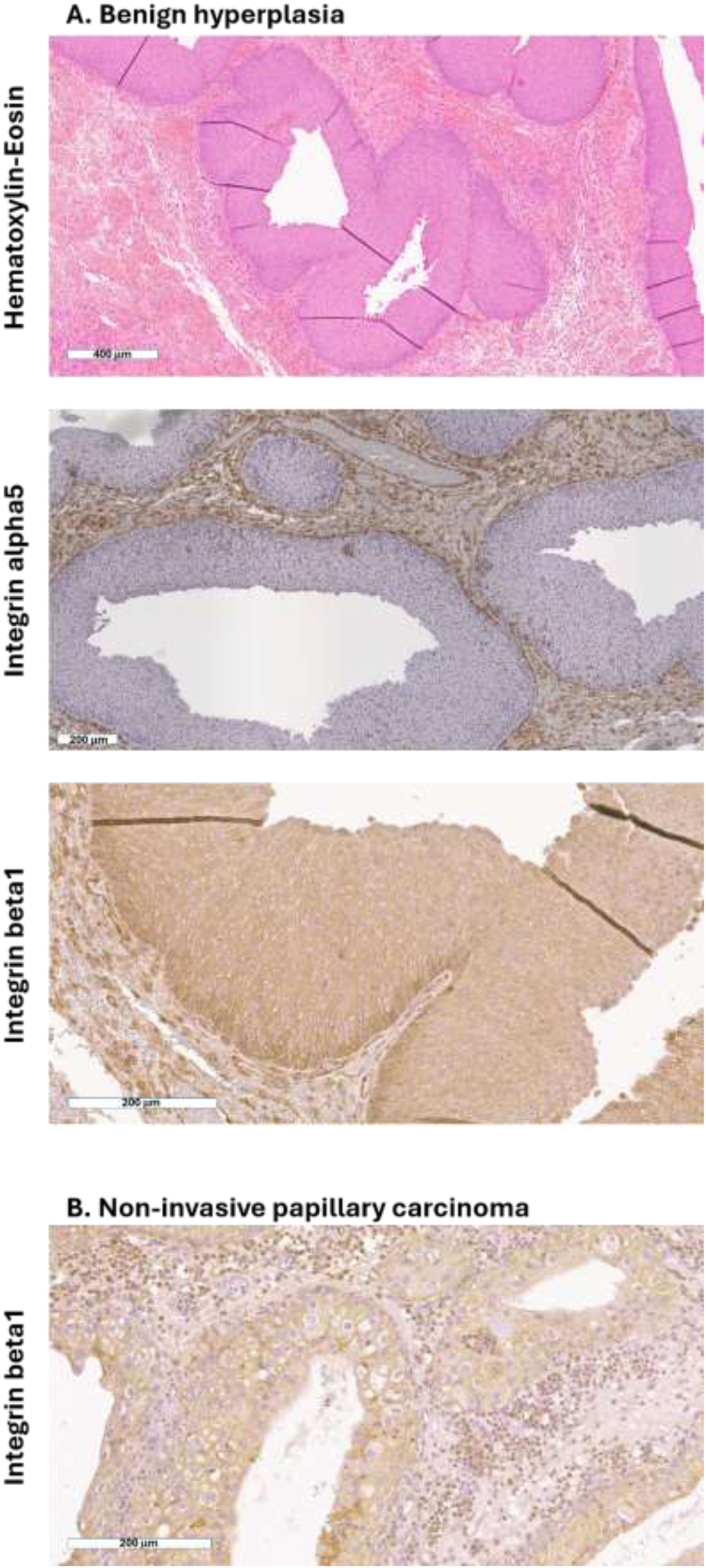
Figure 4. Expression of β1 integrin by normal and neoplastic canine urothelial cells. Representative photomicrographs of (A) hematoxylin–eosin and immunohistochemistry for α5 and β1 integrins of canine bladder section with benign hyperplasia and (B) immunohistochemistry for β1 integrin of canine bladder section with non-invasive papillary carcinoma. Scale bar is reported at the bottom left of each panel.
Discussion
Outcomes from this study show that the integrin α5β1 is a marker of high-grade non-invasive UC, both in humans and dogs. The integrin α5β1 is specifically expressed by the high-grade neoplastic urothelial cells, and its expression is not mediated neither by pro-inflammatory environment nor by the process of benign hyperplasia, nor by previous intravesical immune-/chemotherapy. We have recently reported that the integrin α5β1 is also expressed by the orthotopic bladder cancer in the MB49 murine model but not by the normal murine epithelium (19). The second outcome of this study is that the specific expression of the integrin α5β1 by high-grade bladder cancer cells is conserved among the mouse, dogs, and human species.
The integrin α5β1 was reported to be expressed also in other neoplasia of epithelial origin such as ovarian and cervical tumors in which 80% and 84% of tumors are positive for the expression of integrin α5β1 and significantly correlate with higher clinical stage (29, 30). We here report that 81% of the high-grade NMIBC express the integrin α5β1 but only when the neoplastic cells are in the luminal area, while neoplastic cells in the lamina propria (T1 stage) do not express the integrin α5β1. The expression of the integrin α5β1 by the malignant cells is likely to represent one of the steps of the tumorigenesis of epithelial tumors. It has been reported that silencing of α5 integrin in tumor cells strongly reduces lung tumor spreading (31). Concerning the mechanism, it has been previously reported that α5β1 and αVβ3 integrins directly activate matrix metalloproteinases and thus participate in the degradation of the components of the extracellular matrix of the basement membrane to support tumor invasion (32, 33). Once in the lamina propria, the lost expression of the integrin α5β1 could be part of a process to facilitate cancer cell movement, while degradation of the basement membrane of the endothelium by the integrin αVβ3 supports intravasation and spreading of the disease (34). Overall, the expression of the integrin α5β1 by the high-grade malignant cells is likely to represent one of the steps in the progression of epithelial tumors (35).
The identification of the integrin α5β1 expressed by high-grade bladder cancer cells located in the urothelium can be used for the delivery of contrast agents aimed to carry out targeted diagnostic imaging solutions in either therapy-naive patients or patients with relapsing high-grade UC. Not being expressed by the urothelial cells in the conditions of hyperplasia, cystitis, and chronic inflammation, but being specific for the high-grade cancer cells, the targeting of integrin α5β1 could overcome the limitation of white and blue light cystoscopies that detects tumor and areas of inflammation that arise following TURBT or BCG instillation (36–38).
The integrin α5β1 can represent a target that can be exploited using molecules recognizing the heterodimer, such as cyclic peptide (19, 20), peptidometic ligand (39) or antibody (40), or the integrin α5 subunit through an aptamer (41). Depending on the resolution of the diagnostic imaging techniques, lesions of different sizes can be diagnosed. Peptidomimetic ligand that delivers 68gadolinium was developed as a contrast agent for PET imaging (39). The application of engineered gold nanorods targeting the integrin α5β1 as the contrast agent in photoacoustic imaging has recently demonstrated to diagnose bladder cancer lesions <1mm in the preclinical murine model (19, 20, 41). Deployment of this solution in the clinical scenario could offer a novel strategy that complements the TURBT approach, which allows for more accurate detection of the disease and identification of the residual disease in patients undergoing i) second-look TURBT and ii) enrolled in bladder-sparing protocols (42). Likewise, the integrin α5β1 also represents a promising therapeutic target for the gold nanorods-assisted hyperthermia of orthotopic bladder cancer (20).
The integrin α5β1 can be used to deliver a targeted contrast agent for diagnosis or therapy against high-grade UC both in humans and dogs and to reduce the high tumor recurrence rate observed in both species. Dogs and humans UC share many characteristics, but the shorter lifespan of dogs mean that tumors develop, metastasize, and respond to therapy more quickly (3). Given the close similarity between high-grade UC in humans and dogs, but the shorter lifespan of dogs, the spontaneous high-grade UC in dogs can be a helpful model to speed up the transition of drugs tested in murine models in clinical trials, with results that can impact both humans and dogs.
Data availability statement
The original contributions presented in the study are included in the article/supplementary material. Further inquiries can be directed to the corresponding author.
Ethics statement
The studies involving humans were approved by Ethics Committee IRCCS Ospedale San Raffaele, Milan, Italy. The studies were conducted in accordance with the local legislation and institutional requirements. The study on the human patient cohort was conducted in compliance with the principles of the Declaration of Helsinki, and all patients signed an informed consent agreement to deliver their own anonymous information for future studies. This study and all experimental protocols were approved by the Institutional Review Board (Ethics Committee IRCCS Ospedale San Raffaele, Milan, Italy). For the canine sample, written informed consent for participation was not required from the participants or the participants’ legal guardians/next of kin in accordance with the national legislation and institutional requirements. Ethical approval was not required for the studies involving animals in accordance with the local legislation and institutional requirements because all canine samples used in the study were collected from canine patients referred to the Veterinary Teaching Hospital (VTH) for routine diagnostic or wellness visits. Therefore, in accordance with the guidelines of our institution (University of Milan), formal approval from the Institutional Animal Care and Use Committee was not required, as the activity falls within the exclusions specified under Article 2 of Legislative Decree 26/2014 (EC decision 02-2016). Written informed consent was obtained from the owners for the participation of their animals in this study.
Author contributions
MA: Conceptualization, Data curation, Formal analysis, Funding acquisition, Investigation, Methodology, Writing – original draft. RL: Investigation, Writing – review & editing. MC: Investigation, Writing – review & editing. FS: Investigation, Writing – review & editing. IL: Data curation, Writing – review & editing. CV: Data curation, Writing – review & editing. AC: Writing – review & editing. DDZ: Data curation, Resources, Writing – review & editing. ASt: Resources, Writing – review & editing. CG: Resources, Writing – review & editing. AR: Resources, Writing – review & editing. MG: Resources, Writing – review & editing. FM: Writing – review & editing. ASa: Writing – review & editing. MM: Writing – review & editing.
Funding
The author(s) declare that financial support was received for the research and/or publication of this article. This study was funded by the European Union’s Horizon EUROPE; PHIRE project is funded under grant agreement no. 101113193 (https://cordis.europa.eu/project/id/101113193). The funding source had no role in the design of this study, data interpretation, or writing of the manuscript. Views and opinions expressed are those of the author(s) and do not necessarily reflect those of the EU or the EIC.
Acknowledgments
We thank Dr. Rossana Norata and Dr. Martina Rocchi (Department of Pathology, IRCCS Ospedale San Raffaele) and Dr. Amleto Fiocchi (Animal Histopathology, IRCCS Ospedale San Raffaele) for their technical support.
Conflict of interest
The authors declare that the research was conducted in the absence of any commercial or financial relationships that could be construed as a potential conflict of interest.
Generative AI statement
The author(s) declare that no Generative AI was used in the creation of this manuscript.
Correction note
A correction has been made to this article. Details can be found at: 10.3389/fonc.2025.1640082.
Publisher’s note
All claims expressed in this article are solely those of the authors and do not necessarily represent those of their affiliated organizations, or those of the publisher, the editors and the reviewers. Any product that may be evaluated in this article, or claim that may be made by its manufacturer, is not guaranteed or endorsed by the publisher.
References
1. Strandgaard T, Nordentoft I, Birkenkamp-Demtroder K, Salminen L, Prip F, Rasmussen J, et al. Field cancerization is associated with tumor development, T-cell exhaustion, and clinical outcomes in bladder cancer. Eur Urol. (2024) 85:82–92. doi: 10.1016/j.eururo.2023.07.014
2. Venegoni C, Pederzoli F, Locatelli I, Alchera E, Martinez-Vidal L, Di Coste A, et al. Topographic modification of the extracellular matrix precedes the onset of bladder cancer. Matrix Biol Plus. (2024) 23:100154. doi: 10.1016/j.mbplus.2024.100154
3. Arnold C. Sick as a dog: how understanding canine diseases will save human lives. Nat Med. (2022) 28:1970–3. doi: 10.1038/s41591-022-02025-5
4. Ferlay J, Soerjomataram I, Dikshit R, Eser S, Mathers C, Rebelo M, et al. Cancer incidence and mortality worldwide: sources, methods and major patterns in globocan 2012. Int J Cancer. (2015) 136:E359–86. doi: 10.1002/ijc.29210
5. Knapp DW, Dhawan D, Ramos-Vara JA, Ratliff TL, Cresswell GM, Utturkar S, et al. Naturally-occurring invasive urothelial carcinoma in dogs, a unique model to drive advances in managing muscle invasive bladder cancer in humans. Front Oncol. (2019) 9:1493. doi: 10.3389/fonc.2019.01493
6. Fulkerson CM and Knapp DW. Management of transitional cell carcinoma of the urinary bladder in dogs: A review. Vet J. (2015) 205:217–25. doi: 10.1016/j.tvjl.2015.01.017
7. Available online at: https://uroweb.org/guidelines/non-muscle-invasive-bladder-cancer/related-content (Accessed February 1, 2025).
8. Rangel MMM, Linhares LCM, de Oliveira KD, Suzuki DOH, Maglietti FH, and de Nardi AB. Evaluation of the safety and feasibility of electrochemotherapy with intravenous bleomycin as local treatment of bladder cancer in dogs. Sci Rep. (2023) 13:21078. doi: 10.1038/s41598-023-45433-4
9. Marvel SJ, Seguin B, Dailey DD, and Thamm DH. Clinical outcome of partial cystectomy for transitional cell carcinoma of the canine bladder. Vet Comp Oncol. (2017) 15:1417–27. doi: 10.1111/vco.12286
10. Bradbury ML, Mullin CM, Gillian SD, Weisse C, Bergman PJ, Morges MA, et al. Clinical outcomes of dogs with transitional cell carcinoma receiving medical therapy, with and without partial cystectomy. Can Vet J. (2021) 62:133–40.
11. Abbo AH, Jones DR, Masters AR, Stewart JC, Fourez L, and Knapp DW. Phase I clinical trial and pharmacokinetics of intravesical mitomycin C in dogs with localized transitional cell carcinoma of the urinary bladder. J Vet Intern Med. (2010) 24:1124–30. doi: 10.1111/j.1939-1676.2010.0569.x
12. Del Giudice F, Flammia RS, Chung BI, Moschini M, Pradere B, Mari A, et al. Compared efficacy of adjuvant intravesical bcg-tice vs. Bcg-rivm for high-risk non-muscle invasive bladder cancer (Nmibc): A propensity score matched analysis. Cancers (Basel). (2022) 14(4):887–98. doi: 10.3390/cancers14040887
13. Cambier S, Sylvester RJ, Collette L, Gontero P, Brausi MA, van Andel G, et al. Eortc nomograms and risk groups for predicting recurrence, progression, and disease-specific and overall survival in non-muscle-invasive stage ta-T1 urothelial bladder cancer patients treated with 1–3 years of maintenance bacillus calmette-guerin. Eur Urol. (2016) 69:60–9. doi: 10.1016/j.eururo.2015.06.045
14. Schrier BP, Hollander MP, van Rhijn BW, Kiemeney LA, and Witjes JA. Prognosis of muscle-invasive bladder cancer: difference between primary and progressive tumours and implications for therapy. Eur Urol. (2004) 45:292–6. doi: 10.1016/j.eururo.2003.10.006
15. Mrofchak R, Madden C, Evans MV, Kisseberth WC, Dhawan D, Knapp DW, et al. Urine and fecal microbiota in a canine model of bladder cancer and comparison of canine and human urine microbiota. All Life. (2022) 15:1245–63. doi: 10.1080/26895293.2022.2154858
16. Pederzoli F, Ferrarese R, Amato V, Locatelli I, Alchera E, Luciano R, et al. Sex-specific alterations in the urinary and tissue microbiome in therapy-naive urothelial bladder cancer patients. Eur Urol Oncol. (2020) 3:784–8. doi: 10.1016/j.euo.2020.04.002
17. Coelho LP, Kultima JR, Costea PI, Fournier C, Pan Y, Czarnecki-Maulden G, et al. Similarity of the dog and human gut microbiomes in gene content and response to diet. Microbiome. (2018) 6:72. doi: 10.1186/s40168-018-0450-3
18. Pederzoli F, Riba M, Venegoni C, Marandino L, Bandini M, Alchera E, et al. Stool microbiome signature associated with response to neoadjuvant pembrolizumab in patients with muscle-invasive bladder cancer. Eur Urol. (2024) 85:417–21. doi: 10.1016/j.eururo.2023.12.014
19. Alchera E, Monieri M, Maturi M, Locatelli I, Locatelli E, Tortorella S, et al. Early diagnosis of bladder cancer by photoacoustic imaging of tumor-targeted gold nanorods. Photoacoustics. (2022) 28:100400. doi: 10.1016/j.pacs.2022.100400
20. Armanetti P, Locatelli I, Venegoni C, Alchera E, Campanella B, Pederzoli F, et al. Gold nanorod-assisted theranostic solution for nonvisible residual disease in bladder cancer. Proc Natl Acad Sci U.S.A. (2024) 121:e2411583121. doi: 10.1073/pnas.2411583121
21. Magers MJ, Lopez-Beltran A, Montironi R, Williamson SR, Kaimakliotis HZ, and Cheng L. Staging of bladder cancer. Histopathology. (2019) 74:112–34. doi: 10.1111/his.13734
22. Netto GJ, Amin MB, Berney DM, Comperat EM, Gill AJ, Hartmann A, et al. The 2022 world health organization classification of tumors of the urinary system and male genital organs-part B: prostate and urinary tract tumors. Eur Urol. (2022) 82:469–82. doi: 10.1016/j.eururo.2022.07.002
23. Liptak JM, Brutscher SP, Monnet E, Dernell WS, Twedt DC, Kazmierski KJ, et al. Transurethral resection in the management of urethral and prostatic neoplasia in 6 dogs. Vet Surg. (2004) 33:505–16. doi: 10.1111/j.1532-950X.2004.04067.x
24. Brambilla E, Govoni VM, Cavalca AMB, Laufer-Amorim R, Fonseca-Alves CE, and Grieco V. Grading systems for canine urothelial carcinoma of the bladder: A comparative overview. Animals-Basel. (2022) 12:1455–66. doi: 10.3390/ani12111455
25. Tateo V, Mollica V, Rizzo A, Santoni M, and Massari F. Re: who classification of tumours volume 8: urinary and male genital tumours. Eur Urol. (2023) 84:348–9. doi: 10.1016/j.eururo.2023.04.030
26. Southgate J, Kennedy W, Hutton KA, and Trejdosiewicz LK. Expression and in vitro regulation of integrins by normal human urothelial cells. Cell Adhes Commun. (1995) 3:231–42. doi: 10.3109/15419069509081289
27. Sun L, Guo S, Xie Y, and Yao Y. The characteristics and the multiple functions of integrin beta1 in human cancers. J Transl Med. (2023) 21:787. doi: 10.1186/s12967-023-04696-1
28. Available online at: https://www.proteinatlas.org/ENSG00000150093-ITGB1/tissue/urinary+bladder (Accessed February 1, 2025).
29. Li Q, Liu S, Lin B, Yan L, Wang Y, Wang C, et al. Expression and correlation of lewis Y antigen and integrins alpha5 and beta1 in ovarian serous and mucinous carcinoma. Int J Gynecol Cancer. (2010) 20:1482–9. doi: 10.1111/IGC.0b013e3181ea7ecb
30. Wang HY, Chen Z, Wang ZH, Wang H, and Huang LM. Prognostic significance of alpha5beta1-integrin expression in cervical cancer. Asian Pac J Cancer Prev. (2013) 14:3891–5. doi: 10.7314/apjcp.2013.14.6.3891
31. Roman J, Ritzenthaler JD, Roser-Page S, Sun X, and Han S. Alpha5beta1-integrin expression is essential for tumor progression in experimental lung cancer. Am J Respir Cell Mol Biol. (2010) 43:684–91. doi: 10.1165/rcmb.2009-0375OC
32. Alphonso A and Alahari SK. Stromal cells and integrins: conforming to the needs of the tumor microenvironment. Neoplasia. (2009) 11:1264–71. doi: 10.1593/neo.91302
33. Mierke CT, Frey B, Fellner M, Herrmann M, and Fabry B. Integrin alpha5beta1 facilitates cancer cell invasion through enhanced contractile forces. J Cell Sci. (2011) 124:369–83. doi: 10.1242/jcs.071985
34. Hamidi H and Ivaska J. Every step of the way: integrins in cancer progression and metastasis. Nat Rev Cancer. (2018) 18:533–48. doi: 10.1038/s41568-018-0038-z
35. Hou J, Yan D, Liu Y, Huang P, and Cui H. The roles of integrin alpha5beta1 in human cancer. Onco Targets Ther. (2020) 13:13329–44. doi: 10.2147/OTT.S273803
36. Mowatt G, N’Dow J, Vale L, Nabi G, Boachie C, Cook JA, et al. Photodynamic diagnosis of bladder cancer compared with white light cystoscopy: systematic review and meta-analysis. Int J Technol Assess Health Care. (2011) 27:3–10. doi: 10.1017/S0266462310001364
37. Draga RO, Grimbergen MC, Kok ET, Jonges TN, van Swol CF, and Bosch JL. Photodynamic diagnosis (5-aminolevulinic acid) of transitional cell carcinoma after bacillus calmette-guerin immunotherapy and mitomycin C intravesical therapy. Eur Urol. (2010) 57:655–60. doi: 10.1016/j.eururo.2009.09.037
38. Ray ER, Chatterton K, Khan MS, Chandra A, Thomas K, Dasgupta P, et al. Hexylaminolaevulinate fluorescence cystoscopy in patients previously treated with intravesical bacille calmette-guerin. BJU Int. (2010) 105:789–94. doi: 10.1111/j.1464-410X.2009.08839.x
39. D’Alessandria C, Pohle K, Rechenmacher F, Neubauer S, Notni J, Wester HJ, et al. In vivo biokinetic and metabolic characterization of the (6)(8)Ga-labelled alpha5beta1-selective peptidomimetic fr366. Eur J Nucl Med Mol Imaging. (2016) 43:953–63. doi: 10.1007/s00259-015-3218-z
40. Xu Y, Li Y, Pan J, Kang X, Zhang X, Feng X, et al. Em2d9, a monoclonal antibody against integrin alpha5beta1, has potent antitumor activity on endometrial cancer in vitro and in vivo. Cancer Lett. (2020) 483:66–74. doi: 10.1016/j.canlet.2020.02.019
41. Venegoni C, Tortorella S, Caliendo A, Locatelli I, Coste AD, Locatelli E, et al. Urine-stable aptamer-conjugated gold nanorods for the early detection of high-grade bladder cancer residual disease. Adv Healthc Mater. (2025) 14(10):e2403314. doi: 10.1002/adhm.202403314
Keywords: urothelial cancer, human, dog, integrin, marker, bladder
Citation: Lucianò R, Colecchia M, Sanvito F, Locatelli I, Venegoni C, Di Coste A, Zani DD, Stranieri A, Giudice C, Rigillo A, Gambini M, Montorsi F, Salonia A, Moschini M and Alfano M (2025) High-grade non-muscle invasive urothelial carcinoma in dogs and humans share specific expression of integrin α5β1. Front. Oncol. 15:1590073. doi: 10.3389/fonc.2025.1590073
Received: 08 March 2025; Accepted: 24 April 2025;
Published: 28 May 2025; Corrected: 25 June 2025.
Edited by:
Francesca Sanguedolce, University of Foggia, ItalyReviewed by:
Elisabeth Huijbers, VU Medical Center, NetherlandsRajesh Kumar, Kerala University of Health Sciences, India
Copyright © 2025 Lucianò, Colecchia, Sanvito, Locatelli, Venegoni, Di Coste, Zani, Stranieri, Giudice, Rigillo, Gambini, Montorsi, Salonia, Moschini and Alfano. This is an open-access article distributed under the terms of the Creative Commons Attribution License (CC BY). The use, distribution or reproduction in other forums is permitted, provided the original author(s) and the copyright owner(s) are credited and that the original publication in this journal is cited, in accordance with accepted academic practice. No use, distribution or reproduction is permitted which does not comply with these terms.
*Correspondence: Massimo Alfano, YWxmYW5vLm1hc3NpbW9AaHNyLml0
†ORCID: Massimo Alfano, orcid.org/0000-0002-6904-9158