 Antonin Schmitt
 Baptiste Bouillet2
 François Ghinringhelli
- 1Université Bourgogne Europe, Centre Georges-François Leclerc, Service Pharmacie, INSERM, CTM UMR 1231, TIRECS, Dijon, France
- 2Université Bourgogne Europe, Centre Georges-François Leclerc, Service Oncologie Médicale, INSERM, CTM UMR 1231, TIRECS, Dijon, France
- 3Université Bourgogne Europe, INSERM, CTM UMR 1231, TIRECS, Dijon, France
- 4Pharmacology and Toxicology Laboratory, Besançon University Hospital, Dijon, France
Fluoropyrimidines, including 5-fluorouracil (5-FU) and its derivatives, remain the standard first-line treatment for metastatic colorectal cancer (mCRC). In recent years, trifluridine/tipiracil (TAS-102), an orally administered combination drug, has become a common third-line therapy for mCRC and could increasingly be used as first-line treatment. We report, for the first time, the case of an mCRC patient presenting discrepancies in uracilemia between measurements taken during (43.0 µg/L) and outside trifluridine/tipiracil treatment (7.3 and 4.5 µg/L). This inconsistency could be attributed to the metabolism of trifluridine into 5-carboxyuracil (5-CU), which can interfere with dihydropyrimidine dehydrogenase (DPD) phenotyping and cause falsely elevated uracilemia. This can lead to unnecessary reduction in the dose of fluoropyrimidines. Clinicians should be aware of this potential interaction when performing DPD phenotyping in patients treated with trifluridine/tipiracil, ensuring that testing is performed either before the treatment begins or after it has finished, or when genotyping DPYD.
Introduction
For patients with unresectable metastatic colorectal cancer (mCRC), 5-fluorouracil (5-FU) or capecitabine remains the backbone of standard first-line treatment in combination with oxaliplatin and/or irinotecan, as well as a targeted biological therapy such as bevacizumab, cetuximab, or panitumumab, unless contraindicated (1). However, therapeutic options are limited after progression in the second line of treatment. European Society of Medical Oncology (ESMO) guidelines state that trifluridine/tipiracil may be a third-line option (2). Median overall survival was improved from 5.3 to 7.1 months as compared to placebo in the pivotal RECOURSE trial (3). Trifluridine/tipiracil (TAS-102) is an orally administered combination drug. It is administered in 28-day cycles, each comprising 5 days of treatment followed by a 2-day rest period for 2 weeks and then a 14-day rest period. Trifluridine is the active compound in the combination, while tipiracil increases its bioavailability.
Trifluridine/tipiracil may also be used as a first-line treatment for patients with dihydropyrimidine dehydrogenase (DPD) deficiency. Indeed, despite its homology with 5-FU, trifluridine is not metabolised by this enzyme and can therefore be safely administered to deficient patients (4). However, these patients should never receive fluoropyrimidine-based treatment due to their enzyme deficiency.
Nevertheless, some patients may be considered ineligible for the usual intensive fluoropyrimidine-based chemotherapy as a first-line treatment due to their age, frailty, altered performance status, or comorbidities. Several recent studies have shown that trifluridine/tipiracil + bevacizumab could be a viable first-line therapy for these patients (5, 6). Nevertheless, these patients may require fluoropyrimidine-based therapies as a second-line treatment and beyond (at least 5-FU monotherapy), which would necessitate DPD phenotyping via uracilemia measurement in some countries, such as France. A threshold uracilemia value set at 16 µg/L helps discriminate between partially deficient patients (uracilemia between 16 and 150 µg/L) and completely deficient patients (uracilemia above 150 µg/L).
Several sources of variability affect uracilemia, including circadian variation, food intake, and sampling conditions (7–9). It has also recently been shown that uracilemia can be falsely increased when measured in patients receiving fluoropyrimidine (10). This is due to the competition between uracil and fluoropyrimidine for DPD. As trifluridine is not metabolised by DPD, an increase in uracilemia in patients receiving trifluridine/tipiracil was not anticipated. Nevertheless, we present, for the first time, the case of a falsely elevated uracilemia in a patient treated with trifluridine/tipiracil.
Case description
The patient is a 62-year-old married farmer with two children, one of whom still lives with him and his wife. He has no past medical history. His father died of Hodgkin’s disease at the age of 50, and his mother died of uterine cancer at the age of 70. All patient-specific information has been de-identified to protect confidentiality. At the end of 2021, the patient experienced a gradual onset of fatigue, loss of appetite, weight loss, and diarrhoea. Imaging revealed metastatic rectal adenocarcinoma with hepatic and nodal spread. Biopsies confirmed a well-differentiated adenocarcinoma MSS, KRAS, and BRAF wild-type. DPYD genotyping was normal, and the patient was *28-homozygous for UGT1A1. Uracilemia in March 2022 was 7.3 µg/L (Table 1), which allowed for the initiation of 5-FU treatment. Initial treatment included FOLFOX and panitumumab from April 2022 to December 2023 with no digestive or haematological side effects observed. Only grade 2 neuropathy and skin rash were observed. Following an initial response to treatment, the tumour became resistant to therapy, prompting the initiation of a treatment with FOLFIRI and aflibercept. However, this treatment was frequently interrupted due to digestive side effects (diarrhoea and weight loss). The treatment induced tumour stabilisation until January 2024. A few weeks later, pulmonary, hepatic, and bone progression was observed. The patient was then treated with trifluridine/tipiracil and bevacizumab. Due to disease progression and the use of bevacizumab, rectal bleeding required haemostatic radiotherapy. As 5-FU re-administration was being considered while the patient was receiving trifluridine/tipiracil and bevacizumab, uracil concentration was measured at 43.0 µg/L on December 5, 2024 (Table 1). It was therefore decided to switch to regorafenib. Fifteen days after stopping trifluridine/tipiracil and bevacizumab, just before starting regorafenib, uracilemia was tested again due to the discrepancy between two measurements and found to be 4.5 µg/L.
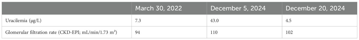
Table 1. Uracilemia values.
Patient perspective
The patient died on January 26, 2025, following cardiorespiratory arrest due to haemorrhagic shock linked to satellite rectal bleeding from his metastatic rectal tumour. The patient’s perspectives were therefore not collected.
Discussion
DPD activity should be evaluated before any fluoropyrimidine administration. This can be achieved through DPYD genotyping, which enables the early detection of genetic variants that predispose patients to severe toxicity. However, genotype testing may miss rare or unknown mutations in the DPYD gene and does not fully predict the severity of toxicity, as other genetic or environmental factors can influence the clinical outcome. Although unsuitable for daily care, the gold standard approach consists of measuring DPD activity in peripheral blood mononuclear cells (PBMCs) (11). Phenotyping by uracil concentration measurement is the method approved by the French health authorities [Institut du Cancer/Haute Autorité de Santé (INCa/HAS)]. Uracilemia is sensitive to several factors, the most important of which are pre-analytical and analytical issues (9), sampling while patients are being treated with fluoropyrimidines (10), and renal insufficiency (12, 13). All of these factors were either controlled or within the range in this patient. Other less significant sources of variability are tumour lysis syndrome (14), a high-meat diet (8), and the nycthemeral cycle (9). They were not all controlled in our case, but they have a limited impact. Thus, none of these sources of variability could explain the differences in uracil measurements observed in our patient.
Trifluridine/tipiracil is a well-known and useful option for patients with DPD deficiency since the main active compound (trifluridine) is a fluorinated pyrimidine that, like 5-FU, disrupts DNA. However, unlike 5-FU, trifluridine is not metabolised by DPD. Nevertheless, a small amount of trifluridine is metabolised into 5-carboxyuracil (5-CU) (15). 5-CU can be decarboxylated by iso-orotate decarboxylase (an enzyme of the thymidine salvage pathway) to form uracil, which could possibly explain the increase in uracil levels during treatment with trifluridine/tipiracil (16). Although 5-CU levels in plasma are low, it appears to be sufficient to increase uracilemia at least up to almost 35–40 µg/L. Unfortunately, no 5-CU concentrations were available in this case.
Thus, despite the lack of risk of using trifluridine/tipiracil in DPD-deficient patients, clinicians need to be aware of the potential interaction between the trifluridine metabolite and DPD phenotyping, as it may falsely give a deficient result if the patient is treated with trifluridine/tipiracil at the time of uracil measurement. Therefore, if DPD phenotyping is to be performed, it should always be performed at the time of mCRC diagnosis or at least at an extended period after any intake of fluoropyrimidine or trifluridine/tipiracil. Genotyping of DPYD could alternatively be preferred in such situations.
Data availability statement
The original contributions presented in the study are included in the article/supplementary material. Further inquiries can be directed to the corresponding author.
Ethics statement
Ethical approval was not required for the studies involving humans because the manuscript is a case report presenting routine collected data. Only written informed consent for publication was gathered. The studies were conducted in accordance with the local legislation and institutional requirements. The participants provided their written informed consent to participate in this study. Written informed consent was obtained from the individual(s) next of kin for the publication of any potentially identifiable images or data included in this article.
Author contributions
AS: Conceptualization, Formal analysis, Supervision, Writing – original draft, Writing – review & editing. BB: Data curation, Investigation, Writing – original draft, Writing – review & editing. BR: Data curation, Investigation, Writing – original draft, Writing – review & editing. FG: Data curation, Investigation, Writing – original draft, Writing – review & editing.
Funding
The author(s) declare that no financial support was received for the research, and/or publication of this article.
Conflict of interest
The authors declare that the research was conducted in the absence of any commercial or financial relationships that could be construed as a potential conflict of interest.
The author(s) declared that they were an editorial board member of Frontiers, at the time of submission. This had no impact on the peer review process and the final decision.
Generative AI statement
The author(s) declare that no Generative AI was used in the creation of this manuscript.
Publisher’s note
All claims expressed in this article are solely those of the authors and do not necessarily represent those of their affiliated organizations, or those of the publisher, the editors and the reviewers. Any product that may be evaluated in this article, or claim that may be made by its manufacturer, is not guaranteed or endorsed by the publisher.
References
1. Cervantes A, Adam R, Roselló S, Arnold D, Normanno N, Taïeb J, et al. Metastatic colorectal cancer: ESMO Clinical Practice Guideline for diagnosis, treatment and follow-up. Ann Oncol. (2023) 34:10–32. doi: 10.1016/j.annonc.2022.10.003
2. Van Cutsem E, Cervantes A, Adam R, Sobrero A, Van Krieken JH, Aderka D, et al. ESMO consensus guidelines for the management of patients with metastatic colorectal cancer. Ann Oncol Off J Eur Soc Med Oncol. (2016) 27:1386–422. doi: 10.1093/annonc/mdw235
3. Mayer RJ, Van Cutsem E, Falcone A, Yoshino T, Garcia-Carbonero R, Mizunuma N, et al. Randomized trial of TAS-102 for refractory metastatic colorectal cancer. N Engl J Med. (2015) 372:1909–19. doi: 10.1056/NEJMoa1414325
4. Schouten JF, Willems J, Sanders SJWJ, Creemers GJ, and Deenen MJ. Standard-dose trifluridine/tipiracil as safe treatment alternative in metastatic colorectal cancer patients with DPD deficiency. Clin Colorectal Cancer. (2021) 20:359–63. doi: 10.1016/j.clcc.2021.09.004
5. Van Cutsem E, Danielewicz I, Saunders MP, Pfeiffer P, Argilés G, Borg C, et al. First-line trifluridine/tipiracil + bevacizumab in patients with unresectable metastatic colorectal cancer: final survival analysis in the TASCO1 study. Br J Cancer. (2022) 126:1548–54. doi: 10.1038/s41416-022-01737-2
6. André T, Falcone A, Shparyk Y, Moiseenko F, Polo-Marques E, Csöszi T, et al. Trifluridine–tipiracil plus bevacizumab versus capecitabine plus bevacizumab as first-line treatment for patients with metastatic colorectal cancer ineligible for intensive therapy (SOLSTICE): a randomised, open-label phase 3 study. Lancet Gastroenterol Hepatol. (2023) 8:133–44. doi: 10.1016/S2468-1253(22)00334-X
7. Jiang H, Lu J, and Ji J. Circadian rhythm of dihydrouracil/uracil ratios in biological fluids: a potential biomarker for dihydropyrimidine dehydrogenase levels. Br J Pharmacol. (2004) 141:616–23. doi: 10.1038/sj.bjp.0705651
8. Henricks LM, Jacobs BAW, Meulendijks D, Pluim D, van den Broek D, de Vries N, et al. Food-effect study on uracil and dihydrouracil plasma levels as marker for dihydropyrimidine dehydrogenase activity in human volunteers. Br J Clin Pharmacol. (2018) 84:2761–9. doi: 10.1111/bcp.13719
9. Maillard M, Launay M, Royer B, Guitton J, Gautier-Veyret E, Broutin S, et al. Quantitative impact of pre-analytical process on plasma uracil when testing for dihydropyrimidine dehydrogenase deficiency. Br J Clin Pharmacol. (2023) 89(2):762–72. doi: 10.1111/bcp.15536
10. Thomas F, Maillard M, Launay M, Tron C, Etienne-Grimaldi MC, Gautier-Veyret E, et al. Artificial increase of uracilemia during fluoropyrimidine treatment can lead to DPD deficiency misinterpretation. Ann Oncol Off J Eur Soc Med Oncol. (2021) 32:810–1. doi: 10.1016/j.annonc.2021.02.020
11. Hodroj K, Barthelemy D, Lega JC, Grenet G, Gagnieu MC, Walter T, et al. Issues and limitations of available biomarkers for fluoropyrimidine-based chemotherapy toxicity, a narrative review of the literature. ESMO Open. (2021) 6(3):100125. doi: 10.1016/j.esmoop.2021.100125
12. Royer B, Launay M, Ciccolini J, Derain L, Parant F, Thomas F, et al. Impact of renal impairment on dihydropyrimidine dehydrogenase (DPD) phenotyping. ESMO Open. (2023) 8:101577. doi: 10.1016/j.esmoop.2023.101577
13. Callon S, Brugel M, Botsen D, Royer B, Slimano F, Feliu C, et al. Renal impairment and abnormal liver function tests in pre-therapeutic phenotype-based DPD deficiency screening using uracilemia: a comprehensive population-based study in 1138 patients. Ther Adv Med Oncol. (2023) 15:17588359221148536. doi: 10.1177/17588359221148536
14. Launay M, Guitton J, Balluet R, Moreau A, Phelip JM, Tholance Y, et al. Clinical considerations for DPD deficiency testing in advanced cancer patients: tumor lysis syndrome should be considered as a major interference. Ann Oncol Off J Eur Soc Med Oncol. (2022) 33:850–2. doi: 10.1016/j.annonc.2022.04.073
15. Temmink OH, Emura T, De Bruin M, Fukushima M, and Peters GJ. Therapeutic potential of the dual-targeted TAS-102 formulation in the treatment of gastrointestinal Malignancies. Cancer Sci. (2007) 98:779–89. doi: 10.1111/j.1349-7006.2007.00477.x
Keywords: trifluridine, tipiracil, DPD deficiency, metastatic colorectal cancer, case report, uracilemia
Citation: Schmitt A, Bouillet B, Royer B and Ghinringhelli F (2025) Trifluridine- and tipiracil-induced DPD inhibition mimicking DPD deficiency: a case report. Front. Oncol. 15:1591120. doi: 10.3389/fonc.2025.1591120
Received: 10 March 2025; Accepted: 21 July 2025;
Published: 12 August 2025.
Edited by:
Khaled Abdelkawy Ibrahim, Kafrelsheikh University, EgyptReviewed by:
Barathan Muttiah, University of Malaya, MalaysiaFrancisco Abad-Santos, Universidad Autónoma de Madrid, Spain
Copyright © 2025 Schmitt, Bouillet, Royer and Ghinringhelli. This is an open-access article distributed under the terms of the Creative Commons Attribution License (CC BY). The use, distribution or reproduction in other forums is permitted, provided the original author(s) and the copyright owner(s) are credited and that the original publication in this journal is cited, in accordance with accepted academic practice. No use, distribution or reproduction is permitted which does not comply with these terms.
*Correspondence: Antonin Schmitt, YXNjaG1pdHRAY2dmbC5mcg==