 Dongpo Zhang1Jun Li1Tao Sun2Ling Zhang3Lian Wang1Quan Gan1
 Xiaoxiao Xing1Yong Zhang1Yue Wang1
 Daixiang Liao1*
 Junyi Li1*
- 1Department of Surgery, Guang’anmen Hospital, China Academy of Chinese Medical Sciences, Beijing, China
- 2Department of Anorectal Surgery, Guang’anmen Hospital (Baoding), China Academy of Chinese Medical Sciences, Beijing, China
- 3Department of Pathology, Guang’anmen Hospital, China Academy of Chinese Medical Sciences, Beijing, China
Background: Prostate cancer, the most prevalent male malignancy in Western countries, seldom presents as secondary rectal linitis plastica (RLP).
Case presentation: We present an 82-year-old man with a 6-month history of altered bowel habits, narrowed stools, and mucous discharge, with absent lower urinary tract symptoms. Serum Prostate Specific Antigen (PSA) was markedly elevated (392 ng/mL). Imaging demonstrated circumferential rectal thickening and a prostatic mass invading the bladder. MRI revealed a “target sign” with associated diffusion restriction. Colonoscopy identified circumferential mucosal protrusions resembling grape-like clusters (Nice Band Imaging (NBI) International Colorectal Endoscopic (NICE) type 3). Deep biopsies confirmed prostatic adenocarcinoma (Gleason score 4 + 3 = 7).
Diagnosis: A multidisciplinary team confirmed the diagnosis of prostate cancer with secondary RLP.
Treatment: Combination therapy (prophylactic colostomy, leuprorelin, and abiraterone) reduced PSA from 392 to 2.16 ng/mL within 8 months.
Conclusions: RLP may mimic various gastrointestinal disorders clinically. Clinicians should consider RLP in elderly men presenting with gastrointestinal symptoms. Definitive diagnosis requires the integration of multi-modality imaging, endoscopy, and histopathological biopsy.
Highlights
● RLP may mimic different diseases in presentation.
● Clinicians should suspect RLP in elderly men with gastrointestinal symptoms. Collaboration across specialties may avoid misdiagnosis.
● The combination of different imaging modalities, endoscopy, and tissue biopsy leads to definitive diagnosis.
Background
Prostate cancer is the most prevalent malignancy and the second leading cause of cancer-related mortality in Western men (1). Bone metastases constitute the predominant metastatic pattern, followed by lymphatic, pulmonary, and hepatic involvement. Clinically, prostate cancer primarily presents with lower urinary tract symptoms: 1) irritative symptoms (urinary frequency, urgency, nocturia, and urge incontinence), 2) obstructive symptoms (hesitancy, weak stream, intermittency, and retention), and 3) local invasion symptoms (testicular pain, painful ejaculation, hematuria, renal insufficiency, and hematospermia). Denonvilliers’ fascia forms the primary anatomical barrier between the rectum and prostate, critically containing tumor spread. The disruption of this fascial layer enables direct prostate cancer invasion into the rectum (2).
Secondary rectal linitis plastica (RLP) represents a rare metastatic manifestation. Its pathogenesis involves direct tumor extension or lymphatic dissemination from prostate cancer to the rectal wall. Conventional rectal invasion by prostate cancer typically demonstrates localized anterior wall involvement on imaging (3), whereas RLP features diffuse circumferential fibrosis and mural thickening throughout the rectum. Histopathological examination reveals diffuse tumor cell infiltration through the submucosal and muscularis propria layers, with prominent desmoplastic stromal reaction (4). Clinically, RLP manifests predominantly with gastrointestinal symptoms including defecatory dysfunction (80%), rectal pain (60%), and weight loss (40%), mimicking primary rectal adenocarcinoma. This report illustrates a rare case of prostate cancer-induced secondary rectal linitis plastica.
Case presentation
An 82-year-old man presented to our department with a 6-month history of altered bowel habits. Figure 1 illustrates the diagnostic and therapeutic timeline. Six months prior, he developed narrowed stools, increased defecation frequency (6–10 daily), defecation-related abdominal pain, persistent post-evacuation urgency, and mucoid stools. Notably, he denied hematochezia, dysuria, night sweats, or weight loss. Initial tumor markers revealed the following: total PSA 392 ng/mL (ref: <4), free PSA 49.7 ng/mL, and free prostate-specific antigen / total prostate-specific antigen (fPSA/tPSA) ratio of 0.13. Contrast-enhanced abdominal computed tomography (CT) demonstrated the following: circumferential wall thickening (max 2.0 cm) at 4 cm from the anal verge and serosal penetration with ill-defined prostate borders in the rectum, enlargement (38 × 53 mm) with extracapsular bladder invasion and seminal vesicle angle obliteration in the prostate, and complication of bilateral hydroureteronephrosis (left, 2.1 cm; right, 1.8 cm). In August 2024, the patient was referred to our surgical department.
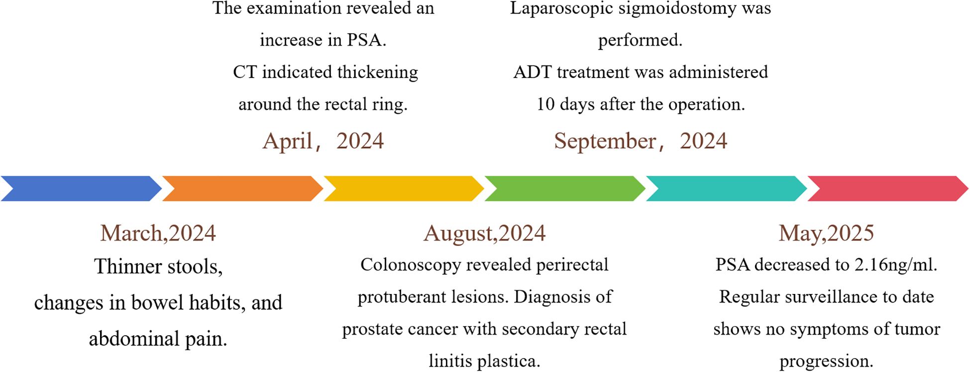
Figure 1. Clinical timeline: symptom onset, diagnosis, and treatment.
Physical examination
Digital rectal examination revealed a fixed stenotic mass at 5 cm from the anal verge, preventing further advancement. The prostate was markedly enlarged and indurated, and blood was noted on digital rectal examination.
Auxiliary examinations
Subsequent contrast-enhanced pelvic MRI revealed a rectal tumor with infiltrative carcinoma and concentric full-thickness rectal wall involvement. Posterior extension demonstrated circumferential encasement of the rectum exhibiting “target sign” (Figure 2). Colonoscopy revealed a circumferential elevated lesion spanning 4–7 cm from the anal verge in the rectum (Figure 3). The lesion exhibited circumferential mucosal protrusions resembling grape-like clusters. Narrow-band imaging chromoendoscopy revealed a NICE type 3 pattern, indicating deep submucosal invasion. The endoscope could not be fully advanced through the stenotic segment (Figure 3). Through colonoscopic biopsy pathology, histopathological examination of colonoscopic biopsies revealed infiltrative atypical glands extending beyond the muscularis mucosae, with prominent nucleoli. Immunohistochemical analysis demonstrated positive PSA, positive prostate-specific membrane antigen (PSMA), negative Carcinoembryonic Antigen (CEA), and a Ki-67 labeling index of 10%–20%. The findings were consistent with metastatic prostatic acinar adenocarcinoma (Gleason score 4 + 3 = 7) (Figure 4).
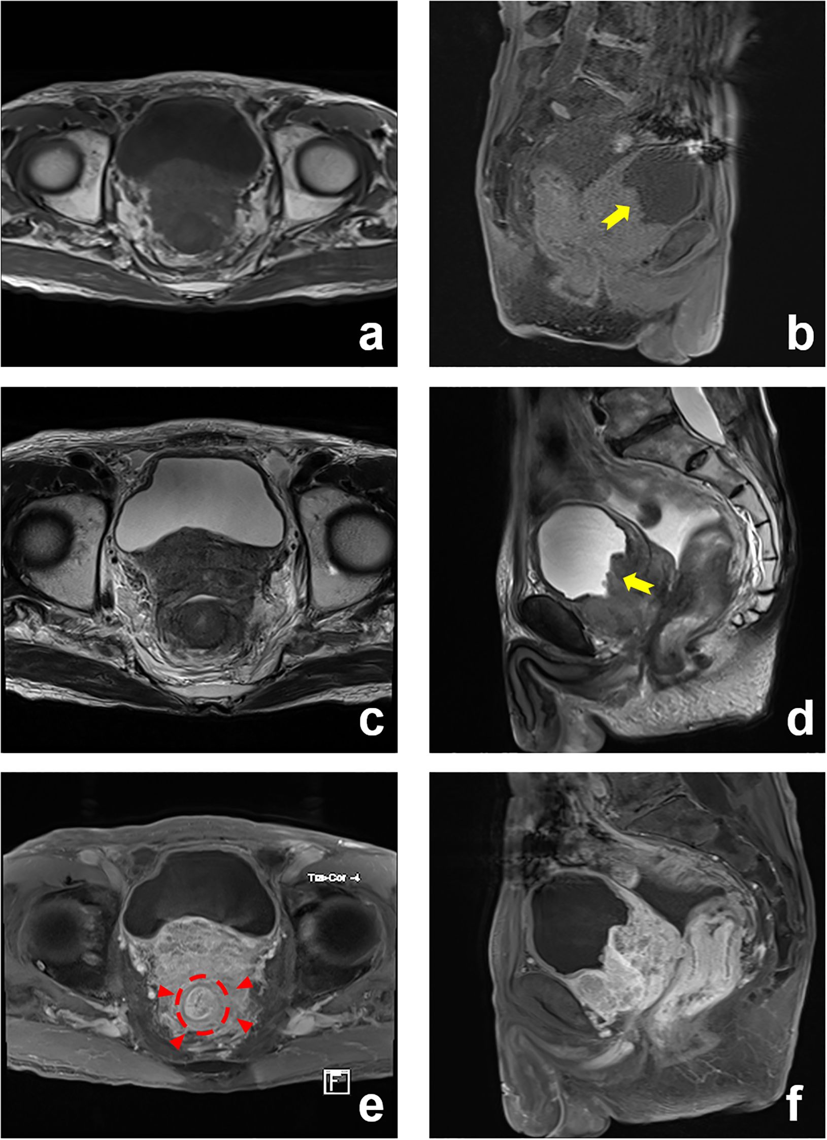
Figure 2. Prostate MRI: enlarged prostate with transition zone enlargement. Posterior extension: encasement of the rectum (“target sign”) (red arrowed). Anterior extension: invasion of the bladder (yellow arrowed). (a) T1-Weighted (T1W), axial; (b) T1-Weighted (T1W), sagittal; (c) T2-Weighted (T2W), axial; (d) T2-Weighted (T2W), sagittal; (e) T1-Weighted (T1W) C+, axial; (f) T1-Weighted (T1W) C+, sagittal.
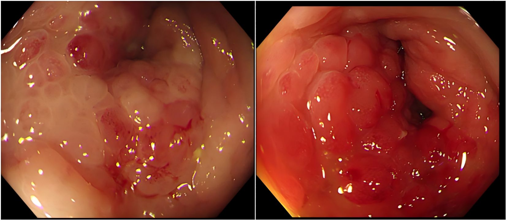
Figure 3. Colonoscopic findings. Colonoscopy demonstrated rectal stenosis with circumferential mucosal protrusions resembling grape-like clusters, characterized by friability and contact bleeding.
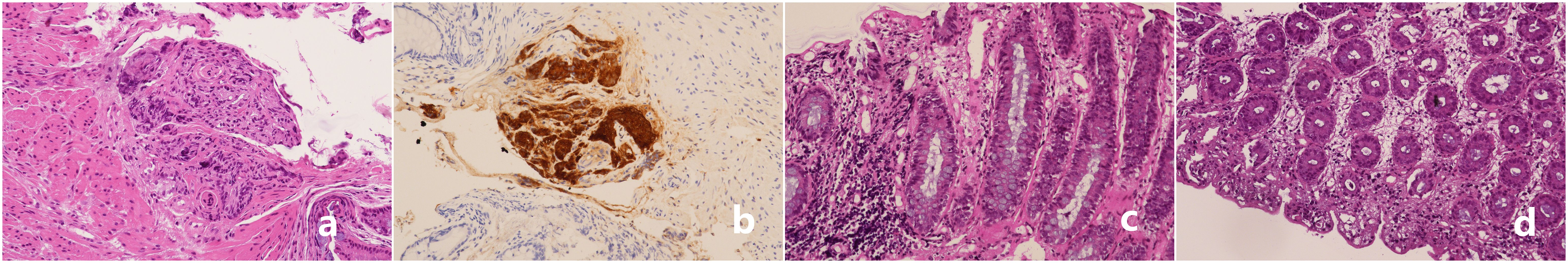
Figure 4. Histopathological findings. (a, b) Tumor cells infiltrating the muscularis mucosae and submucosa, displaying small acinar structures with crowded, disorganized glands and dysplastic acini, consistent with prostatic acinar adenocarcinoma (Gleason score 4 + 3 = 7) (H&E, ×200). (c, d) Adjacent mucosa shows superficial erosion, stromal edema, fibrous hyperplasia, and dense lymphocytic infiltration (H&E, ×100).
Diagnosis
Based on elevated PSA levels, imaging features, and colonoscopic biopsy histopathology, a multidisciplinary team (MDT) comprising specialists in urology, surgical oncology, pathology, and radiology convened for diagnostic assessment. The patient was definitively diagnosed with prostate cancer accompanied by secondary RLP.
Treatment and prognosis
Given locally advanced disease with bladder invasion and rectal lymphatic permeation, definitive surgical management necessitated total pelvic exenteration. The 72-year-old male patient presented with a low body mass index (BMI 16.2 kg/m2) and severe malnutrition. His nutritional status was poor. The patient’s current frail physical condition rendered him unable to tolerate such a major procedure. Consequently, emergency management focused on relieving malignant bowel obstruction. Definitive treatment (either radical surgery or localized radiotherapy) was deferred until nutritional status improved. The MDT consensus recommended diverting sigmoid colostomy for palliation of obstruction. According to the clinical practice guidelines for prostate cancer (5), androgen deprivation therapy (ADT) with leuprorelin and abiraterone was administered to the patient. Laparoscopic sigmoid loop colostomy was performed in September 2024. ADT commenced on postoperative day 10.
Follow-up
Serial PSA monitoring demonstrated progressive decline: 164 (1 month), 53 (3 months), 5.89 (6 months), and 2.16 ng/mL (8 months) post-ADT. The patient elected to forgo surveillance imaging, such as bone scintigraphy and PSMA-positron emission tomography (PET)/CT. The initial ADT regimen (leuprorelin with abiraterone acetate) was maintained. Ongoing surveillance revealed no clinical evidence of disease progression.
Discussion
This case exhibited exclusively gastrointestinal manifestations: malignant bowel obstruction, decreased stool caliber, increased bowel movement frequency, and incomplete defecation, with absent lower urinary tract symptoms. Putative metastatic pathways to the rectum included the following: a) direct invasion through Denonvilliers’ fascia, b) lymphatic permeation, c) retrograde venous spread, and d) iatrogenic implantation post-transrectal biopsy (4). Tumor infiltration induced desmoplastic stromal reaction in the rectal submucosa, causing muscularis propria rigidity and contracture that culminated in luminal stenosis. MRI demonstrated circumferential encasement of the rectum by prostatic cancer, with tumor extension posteriorly involving Denonvilliers’ fascia. This generated concentric layered thickening of the rectal wall, classically termed the “target sign” on imaging (6).
We reviewed similar reported cases in the literature, as shown in Table 1 (3, 4, 6–8). This comparative analysis of seven published cases revealed that RLP secondary to prostate cancer occurs exclusively in elderly men (ages 57–86), presenting primarily with bowel dysfunction (constipation, incontinence, pain, and bleeding) or weight loss, although one case was asymptomatic. Crucially, half of the cases had no prior cancer diagnosis. Diagnosis relied on MRI, PSMA-PET, and histology. ADT was the main reported treatment. Outcomes varied considerably, ranging from symptomatic improvement to death or progression.
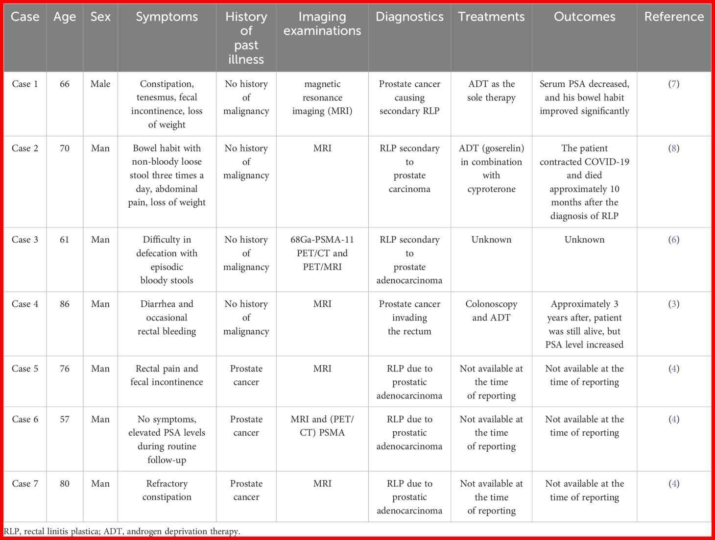
Table 1. Clinical features, diagnostics, treatments, and outcomes of similar cases.
Our case of secondary RLP aligns with the core demographic (elderly man) and clinical presentation (bowel dysfunction) seen in published cases. It underscores the critical diagnostic challenges of frequent absence of prior cancer history (like Cases 1–4) and the pitfall of normal mucosa or superficial biopsies necessitating deep sampling. Radiologically, it confirms the centrality of MRI and adds emphasis on the suggestive “target sign” and Diffusion-Weighted Imaging (DWI) restriction. The unique “grape-like” endoscopic morphology provides a valuable descriptive feature. Therapeutically, it demonstrates a robust early response to a contemporary, intensified hormonal regimen (ADT + abiraterone) and combined with proactive surgical management (colostomy) for obstruction. While sharing the underlying pathology, our case enhances the literature by detailing specific diagnostic features (imaging signs, endoscopic morphology, and biopsy strategy), reporting on a novel treatment combination, and documenting a significant early biochemical response.
PET/CT imaging using isotope-labeled PSMA ligands is essential for diagnosis and prognosis in prostate cancer patients. PSMA-PET/CT is superior to conventional imaging (MRI, CT, and bone scan) in primary staging, mainly in the detection of pelvic lymphadenopathy and distant metastases. PSMA-PET/MRI is an emerging modality that combines metabolic information on PSMA receptor expression in prostate tumors derived from PET, with anatomical and functional information derived from magnetic resonance (MR) in one procedure. PSMA-PET/MR is accurate and reliable in the depiction of nodal and osseous metastases compared with PSMA-PET/CT (9, 10). Unfortunately, the patient elected to forgo surveillance imaging.
RLP poses a challenge by closely mimicking primary rectal adenocarcinoma both clinically (rectal bleeding/mass) and endoscopically. However, the biopsy revealed adenocarcinoma with morphology suggestive of prostatic origin. Immunohistochemistry confirmed prostatic lineage (positive for PSA). The Immunohistochemistry (IHC) profile was definitive in distinguishing metastatic prostate cancer from primary rectal cancer. Based on the elevated PSA, radiological findings, and colonoscopy biopsy pathology, a multi-disciplinary team involving specialists from general surgery, urology, oncology, pathology, and radiology held a discussion. The patient was definitively diagnosed with prostate cancer accompanied by secondary RLP.
Diagnosing prostatic adenocarcinoma with rectal invasion poses significant challenges. RLP typically involves submucosal infiltration by prostate cancer cells, often leaving the overlying rectal mucosa intact or only superficially involved. In approximately 30% of RLP cases, the rectal mucosa appears endoscopically normal due to tumor confinement to the submucosal and muscularis propria, often resulting in false-negative initial biopsies and necessitating deep-tissue sampling for definitive diagnosis (8). Superficial biopsies typically demonstrate mucosal erosion, stromal edema, and reactive fibroinflammatory hyperplasia. Such findings frequently delay definitive diagnosis. Deep targeted biopsies sampling the lesion base or submucosal nodules (grape-cluster morphology) are critical for detecting underlying malignancy (11). Initial external institutional biopsies revealed reactive hyperplasia without malignant evidence. Repeat deep biopsies at our center specifically sampled submucosal and muscularis mucosa layers, confirming invasive prostatic adenocarcinoma.
Conclusion
Secondary RLP in prostate cancer is an aggressive and diagnostically elusive entity. Clinicians should maintain a high index of suspicion in elderly men with rectal wall thickening or suspected rectal tumors, particularly those with a history of prostate cancer. A multimodal diagnostic approach—integrating clinical history, serum PSA, advanced imaging (MRI), and deep tissue sampling—is critical to avoid misdiagnosis and ensure histopathological confirmation.
Limitations
This case report provides unique educational insights into the diagnostic challenges and management of prostate cancer metastasis to the rectum. As a case report, conclusions may not be generalizable, and more clinical studies are needed.
Data availability statement
The original contributions presented in the study are included in the article/Supplementary Material. Further inquiries can be directed to the corresponding author.
Ethics statement
The studies involving humans were approved by Ethics Committee of Guang’anmen Hospital, China Academy of Chinese Medical Sciences. The studies were conducted in accordance with the local legislation and institutional requirements. The human samples used in this study were acquired from a by- product of routine care or industry. Written informed consent for participation was not required from the participants or the participants’ legal guardians/next of kin in accordance with the national legislation and institutional requirements. Written informed consent was obtained from the individual(s) for the publication of any potentially identifiable images or data included in this article. Written informed consent was obtained from the participant/patient(s) for the publication of this case report.
Author contributions
JYL: Supervision, Writing – original draft, Writing – review & editing. DZ: Writing – original draft. TS: Writing – review & editing. LZ: Writing – original draft. LW: Supervision, Writing – original draft. QG: Writing – original draft, Supervision. XX: Writing – original draft. YZ: Conceptualization, Writing – original draft. YW: Supervision, Methodology, Writing – original draft. DL: Writing – review & editing, Writing – original draft, Formal Analysis. JL: Writing – review & editing.
Funding
The author(s) declare that financial support was received for the research and/or publication of this article. This study was funded by the Major research project of the Science and Technology Innovation Project of the Chinese Academy of Traditional Chinese Medicine, Grant/Award Number: CI2021A01903.
Conflict of interest
The authors declare that the research was conducted in the absence of any commercial or financial relationships that could be construed as a potential conflict of interest.
Generative AI statement
The author(s) declare that no Generative AI was used in the creation of this manuscript.
Publisher’s note
All claims expressed in this article are solely those of the authors and do not necessarily represent those of their affiliated organizations, or those of the publisher, the editors and the reviewers. Any product that may be evaluated in this article, or claim that may be made by its manufacturer, is not guaranteed or endorsed by the publisher.
References
1. Siegel RL, Miller KD, Fuchs HE, and Jemal A. Cancer statistics, 2022. CA Cancer J Clin. (2022) 72:7–33. doi: 10.3322/caac.21708
2. Lebret T and Méjean A. Les sites métastatiques atypiques des cancers de la prostate [Rare locations of metastases from prostate cancer. Prog Urol. (2008) 7:S357–64. doi: 10.1016/S1166-7087(08)74567-6
3. Hassine H, Azouz SB, Debbabi H, Cherif D, Yacoub H, Chelly B, et al. Prostate carcinoma mimicking rectal cancer: a case report. J Surg Case Rep. (2024) 2024:rjae046. doi: 10.1093/jscr/rjae046
4. Labra AA, Schiappacasse G, Cocio RA, Torres JT, González FO, Cristi JA, et al. Secondary rectal linitis plastica caused by prostatic adenocarcinoma - magnetic resonance imaging findings and dissemination pathways: A case report. World J Radiol. (2024) 16:473–81. doi: 10.4329/wjr.v16.i9.473
5. Schaeffer EM, Srinivas S, Adra N, An Y, Barocas D, Bitting R, et al. Prostate cancer, version 4.2023, NCCN clinical practice guidelines in oncology. J Natl Compr Canc Netw. (2023) 21:1067–96. doi: 10.6004/jnccn.2023.0050
6. Zhao Q, Dong H, Dong A, and Zuo C. 68 ga-PSMA-11 PET/CT and PET/MRI in rectal linitis plastica secondary to prostate adenocarcinoma. Clin Nucl Med. (2023) 48:282–5. doi: 10.1097/RLU.0000000000004476
7. Khor V, Khairul-Asri MG, Fahmy O, Hamid SA, and Lee CKS. Linitis plastica of the rectum secondary to metastatic prostate cancer: A case report of a rare presentation and literature review. Urol Ann. (2021) 13:442–5. doi: 10.4103/UA.UA_188_20
8. Mommersteeg MC, Kies DA, Laan Jvd, and Wonders J. Linitis plastica of the rectum secondary to prostate carcinoma. BMJ Case Rep. (2022) 15:e248462. doi: 10.1136/bcr-2021-248462
9. Hoffmann MA, Buchholz H-G, Wieler HJ, Rosar F, Miederer M, Fischer N, et al. Dual-time point [68 Ga]Ga-PSMA-11 PET/CT hybrid imaging for staging and restaging of prostate cancer. Cancers (Basel). (2020) 12:2788. doi: 10.3390/cancers12102788
10. Domachevsky L, Bernstine H, Goldberg N, Nidam M, Catalano OA, and Groshar D. Comparison between pelvic PSMA-PET/MR and whole-body PSMA-PET/CT for the initial evaluation of prostate cancer: a proof of concept study. Eur Radiol. (2020) 30:328–36. doi: 10.1007/s00330-019-06353-y
Keywords: prostate cancer, secondary rectal linitis plastica, rectal adenocarcinoma, PSA, MRI
Citation: Zhang D, Li J, Sun T, Zhang L, Wang L, Gan Q, Xing X, Zhang Y, Wang Y, Liao D and Li J (2025) Prostate cancer inducing secondary linitis plastica of the rectum: a rare case report and literature review. Front. Oncol. 15:1597367. doi: 10.3389/fonc.2025.1597367
Received: 21 March 2025; Accepted: 26 June 2025;
Published: 23 July 2025.
Edited by:
Feifei Sun, Shandong University, ChinaReviewed by:
Lin Zhang, Binzhou Center for Disease Control and Prevention, ChinaJunmei Liu, University of Duisburg-Essen, Germany
Copyright © 2025 Zhang, Li, Sun, Zhang, Wang, Gan, Xing, Zhang, Wang, Liao and Li. This is an open-access article distributed under the terms of the Creative Commons Attribution License (CC BY). The use, distribution or reproduction in other forums is permitted, provided the original author(s) and the copyright owner(s) are credited and that the original publication in this journal is cited, in accordance with accepted academic practice. No use, distribution or reproduction is permitted which does not comply with these terms.
*Correspondence: Daixiang Liao, MTY4MDIwMDJAMTYzLmNvbQ==; Junyi Li, YnVjbWxqeUAxNjMuY29t