 Liujing Qu1
 Yu Xin2Jieni Feng3Xiaolei Ren3
 Zuming Li3
 Xueru Chen3
 Guangyan Miao4
 Jiankun Chen5*
 Chengming Sun1*
 Yue Lu6,7*
- 1Department of Clinical Laboratory, The Affiliated Yantai Yuhuangding Hospital of Qingdao University, Yantai, China
- 2Department of Medical Laboratory, Qingdao Sixth People’s Hospital, Qingdao, China
- 3The Second Clinical Medical College, Guangzhou University of Chinese Medicine, Guangzhou, China
- 4Department of Molecular, Cell and Cancer Biology, University of Massachusetts Chan Medical School, Worcester, MA, United States
- 5The Third Comprehensive Department, The Second Affiliated Hospital of Guangzhou University of Chinese Medicine (Guangdong Provincial Hospital of Chinese Medicine), Guangzhou, China
- 6State Key Laboratory of Dampness Syndrome of Chinese Medicine, The Second Affiliated Hospital of Guangzhou University of Chinese Medicine (Guangdong Provincial Hospital of Chinese Medicine), Guangzhou, China
- 7Guangdong-Hong Kong-Macau Joint Lab on Chinese Medicine and Immune Disease Research, Guangzhou University of Chinese Medicine, Guangzhou, China
A Correction on
Downregulation of PRKCI inhibits osteosarcoma cell growth by inactivating the Akt/mTOR signaling pathway
By Qu L, Xin Y, Feng J, Ren X, Li Z, Chen X, Miao G, Chen J, Sun C and Lu Y (2024). Front. Oncol. 14:1389136. doi: 10.3389/fonc.2024.1389136
In the published article, there was an error in Figure 5A and Figure 6A as published. During an AI-assisted image integrity review of our published work, we identified inadvertent duplications in Figure 5A and Figure 6A (Migration, PRKCI and shcontrol; Wound healing assay, 0h shcontrol and shPRKCI, 24h Vector and shPRKCI). This was an oversight during figure preparation and does not affect the study’s conclusions. We have since located the original data and prepared a corrected version of the figure. The corrected Figure 5A and Figure 6A and its caption appear below.
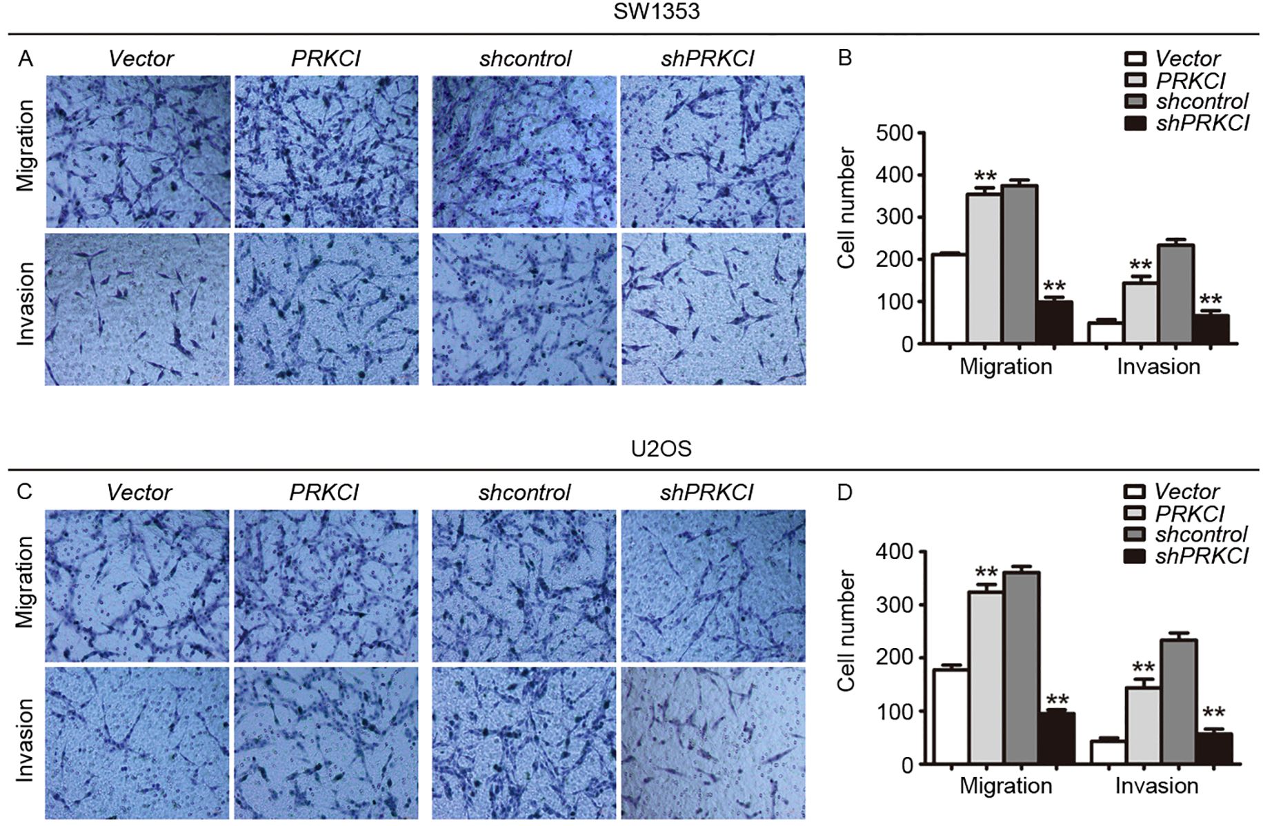
Figure 5. The Transwell system was used to evaluate the effect of PRKCI on the migration and invasion of osteosarcoma cells. (A, C) Transwell migration assay and Matrigel invasion assay for SW1353 cells or U2OS cells after transfection empty vector or PRKCI plasmid for 24 h (shcontrol or shPRKCI for 48 h). Cells were stained with crystal violet (magnification: ×200). (B, D) Quantification of invaded and migrated SW1353 cells or U2OS cells. (Data were based on three independent experiments and shown as the mean ± SEM, **p < 0.01).
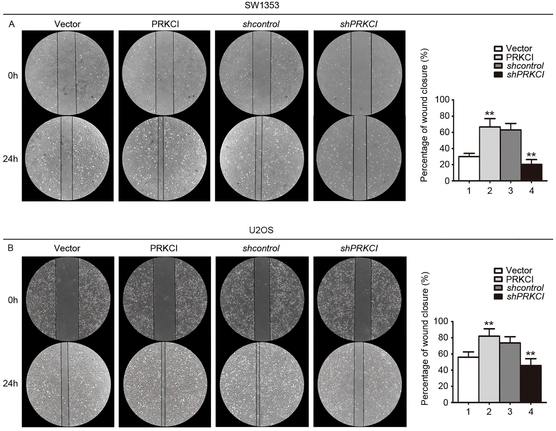
Figure 6. A wound-healing assay was used to evaluate the effect of PRKCI on the migration of osteosarcoma cells. (A, B) Microscopic images of wound-healing assay data for SW1353 and U2OS cells transfected with empty vector or PRKCI plasmid for 24 h (shcontrol or shPRKCI for 48 h). (Data were based on three independent experiments and shown as the mean ± SEM, **p < 0.01).
The authors apologize for this error and state that this does not change the scientific conclusions of the article in any way. The original article has been updated.
Publisher’s note
All claims expressed in this article are solely those of the authors and do not necessarily represent those of their affiliated organizations, or those of the publisher, the editors and the reviewers. Any product that may be evaluated in this article, or claim that may be made by its manufacturer, is not guaranteed or endorsed by the publisher.
Keywords: PRKCI, osteosarcoma, proliferation, Akt/mTOR signaling pathway, therapy
Citation: Qu L, Xin Y, Feng J, Ren X, Li Z, Chen X, Miao G, Chen J, Sun C and Lu Y (2025) Correction: Downregulation of PRKCI inhibits osteosarcoma cell growth by inactivating the Akt/mTOR signaling pathway. Front. Oncol. 15:1643772. doi: 10.3389/fonc.2025.1643772
Received: 09 June 2025; Accepted: 23 June 2025;
Published: 07 July 2025.
Edited and Reviewed by:
Tinghe Yu, Chongqing Medical University, ChinaCopyright © 2025 Qu, Xin, Feng, Ren, Li, Chen, Miao, Chen, Sun and Lu. This is an open-access article distributed under the terms of the Creative Commons Attribution License (CC BY). The use, distribution or reproduction in other forums is permitted, provided the original author(s) and the copyright owner(s) are credited and that the original publication in this journal is cited, in accordance with accepted academic practice. No use, distribution or reproduction is permitted which does not comply with these terms.
*Correspondence: Jiankun Chen, Y2hlbmppYW5rdW5kb2N0b3JAMTI2LmNvbQ==; Chengming Sun, MTg5NTM1Njk4OTdAMTYzLmNvbQ==; Yue Lu, Z3p5bHV5dWVAMTI2LmNvbQ==