 Esteban Zavaleta-Monestel1*
 Sebastián Arguedas-Chacón1
 Jeaustin Mora-Jiménez2
 Kevin Cruz-Mora2
 Abraham Rocha-Romero3
- 1Health Research Department, Clínica Bíblica, San José, Costa Rica
- 2Pharmacy Department, Clínica Bíblica, San José, Costa Rica
- 3Facultad de Farmacia, Universidad de Costa Rica, San José, Costa Rica
Introduction
The global incidence of lung cancer varies depending on the source and year of estimation. However, recent and widely recognized data estimate that in 2022, approximately 2.5 million new cases of lung cancer were diagnosed worldwide, accounting for 12.4% of all cancer diagnoses. Moreover, it was responsible for about 18.7% of cancer-related deaths, making it the leading cause of cancer mortality globally. When excluding non-melanoma skin cancer and melanoma, lung cancer also ranked first in global incidence for that year (1, 2).
There are multiple subtypes of lung cancer; nevertheless, they all share a common characteristic, the rapid progression from early to advanced stages. Therefore, timely diagnosis is essential to initiate treatment as soon as possible and prevent disease advancement (3). In this context, chest imaging plays a crucial role by allowing the early detection of pulmonary nodules. Chest X-ray (CXR) is the primary imaging tool for detecting, assessing, and monitoring cancers such as lung cancer (4).
Although recent studies show that CXR for detecting lung cancer in the general population and symptomatic patients has a sensitivity ranging from 60% to 76%, its specificity is high at 94–95%. This means that about a quarter of early-stage lung cancer cases go undetected, possibly delaying diagnosis and allowing disease progression. Additionally, a negative diagnosis does not reliably exclude the presence of lung cancer (5).
These limitations have led to the development of computer-aided detection (CAD) systems based on artificial intelligence (AI) to enhance the automated analysis of CXR. Among these solutions is ChestView, developed by the company Gleamer, which employs deep learning algorithms trained with millions of clinical images. This tool does not aim to replace radiologists but rather to act as a diagnostic copilot, offering a second automated reading that facilitates early detection of findings suggestive of lung cancer (6).
Features of Gleamer’s ChestView
ChestView, developed by the French company Gleamer, is a computer-aided detection (CAD) solution based on artificial intelligence specifically designed for chest X-ray analysis. This tool is CE certified under the European Medical Device Regulation (EU MDR, class IIa), endorsing its clinical use in hospital settings. ChestView uses a deep convolutional neural network (CNN) based on the Detectron2 architecture, which is an open-source framework created by Facebook AI Research for object detection in medical images. The algorithm is trained to identify five common thoracic abnormalities: pneumothorax, pleural effusion, consolidation, mediastinal or hilar mass, and pulmonary nodule (6).
Current evidence of ChestView as an AI clinical support tool
Artificial intelligence has recently been implemented to support the detection of thoracic diseases in chest radiographs. Bennani et al. (2023) evaluated its utility through a retrospective study of over 500 patient X-rays, focusing on identifying anomalies such as pneumothorax, pleural effusion, consolidation, mediastinal and hilar masses, and pulmonary nodules (7).
Key findings included an average absolute increase of 12% in sensitivity for detecting pulmonary nodules and a significant reduction in average reading time of 81 seconds without AI to 56 seconds with AI (a 31% decrease; P < 0.001) (7).
Furthermore, for pulmonary consolidations, AI achieved an AUC of 0.93, significantly outperforming the average performance of radiologists without AI assistance (AUC 0.71). These results demonstrate that AI not only supports human readers but also has the potential to elevate diagnostic standards in clinical practice (7).
In another study, Chassagnon et al. (2023) included eight radiology residents to evaluate whether a computer-aided detection (CADe) system could serve as a learning tool for interpreting CXR. Key findings showed improved sensitivity (53% vs. 43%; p < 0.001), specificity (94% vs. 90%; p < 0.001), and accuracy (86% vs. 81%; p < 0.001) during the intervention (8).
The key outcomes of ChestView AI support are summarized in Figure 1, which illustrates improvements in sensitivity, specificity, diagnostic accuracy, and interpretation time.
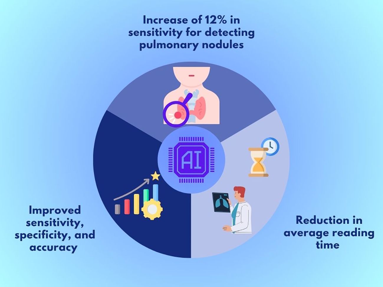
Figure 1. Key outcomes of ChestView AI support in chest X-rays.
Barriers and considerations for clinical adoption
In addition to the methodological considerations mentioned, other factors hinder the safe and effective implementation of AI in daily clinical practice. One such factor is the variable quality of images used in both training and application of models. In real-world contexts, images may contain artifacts, suboptimal positioning, or technical differences that do not present in ideal datasets, negatively affecting the performance of CADe systems (8).
A lack of heterogeneity in training data limits the generalizability of algorithms. Many models have been developed using databases from specific centers, preventing results from being extrapolated to more diverse clinical populations or different technical environments. This lack of representativeness may introduce bias and reduce the applicability of the tool in routine settings (8).
Another consideration is the computational and operational demands associated with implementing these systems in clinical practice. While model training is performed externally by the developer, their clinical use still requires adequate technological infrastructure, which may represent a barrier, particularly in resource-limited institutions (8).
Lastly, although regulatory pathways such as FDA clearance in the US and the MDR in the EU exist, the absence of harmonized and internationally standardized frameworks tailored for AI in radiology remains a critical limitation. This gap creates legal and ethical uncertainty regarding adaptive algorithms, post-market validation, and professional responsibility, complicating their formal integration as diagnostic or educational support tools (8).
Discussion
Gleamer’s ChestView represents a scientifically tangible example of the positive impact AI tools can have on early lung cancer detection using chest radiographs. Current evidence suggests that its use may positively influence diagnostic sensitivity, improve image interpretation accuracy, and optimize diagnostic timing. This not only represents a direct clinical benefit but also an opportunity to align and leverage technological advances for patient health (9).
However, its effective implementation in clinical practice still faces significant limitations, such as the need for advanced technological infrastructure, operational costs, and the lack of standardized regulations to govern its use. These aspects must be considered to avoid diagnostic biases, ensure patient safety, and maximize the added value of these technologies (10).
In conclusion, ChestView has the potential to become a reliable diagnostic tool for medical personnel, especially in clinical contexts where time for accurate interpretation is limited. Its successful future integration will depend on ongoing clinical studies, multicenter validation, and the development of regulatory standards supporting its safe, ethical, and effective use.
Author contributions
EZ-M: Investigation, Supervision, Validation, Writing – original draft, Writing – review & editing. SA-C: Supervision, Validation, Writing – original draft, Writing – review & editing. JM-J: Conceptualization, Investigation, Methodology, Supervision, Validation, Visualization, Writing – original draft, Writing – review & editing. KC-M: Investigation, Methodology, Supervision, Validation, Visualization, Writing – original draft, Writing – review & editing. AR-R: Investigation, Methodology, Visualization, Writing – original draft, Writing – review & editing.
Funding
The author(s) declare that no financial support was received for the research, and/or publication of this article.
Conflict of interest
The authors declare that the research was conducted in the absence of any commercial or financial relationships that could be construed as a potential conflict of interest.
Generative AI statement
The author(s) declare that Generative AI was used in the creation of this manuscript. Generative AI (ChatGPT by OpenAI) was used to assist with language refinement, structural editing, formulation of the abstract, scope statement, and cover letter. The authors reviewed, edited, and take full responsibility for all content.
Any alternative text (alt text) provided alongside figures in this article has been generated by Frontiers with the support of artificial intelligence and reasonable efforts have been made to ensure accuracy, including review by the authors wherever possible. If you identify any issues, please contact us.
Publisher’s note
All claims expressed in this article are solely those of the authors and do not necessarily represent those of their affiliated organizations, or those of the publisher, the editors and the reviewers. Any product that may be evaluated in this article, or claim that may be made by its manufacturer, is not guaranteed or endorsed by the publisher.
References
1. Cancer today. Available online at: https://gco.iarc.who.int/today/ (Accessed July 17, 2025).
2. Bray F, Laversanne M, Sung H, Ferlay J, Siegel RL, Soerjomataram I, et al. Global cancer statistics 2022: GLOBOCAN estimates of incidence and mortality worldwide for 36 cancers in 185 countries. CA Cancer J Clin. (2024) 74:229–63. doi: 10.3322/caac.21834
3. Yuan P, Cao JL, Rustam A, Zhang C, Yuan XS, Bao FC, et al. Time-to-progression of NSCLC from early to advanced stages: an analysis of data from SEER registry and a single institute. Sci Rep. (2016) 6:28477. doi: 10.1038/srep28477
4. Lind Plesner L, Müller FC, Brejnebøl MW, Laustrup LC, Rasmussen F, Nielsen OW, et al. Commercially available chest radiograph AI tools for detecting airspace disease, pneumothorax, and pleural effusion. Radiology. (2023) 308:e231236. doi: 10.1148/radiol.231236
5. A prospective cohort evaluation of the sensitivity and specificity of the chest X-ray for the detection of lung cancer in symptomatic adults - ClinicalKey. Available online at: https://www.clinicalkey.com/!/content/playContent/1-s2.0-S0720048X21004344?returnurl=https:%2F%2Flinkinghub.elsevier.com%2Fretrieve%2Fpii%2FS0720048X21004344%3Fshowall%3Dtrue&referrer=https:%2F%2Fpubmed.ncbi.nlm.nih.gov%2F (Accessed July 17, 2025).
6. ChestView - gleamer. Available online at: https://www.gleamer.ai/solutions/chestview (Accessed July 17, 2025).
7. Bennani S, Regnard NE, Ventre J, Lassalle L, Nguyen T, Ducarouge A, et al. Using AI to improve radiologist performance in detection of abnormalities on chest radiographs. Radiology. (2023) 309:e230860. doi: 10.1148/radiol.230860
8. Chassagnon G, Billet N, Rutten C, Toussaint T, Cassius de Linval Q, Collin M, et al. Learning from the machine: AI assistance is not an effective learning tool for resident education in chest x-ray interpretation. Eur Radiol. (2023) 33:8241–50. doi: 10.1007/s00330-023-10043-1
9. Nam JG, Hwang EJ, Kim J, Park N, Lee EH, Kim HJ, et al. AI improves nodule detection on chest radiographs in a health screening population: A randomized controlled trial. Radiology. (2023) 307(2):e221894. doi: 10.1148/radiol.221894
Keywords: chest X-ray, lung cancer detection, computer-aided detection, diagnostic accuracy, clinical implementation
Citation: Zavaleta-Monestel E, Arguedas-Chacón S, Mora-Jiménez J, Cruz-Mora K and Rocha-Romero A (2025) Opinion paper: artificial intelligence as a clinical ally in lung cancer detection—the case for Gleamer’s ChestView. Front. Oncol. 15:1673369. doi: 10.3389/fonc.2025.1673369
Received: 25 July 2025; Accepted: 30 September 2025;
Published: 20 October 2025.
Edited by:
Arka Bhowmik, Memorial Sloan Kettering Cancer Center, United StatesReviewed by:
Mehmet Dalmis, Meta, NetherlandsCopyright © 2025 Zavaleta-Monestel, Arguedas-Chacón, Mora-Jiménez, Cruz-Mora and Rocha-Romero. This is an open-access article distributed under the terms of the Creative Commons Attribution License (CC BY). The use, distribution or reproduction in other forums is permitted, provided the original author(s) and the copyright owner(s) are credited and that the original publication in this journal is cited, in accordance with accepted academic practice. No use, distribution or reproduction is permitted which does not comply with these terms.
*Correspondence: Esteban Zavaleta-Monestel, ZXphdmFsZXRhQGNsaW5pY2FiaWJsaWNhLmNvbQ==