 Shiqi Chen
 Zhu Wu
 Jing Zhang
 Yuxin Lin
 Jiaqi Xie
 Dehui Yin
 Ye Zhu
- 1Department of Traditional Chinese Medicine, The First Affiliated Hospital of Hainan Medical University, HaiKou, China
- 2College of Traditional Chinese Medicine, Hainan Medical University, HaiKou, China
Ethnopharmacological Relevance: Liver fibrosis is a common pathological consequence of multiple chronic liver diseases, making its pharmacological management a key area of medical research. Diverse classes of therapeutic agents offer distinct advantages and limitations. Notably, combination therapy has emerged as a prominent focus of contemporary investigation due to its potential to enhance treatment outcomes.
Materials and Methods: As of 1 February 2025, a comprehensive literature search was conducted using PubMed and Web of Science, employing keywords related to liver fibrosis and its treatment. In accordance with ConPhyMP guidelines, one author assessed the quality of studies involving botanical drug metabolites.
Results: This review synthesizes findings from 111 research articles, offering an overview of two primary classes of therapeutic agents and their integration with emerging technologies-namely mesenchymal stem cell-derived exosomes and nanoparticles. On one side, it discusses biomedicine-related therapies, including conventional biomedicine medicines, repurposed drugs, and investigational compounds. On the other, it addresses botanical-based treatments, encompassing traditional Chinese medicine (TCM) formulas and botanical drug metabolites. Both categories have shown promising therapeutic efficacy in clinical and preclinical settings.
Conclusion: This review provides a comprehensive and detailed overview of pharmacological strategies for the treatment of liver fibrosis, shows the application and research status of different types of medicines, and provides a comprehensive perspective for current research directions. It points out the limitations of existing research and suggests that the clinical research of various medicines and combination therapies should be strengthened in the future, and the liver fibrosis model should be optimized to promote clinical transformation, which provides an important reference for future research directions.
1 Introduction
Liver fibrosis is a complex pathological response to chronic hepatic injury, characterized by the activation of hepatic stellate cells (HSCs) and excessive extracellular matrix (ECM) deposition, ultimately leading to fibrous tissue formation (Zhang et al., 2021). It represents the liver’s wound-healing mechanism in response to sustained damage and is a shared pathological endpoint across a spectrum of chronic liver diseases, including viral hepatitis and non-alcoholic fatty liver disease (NAFLD) (Henderson et al., 2020)—the latter now recognized as the most prevalent liver disease globally (Kuchay et al., 2020). The extent of hepatic fibrosis is a critical predictor of prognosis and mortality in chronic liver conditions, contributing significantly to global disease burden, healthcare costs, and the rising incidence of cirrhosis-related complications (O'Hara et al., 2020; Sanyal et al., 2023).
Despite its clinical importance, effective treatment of liver fibrosis remains challenging due to its multifactorial and dynamic pathogenesis, which involves complex signaling pathways and pro-fibrotic mediators such as transforming growth factor-beta (TGF-β) (Roehlen et al., 2020). Although several biomedicine pharmacological agents have been developed for liver fibrosis secondary to chronic liver diseases, their efficacy is often suboptimal, and adverse effects are common. Furthermore, many repurposed drugs that show promise in preclinical models have failed to yield meaningful outcomes in clinical trials (Gilgenkrantz et al., 2021). In contrast, certain Traditional Chinese Medicine (TCM) formulas have demonstrated definitive anti-fibrotic effects and are currently approved for clinical use in China (Li, 2020). Additionally, innovative drug delivery strategies—such as mesenchymal stem cell-derived exosomes (MSC-ex) and nanoparticles (NPs)—are being explored to improve hepatic targeting, enhance therapeutic efficacy, and reduce systemic toxicity (Feng et al., 2024; Liu J. et al., 2024).
This review presents a synthesis of recent advances in liver fibrosis treatment, drawing from studies published in the PubMed and Web of Science databases within the past 3 years. It systematically examines developments in biomedicine pharmacotherapy, drug repurposing, novel molecular candidates, botanical metabolites, and TCM-based interventions. These findings highlight the expanding landscape of anti-fibrotic strategies and the growing importance of integrative approaches. In particular, the convergence of MSC-ex and NP technologies with pharmacological agents represents a promising frontier in precision liver therapy. This paper aims to provide a comprehensive overview of current treatment modalities and to identify emerging directions for future research (Figure 1).
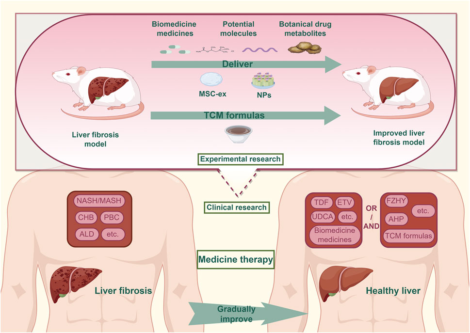
Figure 1. The situation of medicine treatment for liver fibrosis.In clinical practice, the combined treatment of Biomedicine medicines and TCM formulas has been confirmed as an effective strategy to address liver fibrosis caused by common chronic liver diseases. Currently, the field of experimental research are actively advancing the discovery of medicines for the treatment of liver fibrosis, focusing on areas such as repurposed drugs, potential molecules, botanical drug metabolites. MSC-ex and NPs technology provide strong support for translating Experimental research findings into clinical treatments. Abbreviation: NASH, Non-alcoholic Steatohepatitis; MASH, Metabolic Associated Fatty Liver Disease; CHB, Chronic Hepatitis B; PBC, Primary Biliary Cholangitis; ALD, Alcoholic Liver Disease; TDF, Tenofovir Disoproxil Fumarate; ETV:Entecavir; UDCA, Ursodeoxycholic Acid; FZHY, Fuzheng Huayu tablets; AHP, AnluoHuaxian pills; TCM, Traditional Chinese medicine.
2 Methods
A systematic literature review was conducted as of 1 February 2025, encompassing both in vitro and in vivo experimental studies retrieved from PubMed and Web of Science. The search employed keywords such as liver fibrosis, treatment, and Traditional Chinese Medicine. Inclusion criteria were: (1) studies addressing the therapeutic effects or mechanisms of pharmacological agents in liver fibrosis using any combination of the specified keywords; (2) studies presenting original experimental data with clearly defined methodologies; and (3) full-text articles published in English. Exclusion criteria included duplicate records, irrelevant studies based on title and abstract screening, and publications lacking mechanistic insight or sufficient methodological detail. Based on these criteria, 102 studies were selected for analysis. Of these, 18 studies focusing on botanical drug metabolites were further evaluated for methodological quality using the ConPhyMP assessment tool. The evaluation was jointly performed by authors ZJ and LYX, with detailed results provided in the supplementary materials.
3 Pathological mechanisms of hepatic fibrosis
The progression of liver fibrosis is marked by the excessive accumulation of extracellular matrix (ECM) components (Iredale, 2007). Etiological factors such as toxins, metabolic disorders, or viral infections cause hepatocyte damage and immune cell infiltration, which, in turn, activate hepatic stellate cells (HSCs) and promote their differentiation into collagen-producing myofibroblasts (Zhang et al., 2016). Under physiological conditions, ECM synthesis and degradation remain in dynamic balance. However, in chronic liver disease, this balance is disrupted, favoring pro-fibrotic over anti-fibrotic signals. This shift induces myofibroblast proliferation, migration, contraction, and excessive ECM deposition. Moreover, vascular endothelial growth factor A (VEGFA) promotes pathological angiogenesis during fibrosis progression (Elpek, 2014; Shen H. et al., 2022). While producing ECM, activated HSCs also secrete matrix metalloproteinases (MMPs) and tissue inhibitors of metalloproteinases (TIMPs), the latter suppressing MMP activity and further reducing ECM degradation (Tacke and Zimmermann, 2014). In parallel, hepatocyte apoptosis and the release of damage-associated molecular patterns (DAMPs) directly stimulate HSC activation and initiate immune cell recruitment, including lymphocytes and macrophages. Kupffer cells, the liver-resident macrophages, can polarize into M1 or M2 phenotypes and secrete fibrogenic mediators such as transforming growth factor-beta 1 (TGF-β1) and reactive oxygen species (ROS). These mediators, along with ROS from neutrophils, enhance HSC activation and ECM synthesis (Ramachandran et al., 2012; Krenkel and Tacke, 2017). Oxidative stress further exacerbates fibrogenesis through NADPH oxidase (NOX)-mediated ROS production and the release of proinflammatory cytokines such as tumor necrosis factor-alpha (TNF-α) and interleukin-1 beta (IL-1β), which potentiate HSC activation and fibrotic remodeling (Ren et al., 2021). Additionally, epithelial-mesenchymal transition (EMT) contributes to the myofibroblast population (Chen et al., 2020). The TGF-β signaling pathway is pivotal in initiating and sustaining HSC activation and fibrogenesis (Dewidar et al., 2019). Beyond TGF-β, multiple signaling pathways have been implicated in modulating the fibrotic response. A comprehensive overview of the pathogenic mechanisms underlying hepatic fibrosis is illustrated in Figure 2.
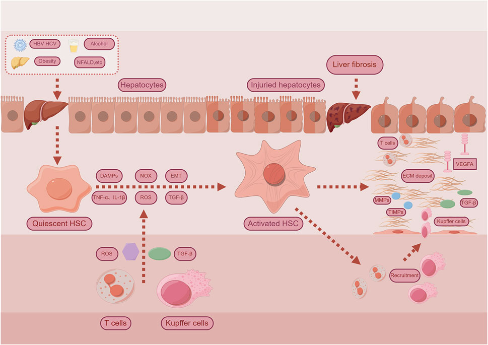
Figure 2. Examples for mechanisms for liver fibrosis. Chronic hepatocyte injury causes release of damage-associated patterns (DAMPs) and so on activate Hepatic stellate cells (HSCs) and recruit immune cells. Complex multidirectional interactions between activated HSCs and Kupffer cells, as well as innate immune cells promote trans-differentiation into proliferative and extracellular matrix (ECM) producing myofibroblasts. Abbreviations: TGF-β, Transforming Growth Factor Beta; NOX, Nicotinamide adenine dinucleotide phosphate-oxidase; TNF-α, Tumor Necrosis Factor-alpha; IL-1β, Interleukin-1-beta; ROS, Reactive Oxygen Species; EMT, Epithelial-Mesenchymal Transition; MMPs, matrix metalloproteinases; TIMPs, Tissue Inhibitors of Metalloproteinases; VEGFA, Vascular Endothelial Growth Factor A.
4 Biomedicine treatment for liver fibrosis
4.1 Biomedicine medicines treatment for liver fibrosis
Clinically, liver fibrosis is commonly associated with non-alcoholic steatohepatitis (NASH), viral hepatitis, type 2 diabetes-related fatty liver disease, and primary biliary cholangitis (PBC). Standard pharmacotherapies—such as entecavir (ETV) for chronic hepatitis B (CHB) and ursodeoxycholic acid for PBC—have shown efficacy in mitigating fibrosis (Table 1). Nonetheless, the lack of fibrosis-specific targeted therapies continues to drive drug discovery efforts (Table 2).
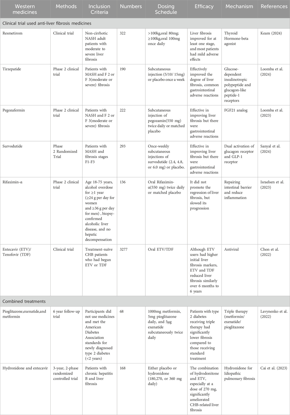
Table 1. Clinical Studies related to the treatment of liver fibrosis by Western medicines.
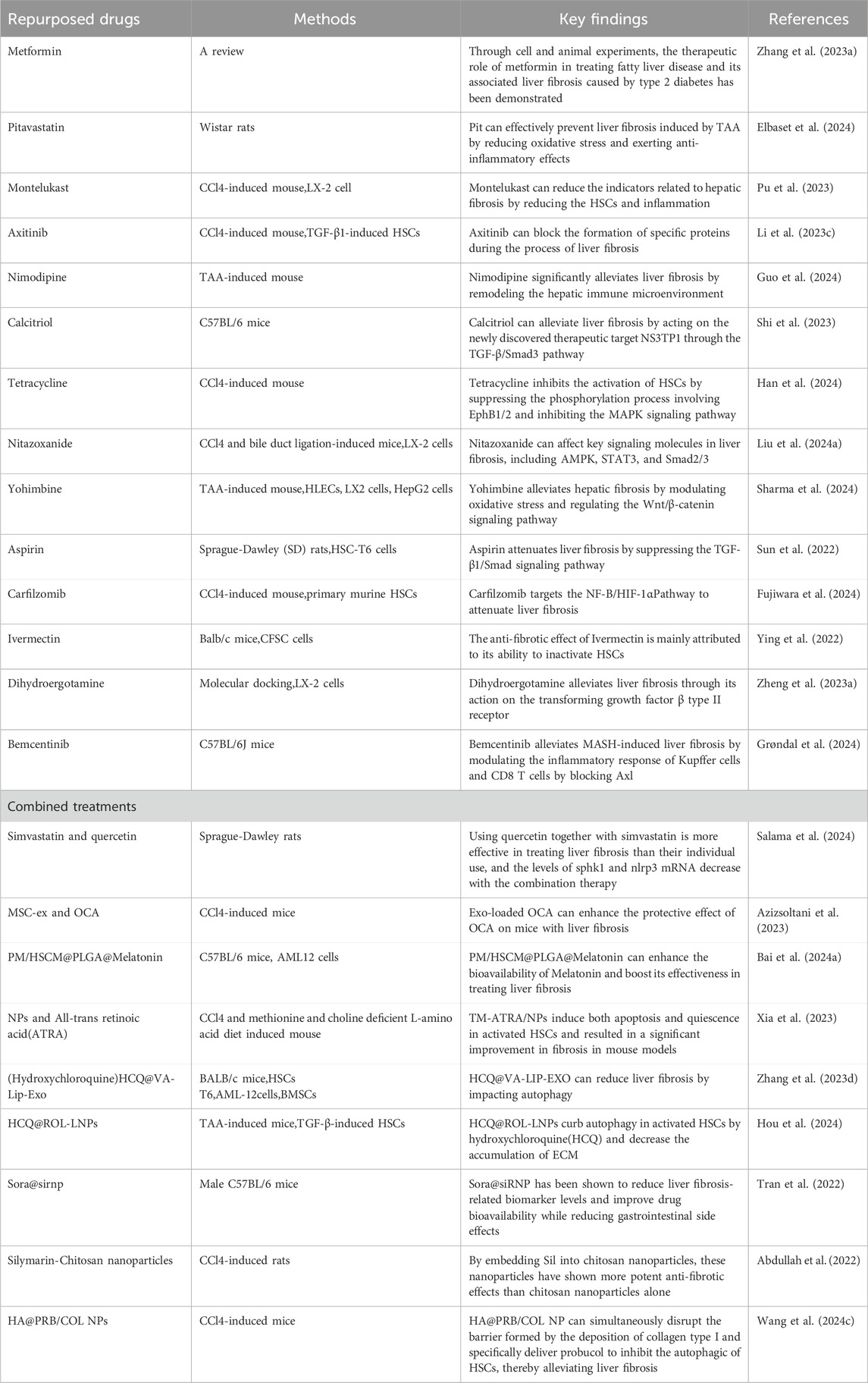
Table 2. Experimental Studies related to the treatment of liver fibrosis by Western medicines.
4.1.1 Clinical trial used anti-liver fibrosis biomedicine medicines
Resmetirom received initial regulatory approval in March 2024 for the treatment of metabolic dysfunction-associated steatohepatitis (MASH)/non-alcoholic steatohepatitis (NASH) and related fibrosis (Keam, 2024). In a phase 2 trial conducted by (Loomba et al., 2024), tirzepatide significantly improved MASH without exacerbating fibrosis after 52 weeks of treatment. In another study, (Loomba et al., 2023), evaluated pegozafermin, a fibroblast growth factor 21 (FGF21) analog, in NASH patients with moderate-to-severe fibrosis. After 24 weeks, fibrosis improvements were observed, although gastrointestinal side effects, notably nausea and diarrhea, were reported. (Sanyal et al., 2024). conducted a 48-week clinical trial assessing survodutide in patients with mild-to-moderate NASH-associated fibrosis. The drug demonstrated superiority over placebo in improving NASH features and preventing fibrosis progression, albeit with significant adverse effects. A follow-up study by (Lavynenko et al., 2022) focused on hepatic fibrosis in individuals with type 2 diabetes. In a large-scale study involving more than 3,000 patients with CHB and hepatic cirrhosis, (Chon et al., 2022), reported that ETV initially led to slight elevations in fibrosis biomarkers. However, long-term treatment (6 months–6 years) with ETV or tenofovir (TDF) yielded comparable efficacy in fibrosis reduction. The study also highlighted that combination therapy with metformin, exenatide, and pioglitazone was more effective in attenuating hepatic fibrosis than conventional stepwise regimens. Finally, in a 5-year randomized, double-blind, placebo-controlled phase 2 trial, (Israelsen et al., 2023), investigated pharmacologic interventions for patients with alcohol-related liver disease. Recent investigations suggest that Rifaximin-α may slow the progression of liver fibrosis, though this finding requires validation in forthcoming multicenter phase 3 clinical trials. At present, clinical studies have been involved in the treatment of liver fibrosis caused by several common liver diseases. However, many of these clinical trials are limited by small sample sizes, which compromises the reliability and generalizability of their findings.
4.1.2 Repurposed drugs
(Zhang et al., 2023a) reviewed therapeutic advances related to metformin in treating fatty liver disease and liver fibrosis in patients with type 2 diabetes, emphasizing both clinical potential and underlying mechanisms. (Elbaset et al., 2024). demonstrated that pitavastatin possesses anti-fibrotic properties by attenuating oxidative stress and inflammatory signaling via modulation of the NF-κB and PI3K/AKT pathways. Similarly, (Pu et al., 2023), showed that montelukast effectively reduces fibrosis in murine models by inhibiting hepatic stellate cell (HSC) activation and inflammatory responses. (Li et al., 2023c). found that axitinib mitigates liver inflammation and fibrosis through dual mechanisms: suppression of HSC activation and enhancement of mitochondrial complexes I and III function. Further evidence supports the anti-fibrotic potential of other repurposed agents. (Guo et al., 2024). reported that nimodipine alleviates hepatic inflammation and fibrosis by modulating the liver’s immune microenvironment. (Shi et al., 2023). found that calcitriol regulates HSC activation, proliferation, and differentiation by downregulating NS3TP1 expression. (Han et al., 2024).showed that tetracycline inhibits activated HSCs by targeting EphB1/2 phosphorylation and the MAPK1/2 pathway. According to (Liu K. X. et al., 2024), nitazoxanide acts on key fibrotic signaling molecules including AMPK, STAT3, and Smad2/3, with follow-up studies confirming its efficacy in ameliorating fibrosis through these mechanisms. (Sharma et al., 2024). demonstrated that yohimbine reduces fibrosis and inflammation by modulating the JNK/Wnt/β-catenin pathway. Early evidence by (Jiang et al., 2016) indicated that aspirin decreases fibrosis indices in adult chronic liver disease patients in the US (Sun et al., 2022), a mechanism later confirmed in rat models via modulation of the TGF-β/Smad pathway. (Fujiwara et al., 2024). reported that carfilzomib reduces fibrotic marker expression in HSCs and attenuates CCl4-induced liver fibrosis. (Ying et al., 2022). identified ivermectin as an anti-fibrotic agent acting primarily through modulation of HSCs. (Zheng K. X. et al., 2023). employed molecular docking to identify dihydroergotamine as a potential TGF pathway modulator, with subsequent cellular assays confirming its anti-fibrotic effect.
Many of these agents, initially developed for unrelated conditions, demonstrate efficacy by modulating key pathogenic pathways involved in hepatic fibrosis. For example, the well-established links between diabetes, dyslipidemia, and liver fibrosis suggest that hypoglycemic and hypolipidemic drugs may exert secondary anti-fibrotic effects. However, it is important to recognize that commonly used experimental models, such as CCl4 and TAA-induced liver fibrosis, do not fully replicate the pathophysiological complexity of human liver fibrosis. This discrepancy may hinder the direct translation of preclinical findings into clinical practice, highlighting the need for more physiologically relevant models in fibrosis research.
4.1.3 Combined treatments
In a 3-year, two-phase randomized controlled trial involving 168 patients, (Cai et al., 2023), demonstrated that Hydronidone combined with ETV significantly improved liver fibrosis associated with chronic hepatitis B, with 270 mg identified as the optimal dosage. (Salama et al., 2024). reported that combination therapy with Quercetin and Simvastatin was more effective than monotherapy in treating liver fibrosis, as evidenced by reduced expression of sphk1 and nlrp3 mRNA, indicating inhibition of the SphK1/NLRP3 signaling pathway. (Azizsoltani et al., 2023). explored mesenchymal stem cell-derived exosomes (MSC-ex) as a delivery platform for Obeticholic Acid (OCA), enhancing targeted delivery and mitigating side effects while effectively alleviating liver fibrosis in mouse models. Similarly, (Bai Y. et al., 2024), employed nanotechnology to improve Melatonin bioavailability, developing PM@PLGA and HSCM@PLGA nanoparticles that exhibited enhanced anti-fibrotic efficacy in animal models. (Xia et al., 2023). designed biomimetic nanoparticles (TM-ATRA/NPs) encapsulating all-trans retinoic acid (ATRA) within LX2 cell membrane-derived vesicles expressing TRAIL, which induced apoptosis and quiescence in activated hepatic stellate cells (HSCs), thereby ameliorating fibrosis in a mouse model. (Zhang Y. W. et al., 2023). developed HCQ@VA-Lip-Exo, a vitamin A-modified hybrid nanobiomimetic delivery system that effectively targeted activated HSCs, suppressed autophagy, and reduced extracellular matrix (ECM) synthesis and deposition. (Hou et al., 2024). further demonstrated that HCQ@ROL-LNPs selectively inhibited activated HSCs. (Tran et al., 2022). showed that sora@sirnp nanoparticles enhanced sorafenib bioavailability, producing anti-fibrotic effects while reducing gastrointestinal side effects. In another nanomedicine approach, a previous study identified silymarin-chitosan nanoparticles as a potent anti-fibrotic nanoformulation (Abdullah et al., 2022) that upregulated protective liver miRNAs and downregulated fibrosis-related markers such as TGFβR1, COL3A1, and TGFβR2. (Wang X. et al., 2024). developed HA@PRB/COL nanoparticles, a hyaluronic acid-based delivery system that targeted HSCs via CD44 receptors and facilitated ECM collagen I degradation through collagenases (COLs), effectively preventing HSC activation. Overall, combining conventional biomedicine medicines with MSC-ex or nanoparticles holds considerable promise for treating liver fibrosis by enhancing bioavailability, improving targeting, and reducing side effects. However, current studies often emphasize efficacy while overlooking adverse effects, limiting the comprehensiveness of research.
4.2 Potential molecules treatment for liver fibrosis
The evolving landscape of liver fibrosis research underscores the limitations of conventional biomedicine therapies in achieving cellular specificity. In response, there is growing interest in developing novel therapeutics based on protein and nucleic acid molecules (Table 3).
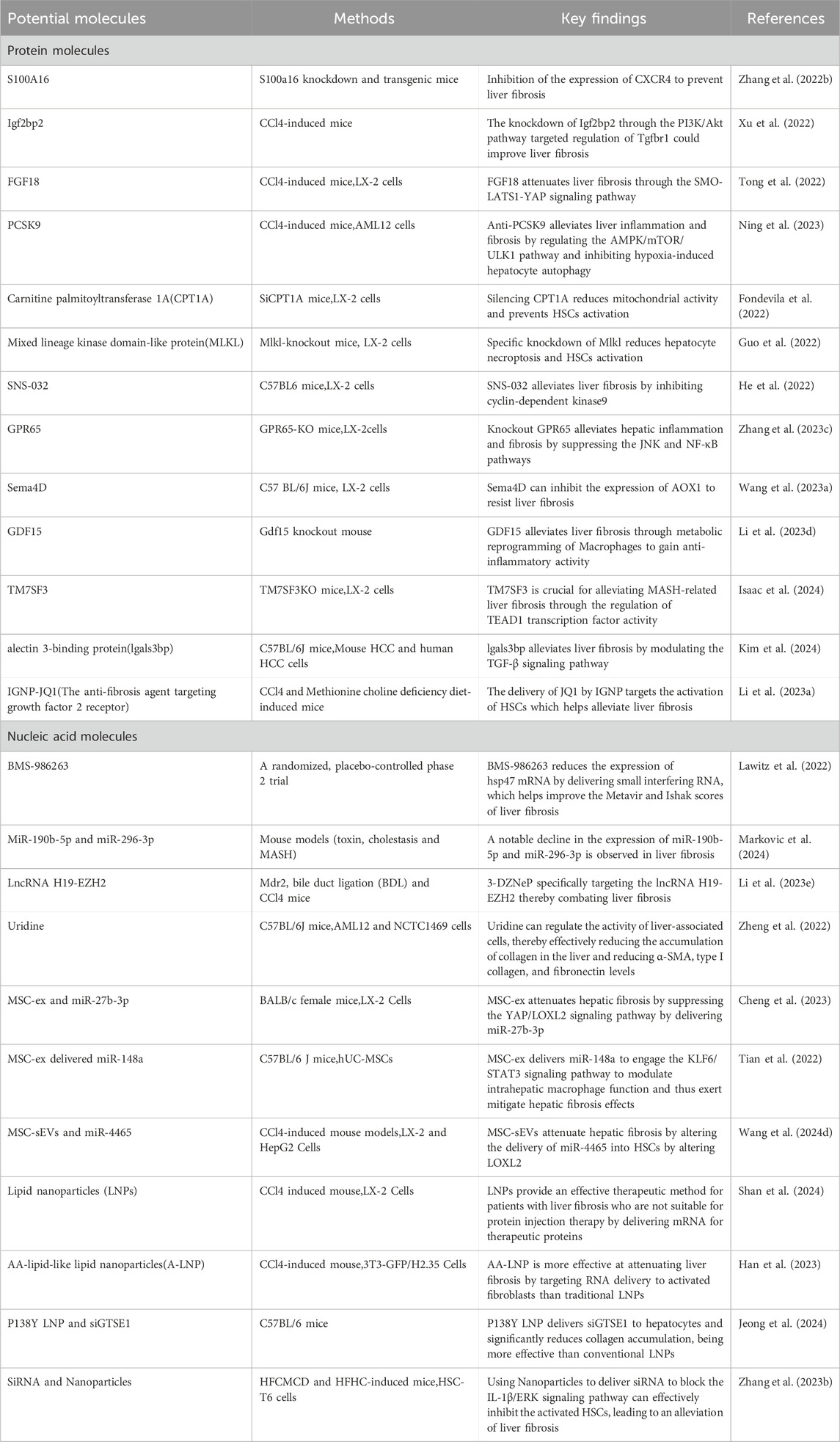
Table 3. Studies related to the treatment of liver fibrosis by Potential molecules.
4.2.1 Protein molecules
(Zhang et al., 2022b) identified S100A16 as a suppressor of CXCR4 expression, which attenuated hepatic stellate cell activation by inhibiting the ERK1/2 and AKT signaling pathways. (Xu et al., 2022). demonstrated that silencing Igf2bp2 ameliorated fibrosis by modulating Tgfbr1 through the PI3K/Akt pathway. (Tong et al., 2022). revealed that FGF18 mitigates liver fibrosis via the SMO-LATS1-YAP signaling axis. Similarly, (Ning et al., 2023), showed that anti-PCSK9 antibodies reduce liver inflammation and fibrosis by modulating the AMPK/mTOR/ULK1 pathway and inhibiting hypoxia-induced hepatocyte autophagy. (Fondevila et al., 2022). reported that CPT1A silencing diminished mitochondrial activity, thereby suppressing HSC activation. (Guo et al., 2022). showed that knockdown of Mlkl specifically reduced hepatocyte necroptosis and HSC activation. (He et al., 2022). proposed that SNS-032, a cyclin-dependent kinase nine inhibitor, alleviates fibrosis by inducing apoptosis and suppressing activated HSCs. (Zhang K. et al., 2023). demonstrated that GPR65 knockout reduces hepatic inflammation and fibrosis through inhibition of the JNK and NF-κB pathways. Finally, (Wang L. et al., 2023), found that Sema4D knockdown downregulates AOX1 and RARA, thereby modulating T helper cell balance and counteracting liver fibrosis. (Li X. et al., 2023). demonstrated that GDF15 mitigates liver fibrosis progression by reprogramming macrophage metabolism to favor anti-inflammatory phenotypes. Similarly, (Isaac et al., 2024), highlighted the role of TM7SF3 in attenuating nonalcoholic steatohepatitis (NASH)-related liver fibrosis through regulation of the transcription factor TEAD1. (Grøndal et al., 2024). showed that Bemcentinib alleviates MASH-induced liver fibrosis by targeting AXL, thereby modulating the inflammatory responses of Kupffer cells and CD8+ T cells. In another study, (Kim et al., 2024), identified Galectin-3-binding protein as a potential biomarker for distinguishing stages of hepatic fibrosis and demonstrated its anti-fibrotic effects via modulation of the TGF-β1 signaling pathway. Additionally, (Li F. et al., 2023), investigated the effects of riociguat-pretreated IGNP-JQ1 on hepatic stellate cells (HSCs) in two murine models of liver fibrosis. Their findings revealed that this intervention significantly reduced hepatic fibrosis by enhancing substance exchange efficiency within liver tissue. Collectively, these studies have found that different molecular targets have intervening effects on liver fibrosis, but further exploration of their specific mechanisms of action is still needed.
4.2.2 Nucleic acid molecules
Nucleic acids, particularly small interfering RNA (siRNA) and microRNA (miRNA), play pivotal roles in regulating liver fibrosis by modulating gene expression and signaling pathways. These molecules have emerged as promising therapeutic agents capable of halting fibrosis progression through targeted gene silencing. (Lawitz et al., 2022). administered BMS-986263—an siRNA targeting HSP47 mRNA—to 61 HCV-SVR patients with advanced liver fibrosis, reporting improvements in both METAVIR and Ishak scores. Infusion-related reactions were the most common adverse events. (Markovic et al., 2024). observed significant reductions in miR-190b-5p and miR-296-3p levels in murine and human liver fibrosis models, identifying hyaluronan synthase 2 (HAS2) and integrin alpha-6 as novel therapeutic targets. (Zhao et al., 2019). provided a comprehensive review of siRNA- and miRNA-based strategies for liver fibrosis, elucidating their mechanisms and therapeutic potential. Beyond canonical nucleic acids, researchers are also investigating related biological processes. (Li X. J. et al., 2023). showed that H19-regulated EZH2 reprograms H3K27me3, activating HSCs, promoting epithelial–mesenchymal transition (EMT), and triggering Wnt/β-catenin signaling—suggesting that disrupting this interaction could represent a novel therapeutic strategy. (Zheng et al., 2022). found that uridine modulates hepatic cell activity, reducing collagen accumulation and downregulating the expression of α-SMA, type I collagen, and fibronectin. (Cheng et al., 2023). reported that mesenchymal stem cell (MSC)-derived exosomes alleviate hepatic fibrosis by delivering miR-27b-3p to inhibit the YAP/LOXL2 signaling axis. Similarly, (Tian et al., 2022), showed that MSC-derived exosomes transporting miR-148a modulate intrahepatic macrophage activity via the KLF6/STAT3 pathway, thereby reducing fibrosis. (Wang Y. et al., 2024). further demonstrated that MSC-derived small extracellular vesicles modulate LOXL2 to facilitate miR-4465 delivery into HSCs, contributing to anti-fibrotic effects. Several nanoparticle-based delivery platforms have also shown promise. (Shan et al., 2024). used retinoid-derived lipid nanoparticles (LNPs) to deliver therapeutic protein mRNA in three NASH-associated liver fibrosis models, achieving enhanced protein retention and reduced systemic toxicity. (Han et al., 2023). developed AA-lipid-like LNPs that improved RNA delivery efficiency to activated fibroblasts and exhibited superior transmission compared to conventional LNPs. (Jeong et al., 2024). demonstrated that engineered LNPs delivering siGTSE1 to hepatocytes significantly reduced collagen accumulation. In a related study, (Zhang C. et al., 2023), observed IL-1β upregulation in two mature NASH mouse models and showed that siRNA-loaded nanoparticles targeting the IL-1β/ERK pathway effectively inhibited HSC activation and attenuated fibrosis. By targeting specific genes, nucleic acid molecules can regulate gene expression and signaling pathways, and achieve precise gene silencing. Not only do they improve symptoms, but they may also slow or even reverse the fibrosis process. However, the full mechanism of their role in disease progression needs to be further explored.
5 Botanical drug metabolites treatment for liver fibrosis
In medicinal research, clinical observations and empirical findings are fundamental to drug development. Botanical drugs has accumulated extensive clinical experience in managing liver fibrosis, offering a valuable foundation for pharmacological investigations. This body of knowledge suggests that specific botanical drug metabolites may hold therapeutic potential against hepatic fibrosis. Numerous reviews have explored phytotherapeutic strategies for hepatic fibrosis, often emphasizing key botanical drug metabolites such as polyphenols, phenolic acids, and flavonoids, along with their underlying mechanisms of action (Wenbo et al., 2024; Zhao B. et al., 2024). To advance this field, a phenotypic perspective on hepatic fibrosis—alongside a synthesis of recent mechanistic insights—may offer new avenues for therapeutic exploration. Table 4 presents a summary of studies investigating botanical drug metabolites in the treatment of hepatic fibrosis, with plant species cross-referenced against the MPNS database (https://powo.science.kew.org/).
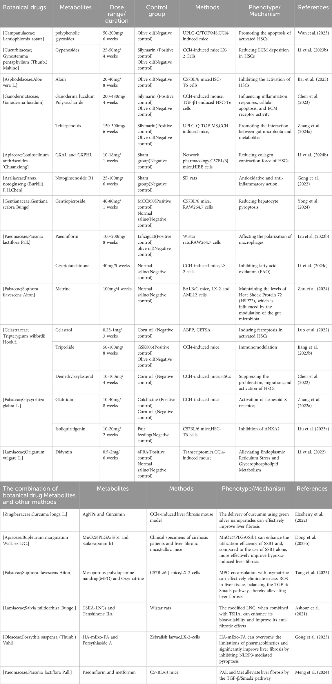
Table 4. Studies related to the treatment of liver fibrosis by Botanical drug Metabolites.
5.1 Effects of botanical drug metabolites on the phenotype or mechanism of liver fibrosis
Aloin has demonstrated anti-fibrotic effects by inhibiting hepatic stellate cell (HSC) activation and attenuating CCL4-and TGF-β1-induced inflammatory responses in both in vitro and in vivo models (Bai et al., 2023). Activated HSCs contribute to fibrosis by promoting apoptosis, extracellular matrix (ECM) accumulation, and hepatocyte pyroptosis. (Wan et al., 2023). reported that the total polyphenolic glycosides of Lamiophlomis rotata suppress HSC proliferation via inhibition of the TGF-β/Smad pathway and enhance apoptosis of activated HSCs, thereby ameliorating liver fibrosis. Similarly, Gypenosides were shown to inhibit TGF-β-induced HSC activation and reduce ECM deposition, leading to fibrosis attenuation in vivo (Li et al., 2023b; Yong et al., 2024) found that Gentiopicroside (GPS), derived from Gentiana scabra Bunge, inhibits HSC activation by blocking the TLR4 and NLRP3 signaling pathways and suppressing hepatocyte pyroptosis. In a related study, (Luo et al., 2022), demonstrated that Celastrol alleviates hepatic fibrosis by inducing ferroptosis in activated HSCs through modulation of peroxiredoxins and heme oxygenase-1 (HO-1).
Emerging evidence suggests that botanical drug metabolites often target multiple fibrotic phenotypes simultaneously. For instance, (Chen et al., 2023), reported that Ganoderma lucidum polysaccharide (GLP) attenuates hepatic fibrosis by regulating inflammation, cell cycle progression, apoptosis, and ECM-receptor interactions. In another study, (Chen et al., 2022), showed that demethylzeylasteral inhibits HSC proliferation, migration, and activation, and downregulates fibrogenic gene expression by suppressing FAK and AKT phosphorylation through inhibition of the AGAP2-mediated signaling pathway.
Botanical metabolites also exert anti-fibrotic effects by reducing oxidative stress and inflammation driven by activated HSCs. Notoginsenoside R1, a metabolite of Panax notoginseng, alleviates fibrosis by suppressing HSC activation and downregulating NF-κB and MAPK signaling pathways, thereby exerting both antioxidant and anti-inflammatory effects (Gong et al., 2022). Some studies have extended beyond HSC-focused mechanisms. For example, CXAL and CXPHL, metabolites of Chuanxiong Rhizoma, mitigate hepatic fibrosis by modulating the CTCF–c-MYC–H19 pathway and reducing HSC-mediated collagen contraction (Li Y. et al., 2024). Additionally, (Liu Y. et al., 2023), demonstrated that Paeoniflorin exerts anti-fibrotic effects by modulating macrophage polarization via the NF-κB/HIF-1α signaling pathway.
This study also revealed novel mechanisms involved in the pathogenesis and mitigation of liver fibrosis. Triterpenoids have been shown to attenuate hepatic fibrosis by modulating NF-κB and TGF-β1/Smads signaling pathways, as well as by increasing the abundance of the gut microbiota genus Ruminococcus (Zhang J. et al., 2024). Similarly, Kuhuang alleviates liver fibrosis by influencing the gut microbiota’s regulation of hepatic interferon signaling and bile acid metabolism (Shen B. et al., 2022; Li Z. et al., 2024) reported that Cryptotanshinone (CTS) was reported to reduce liver fibrosis through the inhibition of p-STAT3/CPT1A-mediated fatty acid oxidation. (Jiang S. et al., 2023). showed that Triptolide exerts its anti-fibrotic effects by modulating T-helper (Th) and CD4+ T cell differentiation. According to (Li et al., 2022), Didymin, a metabolite derived from Origanum vulgare L., primarily mitigates hepatic fibrosis by reducing endoplasmic reticulum stress and altering glycerophospholipid metabolism. In addition to small-molecule metabolites, several protein-related mechanisms have emerged as promising anti-fibrotic targets. Glabridin alleviates inflammation and oxidative stress via PPARγ activation, thereby suppressing fibrotic progression (Zhang L. et al., 2022; Zhang L. et al., 2024) demonstrated that Astragalus saponins, through activation of the FXR receptor, downregulate key proteins implicated in hepatic fibrosis, particularly in cholestatic liver disease models. Isoliquiritigenin (ISL) inhibits ANXA2 expression and blocks the sphks/S1P/IL-17 signaling axis while suppressing STAT3 phosphorylation, leading to reduced α-SMA expression and fibrosis reversal (Liu N. et al., 2023). The investigation of botanical drug metabolites has expanded rapidly in recent years (Dan et al., 2024). However, focusing solely on mechanistic studies is insufficient to address clinical demands. If botanical drug metabolites that can act on the initial causes of primary liver diseases and effectively target liver fibrosis are successfully developed, for example, identifying botanical drug metabolites capable of simultaneously ameliorating cholestasis and liver fibrosis would substantially enhance research significance and translational potential. Mirroring the successful development trajectories of artemisinin and tanshinone, such efforts are critical to advancing liver fibrosis treatment. In contrast, existing agents such as colchicine, lificigat, and 4PBA lack fibrosis-specific efficacy, have limited clinical validation, and show weak mechanistic alignment, rendering them suboptimal as positive control drugs in experimental models.
5.2 Combined application
Combination strategies have demonstrated enhanced therapeutic efficacy. (Elzoheiry et al., 2022). addressed curcumin’s poor bioavailability using green silver nanoparticles (AgNPs) for targeted delivery in a mouse model, which resulted in significantly reduced liver fibrosis severity. (Dong H. et al., 2023). developed a multifunctional nanosystem (MnO2@PLGA/Ssb1), which integrates MnO2 with saikosaponin b1 (Ssb1) to augment its anti-fibrotic activity by scavenging excess H2O2 and relieving hypoxic stress. (Tang et al., 2023). utilized mesoporous polydopamine (MPO) for targeted drug delivery in both animal and cell models. MPO effectively reduced hepatic reactive oxygen species (ROS) and modulated the TGF-β/Smad signaling pathway, thereby attenuating fibrosis. Encapsulation of oxymatrine in MPO further enhanced therapeutic efficacy by restoring TGF-β/Smad signaling balance and reducing fibrotic progression. Similarly, (Ashour et al., 2021), demonstrated that lipid nanocapsules encapsulating Tanshinone IIA (TSIIA-LNCs) exerted superior anti-fibrotic effects compared to the free compound. (Gong et al., 2023). employed milk-derived exosomes modified with hyaluronic acid (HA-mExo-FA) to deliver Forsythiaside A. This nanocomplex targeted CD44 receptors on activated hepatic stellate cells, significantly inhibiting TGF-β1-induced LX2 cell proliferation, downregulating α-SMA expression, and promoting apoptosis (Meng et al., 2024). We found that many studies have been carried out on the combined application of mesenchymal stem cell exosomes (MSC-ex) or nanoparticle-based systems with botanical drug metabolites, potential molecules, and biomedicine medicines, however, there is still a lack of systematic comparative studies on the therapeutic effects of MSC-ex, nanoparticle-based systems and these medicines in the treatment of liver fibrosis. In the future, it is of great significance to carry out relevant comparative research.
6 Traditional Chinese medicine formulas treatment for liver fibrosis
Liver fibrosis, as a chronic progressive condition, has long been managed using TCM formulas, many of which are clinically validated. Increasingly, clinical studies are evaluating the integrative potential of TCM and biomedicine medicines in treating liver fibrosis. The TCM literature extensively documents the therapeutic efficacy of classical and empirically derived formulas. Recent rigorous investigations have elucidated the molecular mechanisms underlying these TCM-based interventions (see Table 5), further validating their relevance in modern hepatology.
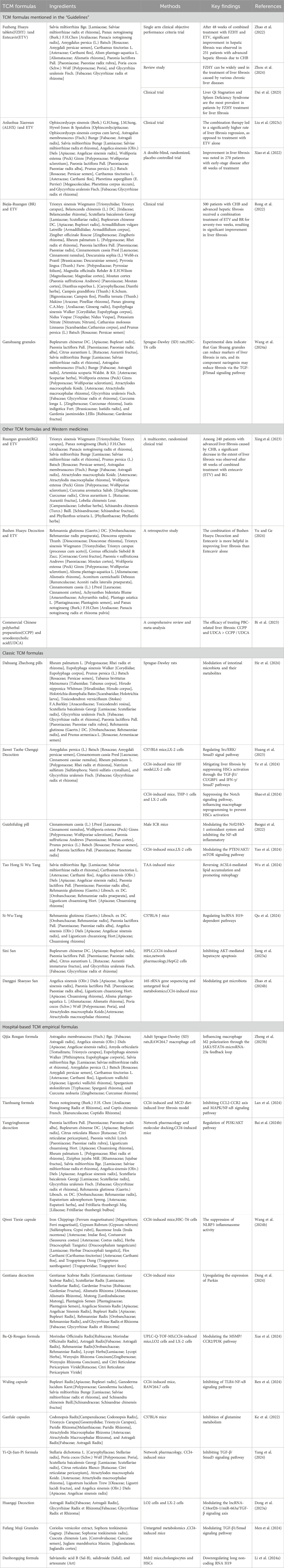
Table 5. Studies exploring the therapeutic effects of TCM formulas on liver fibrosis.
6.1 The “guidelines for diagnosis and treatment of hepatic fibrosis with integrated traditional Chinese and Western medicine (2019 edition)”
(Xu and Liu, 2020) highlighted several patented TCM formulas with demonstrated efficacy. In a single-arm clinical trial using objective performance criteria, 251 patients with chronic hepatitis B (CHB) and advanced liver fibrosis underwent 48 weeks of combined therapy with Fuzheng Huayu (FZHY) tablets and entecavir (ETV). The FZHY group showed a significantly higher rate of fibrosis improvement—15% greater than with ETV monotherapy (Zhao et al., 2022). A comprehensive review (Zhou et al., 2024) concluded that FZHY is broadly applicable for treating liver fibrosis across various chronic liver diseases, with numerous animal and cellular studies confirming its effectiveness in targeting all known pathogenic mechanisms of fibrosis. (Dai et al., 2023). reported that Liver Qi Stagnation and Spleen Deficiency Syndrome were the predominant TCM syndromes among patients treated with FZHY. In a study of 400 CHB patients (Liu Y. Q. et al., 2023), compared ETV monotherapy with a combination of ETV and Anluohua Xianwan (ALHX), finding a significantly higher rate of fibrosis regression in the combination group. Similarly, a randomized controlled trial involving 270 CHB patients with early-stage liver fibrosis demonstrated that 48 weeks of Anluo Huaxian Pills (AHP) significantly improved fibrosis compared to placebo, with no notable adverse effects observed in the AHP group (Xiao et al., 2022). In another multicenter, randomized, double-blind, placebo-controlled trial, (Rong et al., 2022), showed that 72 weeks of ETV combined with Biejia-Ruangan (BR) significantly enhanced fibrosis regression in 500 patients with advanced CHB-related liver fibrosis. Preclinical studies support these findings. In rat models, Ganshuang Granules (GSG) reduced key fibrosis biomarkers, and in vitro studies indicated that its active compound, Naringin, may attenuate fibrosis by modulating the TGF-β/Smad signaling pathway (Wang F. et al., 2024). Although FZHY has been extensively studied-particularly in patients with Liver Qi Stagnation and Spleen Deficiency-the international persuasiveness of this evidence remains limited. Future research should focus on improving trial design and further exploring TCM syndrome differentiation to strengthen the global acceptance and methodological rigor of related studies. Meanwhile, clinical research on other formulas such as GSG and Biejia-Ruangan remains scarce, with even fewer studies elucidating their mechanisms of action.
6.2 Integration of other TCM formulas with biomedicine medicines for liver fibrosis
(Xing et al., 2023) evaluated 240 patients with advanced CHB-related fibrosis and found that 48 weeks of combination therapy with ETV and Ruangan Granule (RG) significantly reduced fibrosis severity. In a retrospective study, (Yu and Ge, 2024), reported that Bushen Huayu Decoction combined with ETV produced greater reductions in fibrosis markers than ETV alone, with statistically significant improvements observed at both 2 and 4 weeks. (Bi et al., 2023), including 22 randomized controlled trials, showed that commercial Chinese polyherbal preparation (CCPP) combined with ursodeoxycholic acid (UDCA) was more effective for primary biliary cholangitis (PBC)-related fibrosis than either treatment alone. In the future, more high-quality clinical studies can be carried out to further verify the efficacy of integrated traditional Chinese and biomedicine medicines, and provide patients with more effective treatment options.
6.3 Classic TCM formulas
Mechanistic studies continue to advance understanding of TCM therapies. (He et al., 2024). found that Dahuang Zhechong pills ameliorate liver fibrosis by modulating gut microbiota and associated metabolites. (Huang et al., 2023). demonstrated that Jiawei Taohe Chengqi Decoction inhibits HSC activation via the Src/ERK/Smad3 signaling pathway. (Ye et al., 2024). further showed that this decoction mitigates fibrosis by targeting activated HSCs through both the TGF-β1/CUGBP1 and IFN-γ/Smad7 pathways. In addition, (Shao et al., 2024), revealed that the same decoction suppresses HSC activation via inhibition of the Notch signaling pathway and modulation of macrophage polarization. The Guizhifuling Pill exhibits hepatoprotective effects by regulating the Nrf2/HO-1 antioxidant axis and inhibiting the NF-κB inflammatory signaling pathways (Baogui et al., 2022), as well as modulating the PTEN/AKT/mTOR signaling pathway (Yao et al., 2024; Wu et al., 2024) demonstrated that Tao-Hong-Si-Wu-Tang attenuates liver fibrosis by restoring lipid metabolism, inhibiting long-chain acyl-CoA synthetase 4 (ACSL4), and promoting mitophagy. Similarly, (Qu et al., 2024), reported that Si-Wu-Tang reduces ECM deposition and liver fibrosis through the H19-associated miRNA pathway. (Wang S. J. et al., 2023). reported that Xiaochaihu Tang alleviates hepatic fibrosis through leptin and Nrf2 signaling pathways. (Jiang M. et al., 2023). showed that Isorhamnetin, a key metabolite of Sini San, prevents the AKT-mediated suppression of FXR expression. (Zhao Y. et al., 2024). that Danggui Shaoyao San ameliorates hepatic fibrosis by modulating gut microbiota and their metabolites, particularly short-chain fatty acids (SCFAs) and bile acids (BAs). Classic TCM formulas have been extensively studied and applied to a variety of diseases, and their best indications are not limited to liver fibrosis. In the future, clinical studies need to be strengthened to better determine its optimal application in the treatment of liver fibrosis.
6.4 Hospital-based TCM empirical formulas
The Qijia Rougan formula exerts anti-fibrotic effects by regulating the JAK1/STAT6/microRNA-23a axis through M2 macrophage polarization (Zheng Y. et al., 2023). The Tianhuang formula reduces liver fibrosis by targeting the CCL2/CCR2 axis and downregulating the MAPK-NF-κB pathway (Lan et al., 2024). Yangyinghuoxue decoction mitigates fibrosis via PI3K/Akt pathway activation (Bai Y. M. et al., 2024). Qiwei Tiexie capsule effectively combats hepatic fibrosis by suppressing NLRP3 inflammasome activation while enhancing hydroxyproline content and glutathione peroxidase activity in vivo (Wang S. et al., 2024). Gentiana decoction, as reported by (Deng et al., 2024), inhibits HSC activation through the Parkin signaling pathway. The Danhongqing formula exhibits anti-fibrotic activity by downregulating long non-coding RNA H19, promoting LX-2 cell apoptosis, and inhibiting HSC activation (Li M. et al., 2024; Xue et al., 2024) showed that the Ba-Qi-Rougan formula suppresses activated HSCs via the CCR2/PI3K/AKT signaling pathway. Wuling capsules activate the NF-κB signaling cascade, a critical regulator of inflammation and immunity, thereby reducing hepatic fibrosis (Ren et al., 2024). Ganfule capsules inhibit glutamine metabolism linked to NF-κB signaling, contributing to anti-fibrotic effects (Ke et al., 2022). Fufang Muji granules improve hepatic fibrosis by modulating the TGF-β1/Smad pathway, inhibiting apoptosis, regulating metabolism, and alleviating oxidative stress and inflammation (Men et al., 2024). The Yi-Qi-Jian-Pi formula reverses liver fibrosis by inhibiting the TGF-β/Smad3 axis and modulating the abundance of Calditerrivibrio nitroreducens, which negatively correlates with 18β-glycyrrhetinic acid (Yang et al., 2024). Huangqi Decoction exerts anti-fibrotic effects by regulating the lnc-C18orf26-1/miR-663a/TGF-β axis (Dong B. S. et al., 2023).
Currently, the majority of PubMed-indexed studies on TCM formulas are experimental, focusing primarily on elucidating molecular mechanisms. These findings underscore a pressing need for the global research community to clarify the mechanisms of action of TCM formulas through robust scientific inquiry formulas. Future efforts should prioritize rigorous clinical trials to validate these formulas and improve their scientific credibility and global acceptance. Furthermore, formulas derived from botanical drug metabolites may offer enhanced therapeutic efficacy compared to traditional TCM formulas. As such, expanding clinical trials to evaluate both metabolite-based formulas is of substantial significance.
7 Discussion
Liver fibrosis, a common manifestation of chronic liver diseases, remains a major clinical challenge due to the absence of targeted anti-fibrotic therapies. The diversity of potential therapeutic agents highlights the urgent need for continued research. This review systematically compiles clinical and experimental studies and demonstrates the efficacy of various treatment medicines.
Previous reviews have primarily concentrated on fundamental research involving individual compounds, such as single drugs, botanical drug metabolites, or potential therapeutic molecules (Zhang A. et al., 2023; Dan et al., 2024; Wenbo et al., 2024), meta-analyses of clinical studies on TCM formulas (Bi et al., 2023; Zhou et al., 2024),as well as. However, relatively few have focused on clinical trials. This review adopts a broader scope, aiming to identify current trends, major research directions, and recent advancements in liver fibrosis. By first offering a comprehensive overview and subsequently exploring targeted areas in depth, this approach is intended to enhance the efficiency of drug development and guide liver fibrosis research with greater precision. Moreover, it provides future researchers with a structured framework to support more informed decision-making in the development of hepatic fibrosis therapies. Nonetheless, this review has several limitations.
1. Broad coverage with limited depth: While this review offers an extensive overview of therapeutic strategies for liver fibrosis—including conventional biomedicine medicines, repurposed agents, potential small molecules, botanical drug metabolites, and TCM formulas, as well as their integration with MSC-ex and NPs—the level of detail is constrained by space. Consequently, although the current status of each category is objectively summarized, the depth of analysis remains limited.
2. Lack of standardized inclusion criteria for TCM formulas: Although clinical and preclinical studies of TCM formulas are thoroughly discussed, the absence of unified criteria for evaluating efficacy limits the reproducibility and comparability of findings. Additionally, many clinical studies fail to investigate underlying mechanisms in detail, while mechanistic studies often lack validation from clinical data, resulting in a disconnect between basic and clinical research.
3. Insufficient elaboration on phenotypes and mechanisms: The review identifies several relevant phenotypes and mechanistic pathways associated with botanical drug metabolites in the treatment of liver fibrosis. However, the limited scope and brevity preclude a comprehensive synthesis. The primary aim is to propose a novel framework for organizing existing findings, offering new insights to aid in the identification of highly effective botanical metabolites.
4. Limited research on combination therapies: The clinical research of the combination of TCM formulas and Biomedicine medicines and the experimental research related to MSC-ex and NPs are relatively abundant, However, clinical trial data for the latter are insufficient. Moreover, the number of combined application articles included in this review is limited. This situation makes it difficult to conduct effective comparative analyses of different combination treatments.
To address these limitations, we propose the following research priorities for future studies.
1. Focused investigation into high-efficacy treatments: Building on the comparative analysis of current therapeutic strategies presented in this review, future studies should concentrate on a specific treatment modality to accelerate clinical translation and improve the adoption of effective therapies for liver fibrosis.
2. Standardization and clinical validation of TCM formulas: There is an urgent need for multicenter, large-scale clinical trials grounded in evidence-based methodologies and supported by bioinformatics tools. These efforts should aim to standardize TCM formulas, establish unified clinical efficacy evaluation systems, optimize prescriptions, and verify both the efficacy and safety of specific formulas, including determination of appropriate dosages.
3. Clinical advancement of botanical drug metabolites: Once specific phenotypes and mechanisms of action are elucidated, clinical trials must be promptly initiated to bridge the gap between basic research and clinical application. Although mechanistic studies in this area are abundant, the lack of corresponding clinical trials hampers comprehensive evaluation and practical advancement of botanical drug metabolites in liver fibrosis therapy.
4. Enrich Clinical Trials of Combination Therapies: To strengthen the evidence base for integrated traditional Chinese and biomedicine medicine treatments, future efforts should prioritize multicenter, randomized controlled trials (RCTs). These trials must be rigorously designed to generate reliable, evidence-based outcomes. Simultaneously, advanced preclinical studies and clinical trial frameworks for MSC-ex and NPs should be refined to optimize their application in combination therapies, thereby enhancing both therapeutic efficacy and safety.
8 Conclusion
This review provides a comprehensive analysis of the pathological mechanisms underlying liver fibrosis, systematically sorts out various therapeutic medicines, and analyzes its research status. However, there are some gaps in existing studies, which can easily lead to one-sided research results and affect the comprehensive grasp of the treatment of liver fibrosis. Moreover, there is a critical need to expand the scale and depth of clinical trials, continuously refine liver fibrosis models, and work toward greater standardization and normalization in pharmacological research. These efforts will collectively accelerate the development of effective, targeted treatments for liver fibrosis.
Author contributions
SC: Conceptualization, Data curation, Formal Analysis, Funding acquisition, Investigation, Methodology, Project administration, Resources, Software, Supervision, Validation, Visualization, Writing – original draft, Writing – review and editing. ZW: Conceptualization, Data curation, Formal Analysis, Funding acquisition, Investigation, Methodology, Project administration, Resources, Software, Supervision, Validation, Visualization, Writing – original draft, Writing – review and editing. JZ: Data curation, Supervision, Writing – review and editing. YL: Software, Supervision, Writing – review and editing. JX: Investigation, Methodology, Writing – review and editing. DY: Conceptualization, Data curation, Formal Analysis, Funding acquisition, Investigation, Methodology, Project administration, Resources, Software, Supervision, Validation, Writing – review and editing. YZ: Conceptualization, Data curation, Formal Analysis, Funding acquisition, Investigation, Methodology, Project administration, Resources, Software, Supervision, Validation, Writing – review and editing, Visualization.
Funding
The author(s) declare that financial support was received for the research and/or publication of this article. This work was supported by the National Natural Science Foundation of China (Grant No. 82360917).
Acknowledgments
The material in all the pictures of this article was provided friendly by Figdraw.
Conflict of interest
The authors declare that the research was conducted in the absence of any commercial or financial relationships that could be construed as a potential conflict of interest.
Generative AI statement
The author(s) declare that no Generative AI was used in the creation of this manuscript.
Publisher’s note
All claims expressed in this article are solely those of the authors and do not necessarily represent those of their affiliated organizations, or those of the publisher, the editors and the reviewers. Any product that may be evaluated in this article, or claim that may be made by its manufacturer, is not guaranteed or endorsed by the publisher.
Supplementary material
The Supplementary Material for this article can be found online at: https://www.frontiersin.org/articles/10.3389/fphar.2025.1582258/full#supplementary-material
References
Abdullah, A. S., Sayed, I., El-Torgoman, A. M. A., Kalam, A., Wageh, S., and Kamel, M. A. (2022). Green synthesis of silymarin-chitosan nanoparticles as a new nano formulation with enhanced anti-fibrotic effects against liver fibrosis. Int. J. Mol. Sci. 23, 5420. doi:10.3390/ijms23105420
Ashour, A. A., El-Kamel, A. H., Abdelmonsif, D. A., Khalifa, H. M., and Ramadan, A. A. (2021). Modified lipid nanocapsules for targeted tanshinone IIA delivery in liver fibrosis. Int. J. Nanomedicine 16, 8013–8033. doi:10.2147/ijn.S331690
Azizsoltani, A., Hatami, B., Zali, M. R., Mahdavi, V., Baghaei, K., and Alizadeh, E. (2023). Obeticholic acid-loaded exosomes attenuate liver fibrosis through dual targeting of the FXR signaling pathway and ECM remodeling. Biomed. Pharmacother. 168, 115777. doi:10.1016/j.biopha.2023.115777
Bai, J., Qian, B., Cai, T., Chen, Y., Li, T., Cheng, Y., et al. (2023). Aloin attenuates oxidative stress, inflammation, and CCl(4)-Induced liver fibrosis in mice: possible role of TGF-β/Smad signaling. J. Agric. Food Chem. 71, 19475–19487. doi:10.1021/acs.jafc.3c01721
Bai, Y., Chen, J., Zhang, S., Xu, G., Mao, Z., Ding, Y., et al. (2024a). Inflammation-responsive cell membrane-camouflaged nanoparticles against liver fibrosis via regulating endoplasmic reticulum stress and oxidative stress. Adv. Mater 36, e2310443. doi:10.1002/adma.202310443
Bai, Y. M., Liang, S., and Zhou, B. (2024b). Yangyinghuoxue decoction exerts a treatment effect on hepatic fibrosis by PI3K/AKT pathway in rat model: based on the network pharmacology and molecular docking. Aging (Albany NY) 16, 3773–3789. doi:10.18632/aging.205559
Baogui, X. U., Jiawen, Z., Xiaoxiao, T., Falei, Y., Zhongliang, L., Zuisu, Y., et al. (2022). Antihepatofibrotic effect of guizhifuling pill on carbon tetrachloride-induced liver fibrosis in mice. J. Tradit. Chin. Med. 42, 715–722. doi:10.19852/j.cnki.jtcm.20220707.003
Bi, Y., Shi, K., Chen, J., and Wang, X. (2023). Curative effect of anti-fibrosis Chinese patent medicines combined with ursodeoxycholic acid for primary biliary cholangitis: a systematic review and meta-analysis. Front. Pharmacol. 14, 1159222. doi:10.3389/fphar.2023.1159222
Cai, X., Liu, X., Xie, W., Ma, A., Tan, Y., Shang, J., et al. (2023). Hydronidone for the treatment of liver fibrosis related to chronic hepatitis B: a phase 2 randomized controlled trial. Clin. Gastroenterol. Hepatol. 21, 1893–1901.e7. doi:10.1016/j.cgh.2022.05.056
Chen, C., Chen, J., Wang, Y., Fang, L., Guo, C., Sang, T., et al. (2023). Ganoderma lucidum polysaccharide inhibits HSC activation and liver fibrosis via targeting inflammation, apoptosis, cell cycle, and ECM-Receptor interaction mediated by TGF-β/Smad signaling. Phytomedicine 110, 154626. doi:10.1016/j.phymed.2022.154626
Chen, K., Guo, W., Li, R., Han, Y., Gao, Q., and Wang, S. (2022). Demethylzeylasteral attenuates hepatic stellate cell activation and liver fibrosis by inhibiting AGAP2 mediated signaling. Phytomedicine 105, 154349. doi:10.1016/j.phymed.2022.154349
Chen, Y., Fan, Y., Guo, D. Y., Xu, B., Shi, X. Y., Li, J. T., et al. (2020). Study on the relationship between hepatic fibrosis and epithelial-mesenchymal transition in intrahepatic cells. Biomed. Pharmacother. 129, 110413. doi:10.1016/j.biopha.2020.110413
Cheng, F., Yang, F., Wang, Y., Zhou, J., Qian, H., and Yan, Y. (2023). Mesenchymal stem cell-derived exosomal miR-27b-3p alleviates liver fibrosis via downregulating YAP/LOXL2 pathway. J. Nanobiotechnology 21, 195. doi:10.1186/s12951-023-01942-y
Chon, Y. E., Kim, S. U., Seo, Y. S., Lee, H. W., Lee, H. A., Kim, M. N., et al. (2022). Long-term effects of entecavir and tenofovir treatment on the fibrotic burden in patients with chronic hepatitis B. J. Gastroenterol. Hepatol. 37, 200–207. doi:10.1111/jgh.15678
Dai, Y. K., Fan, H. N., Zhao, Z. M., Shen, L., and Liu, C. H. (2023). Syndrome of liver depression and spleen deficiency is a primary TCM syndrome of response to entecavir + FuZheng HuaYu in patients with HBV-Related liver fibrosis. Heliyon 9, e22216. doi:10.1016/j.heliyon.2023.e22216
Dan, L., Hao, Y., Song, H., Wang, T., Li, J., He, X., et al. (2024). Efficacy and potential mechanisms of the main active ingredients of Astragalus mongholicus in animal models of liver fibrosis: a systematic review and meta-analysis. J. Ethnopharmacol. 319, 117198. doi:10.1016/j.jep.2023.117198
Deng, J., Long, J., Yang, Y., Yang, F., and Wei, Y. (2024). Gentiana decoction inhibits liver fibrosis and the activation of hepatic stellate cells via upregulating the expression of parkin. Fitoterapia 178, 106170. doi:10.1016/j.fitote.2024.106170
Dewidar, B., Meyer, C., Dooley, S., and Meindl-Beinker, A. N. (2019). TGF-β in hepatic stellate cell activation and liver fibrogenesis-updated 2019. Cells 8, 1419. doi:10.3390/cells8111419
Dong, B. S., Liu, F. Q., Yang, W. N., Li, X. D., Shi, M. J., Li, M. R., et al. (2023a). Huangqi decoction, a compound Chinese herbal medicine, inhibits the proliferation and activation of hepatic stellate cells by regulating the long noncoding RNA-C18orf26-1/microRNA-663a/transforming growth factor-β axis. J. Integr. Med. 21, 47–61. doi:10.1016/j.joim.2022.11.002
Dong, H., Han, X., Hao, M., Yang, Q., Lyu, Q., Tang, D., et al. (2023b). Nanodrug rescues liver fibrosis via synergistic therapy with H(2)O(2) depletion and saikosaponin b1 sustained release. Commun. Biol. 6, 184. doi:10.1038/s42003-023-04473-2
Elbaset, M. A., Mohamed, B., Hessin, A., Abd El-Rahman, S. S., Esatbeyoglu, T., Afifi, S. M., et al. (2024). Nrf2/HO-1, NF-κB and PI3K/Akt signalling pathways decipher the therapeutic mechanism of pitavastatin in early phase liver fibrosis in rats. J. Cell Mol. Med. 28, e18116. doi:10.1111/jcmm.18116
Elpek, G. (2014). Cellular and molecular mechanisms in the pathogenesis of liver fibrosis: an update. World J. Gastroenterol. 20, 7260–7276. doi:10.3748/wjg.v20.i23.7260
Elzoheiry, A., Ayad, E., Omar, N., Elbakry, K., and Hyder, A. (2022). Anti-liver fibrosis activity of curcumin/chitosan-coated green silver nanoparticles. Sci. Rep. 12, 18403. doi:10.1038/s41598-022-23276-9
Feng, X., Feng, B., Zhou, J., Yang, J., Pan, Q., Yu, J., et al. (2024). Mesenchymal stem cells alleviate mouse liver fibrosis by inhibiting pathogenic function of intrahepatic B cells. Hepatology 81, 1211–1227. doi:10.1097/hep.0000000000000831
Fondevila, M. F., Fernandez, U., Heras, V., Parracho, T., Gonzalez-Rellan, M. J., Novoa, E., et al. (2022). Inhibition of carnitine palmitoyltransferase 1A in hepatic stellate cells protects against fibrosis. J. Hepatol. 77, 15–28. doi:10.1016/j.jhep.2022.02.003
Fujiwara, A., Takemura, K., Tanaka, A., Matsumoto, M., Katsuyama, M., Okanoue, T., et al. (2024). Carfilzomib shows therapeutic potential for reduction of liver fibrosis by targeting hepatic stellate cell activation. Sci. Rep. 14, 19288. doi:10.1038/s41598-024-70296-8
Gilgenkrantz, H., Mallat, A., Moreau, R., and Lotersztajn, S. (2021). Targeting cell-intrinsic metabolism for antifibrotic therapy. J. Hepatol. 74, 1442–1454. doi:10.1016/j.jhep.2021.02.012
Gong, L., Zhou, H., Zhang, S., Wang, C., Fu, K., Ma, C., et al. (2023). CD44-Targeting drug delivery system of exosomes loading forsythiaside A combats liver fibrosis via regulating NLRP3-Mediated pyroptosis. Adv. Healthc. Mater 12, e2202228. doi:10.1002/adhm.202202228
Gong, X., Shan, L., Cao, S., Li, K., Wu, Y., and Zhang, Q. (2022). Notoginsenoside R1, an active compound from Panax notoginseng, inhibits hepatic stellate cell activation and liver fibrosis via MAPK signaling pathway. Am. J. Chin. Med. 50, 511–523. doi:10.1142/s0192415x22500197
Grøndal, S. M., Tutusaus, A., Boix, L., Reig, M., Blø, M., Hodneland, L., et al. (2024). Dynamic changes in immune cell populations by AXL kinase targeting diminish liver inflammation and fibrosis in experimental MASH. Front. Immunol. 15, 1400553. doi:10.3389/fimmu.2024.1400553
Guo, Q., Yang, A., Zhao, R., Zhao, H., Mu, Y., Zhang, J., et al. (2024). Nimodipine ameliorates liver fibrosis via reshaping liver immune microenvironment in TAA-Induced in mice. Int. Immunopharmacol. 138, 112586. doi:10.1016/j.intimp.2024.112586
Guo, R., Jia, X., Ding, Z., Wang, G., Jiang, M., Li, B., et al. (2022). Loss of MLKL ameliorates liver fibrosis by inhibiting hepatocyte necroptosis and hepatic stellate cell activation. Theranostics 12, 5220–5236. doi:10.7150/thno.71400
Han, X., Gong, N., Xue, L., Billingsley, M. M., El-Mayta, R., Shepherd, S. J., et al. (2023). Ligand-tethered lipid nanoparticles for targeted RNA delivery to treat liver fibrosis. Nat. Commun. 14, 75. doi:10.1038/s41467-022-35637-z
Han, Y., Song, H., Li, Y., Li, R., Chen, L., Gao, B., et al. (2024). The combination of tetracyclines effectively ameliorates liver fibrosis via inhibition of EphB1/2. Int. Immunopharmacol. 126, 111261. doi:10.1016/j.intimp.2023.111261
He, X., Liang, J., Li, X., Wang, Y., Zhang, X., Chen, D., et al. (2024). Dahuang zhechong pill ameliorates hepatic fibrosis by regulating gut microbiota and metabolites. J. Ethnopharmacol. 321, 117402. doi:10.1016/j.jep.2023.117402
He, X. L., Hu, Y. H., Chen, J. M., Zhang, D. Q., Yang, H. L., Zhang, L. Z., et al. (2022). SNS-032 attenuates liver fibrosis by anti-active hepatic stellate cells via inhibition of cyclin dependent kinase 9. Front. Pharmacol. 13, 1016552. doi:10.3389/fphar.2022.1016552
Henderson, N. C., Rieder, F., and Wynn, T. A. (2020). Fibrosis: from mechanisms to medicines. Nature 587, 555–566. doi:10.1038/s41586-020-2938-9
Hou, L. S., Zhai, X. P., Zhang, Y. W., Xing, J. H., Li, C., Zhou, S. Y., et al. (2024). Targeted inhibition of autophagy in hepatic stellate cells by hydroxychloroquine: an effective therapeutic approach for the treatment of liver fibrosis. Liver Int. 44, 1937–1951. doi:10.1111/liv.15915
Huang, Y., Wang, Z. L., He, Y., Ye, L. M., Guo, W. Q., and Zhang, J. J. (2023). Jiawei taohe chengqi decoction attenuates hepatic fibrosis by preventing activation of HSCs through regulating Src/ERK/Smad3 signal pathway. J. Ethnopharmacol. 305, 116059. doi:10.1016/j.jep.2022.116059
Iredale, J. P. (2007). Models of liver fibrosis: exploring the dynamic nature of inflammation and repair in a solid organ. J. Clin. Invest 117, 539–548. doi:10.1172/jci30542
Isaac, R., Bandyopadhyay, G., Rohm, T. V., Kang, S., Wang, J., Pokhrel, N., et al. (2024). TM7SF3 controls TEAD1 splicing to prevent MASH-Induced liver fibrosis. Cell Metab. 36, 1030–1043.e7. doi:10.1016/j.cmet.2024.04.003
Israelsen, M., Madsen, B. S., Torp, N., Johansen, S., Hansen, C. D., Detlefsen, S., et al. (2023). Rifaximin-α for liver fibrosis in patients with alcohol-related liver disease (GALA-RIF): a randomised, double-blind, placebo-controlled, phase 2 trial. Lancet Gastroenterol. Hepatol. 8, 523–532. doi:10.1016/s2468-1253(23)00010-9
Jeong, M., Shin, S., Lee, G., Lee, Y., Park, S. B., Kang, J., et al. (2024). Engineered lipid nanoparticles enable therapeutic gene silencing of GTSE1 for the treatment of liver fibrosis. J. Control Release 374, 337–348. doi:10.1016/j.jconrel.2024.08.012
Jiang, M., Huang, C., Wu, Q., Su, Y., Wang, X., Xuan, Z., et al. (2023a). Sini san ameliorates CCl4-induced liver fibrosis in mice by inhibiting AKT-Mediated hepatocyte apoptosis. J. Ethnopharmacol. 303, 115965. doi:10.1016/j.jep.2022.115965
Jiang, S., Feng, J., Jiang, Y., Lu, Z., Kong, J., Li, X., et al. (2023b). Triptolide attenuates CCL(4)-induced liver fibrosis by regulating the differentiation of CD(4)(+) T cells in mice. Int. Immunopharmacol. 125, 111206. doi:10.1016/j.intimp.2023.111206
Jiang, Z. G., Feldbrügge, L., Tapper, E. B., Popov, Y., Ghaziani, T., Afdhal, N., et al. (2016). Aspirin use is associated with lower indices of liver fibrosis among adults in the United States. Aliment. Pharmacol. Ther. 43, 734–743. doi:10.1111/apt.13515
Ke, C., Gao, J., Tu, J., Wang, Y., Xiao, Y., Wu, Y., et al. (2022). Ganfule capsule alleviates bile duct ligation-induced liver fibrosis in mice by inhibiting glutamine metabolism. Front. Pharmacol. 13, 930785. doi:10.3389/fphar.2022.930785
Kim, D. H., Sung, M., Park, M. S., Sun, E. G., Yoon, S., Yoo, K. H., et al. (2024). Galectin 3-binding protein (LGALS3BP) depletion attenuates hepatic fibrosis by reducing transforming growth factor-β1 (TGF-β1) availability and inhibits hepatocarcinogenesis. Cancer Commun. (Lond) 44, 1106–1129. doi:10.1002/cac2.12600
Krenkel, O., and Tacke, F. (2017). Liver macrophages in tissue homeostasis and disease. Nat. Rev. Immunol. 17, 306–321. doi:10.1038/nri.2017.11
Kuchay, M. S., Choudhary, N. S., Mishra, S. K., and Misra, A. (2020). Nonalcoholic fatty liver disease should be considered for treatment allocation in standard management algorithms for type 2 diabetes. Diabetes Metab. Syndr. 14, 2233–2239. doi:10.1016/j.dsx.2020.11.015
Lan, T., Chen, B., Hu, X., Cao, J., Chen, S., Ding, X., et al. (2024). Tianhuang formula ameliorates liver fibrosis by inhibiting CCL2-CCR2 axis and MAPK/NF-κB signaling pathway. J. Ethnopharmacol. 321, 117516. doi:10.1016/j.jep.2023.117516
Lavynenko, O., Abdul-Ghani, M., Alatrach, M., Puckett, C., Adams, J., Abdelgani, S., et al. (2022). Combination therapy with pioglitazone/exenatide/metformin reduces the prevalence of hepatic fibrosis and steatosis: the efficacy and durability of initial combination therapy for type 2 diabetes (EDICT). Diabetes Obes. Metab. 24, 899–907. doi:10.1111/dom.14650
Lawitz, E. J., Shevell, D. E., Tirucherai, G. S., Du, S., Chen, W., Kavita, U., et al. (2022). BMS-986263 in patients with advanced hepatic fibrosis: 36-Week results from a randomized, placebo-controlled phase 2 trial. Hepatology 75, 912–923. doi:10.1002/hep.32181
Li, F., Zhao, Y., Cheng, Z., Wang, Y., Yue, Y., Cheng, X., et al. (2023a). Restoration of sinusoid fenestrae followed by targeted nanoassembly delivery of an anti-fibrotic agent improves treatment efficacy in liver fibrosis. Adv. Mater 35, e2212206. doi:10.1002/adma.202212206
Li, H. (2020). Advances in anti hepatic fibrotic therapy with traditional Chinese medicine herbal formula. J. Ethnopharmacol. 251, 112442. doi:10.1016/j.jep.2019.112442
Li, H., Wang, H., Yang, A., Xue, M., Wang, J., Lv, Q., et al. (2023b). Gypenosides synergistically reduce the extracellular matrix of hepatic stellate cells and ameliorate hepatic fibrosis in mice. Molecules 28, 5448. doi:10.3390/molecules28145448
Li, H., Zhang, R., Hu, Y., Li, J., Yang, Y., Wu, D., et al. (2023c). Axitinib attenuates the progression of liver fibrosis by restoring mitochondrial function. Int. Immunopharmacol. 122, 110555. doi:10.1016/j.intimp.2023.110555
Li, M., Zhou, Y., Zhu, H., Xu, L. M., and Ping, J. (2024a). Danhongqing formula alleviates cholestatic liver fibrosis by downregulating long non-coding RNA H19 derived from cholangiocytes and inhibiting hepatic stellate cell activation. J. Integr. Med. 22, 188–198. doi:10.1016/j.joim.2024.03.006
Li, X., Huai, Q., Zhu, C., Zhang, X., Xu, W., Dai, H., et al. (2023d). GDF15 ameliorates liver fibrosis by metabolic reprogramming of macrophages to acquire anti-inflammatory properties. Cell Mol. Gastroenterol. Hepatol. 16, 711–734. doi:10.1016/j.jcmgh.2023.07.009
Li, X. J., Zhou, F., Li, Y. J., Xue, X. Y., Qu, J. R., Fan, G. F., et al. (2023e). LncRNA H19-EZH2 interaction promotes liver fibrosis via reprogramming H3K27me3 profiles. Acta Pharmacol. Sin. 44, 2479–2491. doi:10.1038/s41401-023-01145-z
Li, Y., Li, C., Xiong, Y., Fang, B., Lin, X., and Huang, Q. (2022). Didymin ameliorates liver fibrosis by alleviating endoplasmic reticulum stress and glycerophospholipid metabolism: based on transcriptomics and metabolomics. Drug Des. Devel Ther. 16, 1713–1729. doi:10.2147/dddt.S351092
Li, Y., Li, F., Ding, M., Ma, Z., Li, S., Qu, J., et al. (2024b). Chuanxiong rhizoma extracts prevent liver fibrosis via targeting CTCF-c-MYC-H19 pathway. Chin. Herb. Med. 16, 82–93. doi:10.1016/j.chmed.2023.07.003
Li, Z., Zheng, Y., Zhang, L., and Xu, E. (2024c). Cryptotanshinone alleviates liver fibrosis via inhibiting STAT3/CPT1A-dependent fatty acid oxidation in hepatic stellate cells. Chem. Biol. Interact. 399, 111119. doi:10.1016/j.cbi.2024.111119
Liu, J., Liu, J., Mu, W., Ma, Q., Zhai, X., Jin, B., et al. (2024a). Delivery strategy to enhance the therapeutic efficacy of liver fibrosis via nanoparticle drug delivery systems. ACS Nano 18, 20861–20885. doi:10.1021/acsnano.4c02380
Liu, K. X., Wang, Z. Y., Ying, Y. T., Wei, R. M., Dong, D. L., and Sun, Z. J. (2024b). The antiprotozoal drug nitazoxanide improves experimental liver fibrosis in mice. Biochem. Pharmacol. 224, 116205. doi:10.1016/j.bcp.2024.116205
Liu, N., Liu, M., Jiang, M., Li, Z., Chen, W., Wang, W., et al. (2023a). Isoliquiritigenin alleviates the development of alcoholic liver fibrosis by inhibiting ANXA2. Biomed. Pharmacother. 159, 114173. doi:10.1016/j.biopha.2022.114173
Liu, Y., He, C. Y., Yang, X. M., Chen, W. C., Zhang, M. J., Zhong, X. D., et al. (2023b). Paeoniflorin coordinates macrophage polarization and mitigates liver inflammation and fibrogenesis by targeting the NF-[Formula: see text]B/HIF-1α pathway in CCl(4)-Induced liver fibrosis. Am. J. Chin. Med. 51, 1249–1267. doi:10.1142/s0192415x2350057x
Liu, Y. Q., Zhang, C., Li, J. W., Cao, L. H., Zhang, Z. Q., Zhao, W. F., et al. (2023c). An-luo-hua-xian pill improves the regression of liver fibrosis in chronic hepatitis B patients treated with entecavir. J. Clin. Transl. Hepatol. 11, 304–313. doi:10.14218/jcth.2022.00091
Loomba, R., Hartman, M. L., Lawitz, E. J., Vuppalanchi, R., Boursier, J., Bugianesi, E., et al. (2024). Tirzepatide for metabolic dysfunction-associated steatohepatitis with liver fibrosis. N. Engl. J. Med. 391, 299–310. doi:10.1056/NEJMoa2401943
Loomba, R., Sanyal, A. J., Kowdley, K. V., Bhatt, D. L., Alkhouri, N., Frias, J. P., et al. (2023). Randomized, controlled trial of the FGF21 analogue pegozafermin in NASH. N. Engl. J. Med. 389, 998–1008. doi:10.1056/NEJMoa2304286
Luo, P., Liu, D., Zhang, Q., Yang, F., Wong, Y. K., Xia, F., et al. (2022). Celastrol induces ferroptosis in activated HSCs to ameliorate hepatic fibrosis via targeting peroxiredoxins and HO-1. Acta Pharm. Sin. B 12, 2300–2314. doi:10.1016/j.apsb.2021.12.007
Markovic, J., Li, R., Khanal, R., Peng, Q., Möbus, S., Yuan, Q., et al. (2024). Identification and functional validation of miR-190b-5p and miR-296-3p as novel therapeutic attenuators of liver fibrosis. J. Hepatol. 82, 301–314. doi:10.1016/j.jhep.2024.08.014
Men, L., Gu, Z., Wang, E., Li, J., Li, Z., Li, K., et al. (2024). Fufang muji granules ameliorate liver fibrosis by reducing oxidative stress and inflammation, inhibiting apoptosis, and modulating overall metabolism. Metabolites 14, 446. doi:10.3390/metabo14080446
Meng, L., Lv, H., Kong, Q., Li, S., Jiang, N., Yu, C., et al. (2024). The combination of paeoniflorin and metformin synergistically inhibits the progression of liver fibrosis in mice. Eur. J. Pharmacol. 981, 176917. doi:10.1016/j.ejphar.2024.176917
Ning, L., Zou, Y., Li, S., Cao, Y., Xu, B., Zhang, S., et al. (2023). Anti-PCSK9 treatment attenuates liver fibrosis via inhibiting hypoxia-induced autophagy in hepatocytes. Inflammation 46, 2102–2119. doi:10.1007/s10753-023-01865-8
O'hara, J., Finnegan, A., Dhillon, H., Ruiz-Casas, L., Pedra, G., Franks, B., et al. (2020). Cost of non-alcoholic steatohepatitis in Europe and the USA: the GAIN study. JHEP Rep. 2, 100142. doi:10.1016/j.jhepr.2020.100142
Pu, S., Zhang, J., Ren, C., Zhou, H., Wang, Y., Wu, Y., et al. (2023). Montelukast prevents mice against carbon tetrachloride- and methionine-choline deficient diet-induced liver fibrosis: reducing hepatic stellate cell activation and inflammation. Life Sci. 325, 121772. doi:10.1016/j.lfs.2023.121772
Qu, J., Xue, X., Wang, Z., Ma, Z., Jia, K., Li, F., et al. (2024). Si-wu-tang attenuates liver fibrosis via regulating lncRNA H19-dependent pathways involving cytoskeleton remodeling and ECM deposition. Chin. J. Nat. Med. 22, 31–46. doi:10.1016/s1875-5364(24)60560-1
Ramachandran, P., Pellicoro, A., Vernon, M. A., Boulter, L., Aucott, R. L., Ali, A., et al. (2012). Differential Ly-6C expression identifies the recruited macrophage phenotype, which orchestrates the regression of murine liver fibrosis. Proc. Natl. Acad. Sci. U. S. A. 109, E3186–E3195. doi:10.1073/pnas.1119964109
Ren, L., Li, J., Liu, L., Wu, W., Zhao, D., Zhang, K., et al. (2021). Resolving hepatic fibrosis via suppressing oxidative stress and an inflammatory response using a novel hyaluronic acid modified nanocomplex. Biomater. Sci. 9, 8259–8269. doi:10.1039/d1bm01499d
Ren, S., Zhou, R., Tang, Z., Song, Z., Li, N., Shi, X., et al. (2024). Wuling capsule modulates macrophage polarization by inhibiting the TLR4-NF-κB signaling pathway to relieve liver fibrosis. Int. Immunopharmacol. 129, 111598. doi:10.1016/j.intimp.2024.111598
Roehlen, N., Crouchet, E., and Baumert, T. F. (2020). Liver fibrosis: mechanistic concepts and therapeutic perspectives. Cells 9, 875. doi:10.3390/cells9040875
Rong, G., Chen, Y., Yu, Z., Li, Q., Bi, J., Tan, L., et al. (2022). Synergistic effect of Biejia-Ruangan on fibrosis regression in patients with chronic hepatitis B treated with entecavir: a multicenter, randomized, double-blind, placebo-controlled trial. J. Infect. Dis. 225, 1091–1099. doi:10.1093/infdis/jiaa266
Salama, Y. A., Hassan, H. M., El-Gayar, A. M., and Abdel-Rahman, N. (2024). Combined quercetin and simvastatin attenuate hepatic fibrosis in rats by modulating SphK1/NLRP3 pathways. Life Sci. 337, 122349. doi:10.1016/j.lfs.2023.122349
Sanyal, A. J., Bedossa, P., Fraessdorf, M., Neff, G. W., Lawitz, E., Bugianesi, E., et al. (2024). A phase 2 randomized trial of survodutide in MASH and fibrosis. N. Engl. J. Med. 391, 311–319. doi:10.1056/NEJMoa2401755
Sanyal, A. J., Castera, L., and Wong, V. W. (2023). Noninvasive assessment of liver fibrosis in NAFLD. Clin. Gastroenterol. Hepatol. 21, 2026–2039. doi:10.1016/j.cgh.2023.03.042
Shan, X., Zhao, Z., Lai, P., Liu, Y., Li, B., Ke, Y., et al. (2024). RNA nanotherapeutics with fibrosis overexpression and retention for MASH treatment. Nat. Commun. 15, 7263. doi:10.1038/s41467-024-51571-8
Shao, C., Xu, H., Sun, X., Pan, Y., Liang, X., Huang, J., et al. (2024). Jiawei taohe chengqi decoction inhibition of the notch signal pathway affects macrophage reprogramming to inhibit HSCs activation for the treatment of hepatic fibrosis. J. Ethnopharmacol. 321, 117486. doi:10.1016/j.jep.2023.117486
Sharma, N., Sistla, R., and Andugulapati, S. B. (2024). Yohimbine ameliorates liver inflammation and fibrosis by regulating oxidative stress and Wnt/β-catenin pathway. Phytomedicine 123, 155182. doi:10.1016/j.phymed.2023.155182
Shen, B., Zhou, C., Gu, T., Shen, Z., Guo, Y., Dai, W., et al. (2022a). Kuhuang alleviates liver fibrosis by modulating gut microbiota-mediated hepatic IFN signaling and bile acid synthesis. Front. Pharmacol. 13, 1080226. doi:10.3389/fphar.2022.1080226
Shen, H., Yu, H., Li, Q. Y., Wei, Y. T., Fu, J., Dong, H., et al. (2022b). Hepatocyte-derived VEGFA accelerates the progression of non-alcoholic fatty liver disease to hepatocellular carcinoma via activating hepatic stellate cells. Acta Pharmacol. Sin. 43, 2917–2928. doi:10.1038/s41401-022-00907-5
Shi, L., Zhou, L., Han, M., Zhang, Y., Zhang, Y., Yuan, X. X., et al. (2023). Calcitriol attenuates liver fibrosis through hepatitis C virus nonstructural protein 3-transactivated protein 1-mediated TGF β1/Smad3 and NF-κB signaling pathways. World J. Gastroenterol. 29, 2798–2817. doi:10.3748/wjg.v29.i18.2798
Sun, Y., Liu, B., Xie, J., Jiang, X., Xiao, B., Hu, X., et al. (2022). Aspirin attenuates liver fibrosis by suppressing TGF-β1/Smad signaling. Mol. Med. Rep. 25, 181. doi:10.3892/mmr.2022.12697
Tacke, F., and Zimmermann, H. W. (2014). Macrophage heterogeneity in liver injury and fibrosis. J. Hepatol. 60, 1090–1096. doi:10.1016/j.jhep.2013.12.025
Tang, Z., Li, X., Tian, L., Sun, Y., Zhu, X., and Liu, F. (2023). Mesoporous polydopamine based biominetic nanodrug ameliorates liver fibrosis via antioxidation and TGF-β/SMADS pathway. Int. J. Biol. Macromol. 248, 125906. doi:10.1016/j.ijbiomac.2023.125906
Tian, S., Zhou, X., Zhang, M., Cui, L., Li, B., Liu, Y., et al. (2022). Mesenchymal stem cell-derived exosomes protect against liver fibrosis via delivering miR-148a to target KLF6/STAT3 pathway in macrophages. Stem Cell Res. Ther. 13, 330. doi:10.1186/s13287-022-03010-y
Tong, G., Chen, X., Lee, J., Fan, J., Li, S., Zhu, K., et al. (2022). Fibroblast growth factor 18 attenuates liver fibrosis and HSCs activation via the SMO-LATS1-YAP pathway. Pharmacol. Res. 178, 106139. doi:10.1016/j.phrs.2022.106139
Tran, H. T., Vong, L. B., Nishikawa, Y., and Nagasaki, Y. (2022). Sorafenib-loaded silica-containing redox nanoparticles for oral anti-liver fibrosis therapy. J. Control Release 345, 880–891. doi:10.1016/j.jconrel.2022.04.002
Wan, G., Chen, Z., Lei, L., Geng, X., Zhang, Y., Yang, C., et al. (2023). The total polyphenolic glycoside extract of lamiophlomis rotata ameliorates hepatic fibrosis through apoptosis by TGF-β/Smad signaling pathway. Chin. Med. 18, 20. doi:10.1186/s13020-023-00723-x
Wang, F., Gan, J., Li, R., Yang, R., Mao, X., Liu, S., et al. (2024a). Naringin from ganshuang granule inhibits inflammatory to relieve liver fibrosis through TGF-β-Smad signaling pathway. PLoS One 19, e0304185. doi:10.1371/journal.pone.0304185
Wang, L., Li, D., Zhu, Z., Liao, Y., Wu, J., Liu, Y., et al. (2023a). Knockout of Sema4D alleviates liver fibrosis by suppressing AOX1 expression. Pharmacol. Res. 195, 106886. doi:10.1016/j.phrs.2023.106886
Wang, S., Ye, F., Ren, Q., Sun, S., Xia, W., Wang, Z., et al. (2024b). The anti-liver fibrosis effect of Tibetan medicine (qiwei tiexie capsule) is related to the inhibition of NLRP3 inflammasome activation in vivo and in vitro. J. Ethnopharmacol. 319, 117283. doi:10.1016/j.jep.2023.117283
Wang, S. J., Ye, W., Li, W. Y., Tian, W., Zhang, M., Sun, Y., et al. (2023b). Effects and mechanisms of xiaochaihu tang against liver fibrosis: an integration of network pharmacology, molecular docking and experimental validation. J. Ethnopharmacol. 303, 116053. doi:10.1016/j.jep.2022.116053
Wang, X., Zhang, W., Zeng, S., Wang, L., and Wang, B. (2024c). Collagenase type I and probucol-loaded nanoparticles penetrate the extracellular matrix to target hepatic stellate cells for hepatic fibrosis therapy. Acta Biomater. 175, 262–278. doi:10.1016/j.actbio.2023.12.027
Wang, Y., Chen, Y., Yang, F., Yu, X., Chu, Y., Zhou, J., et al. (2024d). MiR-4465-modified mesenchymal stem cell-derived small extracellular vesicles inhibit liver fibrosis development via targeting LOXL2 expression. J. Zhejiang Univ. Sci. B 25, 594–604. doi:10.1631/jzus.B2300305
Wenbo, Z., Jianwei, H., Hua, L., Lei, T., Guijuan, C., and Mengfei, T. (2024). The potential of flavonoids in hepatic fibrosis: a comprehensive review. Phytomedicine 133, 155932. doi:10.1016/j.phymed.2024.155932
Wu, J., Gong, L., Li, Y., Qu, J., Yang, Y., Wu, R., et al. (2024). Tao-hong-si-wu-tang improves thioacetamide-induced liver fibrosis by reversing ACSL4-mediated lipid accumulation and promoting mitophagy. J. Ethnopharmacol. 333, 118456. doi:10.1016/j.jep.2024.118456
Xia, S., Liu, Z., Cai, J., Ren, H., Li, Q., Zhang, H., et al. (2023). Liver fibrosis therapy based on biomimetic nanoparticles which deplete activated hepatic stellate cells. J. Control Release 355, 54–67. doi:10.1016/j.jconrel.2023.01.052
Xiao, H. M., Shi, M. J., Jiang, J. M., Cai, G. S., Xie, Y. B., Tian, G. J., et al. (2022). Efficacy and safety of AnluoHuaxian pills on chronic hepatitis B with normal or minimally elevated alanine transaminase and early liver fibrosis: a randomized controlled trial. J. Ethnopharmacol. 293, 115210. doi:10.1016/j.jep.2022.115210
Xing, Y., Zhong, W., Peng, D., Han, Z., Zeng, H., Wang, Y., et al. (2023). Chinese herbal formula ruangan granule enhances the efficacy of entecavir to reverse advanced liver fibrosis/early cirrhosis in patients with chronic HBV infection: a multicenter, randomized clinical trial. Pharmacol. Res. 190, 106737. doi:10.1016/j.phrs.2023.106737
Xu, L. M., and Liu, P.Hepatology Committee of Chinese Association of Integrative Medicine, China (2020). Guidelines for diagnosis and treatment of hepatic fibrosis with integrated traditional Chinese and Western medicine (2019 edition). J. Integr. Med. 18, 203–213. doi:10.1016/j.joim.2020.03.001
Xu, Z., He, B., Jiang, Y., Zhang, M., Tian, Y., Zhou, N., et al. (2022). Igf2bp2 knockdown improves CCl(4)-induced liver fibrosis and TGF-β-activated mouse hepatic stellate cells by regulating Tgfbr1. Int. Immunopharmacol. 110, 108987. doi:10.1016/j.intimp.2022.108987
Xue, Y., Zhu, W., Qiao, F., Yang, Y., Qiu, J., Zou, C., et al. (2024). Ba-qi-rougan formula alleviates hepatic fibrosis by suppressing hepatic stellate cell activation via the MSMP/CCR2/PI3K pathway. J. Ethnopharmacol. 329, 118169. doi:10.1016/j.jep.2024.118169
Yang, S., Cheng, Y., Wang, X., Yue, S., Wang, X., Tang, L., et al. (2024). Chinese herbal decoction, yi-qi-jian-pi formula exerts anti-hepatic fibrosis effects in mouse models of CCl(4)-induced liver fibrosis. Heliyon 10, e26129. doi:10.1016/j.heliyon.2024.e26129
Yao, H., He, Q., Xiang, L., Liu, S., Yang, Z., Li, X., et al. (2024). Guizhi fuling wan attenuates tetrachloromethane-induced hepatic fibrosis in rats via PTEN/AKT/mTOR signaling pathway. J. Ethnopharmacol. 334, 118593. doi:10.1016/j.jep.2024.118593
Ye, L., Huang, J., Liang, X., Guo, W., Sun, X., Shao, C., et al. (2024). Jiawei taohe chengqi decoction attenuates CCl(4) induced hepatic fibrosis by inhibiting HSCs activation via TGF-β1/CUGBP1 and IFN-γ/Smad7 pathway. Phytomedicine 133, 155916. doi:10.1016/j.phymed.2024.155916
Ying, H., Li, L., Zhao, Y., and Ni, F. (2022). Ivermectin attenuates CCl(4)-Induced liver fibrosis in mice by suppressing hepatic stellate cell activation. Int. J. Mol. Sci. 23, 16043. doi:10.3390/ijms232416043
Yong, Q., Huang, C., Chen, B., An, J., Zheng, Y., Zhao, L., et al. (2024). Gentiopicroside improves NASH and liver fibrosis by suppressing TLR4 and NLRP3 signaling pathways. Biomed. Pharmacother. 177, 116952. doi:10.1016/j.biopha.2024.116952
Yu, W., and Ge, C. Q. (2024). Effects of bushen huayu decoction combined with entecavir on liver function and hepatic fibrosis in patients with compensated cirrhosis. Am. J. Transl. Res. 16, 4163–4173. doi:10.62347/qdxj3369
Zhang, A., Qian, F., Li, Y., Li, B., Yang, F., Hu, C., et al. (2023a). Research progress of metformin in the treatment of liver fibrosis. Int. Immunopharmacol. 116, 109738. doi:10.1016/j.intimp.2023.109738
Zhang, C., Teng, Y., Li, F., Ho, W., Bai, X., Xu, X., et al. (2023b). Nanoparticle-mediated RNA therapy attenuates nonalcoholic steatohepatitis and related fibrosis by targeting activated hepatic stellate cells. ACS Nano 17, 14852–14870. doi:10.1021/acsnano.3c03217
Zhang, C. Y., Yuan, W. G., He, P., Lei, J. H., and Wang, C. X. (2016). Liver fibrosis and hepatic stellate cells: etiology, pathological hallmarks and therapeutic targets. World J. Gastroenterol. 22, 10512–10522. doi:10.3748/wjg.v22.i48.10512
Zhang, J., Wang, W., Cui, X., Zhu, P., Li, S., Yuan, S., et al. (2024a). Ganoderma lucidum ethanol extracts ameliorate hepatic fibrosis and promote the communication between metabolites and gut microbiota g_Ruminococcus through the NF-κB and TGF-β1/Smads pathways. J. Ethnopharmacol. 322, 117656. doi:10.1016/j.jep.2023.117656
Zhang, K., Zhang, M. X., Meng, X. X., Zhu, J., Wang, J. J., He, Y. F., et al. (2023c). Targeting GPR65 alleviates hepatic inflammation and fibrosis by suppressing the JNK and NF-κB pathways. Mil. Med. Res. 10, 56. doi:10.1186/s40779-023-00494-4
Zhang, L., Shi, J., Shen, Q., Fu, Y., Qi, S., Wu, J., et al. (2024b). Astragalus saponins protect against extrahepatic and intrahepatic cholestatic liver fibrosis models by activation of farnesoid X receptor. J. Ethnopharmacol. 318, 116833. doi:10.1016/j.jep.2023.116833
Zhang, L., Zhang, H., Gu, J., Xu, W., Yuan, N., Sun, J., et al. (2022a). Glabridin inhibits liver fibrosis and hepatic stellate cells activation through suppression of inflammation and oxidative stress by activating PPARγ in carbon tetrachloride-treated mice. Int. Immunopharmacol. 113, 109433. doi:10.1016/j.intimp.2022.109433
Zhang, M., Serna-Salas, S., Damba, T., Borghesan, M., Demaria, M., and Moshage, H. (2021). Hepatic stellate cell senescence in liver fibrosis: characteristics, mechanisms and perspectives. Mech. Ageing Dev. 199, 111572. doi:10.1016/j.mad.2021.111572
Zhang, W. S., Zhang, R., Ge, Y., Wang, D., Hu, Y., Qin, X., et al. (2022b). S100a16 deficiency prevents hepatic stellate cells activation and liver fibrosis via inhibiting CXCR4 expression. Metabolism 135, 155271. doi:10.1016/j.metabol.2022.155271
Zhang, Y. W., Hou, L. S., Xing, J. H., Zhang, T. R., Zhou, S. Y., and Zhang, B. L. (2023d). Two-membrane hybrid nanobiomimetic delivery system for targeted autophagy inhibition of activated hepatic stellate cells to synergistically treat liver fibrosis. ACS Appl. Mater Interfaces 15, 50863–50877. doi:10.1021/acsami.3c11046
Zhao, B., Liu, K., Liu, X., Li, Q., Li, Z., Xi, J., et al. (2024a). Plant-derived flavonoids are a potential source of drugs for the treatment of liver fibrosis. Phytother. Res. 38, 3122–3145. doi:10.1002/ptr.8193
Zhao, Y., Zhao, M., Zhang, Y., Fu, Z., Jin, T., Song, J., et al. (2024b). Bile acids metabolism involved in the beneficial effects of danggui shaoyao san via gut microbiota in the treatment of CCl(4) induced hepatic fibrosis. J. Ethnopharmacol. 319, 117383. doi:10.1016/j.jep.2023.117383
Zhao, Z., Lin, C. Y., and Cheng, K. (2019). siRNA- and miRNA-based therapeutics for liver fibrosis. Transl. Res. 214, 17–29. doi:10.1016/j.trsl.2019.07.007
Zhao, Z. M., Zhu, C. W., Huang, J. Q., Li, X. D., Zhang, Y. X., Liang, J., et al. (2022). Efficacy and safety of fuzheng huayu tablet on persistent advanced liver fibrosis following 2 years entecavir treatment: a single arm clinical objective performance criteria trial. J. Ethnopharmacol. 298, 115599. doi:10.1016/j.jep.2022.115599
Zheng, K. X., Yuan, S. L., Dong, M., Zhang, H. L., Jiang, X. X., Yan, C. L., et al. (2023a). Dihydroergotamine ameliorates liver fibrosis by targeting transforming growth factor β type II receptor. World J. Gastroenterol. 29, 3103–3118. doi:10.3748/wjg.v29.i20.3103
Zheng, W. V., Li, Y., Cheng, X., Xu, Y., Zhou, T., Li, D., et al. (2022). Uridine alleviates carbon tetrachloride-induced liver fibrosis by regulating the activity of liver-related cells. J. Cell Mol. Med. 26, 840–854. doi:10.1111/jcmm.17131
Zheng, Y., Ji, S., Li, X., and Wen, L. (2023b). Qijia rougan formula ameliorates ECM deposition in hepatic fibrosis by regulating the JAK1/STAT6-microRNA-23a feedback loop in macrophage M2 polarization. Biomed. Pharmacother. 168, 115794. doi:10.1016/j.biopha.2023.115794
Keywords: liver fibrosis, medicine therapy, biomedicine medicines, repurposed drugs, potential molecules, botanical drug metabolites, traditional Chinese medicine formulas, combined treatment
Citation: Chen S, Wu Z, Zhang J, Lin Y, Xie J, Yin D and Zhu Y (2025) Research and application of medicines for treating liver fibrosis: current status and prospects. Front. Pharmacol. 16:1582258. doi: 10.3389/fphar.2025.1582258
Received: 24 February 2025; Accepted: 16 June 2025;
Published: 09 July 2025.
Edited by:
Xiang Tao, Fudan University, ChinaReviewed by:
Bing Yu, Second Military Medical University, ChinaJunkai Hu, University of Maryland, United States
Copyright © 2025 Chen, Wu, Zhang, Lin, Xie, Yin and Zhu. This is an open-access article distributed under the terms of the Creative Commons Attribution License (CC BY). The use, distribution or reproduction in other forums is permitted, provided the original author(s) and the copyright owner(s) are credited and that the original publication in this journal is cited, in accordance with accepted academic practice. No use, distribution or reproduction is permitted which does not comply with these terms.
*Correspondence: Ye Zhu, enk2ODk2QDE2My5jb20=; Dehui Yin, eTExMTExMUAxMjYuY29t
†These authors have contributed equally to this work and share first authorship