 Serpil Demirci Kayıran1
 Ömerül Faruk Tavlı2
 Emel Mataracı Kara3Alevcan Kaplan4Hasan Şahin5
 Mehmet Boğa6
 Esra Eroğlu Özkan7*
- 1Department of Pharmaceutical Botany, Faculty of Pharmacy, Çukurova University, Adana, Türkiye
- 2Department of Pharmacognosy, Faculty of Pharmacy, Biruni University, Istanbul, Türkiye
- 3Department of Pharmaceutical Microbiology, Faculty of Pharmacy, Istanbul University, Istanbul, Türkiye
- 4Department of Crop and Animal Production, Sason Vocational School, Batman University, Batman, Türkiye
- 5Department of Pharmacognosy, Faculty of Pharmacy, Dicle University, Diyarbakır, Türkiye
- 6Department of Analytical Chemistry, Faculty of Pharmacy, Dicle University, Diyarbakır, Türkiye
- 7Department of Pharmacognosy, Faculty of Pharmacy, Istanbul University, Istanbul, Türkiye
Introduction: Hypericum empetrifolium subsp. empetrifolium Willd. (Hypericaceae), traditionally used in folk medicine, was investigated for its diverse biological activities. The study was driven by the increasing global health and economic burden posed by fungal infections, highlighting the urgent need for novel antifungal agents.
Methods: Plant materials were collected from distinct regions in Türkiye. Methanolic extracts were prepared and tested in vitro for antimicrobial and enzyme inhibition activities. Minimum inhibitory concentration (MIC) and IC50 values were determined using standard microdilution and spectrophotometric assays. Phytochemical profiling of the extracts was performed using HPLC-DAD and LC-HR/MS to identify major chemical constituents and investigate variation due to geographic and environmental factors.
Results:: Methanolic extracts exhibited potent antifungal activity against Candida strains, with MIC values as low as 4.88 μg/mL. Enzyme inhibition assays revealed strong activity for HE-1, with IC50 values of 8.16 μg/mL for acetylcholinesterase and 2.46 μg/mL for butyrylcholinesterase. The extracts also significantly inhibited α-glucosidase (IC50 ≈ 26–31 μg/mL), outperforming acarbose, and showed moderate inhibition against elastase. Phytochemical profiling indicated notable variations in flavonoid and phenolic acid content, likely influenced by geographic and environmental factors.
Discussion:: These findings suggest that H. empetrifolium subsp. empetrifolium extracts possess promising antifungal, antidiabetic, anti-aging, and neuroprotective properties. The observed bioactivities and phytochemical richness support further exploration of this species as a potential source of therapeutic agents.
1 Introduction
In 2019 alone, the economic burden of fungal disease-related illnesses and deaths in the United States was estimated to exceed USD 24.3 billion (Kumar et al., 2022; Benedict et al., 2022). Invasive candidiasis and aspergillosis accounted for 30
Invasive candidiasis is a severe fungal infection that can affect various organs, often occurring in immunocompromised patients with weakened host defense mechanisms (Lass-Flörl et al., 2024). The presence of invasive candidiasis is associated with a high mortality rates, with an attributable death rate of 49
Beyond drug resistance, existing antifungal agents—including polyenes (e.g., amphotericin B), triazoles (e.g., fluconazole), and echinocandins (e.g., caspofungin)—exhibit high toxicity and a narrow therapeutic index. Their limited oral bioavailability significantly restricts treatment efficacy (Hasim and Coleman, 2019; Gómez-López, 2020). Broad-spectrum triazoles, such as posaconazole and voriconazole, face further limitations due to drug-drug interactions, variable bioavailability, acute adverse events, and emerging resistance (Miceli and Kauffman, 2015).
In addition to systemic antifungals, topical imidazole derivatives such as clotrimazole remain a mainstay for superficial candidiasis. Clotrimazole exerts fungistatic and fungicidal effects via inhibition of ergosterol biosynthesis, yet it presents notable limitations including poor systemic bioavailability, limited tissue penetration, and the need for frequent application, which can compromise patient adherence (Ivanov et al., 2022; Zhou et al., 2016). Although resistance is relatively rare, emerging cases—particularly involving non-Candida albicans species—have been reported, often associated with efflux pump overexpression and ERG11 mutations (Mendling et al., 2020; Keshwania et al., 2023). Local adverse effects such as burning and irritation may also reduce tolerability (Hajizadeh et al., 2025). While clotrimazole remains a valuable agent for localized infections, its pharmacokinetic constraints and formulation-dependent efficacy necessitate cautious use in complicated or recurrent cases (Mendling et al., 2020; Kaur and Kakkar, 2010). These challenges highlight the urgent need for novel antifungal agents with improved efficacy and safety profiles.
Hypericum empetrifolium subsp. empetrifolium Willd. (Hypericaceae) belongs to the section Coridium, which comprises six species (Robson, 1981; Royal Botanic Gardens, Kew, 2025). This shrub thrives in rocky terrains and is widely distributed at low altitudes throughout the Aegean region, particularly in southern mainland Greece and the coastal areas of western Türkiye (Trovato et al., 2001). In Türkiye, H. empetrifolium subsp. empetrifolium is locally known as “sarı piren” or “piren” (“sarı” meaning yellow), and its flower decoctions have been traditionally used for dyeing cloth in western Anatolia (Baytop, 1999). Additionally, the species has been reported to possess significant medicinal properties, including applications for kidney stones and gastric ulcers (Crockett et al., 2008), as well as for treating burn wounds. It also exhibits antispasmodic, laxative, anthelmintic, and antiseptic effects (Ozturk et al., 2013).
Our previous research, as part of an ongoing project investigating plant-derived antimicrobial natural products from the genus Hypericum, demonstrated that the ethanolic extract of the aerial parts of H. empetrifolium exhibited antifungal activity against C. parapsilosis (MIC = 4.88
Apart from our previous findings, other research groups have also reported various bioactivities of H. empetrifolium. Couladis et al. reported the methanolic extract of the plant having cytotoxic effect in human colon carcinoma and human hepatoma cell lines (Couladis et al., 2002). Trovato and colleagues demonstrated the methanolic extract of the plant’s aerial parts exhibited a significant anti-inflammatory activity and analgesic effects in vivo writhing test (Trovato et al., 2001). Subsequently, Crockett et al. isolated two acylphloroglucinols with moderate to potent in vitro activity against COX-1, COX-2, and 5-LOX (Crockett et al., 2008). Schmidt et al. isolated phloroglucinol derivatives from the petroleum ether extract of the plant, which exhibited in vitro antiproliferative activity against human microvascular endothelial cells (HMEC-1) (Schmidt et al., 2012c; b). Additionally, phytochemical investigations of methanolic extract of Jordanian H. empetrifolium identified hypericin and hyperforin (Tawaha et al., 2010) as well as pseudohypericin, protohypericin, and adhyperfirin (Alali et al., 2009).
In this study, three samples of H. empetrifolium subsp. empetrifolium (sect. Coridium) were collected from different regions in Türkiye and their methanolic extracts were analyzed using HPLC-DAD and LC-HR/MS, respectively. Given that certain key metabolites, such as naphthodianthrones and phloroglucinols, are highly lipophilic and exhibit greater solubility in organic alcohols, methanol was selected as the extraction solvent, as previously recommended (Zeliou et al., 2020). The total phenolic and flavonoid content, antioxidant capacity (i.e., ABTS, DPPH, and CUPRAC). Additionally, antimicrobial activity was evaluated using the microdilution method to determine the antimicrobial potential of H. empetrifolium subsp. empetrifolium against standard yeast, Gram (−), and Gram (+) bacterial strains, and enzyme inhibitory activities, including anti-acetylcholinesterase, anti-butyrylcholinesterase, anti-
This study was conducted to address the urgent need for safer and more effective antifungal agents by investigating the phytochemical richness and bioactivity of H. empetrifolium subsp. empetrifolium collected from different ecological regions of Türkiye. As highlighted above, current antifungal treatments are constrained by toxicity, resistance, and poor bioavailability. In contrast, the demonstrated antifungal and enzyme inhibitory properties of the extracts underscore the therapeutic potential of this species. Notably, H. empetrifolium subsp. empetrifolium has received limited attention in the current literature. Therefore, this research not only expands the phytochemical and pharmacological knowledge of this underexplored species but also fills a critical gap in the literature by linking its diverse bioactivities with ecological variation.
2 Materials and methods
2.1 Preparation of plant extracts
Aerial parts of H. empetrifolium subsp. empetrifolium were collected during the flowering stage from Sandras Mountain, in the Denizli region (HE-1, CUEF 1761; HE-2, CUEF 1762) and Geyik Mountains, in the Konya region (HE-3, CUEF1763) in June 2022, and determined by Dr. Serpil Demirci Kayıran who is an associate professor at Çukurova University Faculty of Pharmacy Department of Pharmaceutical Botany. Voucher specimens were deposited in the Herbarium of Çukurova University Faculty of Pharmacy (Türkiye).
Methanol was chosen as the extraction solvent due to its well-documented efficiency in recovering both polar and moderately lipophilic secondary metabolites, including phenolic acids, flavonoids, naphtodianthrones, and phloroglucinol derivatives, which are known to be abundant in Hypericum species (Ion et al., 2022).
The methanolic extract of H. empetrifolium subsp. empetrifolium aerial parts were used for the phytochemical analysis and biological assays. 10 g of crushed aerial parts of H. empetrifolium subsp. empetrifolium was macerated with 100 mL of analytical-grade methanol. The extracts were filtered and the solvent removed in vacuo, and the residue was lyophilized. The extraction yields were calculated as 13.15
2.2 Chemical analysis
2.2.1 HPLC-DAD analysis
Quantitative analysis was conducted following the European Pharmacopoeia method for the evaluation of hypericin, pseudohypericin, and hyperforin (European Pharmacopoeia, 2008). The analysis was performed on a methanolic extract using an HPLC-DAD system (Shimadzu model 20A, Shimadzu Analytical and Measuring Instruments, Kyoto, Japan), equipped with a pump (LC-20AD), a diode array detector (DAD) (SPD-M20A), and an autosampler (SIL-20AD). Chromatographic separation was achieved using a Thermo-Fisher C18 column (250
The operational conditions for pseudohypericin and hypericin were as follows: a flow rate of 1 mL/min, a column oven temperature of 40°C, an injection volume of 20
For the identification and quantification of pseudohypericin and hypericin, an isocratic solvent system was employed, consisting of solvent A [ethyl acetate/15.6 g/L sodium dihydrogen phosphate (adjusted to pH 2 with phosphoric acid)/methanol (39:41:160, v/v/v)]. The gradient solvent system used for phenolic metabolites and hyperforin consisted of Mobile Phase A (0.3
2.2.2 LC-HR/MS analysis
A previously validated method (Kino et al., 2023) was employed with minor revisions to identify the phenolic constituents of the extracts. For sample preparation, 100 mg of dried extract was diluted with 1.8 mL of methanol, followed by the addition of 0.2 mL of an internal standard solution to obtain a final concentration of 50 mg/mL. The samples were then filtered through a 0.45
LC-HR/MS analysis was performed using a Thermo ORBITRAP Q-EXACTIVE system (Bremen, Germany). Chromatographic separation was achieved on a Fortis UniverSil C18 analytical column (150 mm
Metabolites identification was performed by comparing the retention times of reference standards (95–99
2.3 Chemical tests assessing radical scavenging capacity
2.3.1 Determination of total phenolic content
The total phenolic content of the extracts was quantified using the Folin-Ciocalteu reagent, with pyrocatechol as the standard. A standard solution of pyrocatechol was prepared at 100 ppm concentration. Aliquots of this solution, in volumes of 0–8
2.3.2 Determination of total flavonoid content
The total flavonoid content of the extracts was determined by the aluminum nitrate method, using quercetin as a standard. A quercetin solution was prepared at a concentration of 1,000 ppm. Serial dilutions of this solution, ranging from 0 to 8
2.3.3 1,1-Diphenyl-2-picrylhydrazyl (DPPH) free radical scavenging assay
The DPPH radical scavenging assay was evaluated using 1,1-diphenyl-2-picrylhydrazyl (DPPH) free radical according to the method developed by Blois (1958). Extracts were dissolved in methanol at a concentration of 1 mg/mL to prepare the primary stock solutions. Aliquots of these solutions, in volumes of 2, 5, 10, and 20
2.3.4 2,2′-azino-bis (3-ethylbenzothiazoline-6-sulfonic acid) (ABTS) cation scavenging assay
The ABTS cation radical scavenging assay was evaluated using 2,2′-azino-bis(3-ethylbenzothiazoline-6-sulfonic acid) according to the method developed by Re et al. (1999). Extracts were dissolved in methanol at a concentration of 1 mg/mL to prepare the primary stock solutions. Aliquots of these solutions, in volumes of 2, 5, 10, and 20
2.3.5 Cupric reducing antioxidant capacity (CUPRAC) method
In the CUPRAC assay, the presence of antioxidative metabolites in the samples leads to the reduction of the Cu (II)-Neocuproine (Nc) complex to a Cu(I)-Nc chelate, exhibiting an orange-yellow hue. The absorbance of this chelate formation is then quantified at a wavelength of 450 nm. In the CUPRAC method, according to the procedure developed by Apak et al.; Cu (II), neocuproine (2,9-dimethyl-1,10-phenanthroline), and NH4OAc buffer were added to the sample and standard solutions to achieve final concentrations of 10, 25, 50, and 100
2.4 Antimicrobial activity asssays
The Minimum Inhibitory Concentrations (MIC) of the extracts were determined using the broth microdilution method in accordance with the guidelines of the Clinical and Laboratory Standards Institute (CLSI) (Clinical and Laboratory Standards Institute, 2020). The procedure was originally based on the CLSI M27-A2 standard (1997) (Clinical and Laboratory Standards Institute, 2017) for antifungal susceptibility testing, while the updated fourth edition (2017) was referenced to reflect current methodological standards (Clinical and Laboratory Standards Institute, 1997).
The inocula of the tested bacterial and yeast strains were prepared following CLSI protocols, and the extracts were dissolved in DMSO to prepare stock solutions. Serial dilutions ranging from 1,250 to 0.06
2.5 Enzyme inhibition activity assays
2.5.1 Anti-cholinesterase activity assays
The acetylcholinesterase and the butyrylcholinesterase inhibitory assays were conducted using a slightly modified method described by Ellman (Ozkok et al., 2022). Acetylthiocholine iodide (or butyrylthiocholine iodide) was used as substrate of the reaction and DTNB (5,5′ dithiobis nitrobenzoic acid) was used for the measurement of the anticholinesterase activity. 130
2.5.2 Anti-tyrosinase activity assay
Tyrosinase inhibitory assay was performed according to the method described by Hearing and Jimenez (1987). Initially, the ability of the metabolites to inhibit the diphenolase activity was assessed using L-DOPA as the substrate. Tyrosinase from mushroom (E.C. 1.14.18.1) (30 U, 28 nM) was dissolved in Na-phosphate buffer (pH = 6.8, 50 nM) and the compounds were added to the solution for pre-incubation at room temperature for 10 minutes. The enzymatic reaction was initiated by introducing 0.5 mM of L-DOPA into the mixture, followed by monitoring the absorbance shift at a wavelength of 475 nm at a temperature of 37°C. Kojic acid was used as positive control.
2.5.3 Anti-
A previously described method was used with minor changes for
2.5.4 Antiaging activity assays
2.5.4.1 Anti-hyaluronidase activity assay
The hyaluronidase inhibition assay was conducted using a sensitive spectrophotometric method developed by Tung et al. (1994). Hyaluronidase from bovine testes (E.C. 3.2.1.35) was dissolved in 50 mM Tris-HCl buffer (pH 7.0). Hyaluronic acid sodium salt was used as the substrate and prepared in the same buffer at a concentration of 0.4 mg/mL. The plant extracts were incubated with the enzyme solution at 37°C for 1 h. Following incubation, 10
2.5.4.2 Anti-elastase activity assays
The elastase inhibition assay was conducted using a spectrophotometric method developed by Lee et al. (1999). Porcine pancreatic elastase (E.C. 3.4.21.36) at a concentration of 3.33 mg/mL was used as the enzyme and dissolved in 0.2 mM Tris-HCl buffer (pH 8.0). The substrate, N-Succinyl-Ala-Ala-Ala-p-nitroanilide (SANA), was prepared in the same buffer at a concentration of 1.6 mM. For each reaction, 50
The following formula was used to calculate the percentage of all enzyme inhibitions:
Inhibition
3 Results
3.1 Chemical analysis
3.1.1 HPLC-DAD analysis
In the HPLC-DAD analysis, standards utilized included hypericin, pseudohypericin, and hyperforin; however, none of the extracts contained detectable levels of these metabolites (Supplementary Table S1).
3.1.2 LC-HR/MS analysis
In the LC-HR/MS analysis, 26 standards were utilized: ascorbic acid, (−)-epigallocatechin, (−)-epigallocatechin gallate, chlorogenic acid, fumaric acid, (−)-epicatechin, vanillic acid, p-coumaric acid, rutin, hyperoside, dihydrokaempferol, ellagic acid, quercitrin, myricetin, quercetin, salicylic acid, naringenin, kaempferol, 3′-O-methyl quercetin, apigenin, chrysin, emodin, pyrogallol, senecionine N-oxide, hispidulin 7-glucoside, and chrysoeriol. The detailed results are presented in (Supplementary Table S2).
LC-HR/MS analysis revealed a diverse array of bioactive metabolites in the H. empetrifolium subsp. empetrifolium extracts, with concentrations expressed in
Chlorogenic acid was present at low levels in HE-1 and HE-3 (approximately 10.960
Vanillic acid was quantified at 264.615
Senecionine N-oxide was detected only in HE-1 (0.128
3.2 Chemical tests assessing radical scavenging capacity
3.2.1 Determination of total phenolic content
Total phenolic contents were quantified as pyrocatechol equivalents (PEs) using the calibration curve y = 0.0307 pyrocatechol (
3.2.2 Determination of total flavonoid content
Total flavonoid content was quantified as quercetin equivalents (QEs) using the calibration curve y = 0.0331 quercetin (
3.2.3 1,1-Diphenyl-2-picrylhydrazyl (DPPH) free radical scavenging assay
The antioxidant capacity of the extracts was assessed using the DPPH (2,2-diphenyl-1-picrylhydrazyl) assay, a widely recognized method for quantifying free radical scavenging capacity. This technique evaluates the ability of the extracts to donate hydrogen atoms or electrons to neutralize DPPH radicals, thereby serving as an indicator of their potential antioxidant efficacy (Gulcin and Alwasel, 2023). At a concentration of 100
3.2.4 2,2′-azino-bis (3-ethylbenzothiazoline-6-sulfonic acid) (ABTS) cation scavenging assay
The antioxidant capacity of the extracts was evaluated using the ABTS (2,2′-azino-bis (3-ethylbenzothiazoline-6-sulfonic acid)) assay, a well-established method for quantifying free radical scavenging capacity. In this assay, the ABTS radical cation is generated and its reduction by the extracts is measured, providing an indication of their antioxidant efficacy (Nenadis et al., 2004). The corresponding results are detailed in Supplementary Table S4. At 100
3.2.5 Cupric reducing antioxidant capacity (CUPRAC) method
The antioxidant capacity of the extracts was further evaluated using the CUPRAC (cupric reducing antioxidant capacity) assay, a well-established method for assessing the electron-donating potential of antioxidants (Özyürek et al., 2011). TThe corresponding results are presented in Supplementary Table S4. At 100
3.3 Antimicrobial activity
The antimicrobial activity of H. empetrifolium subsp. empetrifolium extracts (HE-1, HE-2, and HE-3) was evaluated against a range of microbial strains, including Gram (−) bacteria, Gram (+) bacteria, and yeasts (Supplementary Table S5).
Among Gram (−) bacteria, none of the extracts exhibited activity against E. coli ATCC 25922, K. pneumoniae ATCC 4352, or P. mirabilis ATCC 14153, while HE-3 inhibited P. aeruginosa ATCC 27853 at 625
3.4 Enzyme inhibition activity assays
3.4.1 Anti-cholinesterase activity assay
Cholinesterases, such as acetylcholinesterase (AChE) and butyrylcholinesterase (BChE), are essential enzymes responsible for the hydrolysis of acetylcholine, a neuro-transmitter critical for effective synaptic transmission. Inhibition of these enzymes represents a targeted therapeutic strategy in neurodegenerative disorders—particularly Alzheimer’s disease—as it aims to preserve acetylcholine levels and mitigate cognitive decline (Francis et al., 1999; Hampel et al., 2019). This study evaluated the inhibitory potential of H. empetrifolium subsp. empetrifolium extracts on both AChE and BChE, enzymes that are pivotal for neural communication and constitute significant therapeutic targets for these diseases (Ahmad et al., 2024). The results are presented in Supplementary Table S6.
The HE-1 extract exhibited the strongest inhibitory activity against acetylcholinesterase (AChE), with an IC50 value of 8.16
Moreover, the butyrylcholinesterase (BChE) inhibition assays demonstrated that HE-1 exhibited the strongest activity, with an IC50 value of 2.46
3.4.2 Anti-tyrosinase activity assay
This study evaluated the inhibitory effects of H. empetrifolium subsp. empetrifolium extracts on tyrosinase, a key enzyme in melanin biosynthesis and pigment production. Tyrosinase inhibitors are essential for addressing hyperpigmentation and are extensively studied for their potential in cosmetic applications (Samaneh et al., 2019). The IC50 values, representing the concentration of extracts required to inhibit 50
3.4.3 Anti-
This study investigated the inhibitory potential of H. empetrifolium subsp. empetrifolium extracts on
3.4.4 Anti-aging activity assay
3.4.4.1 Anti-hyaluronidase activity assay
This study evaluated the inhibitory potential of H. empetrifolium subsp. empetrifolium extracts on hyaluronidase, an enzyme responsible for degrading hyaluronic acid—a key constituent of the skin’s extracellular matrix that maintains hydration and structural integrity. Given the role of hyaluronidase in skin aging and inflammatory processes, its inhibition represents a promising strategy in anti-aging, and dermatological applications (Jung, 2020). The in vitro hyaluronidase inhibitory activity of HE-1, HE-2, and HE-3 extracts could not be determined, indicating no measurable inhibition under the tested conditions. In contrast, the reference inhibitor ursolic acid exhibited a significant inhibitory effect, with an IC50 value of 78.62
3.4.4.2 Anti-elastase activity assay
This study assessed the inhibitory potential of H. empetrifolium subsp. empetrifolium extracts on elastase, a critical enzyme involved in elastin degradation—a key factor in maintaining skin elasticity. Given elastase’s role in skin aging and connective tissue deterioration, its inhibition is a promising strategy in anti-aging and dermatological applications (Pitasi et al., 2024). In vitro assays revealed that extracts HE-1, HE-2, and HE-3 exhibited IC50 values of 17.04
4 Discussion
Fungal infections represent a major public health challenge, with mortality rates comparable to malaria, tuberculosis, or HIV (Gow et al., 2022). Recent epidemiological studies have documented a rising incidence of candidemia in ICU settings, underscoring the need for novel antifungal therapies (Eun et al., 2020; Zhong et al., 2022). Hypericum species have attracted attention due to their antimicrobial properties, particularly against Gram (+) bacteria, and certain species have demonstrated significant antifungal properties certain fungal pathogens (Tavlı, 2024).
For example, among seven Hypericum species from the And Mountains, Hypericum garciae exhibited outstanding antifungal activity against Candida strains; its methanol extract achieved
Synergistic effects have also been reported; the lipophilic fraction of Hypericum carinatum from Brazil, when combined with fluconazole, reduced MIC values by up to eight-fold against C. krusei and C. famata, although fluconazole alone was more effective against C. parapsilosis and C. neoformans (Meirelles et al., 2017). Additionally, n-hexane extracts from five Hypericum species collected in Brazil showed potent antifungal effects against opportunistic yeasts such as Cryptococcus neoformans (MIC
Methanol extracts from Hypericum humifusum and Hypericum perfoliatum, collected in Tunisia, were effective against C. albicans (MIC = 250
Our previous results indicated that the ethanolic extract obtained from the aerial parts of H. empetrifolium subsp. empetrifolium exhibited significant antifungal activity, with MIC values of 4.88
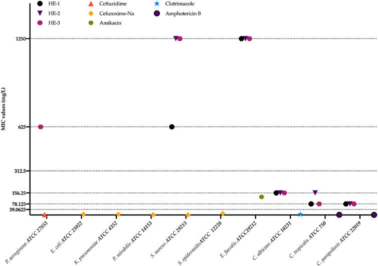
Figure 1. Antimicrobial activity assays. HE-1,2,3: extract codes. Ceftazidime, Cefuroxime-Na, Amikacin, Clotrimazole, and Amphotericin B: standard compounds.
These results were not as robust as those observed in our previous study. This discrepancy may be attributed to extractant solvent differences or variations in the chemical composition of the plants, which can be influenced by differences in their growth conditions.
Although both ethanol and methanol are polar solvents commonly used in phytochemical research, their slight differences in polarity and solvent strength can lead to distinct extraction efficiencies for certain classes of secondary metabolites. Methanol, being slightly more polar than ethanol, may enhance the extraction of certain phenolic acids and low-molecular-weight flavonoids, whereas ethanol might favor more lipophilic or mid-polar metabolites (Chaves et al., 2020). In a comparative study on Hypericum perforatum, Alahmad et al. (2022) observed that while the overall compound profiles were similar across different solvents, water tended to yield higher concentrations of certain phenolic constituents compared to methanol and ethanol (Alahmad et al., 2022). On the other hand, various phloroglucinol derivatives, which have low water solubility, are known for their potent antimicrobial properties (Tavlı, 2024). This suggests that methanol may offer advantages in extracting specific bioactive metabolites, owing to its ability to extract both phenolics and phloroglucinol derivatives effectively.
This variation in solvent polarity likely contributed to the observed differences in antifungal activity between the two studies. Additionally, the chemical composition of the extracts may have been influenced not only by the choice of solvent, but also by ecological and geographical differences in the plant material, including factors such as altitude, climate, and soil conditions. These environmental variables are known to affect the biosynthesis and accumulation of bioactive metabolites in medicinal plants and could therefore play a significant role in the observed phytochemical variation and corresponding biological activities (Çirak et al., 2006; Çirak and Radusiene, 2019; Şenkal and Uskutoglu, 2021; Bálintová et al., 2019; Kurt et al., 2018; Kucharíková et al., 2016; Kladar et al., 2015; Ghasemi Pirbalouti et al., 2011; Hosni et al., 2011; Çırak et al., 2011; Maggi et al., 2010; AI-Rifaee et al., 2010; Toker, 2009; Bagdonaite et al., 2009; Çirak et al., 2008; Mártonfi et al., 2006; Božin et al., 2013).
Phytochemical analysis revealed that the ethanolic extract of H. empetrifolium subsp. empetrifolium was particularly rich in chlorogenic acid, isoquercitrin, malic acid, proto-catechuic acid, quercetin, and fumaric acid. Additionally, several other phenolic metabolites—including salicylic acid, caffeic acid, p-coumaric acid, rutin, nicotiflorin, rosmarinic acid, naringenin, apigenin, and vanillin—were identified at lower concentrations.
Similarly, chemical examination of the H. empetrifolium subsp. empetrifolium extracts revealed marked differences in their phytochemical profiles. For instance, while ascorbic acid levels were relatively similar among the samples notable variations were observed for several key flavonoids and phenolic acids. HE-3 exhibited a substantially higher concentration of (−)-epigallocatechin (285.744
Reactive oxygen species (ROS), including free radicals such as superoxide (
The radical scavenging capacities of the extracts were assessed using three distinct chemical methods.The results were as follows: maximum inhibition values of 82.60
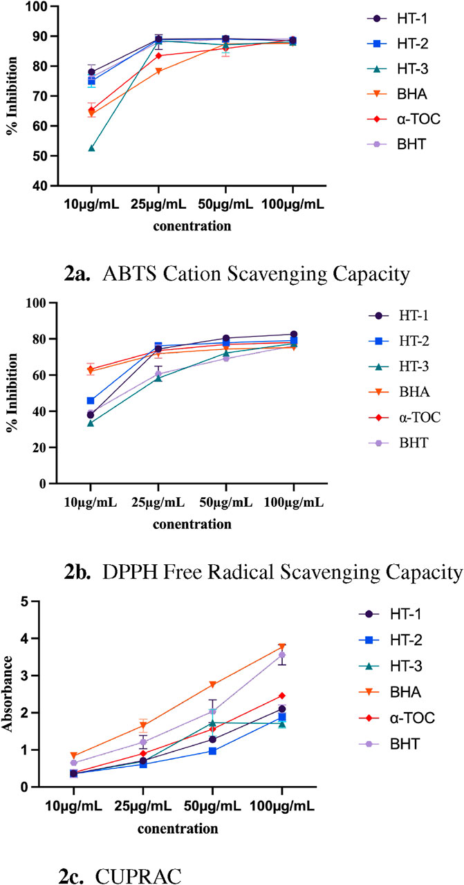
Figure 2. (a) ABTS Cation Scavenging Capacity. (b) DPPH Free Radical Scavenging Capacity. (c) CUPRAC. Chemical tests assessing radical scavenging capacity results. HE-1,2,3: extract codes. BHA,
To date, different investigations in literature have not only highlighted differences among species but also revealed variations in the antioxidant potential of the same species (particularly H. perforatum) sourced from different regions. Among of them, a study showed that among three Hypericum species grown in Türkiye—Hypericum aviculariifolium subsp. depilatum var. depilatum, Hypericum salsugineum, and H. perforatum—the methanolic extracts exhibited DPPH radical scavenging capacity of 88.29
Studies on cholinesterase inhibitors have demonstrated that, beyond their established efficacy in Alzheimer’s disease, these agents may also exert potential effects on mood disorders related to depression and stress. Consequently, cholinesterase inhibitors are being considered for their potential role in the treatment of depression, in addition to neurodegenerative conditions (Fitzgerald et al., 2020). Based on the anticholinesterase activity assays results, H. empetrifolium subsp. empetrifolium extracts—especially HE-1—demonstrate promising potential for the treatment of these conditions. In the present study, the cholinesterase inhibitory activities of the extracts were determined as IC50 values ranging from 8.16
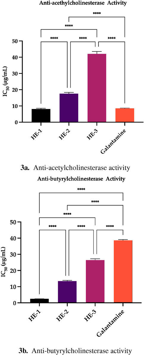
Figure 3. (a) Anti-acetylcholinesterase activity. (b) Anti-butyrylcholinesterase activity. Anti-cholinesterase activity results. HE-1,2,3: extract codes. Galantamine, kojic acid, acarbose, and ursolic acid: standard compounds. Statistical significance levels are indicated as follows: (*)
On the other hands, various studies have explored the cholinesterase inhibitory effects of Hypericum species, shedding light on their potential as natural anticholinesterase agents. Bozin and colleagues evaluated the anti-acetylcholinesterase activity of extracts of H. perforatum, Hypericum maculatum subsp. immaculatum, H. olympicum, Hypericum richeri subsp. grise-bachii, and Hypericum barbatum, reporting that the H. perforatum extract exhibited the highest activity (IC50 = 432.74
The investigation of tyrosinase enzyme inhibitors is of considerable importance for the discovery of novel therapeutic agents targeting a variety of health issues. These inhibitors effectively suppress the activity of tyrosinase—an enzyme critical for melanin synthesis—thereby offering potential benefits against hyperpigmentation and skin discoloration. In the cosmetic industry, such compounds are widely incorporated into skin whitening and spot treatment products, as they play a pivotal role in reducing melanin production and treating hyperpigmentation disorders. Moreover, the oxidative stress-related effects associated with tyrosinase are currently under investigation for their implications in neurodegenerative diseases, particularly in conditions such as Parkinson’s disease (Baber et al., 2023; Fernandes and Kerkar, 2017). Previous studies have demonstrated that extracts from various Hypericum species exhibit significant antityrosinase activity. An extract prepared from H. androsaemum fruits—rich in phenolic metabolites such as shikimic acid and chlorogenic acid—showed notable tyrosinase inhibition (IC50 = 229,1
In the present study, the tyrosinase inhibitory potential of H. empetrifolium subsp. empetrifolium extracts was further evaluated. These findings suggest that although H. empetrifoliumextracts possess antityrosinase activity, their inhibitory potency is substantially lower than that of kojic acid (Figure 4), similar to previous work (Bo et al., 2021).
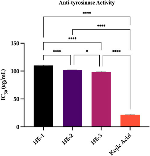
Figure 4. Anti-tyrosinase activity. Kojic acid: standard compound. Statistical significance levels are indicated as follows: (**)
H.ypericumascyron extracts prepared with ethyl acetate and methanol inhibited
Our findings are particularly noteworthy when compared with the existing literature, where various Hypericum species have also been reported to exhibit
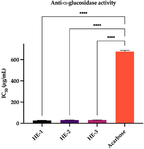
Figure 5. Anti-
Investigating inhibitors of extracellular matrix (ECM) degrading enzymes—such as collagenase, elastase, and hyaluronidase—is critical for developing effective anti-aging cosmeceuticals. These enzymes play a pivotal role in ECM remodeling; their overactivity contributes to the degradation of collagen, elastin, and hyaluronic acid, leading to loss of skin firmness, increased wrinkle formation, and other visible signs of aging. By targeting these enzymes, natural compounds can help preserve the skin’ structural integrity and mitigate age-related changes (Silva et al., 2021).
Studies on various Hypericum species have demonstrated promising inhibitory effects on these ECM-degrading enzymes, suggesting that their bioactive constituents could serve as safer and more effective alternatives to synthetic inhibitors in cosmetic formulations. This line of research is therefore essential not only for understanding the mechanisms behind skin aging but also for the development of innovative treatments that improve skin health and appearance. In studies assessing the anti-aging properties of Hypericum extracts from H. perforatum, H. calycinum, and H. confertum. H. calycinum consistently demonstrated the most potent activity. Its methanol extracts inhibited collagenase, elastase, and hyaluronidase with
The results of present study offer valuable insights into the enzyme inhibitory properties of H. empetrifolium subsp. empetrifolium extracts. Notably, no measurable inhibition of hyaluronidase was observed for HE-1, HE-2, and HE-3 under the tested conditions. This suggests that the bioactive constituents in the HE extracts may lack the necessary affinity or concentration to effectively inhibit hyaluronidase, an enzyme critical for hyaluronic acid degradation.
In contrast, the HE extracts demonstrated moderate elastase inhibitory activity, with
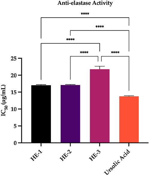
Figure 6. Anti-elastase Activity. Ursolic acid: standard compound. Statistical significance levels are indicated as follows: (****)
Although this study does not include primary ethnobotanical fieldwork, the selection of H. empetrifolium subsp. empetrifolium was based on its reported traditional use in the literature. The work aligns with the core principles of the Four Pillars of Best Practice in Ethnopharmacology (Heinrich et al., 2020), including proper taxonomic identification (with voucher specimen), use of pharmacologically relevant in vitro models, detailed phytochemical profiling, and transparent data reporting. The antifungal and enzyme inhibition assays were selected to reflect plausible therapeutic mechanisms relevant to the species’ ethnomedical context.
5 Conclusion
In conclusion, our study demonstrates that H. empetrifolium subsp. empetrifolium extracts exhibit significant antifungal activity against clinically relevant Candida strains, underscoring their potential as alternative therapeutic agents for fungal infections, their robust antifungal efficacy aligns with the pressing need for novel, effective antifungal agents.
Moreover, the extracts displayed potent
Phytochemical analyses via HPLC-DAD and LC-HR/MS further revealed notable variations in the profiles of key antioxidants, flavonoids, and phenolic metabolites among the samples. These differences, likely resulting from variations in collection locations and environmental conditions, emphasize the impact of growth conditions on the chemical composition and bioactivity of H. empetrifolium subsp. empetrifolium. Although lipophilic metabolites such as hypericin, pseudohypericin, and hyperforin were not detected, the presence of other bioactive phenolic constituents appears to underpin the diverse therapeutic properties observed.
Building upon these promising findings, future research should focus on the targeted isolation and structural elucidation of the most active constituents within H. empetrifolium subsp. empetrifolium extracts, employing advanced spectroscopic and chromatographic techniques. Moreover, standardized extraction protocols must be developed to ensure reproducibility and consistency in bioactivity, which are essential for translational applications. Given the demonstrated in vitro efficacy, comprehensive in vivo studies are crucial to assess pharmacokinetics, bioavailability, and potential toxicity profiles. In parallel, mechanistic investigations at the molecular level—particularly concerning enzyme inhibition and antifungal pathways—would provide deeper insights into the modes of action.
On the other hands, the notable chemical variations observed among samples collected from different geographical locations strongly suggest that environmental conditions significantly influence the phytochemical composition and, consequently, the bioactivity of H. empetrifolium subsp. empetrifolium. Therefore, future studies should include broader sampling from diverse habitats to identify the most favorable ecological conditions or specific locations associated with the highest concentration of bioactive metabolites. This approach would not only optimize raw material sourcing but also support conservation strategies and sustainable use of medicinal plant resources. Finally, the formulation of these extracts into biocompatible delivery systems could pave the way for novel phytopharmaceuticals and cosmeceuticals targeting fungal infections, metabolic disorders, and neurodegenerative diseases.
Data availability statement
The original contributions presented in the study are included in the article/Supplementary Material, further inquiries can be directed to the corresponding author.
Author contributions
SD: Writing – review and editing, Supervision, Methodology, Visualization, Investigation, Writing – original draft, Resources. OT: Software, Funding acquisition, Writing – original draft, Resources, Investigation, Formal Analysis, Writing – review and editing, Methodology, Data curation, Validation. EM: Conceptualization, Investigation, Data curation, Writing – review and editing, Formal Analysis, Methodology, Writing – original draft. AK: Methodology, Writing – review and editing, Investigation, Data curation, Writing – original draft, Formal Analysis. HS: Writing – review and editing, Formal Analysis, Writing – original draft, Data curation, Methodology, Investigation. MB: Methodology, Conceptualization, Investigation, Writing – review and editing, Writing – original draft, Formal Analysis, Data curation. EE: Funding acquisition, Writing – review and editing, Writing – original draft, Supervision, Resources, Investigation, Project administration, Validation, Formal Analysis, Data curation, Methodology, Visualization, Conceptualization.
Funding
The author(s) declare that no financial support was received for the research and/or publication of this article.
Acknowledgments
Even though the authors are based at different institutions and in different cities, working side-by-side on this project has been an absolute delight. From spur-of-the-moment online brainstorming sessions to the uncountable coffees shared virtually, distance quickly turned into synergy and fresh ideas. We are grateful to our families for their patience with our impromptu schedules and to our colleagues for their constant encouragement.
Collaboration really does make the science—and the journey—so much sweeter.
Conflict of interest
The authors declare that the research was conducted in the absence of any commercial or financial relationships that could be construed as a potential conflict of interest.
Generative AI statement
The author(s) declare that no Generative AI was used in the creation of this manuscript.
Publisher’s note
All claims expressed in this article are solely those of the authors and do not necessarily represent those of their affiliated organizations, or those of the publisher, the editors and the reviewers. Any product that may be evaluated in this article, or claim that may be made by its manufacturer, is not guaranteed or endorsed by the publisher.
Supplementary material
The Supplementary Material for this article can be found online at: https://www.frontiersin.org/articles/10.3389/fphar.2025.1618761/full#supplementary-material
References
Ahmad, V., Alotibi, I., Srivastava, S., Imran, M., Amir, M., Ali, M. S., et al. (2024). Computational approaches to evaluate the acetylcholinesterase binding interaction with taxifolin for the management of alzheimer’s disease. Molecules 29, 674. doi:10.3390/molecules29030674
Ai-Rifaee, M., Aburjai, T., and HaddadJ, N. (2010). Hypericin from Hypericum triquetrifolium in wild and under cultivation: variation revealed by genetic distance. Pharmacogn. Mag. 6, 50.
Akyuz, S., Kurt-Celep, I., Inan, Y., Ozdemir, O. E., Celep, E., and Yeşilada, E. (2021). In vitro evaluation of the bioactivity and bioaccessibility of Hypericum olympicum l. South Afr. J. Bot. 142, 316–324. doi:10.1016/j.sajb.2021.06.043
Alahmad, A., Alghoraibi, I., Zein, R., Kraft, S., Drager, G., Walter, J., et al. (2022). Identification of major constituents of Hypericum perforatum L. Extracts in Syria by development of a rapid, simple, and reproducible hplc-esi-q-tof ms analysis and their antioxidant activities. ACS Omega 7, 13475–13493. doi:10.1021/acsomega.1c06335
Alali, F., Tawaha, K., and Gharaibeh, M. (2009). Lc-ms and lc-pda analysis of Hypericum empetrifolium and Hypericum sinaicum. Z. für Naturforsch. C 64, 476–482. doi:10.1515/znc-2009-7-802
Antognoni, F., Lianza, M., Poli, F., Buccioni, M., Santinelli, C., Caprioli, G., et al. (2017). Polar extracts from the berry-like fruits of Hypericum androsaemum L. As a promising ingredient in skin care formulations. J. Ethnopharmacol. 195, 255–265. doi:10.1016/j.jep.2016.11.029
Apak, R., Guclu, K., Ozyurek, M., and Karademir, S. (2004). Novel total antioxidant capacity index for dietary polyphenols and vitamins c and E, using their cupric ion reducing capability in the presence of neocuproine: Cuprac method. J. Agric. Food Chem. 52, 7970–7981. doi:10.1021/jf048741x
Baber, M., Crist, C., Devolve, N., and Patrone, J. (2023). Tyrosinase inhibitors: a perspective. Molecules 28, 5762. doi:10.3390/molecules28155762
Bagdonaite, E., Janulis, V., Ivanauskas, L., and Labokas, J. (2009). Variation in contents of hypericin and flavonoids in Hypericum maculatum (Hypericaceae) from Lithuania. Acta Bot. Hung. 51, 237–244. doi:10.1556/abot.51.2009.3-4.1
Bálintová, M., Bruňáková, K., Petijová, L., and Čellárová, E. (2019). Targeted metabolomic profiling reveals interspecific variation in the genus hypericum in response to biotic elicitors. Plant Physiology Biochem. 135, 348–358. doi:10.1016/j.plaphy.2018.12.024
Barros, F. M. C. d., Poser, G. L. V., Schenkel, E. E. P., Rates, S. M. K., Luciano, S. C., Apel, M. A., et al. (2013). Antifungal and antichemotactic activities and quantification of phenolic compounds in lipophilic extracts of hypericum spp. native to south Brazil. Industrial Crops Prod. 44, 294–299. doi:10.1016/j.indcrop.2012.11.017
Bejaoui, A., Ben Salem, I., Rokbeni, N., Boussaid, M., and Boulila, A. (2017). Bioactive compounds from Hypericum humifusum and hypericum perfoliatum: inhibition potential of polyphenols with acetylcholinesterase and key enzymes linked to type-2 diabetes. Pharm. Biol. 55, 906–911. doi:10.1080/13880209.2016.1270973
Benedict, K., Whitham, H., and Jackson, B. (2022). “Economic burden of fungal diseases in the United States,” in Open forum infectious diseases (Oxford University Press US).
Blois, M. (1958). Antioxidant determinations by the use of a stable free radical. Nature 181, 1199–1200. doi:10.1038/1811199a0
Bo, M., Ersoy, E., Ero, E., Çınar, E., Mataracı Kara, E., Yeşil Çantürk, Y., et al. (2021). Volatile and phenolic profiling of a traditional medicinal plant, Hypericum empetrifolium with in vitro biological activities. J. Ethnopharmacol. 272, 113933. doi:10.1016/j.jep.2021.113933
Božin, B., Kladar, N., Grujić, N., Anačkov, G., Samojlik, I., Gavarić, N., et al. (2013). Impact of origin and biological source on chemical composition, anticholinesterase and antioxidant properties of some st. John's wort species (hypericum spp., hypericaceae) from the central balkans. Molecules 18, 11733–11750. doi:10.3390/molecules181011733
Chaves, J. O., de Souza, M. C., da Silva, L. C., Lachos-Perez, D., Torres-Mayanga, P. C., Machado, A. P. F., et al. (2020). Extraction of flavonoids from natural sources using modern techniques. Front. Chem. 8, 507887. doi:10.3389/fchem.2020.507887
Çirak, C., and Radusiene, J. (2019). Factors affecting the variation of bioactive compounds in hypericum species. Biol. Futura 70, 198–209. doi:10.1556/019.70.2019.25
Çirak, C., Radusiene, J., and Arslan, B. (2008). Variation of bioactive substances in Hypericum montbretii during plant growth. Nat. Prod. Res. 22, 246–252. doi:10.1080/14786410701642623
Çirak, C., Radusiene, J., Janulis, V., and Ivanauskas, L. (2006). Variation of hypericin in st. John’s wort (Hypericum perforatum) from wild populations of northern Turkey. Acta Bot. Hung. 48, 55–64. doi:10.1556/abot.48.2006.1-2.8
Çırak, C., Radušienė, J., Janulis, V., Ivanauskas, L., Çamaş, N., and Ayan, A. K. (2011). Phenolic constituents of Hypericum triquetrifolium turra (Guttiferae) growing in Turkey: variation among populations and plant parts. Turkish J. Biol. 35, 449–456. doi:10.3906/biy-1002-36
Clinical and Laboratory Standards Institute (CLSI) (1997). Reference method for broth dilution antifungal susceptibility testing of yeasts; approved standard. Second Edition. Wayne, PA, USA: Clinical and Laboratory Standards Institute.
Clinical and Laboratory Standards Institute (CLSI) (2017). Reference method for broth dilution antifungal susceptibility testing of yeasts; approved standard—fourth edition (CLSI document M27). Wayne, PA, USA: Clinical and Laboratory Standards Institute.
Clinical and Laboratory Standards Institute (CLSI) (2020). Performance standards for antimicrobial susceptibility testing CLSI document M100 950 west valley road, suite 2500. Wayne, Pennsylvania 19087, USA: Clinical and Laboratory Standards Institute.
Couladis, M., Badisa, R., Baziou, P., Chaudhuri, S., Pilarinou, E., Verykokidou, E., et al. (2002). Antioxidant and cytotoxic activities of hypericum sp. on brine shrimps and human cancer cell lines. Phytotherapy Res. 16, 719–722. doi:10.1002/ptr.1042
Crockett, S., Wenzig, E.-M., Kunert, O., and Bauer, R. (2008). Anti-inflammatory phloroglucinol derivatives from Hypericum empetrifolium. Phytochem. Lett. 1, 37–43. doi:10.1016/j.phytol.2007.12.003
Dirir, A. M., Daou, M., Yousef, A. F., and Yousef, L. F. (2022a). A review of alpha-glucosidase inhibitors from plants as potential candidates for the treatment of type-2 diabetes. Phytochem. Rev. 21, 1049–1079. doi:10.1007/s11101-021-0973-1
Dirir, A. M., Daou, M., Yousef, A. F., and Yousef, L. F. (2022b). A review of alpha-glucosidase inhibitors from plants as potential candidates for the treatment of type-2 diabetes. Phytochem. Rev. 21, 1049–1079. doi:10.1007/s11101-021-09773-1
Dulger, G., and Dulger, B. (2016). Antifungal activity of Hypericum havvae against some medical candida yeast and cryptococcus species. Trop. J. Pharm. Res. 13, 405. doi:10.4314/tjpr.v13i3.14
Ero, E., Özbek Çelik, B., and Mat, A. (2018b). Antimicrobial activities of five endemic hypericum species from Anatolia compared with Hypericum perforatum. J. Res. Pharm. 23, 114–119. doi:10.12991/jrp.2018.115
Ero, E., Yılmaz Özden, T., Özsoy, N., and Mat, A. (2018a). Evaluation of chemical composition, antioxidant and anti-acetylcholinesterase activities of Hypericum neurocalycinum and Hypericum malatyanum. South Afr. J. Bot. 114, 104–110. doi:10.1016/j.sajb.2017.10.022
Ersoy, E., Ero, E., Bo, M., and Mat, A. (2020). Evaluation of in vitro biological activities of three hypericum species (h. Calycinum, H. Confertum, and H. Perforatum) from Turkey. South Afr. J. Bot. 130, 141–147. doi:10.1016/j.sajb.2019.12.017
Ersoy, E., Ero, E., Bo, M., Yılmaz, M., and Mat, A. (2019). Anti-aging potential and anti-tyrosinase activity of three hypericum species with focus on phytochemical composition by Lc–Ms/Ms. Industrial Crops Prod. 141, 111735. doi:10.1016/j.indcrop.2019.111735
Eun, J. K., Eunyoung, L., Yee, G. K., Hye, M. Y., Jae, Y. C., Seong, R. K., et al. (2020). Trends in the epidemiology of candidemia in intensive care units from 2006 to 2017: results from the Korean national healthcare-associated infections surveillance system. Front. Med. 7, 606976. doi:10.3389/fmed.2020.606976
European Pharmacopoeia (2008). St. John’s wort (hyperici herba). Strasbourg, France: Council of Europe.
Fernandes, M., and Kerkar, S. (2017). Microorganisms as a source of tyrosinase inhibitors: a review. Ann. Microbiol. 67, 343–358. doi:10.1007/s13213-017-1261-7
Fitzgerald, P., Hale, P., Ghimire, A., and Watson, B. (2020). Repurposing cholinesterase inhibitors as antidepressants? Dose and stress-sensitivity May be critical to opening possibilities. Front. Behav. Neurosci. 14, 620119. doi:10.3389/fnbeh.2020.620119
Foret, M. K., Lincoln, R., Do Carmo, S., Cuello, A. C., and Cosa, G. (2020). Connecting the “dots”: from free radical lipid autoxidation to cell pathology and disease. Chem. Rev. 120, 12757–12787. doi:10.1021/acs.chemrev.0c00761
Francis, P. T., Palmer, A. M., Snape, M., and Wilcock, G. K. (1999). The cholinergic hypothesis of alzheimer’s disease: a review of progress. J. Neurology, Neurosurg. and Psychiatry 66, 137–147. doi:10.1136/jnnp.66.2.137
Ghasemi Pirbalouti, A., Rahnama, G. H., Malekpoor, F., and Roohi Broujeni, H. R. (2011). Variation in antibacterial activity and phenolic content of Hypericum scabrum L. Populations. J. Med. Plants Res. 5, 4119–4125.
Gómez-López, A. (2020). Antifungal therapeutic drug monitoring: focus on drugs without a clear recommendation. Clin. Microbiol. Infect. 26, 1481–1487. doi:10.1016/j.cmi.2020.05.037
Gow, N. A. R., Johnson, C., Berman, J., Coste, A. T., Cuomo, C. A., Perlin, D. S., et al. (2022). The importance of antimicrobial resistance in medical mycology. Nat. Commun. 13, 5352. doi:10.1038/s41467-022-32249-5
GuillenQuispe, Y., Hwang, S., Wang, Z., Zuo, G., and Lim, S. (2017). Screening in vitro targets related to diabetes in herbal extracts from Peru: identification of active compounds in Hypericum laricifolium juss. By offline high-performance liquid chromatography. Int. J. Mol. Sci. 18, 2512. doi:10.3390/ijms1812512
Gulcin, I., and Alwasel, S. (2023). Dpph radical scavenging assay. Processes 11, 2248. doi:10.3390/pr11082248
Hajizadeh, K., Alivand, Z., Rahmani, V., Mehrannia, L., Nami, S., Shokouhi, B., et al. (2025). Comparing the effects of mycozin and clotrimazole 1% creams on vaginal candidiasis: a triple-blinded randomized controlled trial. Sci. Rep. 15, 2356. doi:10.1038/s41598-024-84389-x
Hampel, H., Mesulam, M.-M., Cuello, A. C., Khachaturian, A. S., Vergallo, A., Farlow, M., et al. (2019). Revisiting the cholinergic hypothesis in alzheimer’s disease: emerging evidence from translational and clinical research. J. Prev. Alzheimer’s Dis. 6, 2–15. doi:10.14283/jpad.2018.43
Hasim, S., and Coleman, J. (2019). Targeting the fungal cell wall: current therapies and implications for development of alternative antifungal agents. Future Med. Chem. 11, 869–883. doi:10.4155/fmc-2018-0465
Hearing, V., and Jimenez, M. (1987). Mammalian tyrosinase–the critical regulatory control point in melanocyte pigmentation. Int. J. Biochem. 19, 1141–1147. doi:10.1016/0020-711x(87)90095-4
Heinrich, M., Appendino, G., Efferth, T., Fürst, R., Izzo, A. A., Kayser, O., et al. (2020). Best practice in research–overcoming common challenges in phytopharmacological research. J. Ethnopharmacol. 246, 112230. doi:10.1016/j.jep.2019.112230
Hernandez, M., Falé, P., Araújo, M., and Serralheiro, M. (2010). Acetylcholinesterase inhibition and antioxidant activity of the water extracts of several hypericum species. Food Chem. 120, 1076–1082. doi:10.1016/j.foodchem.2009.11.055
Hosni, K., Msaada, K., Ben Taârit, M., and Marzouk, B. (2011). Phenological variations of secondary metabolites from Hypericum triquetrifolium turra. Biochem. Syst. Ecol. 39, 43–50. doi:10.1016/j.bse.2011.01.001
Ion, V., Ielciu, I., Cârje, A.-G., Muntean, D. L., Crişan, G., and Păltinean, R. (2022). Hypericum Spp.—An overview of the extraction methods and analysis of compounds. Separations 9, 17. doi:10.3390/separations9010017
Ivanov, M., Ćirić, A., and Stojković, D. (2022). Emerging antifungal targets and strategies. Int. J. Mol. Sci. 23, 2756. doi:10.3390/ijms23052756
Jin, D. X., He, J. F., Zhang, K. Q., Luo, X. G., and Zhang, T. C. (2021). α-glucosidase inhibition action of major flavonoids identified from Hypericum attenuatum choisy and their synergistic effects. Chem. Biodivers. 18, e2100244. doi:10.1002/cbdv.202100244
Jung, H. (2020). Hyaluronidase: an overview of its properties, applications, and side effects. Archives plastic Surg. 47, 297–300. doi:10.5999/aps.2020.00752
Kakouri, E., Trigas, P., Daferera, D., Skotti, E., Tarantilis, P. A., and Kanakis, C. (2023). Chemical characterization and antioxidant activity of nine hypericum species from Greece. Antioxidants 12, 899. doi:10.3390/antiox12040899
Kang, W.-Y., Song, Y.-L., and Zhang, L. (2011). α-glucosidase inhibitory and antioxidant properties and antidiabetic activity of Hypericum ascyron l. Med. Chem. Res. 20, 809–816. doi:10.1007/s00044-010-9391-5
Kaur, I. P., and Kakkar, S. (2010). Topical delivery of antifungal agents. Expert Opin. drug Deliv. 7, 1303–1327. doi:10.1517/17425247.2010.525230
Keshwania, P., Kaur, N., Chauhan, J., Sharma, G., Afzal, O., Alfawaz Altamimi, A. S., et al. (2023). Superficial dermatophytosis across the world’s populations: potential benefits from nanocarrier-based therapies and rising challenges. ACS omega 8, 31575–31599. doi:10.1021/acsomega.3c01988
Kıno, B. K., Dirmenci, T., Alwasel, S. H., Gören, A. C., and Gülçin, İ. (2023). Quantification of main secondary metabolites of Satureja icarica p.h. davis (Lamiaceae) by lc-hrms and evaluation of antioxidant capacities. J. Chem. Metrology 17, 199–214. doi:10.25135/jcm.2311.2956
Kladar, N., Srdenović, B., Grujić, N., Bokić, B., Rat, M., Anačkov, G., et al. (2015). Ecologically and ontogenetically induced variations in phenolic compounds and biological activities of Hypericum maculatum subsp. maculatum, Hypericaceae. Braz. J. Bot. 38, 703–715. doi:10.1007/s40415-015-0177-3
Kucharíková, A., Kimáková, K., Janfelt, C., and Čellárová, E. (2016). Interspecific variation in localization of hypericins and phloroglucinols in the genus hypericum as revealed by desorption electrospray ionization mass spectrometry imaging. Physiol. Plant. 157, 2–12. doi:10.1111/ppl.12422
Kumar, S., Kumar, A., Roudbary, M., Mohammadi, R., Černáková, L., and Rodrigues, C. (2022). Overview on the infections related to rare candida species. Pathogens 11, 963. doi:10.3390/pathogens11090963
Kurt, D., Caliskan, O., Odabaş, M. S., Radusiene, J., and Cirak, C. (2018). Modeling the altitudinal variation in secondary metabolite contents of Hypericum orientale from Turkey. Int. Biol. Biomed. J. 4.
Lass-Flörl, C., Kanj, S., Govender, N., Thompson III, G., Ostrosky-Zeichner, L., and Ma, G. (2024). Invasive candidiasis. Nat. Rev. Dis. Prim. 10, 20. doi:10.1038/s41572-024-00503-3
Lee, K., Kim, J., Cho, J., and Choi, J. (1999). Inhibitory effects of 150 plant extracts on elastase activity, and their anti-inflammatory effects. Int. J. Cosmet. Sci. 21, 71–82. doi:10.1046/j.1467-2494.1999.181638.x
Llauradó Maury, G., Méndez Rodríguez, D., Hendrix, S., Escalona Arranz, J. C., Fung Boix, Y., Pacheco, A., et al. (2020). Antioxidants in plants: a valorization potential emphasizing the need for the conservation of plant biodiversity in Cuba. Antioxidants 9, 1048. doi:10.3390/antiox9111048
López, V., Iannarelli, R., Caprioli, G., and Maggi, F. (2016). Methanolic extract from red berry-like fruits of hypericum androsaemum: chemical characterization and inhibitory potential of central nervous system enzymes. Industrial Crops Prod. 94, 363–367. doi:10.1016/j.indcrop.2016.09.007
Lortholary, O., Renaudat, C., Sitbon, K., Madec, Y., Denoeud-Ndam, L., Wolff, M., et al. (2011). Recent exposure to caspofungin or fluconazole influences the epidemiology of candidemia: a prospective multicenter study involving 2,441 patients. Antimicrob. Agents Chemother. 55, 532–538. doi:10.1128/AAC.01128-10
Lourenço, S., Moldão-Martins, M., and Alves, V. (2019). Antioxidants of natural plant origins: from sources to food industry applications. Molecules 24, 4132. doi:10.3390/molecules24224132
Maggi, F., Tirillini, B., Vittori, S., Sagratini, G., and Papa, F. (2010). Chemical composition and seasonal variation of Hypericum hircinum L. Subsp. majus (Aiton) N. Robson essential oil. J. Essent. Oil Res. 22, 434–443. doi:10.1080/10412905.2010.9700366
Maltaş, E., Uysal, A., Yıldıztugay, E., Alada, M. O., Yıldız, S., and Küçüködük, M. (2013). Investigation of antioxidant and antibacterial activities of some hypericum species. Fresenius Environ. Bull. 22, 862–869.
Mamali, V., Siopi, M., Charpantidis, S., Samonis, G., Tsakris, A., Vrioni, G., et al. (2022). Increasing incidence and shifting epidemiology of candidemia in Greece: results from the first nationwide 10-year survey. J. Fungi 8, 116. doi:10.3390/jof8020116
Mandrone, M., Lorenzi, B., Venditti, A., Guarcini, L., Bianco, A., Sanna, C., et al. (2015). Antioxidant and anti-collagenase activity of Hypericum hircinum l. Industrial Crops Prod. 76, 402–408. doi:10.1016/j.indcrop.2015.07.012
Mártonfi, P., Repčák, M., and Zanvit, P. (2006). Secondary metabolites variation in Hypericum maculatum and its relatives. Biochem. Syst. Ecol. 34, 56–59. doi:10.1016/j.bse.2005.07.008
Meirelles, G. C., Poser, G. L. V., Schenkel, E. P., Rates, S. M. K., de Oliveira, L. F., von Poser, G. L., et al. (2017). Synergistic antifungal activity of the lipophilic fraction of Hypericum carinatum and fluconazole. Rev. Bras. Farmacogn. 27, 118–123. doi:10.1016/j.bjp.2016.08.001
Mendling, W., Atef El Shazly, M., and Zhang, L. (2020). Clotrimazole for vulvovaginal candidosis: more than 45 years of clinical experience. Pharmaceuticals 13, 274. doi:10.3390/ph13100274
Miceli, M., and Kauffman, C. (2015). Isavuconazole: a new broad-spectrum triazole antifungal agent. Clin. Infect. Dis. 61, 1558–1565. doi:10.1093/cid/civ571
Moreno, M., Isla, M., Sampietro, A., and Vattuone, M. A. (2000). Comparison of the free radical-scavenging activity of propolis from several regions of Argentina. J. Ethnopharmacol. 71, 109–114. doi:10.1016/s0378-8741(99)00189-0
Mota Fernandes, C., das Neves, R. N., Arendrup, M. C., Denning, D. W., Alastruey-Izquierdo, A., Ojima, I., et al. (2021). The future of antifungal drug therapy: novel compounds and targets. Antimicrob. Agents Chemother. 65, e01719-20. doi:10.1128/AAC.01719-20
Neha, K., Haider, M. R., Pathak, A., and Yar, M. S. (2019). Medicinal prospects of antioxidants: a review. Eur. J. Med. Chem. 178, 687–704. doi:10.1016/j.ejmech.2019.06.010
Nenadis, N., Wang, L.-F., Tsimidou, M., and Zhang, H.-Y. (2004). Estimation of scavenging activity of phenolic compounds using the abts•+ assay. J. Agric. Food Chem. 52, 4669–4674. doi:10.1021/jf0400056
Ozkok, F., Bo, M., Tuneg, M., Atalay, V., Onul, N., Asgarova, K., et al. (2022). Evaluation of acetyl-and butyrylcholinesterase enzyme inhibitory activities and cytotoxic activities of anthraquinone derivatives. J. Turkish Chem. Soc. Sect. A Chem. 9, 729–740. doi:10.18596/jotcsa.963290
Ozturk, M., Uysal, I., Gucel, S., Altundag, E., Dogan, Y., and Baslar, S. (2013). Medicinal uses of natural dye-yielding plants in Turkey. Res. J. Text. Appar. 17, 69–80. doi:10.1108/rjta-17-02-2013-b010
Özyürek, M., Güçlü, K., Tütem, E., Sözgen Başkan, K., Erça, E., Esin Çelik, S., et al. (2011). A comprehensive review of cuprac methodology. Anal. Methods 3, 2439–2453. doi:10.1039/C1AY05320E
Pammi, S., Suresh, B., and Giri, A. (2022). Antioxidant potential of medicinal plants. J. Crop Sci. Biotechnol. 26, 13–26. doi:10.1007/s12892-022-00159-z
Pandey, A. K., Kumar, S., Pandey, A. K., and Reis, F. (2021). Editorial: combating redox imbalance-associated complications with natural products. Front. Pharmacol. 12, 802750. doi:10.3389/fphar.2021.802750
Pfaller, M., Diekema, D., Turnidge, J., Castanheira, M., and Jones, R. (2019). “Twenty years of the sentry antifungal surveillance program: results for candida species from 1997–2016,” in Open forum infectious diseases (Oxford University Press US).
Pappas, G. P., Lionakis, M. S., Arendrup, M. C., Ostrosky-Zeichner, L., and Kullberg, B. J. (2018). Invasive candidiasis. Nat. Rev. Dis. Prim. 4, 1–20. doi:10.1038/nrdp.2018.26
Pianalto, K., and Alspaugh, J. (2016). New Horizons in antifungal therapy. J. Fungi 2, 26. doi:10.3390/jof2040026
Pisoschi, A. M., Pop, A., Iordache, F., Stanca, L., Predoi, G., and Serban, A. I. (2021). Oxidative stress mitigation by antioxidants - an overview on their chemistry and influences on health status. Eur. J. Med. Chem. 209, 112891. doi:10.1016/j.ejmech.2020.112891
Pitasi, G., Brancale, A., Floris, S., Fais, A., Gitto, R., and Luca, L. D. (2024). Computational approach to identifying new chemical entities as elastase inhibitors with potential antiaging effects. Int. J. Mol. Sci. 25, 11174. doi:10.3390/ijms252011174
Re, R., Pellegrini, N., Proteggente, A., Pannala, A., Yang, M., and Rice-Evans, C. (1999). Antioxidant activity applying an improved abts radical cation decolorization assay. Free Radic. Biol. Med. 26, 1231–1237. doi:10.1016/s0891-5849(98)00315-3
Royal Botanic Gardens, Kew (2025). Hypericum empetrifolium subsp. empetrifolium. plants world online.
Saddiqe, Z., Naeem, I., Abbas, G., and Shahzad, M. (2014). Aerial parts of Hypericum olympicum possess antioxidant, anti-lipid peroxidation and antiglycation activity. Int. J. Phytomedicine 6, 248–255.
Samaneh, Z., Asieh, B., Mahmud Tareq, H. K., Munoz-Munoz, J., Garcia-Molina, F., Garcia-Canovas, F., et al. (2019). A comprehensive review on tyrosinase inhibitors. J. Enzyme Inhibition Med. Chem. 34, 279–309. doi:10.1080/14756366.2018.1545767
Schieber, M., and Navdeep, C. (2014). Ros function in redox signaling and oxidative stress. Curr. Biol. 24, R453–R462. doi:10.1016/j.cub.2014.03.034
Schmidt, J., Lauridsen, M., Dragsted, L., Nielsen, J., and Staerk, D. (2012a). Development of a bioassay-coupled HPLC-SPE-ttNMR platform for identification of α-glucosidase inhibitors in Apple peel (malus ×Domestica borkh.). Food Chem. 135, 1692–1699. doi:10.1016/j.foodchem.2012.05.075
Schmidt, S., Jürgenliemk, G., Schmidt, T., Skaltsa, H., and Heilmann, J. (2012c). Bi-tri-and polycyclic acylphloroglucinols from Hypericum empetrifolium. J. Nat. Prod. 75, 1697–1705. doi:10.1021/np300237n
Schmidt, S., Jürgenliemk, G., Skaltsa, H., and Heilmann, J. (2012b). Phloroglucinol derivatives from Hypericum empetrifolium with antiproliferative activity on endothelial cells. Phytochemistry 77, 218–225. doi:10.1016/j.phytochem.2011.11.014
Selvan, P., Vajravelu, L., Mohanraj, H., and Ramakrishna, M. (2022). Monitoring the spectrum of candidemia and its anti-fungal resistance in a tertiary care Centre–An emerging global alarm. J. Pure % Appl. Microbiol. 16, 2704–2711. doi:10.22207/jpam.16.4.41
Şenkal, B. C., and Uskutoglu, T. (2021). Hypericum taxa of Turkey’s flora and intra-population variation of morpho-agronomic traits in H. heterophyllum vent., an endemic species. J. Inst. Sci. Technol. 11, 743–752. doi:10.21597/jist.795693
Sies, H. (2018). On the history of oxidative stress: concept and some aspects of current development. Curr. Opin. Toxicol. 7, 122–126. doi:10.1016/j.cotox.2018.01.002
Silva, A. R., Taofiq, O., Ferreira, I. C. F. R., and Barros, L. (2021). Hypericum genus cosmeceutical application – a decade comprehensive review on its multifunctional biological properties. Industrial Crops Prod. 159, 113053. doi:10.1016/j.indcrop.2020.113053
Slinkard, K., and Singleton, V. (1977). Total phenol analysis: automation and comparison with manual methods. Am. J. Enology Vitic. 28, 49–55. doi:10.5344/ajev.1977.28.1.49
Tavlı, O. (2024). Pharmacognosic research on some hypericum species growing in Türkiye. Istanbul: Institute of Health Science, Department of Pharmacognosy.
Tawaha, K., Gharaibeh, M., El-Elimat, T., and Alali, F. Q. (2010). Determination of hypericin and hyperforin content in selected Jordanian hypericum species. Industrial Crops Prod. 32, 241–245. doi:10.1016/j.indcrop.2010.04.017
Thomas-Rüddel, D. O., Schlattmann, P., Pletz, M., Kurzai, O., and Bloos, F. (2022). Risk factors for invasive candida infection in critically ill patients: a systematic review and meta-analysis. CHEST 161, 345–355. doi:10.1016/j.chest.2021.08.081
Timsit, J., Ruppé, E., Barbier, F., Tabah, A., and Bassetti, M. (2020). Bloodstream infections in critically ill patients: an expert statement. Intensive care Med. 46, 266–284. doi:10.1007/s00134-020-05950-6
Tocci, N., Perenzoni, D., Iamonico, D., Fava, F., Weil, T., and Mattivi, F. (2018a). Extracts from Hypericum hircinum subsp. majus exert antifungal activity against a panel of sensitive and drug-resistant clinical strains. Front. Pharmacol. 9, 382. doi:10.3389/fphar.2018.00382
Tocci, N., Weil, T., Perenzoni, D., Narduzzi, L., Madriñán, S., Crockett, S., et al. (2018b). Phenolic profile, chemical relationship and antifungal activity of andean hypericum species. Industrial Crops Prod. 112, 32–37. doi:10.1016/j.indcrop.2017.10.030
Toker, Z. (2009). Variation of total hypericin, phenolic and flavonoid compounds in Hypericum triquetrifolium during its phenological cycle. Pharm. Biol. 47, 285–288. doi:10.1080/13880200802578983
Trovato, A., Raneri, E., Kouladis, M., Tzakou, O., Taviano, M., and Galati, E. (2001). Anti-inflammatory and analgesic activity of Hypericum empetrifolium willd. (guttiferae). Il Farm. 56, 455–457. doi:10.1016/S0014-827X(01)01061-8
Tung, J., Mark, G., and Hollis, G. (1994). A microplate assay for hyaluronidase and hyaluronidase inhibitors. Anal. Biochem. 223, 149–152. doi:10.1006/abio.1994.1560
Vallabhaneni, S., Cleveland, A. A., Farley, M. M., Harrison, L. H., Schaffner, W., Beldavs, Z. G., et al. (2015). Epidemiology and risk factors for echinocandin nonsusceptible Candida glabrata bloodstream infections: data from a large multisite population-based candidemia surveillance program, 2008–2014. Open Forum Infect. Dis. 2, ofv163. doi:10.1093/ofid/ofv163
Yn, G. Q., Hwang Sh, W. Z., Zuo, G. L. S., and Lim, S. S. (2017). Screening in vitro targets related to diabetes in herbal extracts from Peru: identification of active compounds in Hypericum laricifolium juss. By offline high-performance liquid chromatography. Int. J. Mol. Sci. 18, 2512. doi:10.3390/ijms18122512
Zeliou, K., Koui, E.-M., Papaioannou, C., Koulakiotis, N. S., Iatrou, G., Tsarbopoulos, A., et al. (2020). Metabolomic fingerprinting and genetic discrimination of four Hypericum taxa from Greece. Phytochemistry 174, 112290. doi:10.1016/j.phytochem.2020.112290
Zheleva-Dimitrova, D., Nedialkov, P., and Kitanov, G. (2010). Radical scavenging and antioxidant activities of methanolic extracts from hypericum species growing in Bulgaria. Pharmacogn. Mag. 6, 74–78. doi:10.4103/0973-1296.62889
Zhong, L., Li, P., Chang, X., Qi, K., Tang, K., Zheng, C., et al. (2022). Incidence, clinical characteristics, risk factors and outcomes of patients with mixed candida/bacterial bloodstream infections: a retrospective study. Ann. Clin. Microbiol. Antimicrob. 21, 45. doi:10.1186/s12941-022-00538-y
Keywords: Hypericum empetrifolium subsp. empetrifolium Willd, anti-candida, anti-cholinesterase, anti-α-glucosidase, anti-aging, HPLC, LC-HR/MS
Citation: Demirci Kayıran S, Tavlı ÖF, Mataracı Kara E, Kaplan A, Şahin H, Boğa M and Eroğlu Özkan E (2025) Hypericum empetrifolium subsp. empetrifolium: an assessment of its antifungal, antidiabetic, anti-aging, and neuroprotective potential. Front. Pharmacol. 16:1618761. doi: 10.3389/fphar.2025.1618761
Received: 26 April 2025; Accepted: 16 June 2025;
Published: 07 July 2025.
Edited by:
Herbert Júnior Dias, Ciência e Tecnologia Goiano, BrazilReviewed by:
Flaviane Maria Galvão Rocha, University of São Paulo, BrazilRamona Paltinean, University of Medicine and Pharmacy Iuliu Hatieganu, Romania
Copyright © 2025 Demirci Kayıran, Tavlı, Mataracı Kara, Kaplan, Şahin, Boğa and Eroğlu Özkan. This is an open-access article distributed under the terms of the Creative Commons Attribution License (CC BY). The use, distribution or reproduction in other forums is permitted, provided the original author(s) and the copyright owner(s) are credited and that the original publication in this journal is cited, in accordance with accepted academic practice. No use, distribution or reproduction is permitted which does not comply with these terms.
*Correspondence: Esra Eroğlu Özkan, ZXNlcm9nbHVAaXN0YW5idWwuZWR1LnRy