 Deming Liu
 Xue Jiang
 Yanxi Li
 Hua Yang
- Chongqing Clinical Research Center for Dermatology, Chongqing Key Laboratory of Integrative Dermatology Research, Key Laboratory of External Therapies of Traditional Chinese Medicine in Eczema, Department of Dermatology, Chongqing Traditional Chinese Medicine Hospital, The First Affiliated Hospital of Chongqing College of Traditional Chinese Medicine, Chongqing, China
Licochalcone A (Lico-A), a flavonoid compound extracted from Glycyrrhiza uralensis, exhibiting multiple pharmacological properties including anti-inflammatory, antibacterial, antioxidant, and antitumor effects. It demonstrates significant therapeutic potential in the field of dermatological treatment. This review focuses on the efficacy of Lico-A in the treatment of acne, atopic dermatitis, rosacea, pigmentation disorders, and skin tumors. Mechanistically, Lico-A targets multiple signaling pathways such as NLRP3 inflammasome, NF-κB, PLC/ERK/STAT3, AP-1, and Nrf2/HO-1, thereby modulating inflammatory cascades, oxidative stress, melanogenesis, and tumorigenic processes. Clinical studies have also confirmed its ability to reduce inflammatory lesions, improve skin barrier function, and suppressing hyperpigmentation. However, current research is limited by geographical bias, a lack of high-quality randomized controlled trials, and reliance on preclinical models. Future studies should focus on multicenter randomized controlled trials, advanced delivery systems, and mechanistic investigations using systems biology. Addressing these gaps could establish Lico-A as a multifunctional, evidence-based dermatologic therapy for inflammatory, infectious, and neoplastic skin disorders.
1 Introduction
Skin diseases are the fourth leading cause of disability worldwide, affecting approximately one-third of the world’s population (Flohr and Hay, 2021; Trakatelli et al., 2023). In 2019, the burden of skin and subcutaneous diseases, measured in disability-adjusted life years, reached 42,883,695.48, underscoring their substantial impact on global health and the consequent need for effective therapeutic interventions (Yakupu et al., 2023). Current treatment modalities for skin diseases include antibiotics, glucocorticoids, immunosuppressants, and biologics. These agents are often limited by transient efficacy, high relapse rates upon discontinuation, and the emergence of drug resistance. Additionally, some medications are associated with hepatotoxicity and nephrotoxicity, highlighting the urgent demand for safer and more effective alternatives.
Recently, plant-derived natural compounds have emerged as promising therapeutic candidates for dermatological conditions, owing to their low skin irritancy, reduced propensity for resistance development, and ability to modulate skin microbiota (Wang et al., 2025). Among these compounds, licochalcone A (Lico-A), a flavonoid isolated from the roots and stems of Glycyrrhiza uralensis Fisch., has emerged as a promising therapeutic candidate for tumors, infectious diseases, and inflammatory disorders due to its broad-spectrum pharmacological activities including anti-inflammatory, antitumor, antimicrobial, antioxidant, and neuroprotective effects (Li et al., 2024; Liu et al., 2018; Wang et al., 2024). Preliminary clinical evidence suggests that Lico-A alleviates symptoms of acne, rosacea, and atopic dermatitis (AD) while preventing disease recurrence (Olloquequi et al., 2023). (Table 1; Figure 1). Its multi-targeted mechanism of action has further fueled interest in its application for dermatological conditions.
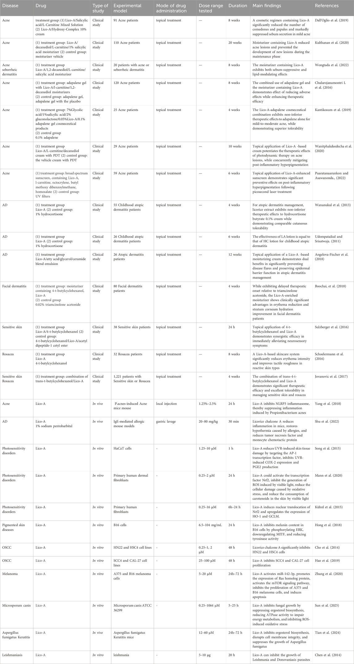
Table 1. Clinical research and mechanism of action of Lico-A.
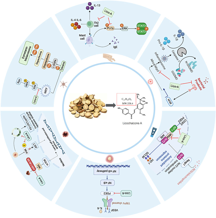
Figure 1. The main targeted signaling pathways and action mechanism of Lico-A in the treatment of dermatologic diseases.
To comprehensively evaluate the therapeutic potential and mechanistic basis of Lico-A in dermatology, we conducted a systematic review of clinical and preclinical studies published in the past decade, sourced from PubMed and Embase. Search terms included “licorice,” “Lico-A,” “skin diseases,” “inflammation,” and “tumors,” among others. This review summarizes the latest advances in the pharmacological research and clinical applications of Lico-A in dermatology, systematically analyzes the advantages and disadvantages of Lico-A in the treatment of various skin diseases and explores future research directions such as its multi-target mechanism and development of novel drug delivery systems.
2 Application of Lico-A in inflammatory dermatological diseases
2.1 Acne vulgaris
Acne vulgaris represents a chronic inflammatory condition of the pilosebaceous unit (Vasam et al., 2023), primarily associated with hyperseborrhea, Cutibacterium acnes colonization, inflammatory cascades, and follicular hyperkeratinization (Reynolds et al., 2024). Lico-A can modulate key pathogenic pathways of acne through multi-target mechanisms, demonstrating significant clinical therapeutic potential. Multiple clinical studies have used topical formulations containing Lico-A for acne patients. Results indicate that these formulations significantly suppress sebum secretion and reduce the formation of comedones, inflammatory papules, and non-inflammatory papules, preventing disease progression (Wongtada et al., 2022; Kulthanan et al., 2020; Dall'Oglio et al., 2019). A randomized controlled trial in an Asian population demonstrated that a moisturizer containing 0.5% Lico-A combined with 0.1% adapalene gel significantly reduced inflammatory and total lesion counts in mild-to-moderate acne, without the initial inflammatory exacerbation commonly observed with retinoid monotherapy (Chularojanamontri et al., 2016). This study confirmed that Lico-A not only mitigates the skin irritation associated with retinoids but also prevents the initial acne flare-up, thereby improving patient adherence.
Additionally, Lico-A has shown beneficial effects when combined with light-based therapies. The combination of Lico-A moisturizer and photodynamic therapy significantly reduced lesion counts in moderate-to-severe acne, enhanced therapeutic efficacy, minimized adverse effects, and markedly decreased post-inflammatory hyperpigmentation (Wanitphakdeedecha et al., 2020). Puaratanaarunkon T’s study further demonstrated that Lico-A cream, when used with picosecond laser treatment, reduced inflammatory papules and hyperpigmentation in acne patients (Puaratanaarunkon and Asawanonda, 2022), outperforming a control group using only conventional ultraviolet filters. These findings further support the safety and efficacy of Lico-A in combination with photodynamic therapy for moderate-to-severe acne.
Further mechanism studies confirmed that Lico-A effectively inhibits P. acnes-induced NLRP3 inflammasome activation, reduces the production of mitochondrial reactive oxygen species (ROS), and subsequently blocks caspase-1 and IL-1β secretion. In murine models, topical application of Lico-A significantly alleviated P. acnes-induced skin inflammation, erythema, and inflammatory cell infiltration, with efficacy comparable to benzoyl peroxide, a clinically standard treatment. Furthermore, Lico-A at concentrations of 10 μM and 20 μM exhibited direct antibacterial activity against P. acnes, reducing its abnormal follicular colonization (Yang et al., 2018). These findings provide a theoretical foundation for the development of Lico-A as a novel anti-acne therapeutic agent.
In summary, Lico-A demonstrates significant efficacy in preventing and treating acne by inhibiting NLRP3 inflammasome activation, reducing mitochondrial ROS production, and exerting direct antibacterial effects. However, current clinical evidence remains limited to small-scale trials with notable geographic bias and lacks long-term safety data. Future research should prioritize larger, high-quality clinical studies and broader mechanistic investigations, including Lico-A’s modulation of C. acnes biofilm formation, sebaceous gland activity, and inflammatory crosstalk.
2.2 Atopic dermatitis
AD is a common chronic relapsing inflammatory disease in dermatology, with type 2 inflammation and barrier dysfunction as important pathogenic mechanisms (Kellogg and Smogorzewski, 2023).The significant inhibition of Th2 cytokines by Lico-A also makes it a potential treatment for AD (Huang et al., 2019; Sherwani et al., 2024).
Multiple clinical studies have demonstrated the efficacy of Lico-A in alleviating AD. Research by Wananukul S et al. revealed that adult AD patients applying Lico-A formulations twice daily for 8 weeks exhibited significant reductions in SCORAD scores, with comparable efficacy to 1% hydrocortisone. Lico-A demonstrated superior outcomes in reducing transepidermal water loss (TEWL), improving skin barrier function, and lowering relapse rates. These findings align with Udompataikul M’s pediatric AD study, collectively indicating that Lico-A not only mitigates AD symptoms but also reduces corticosteroid dependency (Wananukul et al., 2023; Udompataikul and Srisatwaja, 2011). A 12-week double-blind randomized split-face relapse prevention trial further showed that Lico-A emollients reduced relapse rates by 60% in adult AD patients versus controls, significantly alleviating recurrent rash and pruritus while decreasing Staphylococcus aureus colonization and maintaining skin barrier homeostasis, highlighting Lico-A’s long-term therapeutic advantages (Angelova-Fischer et al., 2018). Additionally, a randomized prospective investigator-blinded study confirmed that Lico-A emollients exhibited superior sustained anti-inflammatory effects and skin hydration compared to 0.02% triamcinolone cream in facial AD, suggesting its potential for treating region-specific AD (Boonchai et al., 2018). Mechanistically, beyond Th2 cytokine modulation, Shu et al. (2022) demonstrated in an IgE-mediated murine allergy model that Lico-A suppresses PLC/ERK/STAT3 signaling, thereby inhibiting mast cell activation and reducing tumor necrosis factor-α (TNF-α) and MCP-1 release to attenuate inflammatory responses—further elucidating its anti-allergic mechanisms (Shu et al., 2022).
In conclusion, Lico-A alleviates AD by suppressing mast cell activation through inhibition of Th2 cytokines and the PLC/ERK/STAT3 signaling pathway, thereby reducing TNF-α and MCP-1 release. However, current clinical studies on Lico-A for AD are outdated, with a lack of recent trials. Moreover, existing mechanistic research remains limited, focusing primarily on its anti-inflammatory effects. Future studies should prioritize high-quality randomized controlled trial (RCT) and broader mechanistic investigations, including Lico-A’s effects on skin barrier function and neuroimmune interactions.
2.3 Rosacea and sensitive skin
Rosacea and sensitive skin are common chronic inflammatory dermatoses whose pathogenesis involves skin barrier dysfunction, neurovascular dysregulation, and immune-inflammatory responses (Geng et al., 2024; Sharma et al., 2022; Goh et al., 2023). In recent years, Lico-A has demonstrated unique therapeutic potential for these conditions due to its multi-target pharmacological activities.
Sulzberger et al. (2016); Kolbe et al. (2006) established post-shaving skin irritation models to evaluate the anti-irritant and skin-soothing effects of Lico-A-containing formulations. Their findings revealed that these formulations promoted erythema resolution and significantly reduced inflammatory responses within 24 h post-shaving, demonstrating notable skin-calming properties (Sulzberger et al., 2016; Kolbe et al., 2006). Schoelermann et al. (2016) conducted a clinical study in which skincare products containing Lico-A were applied to patients with mild-to-moderate rosacea. After 4 weeks, the treatment led to significant improvements in facial erythema, telangiectasia, roughness, and tightness, accompanied by reduced TEWL, increased skin hydration, and enhanced barrier function (Schoelermann et al., 2016). To further validate these observations, a large-scale study involving 1,221 patients with sensitive skin and rosacea was conducted to assess the efficacy and tolerability of Lico-A-containing cosmetics. Consistent with previous findings, the results confirmed not only clinical symptom relief but also excellent tolerability with no significant adverse effects (Jovanovic et al., 2017).
Mechanistic studies revealed that even at low concentrations (1.5 μM), Lico-A markedly suppresses TNF-α-induced NF-κB pathway activation and reduces the secretion of inflammatory mediators such as prostaglandin E2 (PGE2). This mechanism is particularly critical, as PGE2 not only directly participates in inflammatory responses but also sensitizes sensory neurons via TRPV1 channel activation, thereby inducing the release of TNF-α, interleukin-6, and vascular endothelial growth factor, which exacerbate vasodilation and non-neurogenic inflammation (Sulzberger et al., 2016). By disrupting this vicious cycle, Lico-A alleviates neurovascular hyperreactivity at its source.
In summary, Lico-A alleviates erythema and dryness in rosacea and sensitive skin by suppressing neurovascular inflammation through inhibition of the NF-κB pathway, thereby reducing PGE2 secretion. However, current clinical studies primarily evaluate Lico-A in combination formulations, with a lack of research on its efficacy as a standalone treatment. Additionally, mechanistic investigations remain limited. Future research should prioritize high-quality RCTs focusing on Lico-A monotherapy, along with further exploration of its mechanisms in neurovascular inflammation regulation.
2.4 Photodermatoses
Photodermatoses are a group of olar radiation-induced or exacerbated cutaneous disorders, including chronic actinic dermatitis, juvenile spring eruption, actinic prurigo and so on (Burfield et al., 2023). Ultraviolet radiation (UVR) and visible light (VIS) serve as primary etiological factors for these photosensitivity disorders (McDonald et al., 2023), inducing pathological consequences through DNA damage, oxidative stress, and inflammatory cascades (Tang et al., 2024). Lico-A has emerged as a promising candidate for photoprotection and photo-dermatoses management due to its dual anti-inflammatory and antioxidant properties.
Molecular studies have elucidated Lico-A’s mechanisms of action. Song et al. (2015) demonstrated that Lico-A specifically targets the AP-1 transcription factor, significantly suppressing UVR-induced COX-2 expression and PGE2 production. Importantly, this mechanism not only attenuates photoinflammatory responses but also disrupts the vicious cycle between inflammation and oxidative stress (Song et al., 2015). Mann et al. (2020) established a high-energy visible light-induced oxidative stress model, confirming that Lico-A’s photoprotective effects involve activation of the nuclear factor erythroid-2-related factor 2 (Nrf2) pathway. This activation mitigates ROS-mediated lipid peroxidation, DNA/protein damage, and prevents VIS-induced cutaneous carotenoid depletion (Mann et al., 2020). Kühnl et al. (2015) further elucidated the antioxidant mechanism of Lico-A, demonstrating its capacity to induce Nrf2 nuclear translocation and subsequent upregulation of critical antioxidant enzymes, including heme oxygenase-1 (HO-1) and glutamate-cysteine ligase (GCLM). Mechanistically, the catalytic products of HO-1 (biliverdin and carbon monoxide) exhibit synergistic antioxidant and anti-inflammatory properties, whereas GCLM maintains cellular redox homeostasis by regulating glutathione biosynthesis (Kühnl et al., 2015). These molecular mechanisms were clinically validated by Lim et al., 2022 demonstrating that Lico-A-containing formulations significantly reduce UVR/VIS-induced ROS generation while improving clinical manifestations including erythema and hyperpigmentation (Lim et al., 2022).
In conclusion, Lico-A exerts therapeutic effects on photodermatoses through dual modulation of Nrf2 activation and AP-1 suppression. However, current research has primarily focused on elucidating its molecular mechanisms, with limited clinical validation. Future studies should prioritize well-designed clinical trials to evaluate the therapeutic potential of Lico-A in photosensitive disorders, thereby providing an evidence-based foundation for its clinical application in photodermatoses.
3 Application of Lico-A in pigmented skin diseases
Excessive melanin production represents the central pathogenic mechanism in cutaneous pigmentation disorders. The microphthalmia-associated transcription factor (MITF) serves as the master regulator of melanogenesis, controlling expression of key enzymes including tyrosinase (TYR), tyrosinase-related protein 1 and tyrosinase-related protein 2. TYR is the key enzyme in melanin synthesis (Qu et al., 2023). And natural skin-lightening agents exert depigmenting effects through multiple pathways, including TYR inhibition, suppression of MAPK signaling, and interference with melanosome transport (Liu et al., 2024).
Hong et al. (2018) applied different concentrations (6.5–104 mg/mL) of Lico-A on melanoma cells B16 to evaluate the role of Lico-A in pigmentary diseases. The results showed that after 24 h, the amount of melanin in B16 cells was significantly reduced and the tyrosinase activity was significantly attenuated. Mechanistically, Lico-A activates ERK phosphorylation, leading to MITF downregulation and consequent tyrosinase inhibition (Hong et al., 2018). Studies indicate that Lico-A exerts its skin-whitening effects primarily through suppression of MITF expression rather than via direct inhibition of tyrosinase enzymatic activity, which is consistent with the findings reported by Jung et al. (2025). The depigmentation mechanism of Lico-A circumvents the pigmentary rebound effect associated with direct tyrosinase inhibition, thereby conferring therapeutic advantages for treating pigmentary disorders.
Wang et al. (2021) developed a Lico-A-loaded glycyrrhiza acid (GA + Lico-A) micelles with enhanced epidermal delivery and depigmentation efficacy, while systematically evaluating the depigmentation efficacy of GA + Lico-A micelles through both in vitro and in vivo experiments. The results demonstrated that GA + Lico-A micelles exhibited superior tyrosinase inhibitory effects and melanin-reducing capacity in B16 cells compared to Lico-A alone. Furthermore, animal studies demonstrated that 12-day consecutive topical application of both (GA + Lico-A) micelles and Lico-A significantly attenuated UVB-induced hyperpigmentation in murine models, within MAPK/ERK pathway distribution patterns correlating precisely with areas of decreased tyrosinase activity (Wang et al., 2025).
Further supporting evidence emerged from a comprehensive comparative analysis of flavonoid compounds conducted by Fu et al., 2005 which identified Lico-A as a particularly potent tyrosinase inhibitor among five tested flavonoids. Structure-activity relationship studies highlighted the critical importance of hydroxyl group positioning for optimal enzyme inhibition, providing molecular-level insights into Lico-A’s depigmentation mechanism (Fu et al., 2005).
While these preclinical studies establish Lico-A as a promising multi-target depigmenting agent through MITF downregulation and tyrosinase inhibition, clinical study remains limited (Figure 1). Further investigation is required to validate its efficacy, safety profile, and precise mechanisms of action in human pigmentation disorders.
4 Application of Lico-A in skin tumors
Cancer represents a group of malignant diseases that pose serious threats to human health and life. Its hallmark characteristics include rapid proliferation, invasion, and migration of abnormal cells (White et al., 2024). Lico-A has been shown to significantly inhibit proliferation and promote apoptosis in various cancer cell types. In dermatological oncology, Lico-A demonstrates particular therapeutic potential for oral squamous cell carcinoma (OSCC) and melanoma.
Lico-A exhibits distinct anti-OSCC mechanisms across different SCC models. In SCC-25 cells, Lico-A inhibits proliferation by inducing cell cycle arrest at S and G2/M phases. Conversely, in HN22 and HSC4 cell lines, Lico-A exerts its anticancer effects through suppression of specificity protein 1 (Sp1) and its downstream targets, including p27, p21, cyclin D1 and surviving (Cho et al., 2014). Further investigations by Hao et al. (2019) revealed that Lico-A exerts antiproliferative effects on SCC4 and CAL-27 OSCC cell lines through modulation of the PI3K/AKT signaling pathway. In vivo studies showed that Lico-A administration significantly reduced tumor burden in murine models of squamous cell carcinoma, concomitant with downregulation of metastasis-associated markers MMP-2 and MMP-9 (Hao et al., 2019). In a parallel study focusing on melanoma, Zhang et al. (2020) demonstrated that Lico-A enhances the expression of Ras homologous protein enriched in the brain by activating miR-142-3p, thereby activating the mTOR signaling pathway, inhibiting proliferation, reducing melanin production, and inducing apoptosis in A375 and B16 melanoma cells (Zhang et al., 2020).
In conclusion, Lico-A demonstrates multi-target anticancer potential in oral squamous cell carcinoma and melanoma through Sp1 suppression, PI3K/AKT pathway blockade, and mTOR pathway modulation (Figure 1). However, its mechanistic variability across models underscores the need for: (1) unified mechanistic studies, (2) advanced in vivo models, (3) early-phase clinical trials to evaluate human safety and efficacy.
5 Application of Lico-A in infections
Microbial infections represent a major etiological factor in dermatological pathologies (Hansen et al., 2022). Lico-A shows broad-spectrum antimicrobial activity against fungi, bacteria, and parasites via multi-target mechanisms.
5.1 Antifungal activity and mechanisms
Lico-A exhibits potent inhibitory activity against Microsporum canis (MIC = 4 μg/mL), with activity significantly superior to that of fluconazole compared to fluconazole (MIC = 64 μg/mL). Its multifaceted mechanism involves ergosterol biosynthesis inhibition-mediated membrane disruption, ATPase activity reduction impairing energy metabolism, and ROS-induced apoptotic oxidative stress. Transcriptomic analyses further reveal that Lico-A modulates critical pathways including cell wall biosynthesis, tricarboxylic acid cycle, and oxidative phosphorylation, resulting in comprehensive fungal growth suppression (Sun et al., 2025). Against Candida albicans and Aspergillus fumigatus, Lico-A inhibits biofilm formation, reduces host cell adhesion, and exerts direct killing effects through hyphal deformation and mitochondrial damage, while activating the host Nrf2/HO-1 pathway to mitigate inflammatory responses (Tian et al., 2024; Li et al., 2022).
5.2 Antibacterial and wound healing properties
Beyond its antifungal effects, Lico-A demonstrates substantial antibacterial activity against Escherichia coli and Staphylococcus aureus. When incorporated into chitosan-hyaluronic acid hydrogels, Lico-A maintains sustained-release properties and antimicrobial efficacy while accelerating wound healing through attenuation of inflammation, promotion of collagen synthesis, and stimulation of angiogenesis (Hou et al., 2024; Qu et al., 2023).
5.3 Antiparasitic efficacy
Lico-A displays potent activity against both promastigote and amastigote forms of Leishmania species. Experimental evidence demonstrates its capacity to significantly reduce infection rates in macrophages and U937 cells, while in vivo administration markedly decreases parasitic burden in hepatic and splenic tissues and prevents cutaneous lesion development in murine models (Chen et al., 1993).
In summary, Lico-A represents a promising antimicrobial agent with broad-spectrum antibacterial activity that exhibits significant antibacterial effects against fungal, bacterial, and parasitic pathogens through membrane disruption, bioenergetic interference, and immune regulation. Future investigations should focus on delivery system optimization to enhance bioavailability and targeting specificity, potentially positioning Lico-A as a next-generation antimicrobial for dermatological applications.
6 Discussion
Lico-A has demonstrated significant therapeutic potential across multiple dermatological conditions including acne vulgaris, AD, and rosacea, with mechanisms of action involving anti-inflammatory, antimicrobial, antioxidant, and antitumor. Clinical studies have confirmed its efficacy in reducing acne lesions, improving skin barrier function, and alleviating erythema, while exhibiting superior tolerability compared to conventional therapies such as corticosteroids and retinoids. These properties have positioned Lico-A as a compound of particular interest to dermatology specialists. However, several limitations persist in current research.
A critical issue is the geographical restriction of clinical evidence, with most studies conducted in Asian populations, raising questions about the generalizability of findings to other ethnic groups. The field also suffers from a paucity of high-quality randomized controlled trials, as existing clinical data primarily derive from small-scale, short-term studies with limited sample sizes. This gap is particularly evident in long-term safety assessments and relapse prevention studies. Furthermore, mechanistic understanding of Lico-A remains largely dependent on preclinical models including in vitro cell cultures and murine studies, which may not fully recapitulate the complexity of human skin pathophysiology. The therapeutic performance of Lico-A is additionally influenced by delivery system selection. While topical formulations such as creams and moisturizers minimize systemic side effects, their efficacy is constrained by poor skin penetration and short residence time. Microemulsion-based systems can enhance epidermal delivery efficiency but face challenges in stability and large-scale production. Hydrogels offer sustained antimicrobial release and wound healing benefits, though their high water content may compromise drug loading capacity.
To advance the clinical translation of Lico-A in dermatology, future investigations should prioritize global multicenter randomized controlled trials incorporating diverse patient populations, long-term follow-up, and rigorous methodological standards. Concurrent optimization of Lico-A formulations - including monotherapy trials and advanced delivery systems such as nanoparticles - is necessary to clarify its standalone therapeutic profile. A paradigm shift toward systems biology and network pharmacology approaches will be crucial to comprehensively elucidate Lico-A’s pleiotropic mechanisms and refine clinical applications. Given the multifactorial etiology of many dermatoses, integrating Lico-A into multimodal combination therapies shows particular promise. Such combinatorial strategies may not only amplify therapeutic outcomes but also mitigate resistance development, a persistent challenge in chronic dermatological management. Finally, clinical implementation must prioritize safety and efficacy evaluations across special populations, particularly vulnerable groups including pediatric and geriatric patients, where careful benefit-risk assessment remains essential.
In summary, realizing the full therapeutic potential of Lico-A in dermatology requires an integrated approach combining systems biology, advanced drug delivery technologies, and innovative combination therapies. Meanwhile, standardized regulatory frameworks and enhanced strategic industry partnerships should be pursued to facilitate commercialization, establishing a clear translational pathway from mechanistic insights to clinically viable therapies. By addressing current gaps, Lico-A may emerge as a cornerstone treatment for diverse dermatological conditions.
Author contributions
DL: Writing – original draft. XJ: Data curation, Writing – original draft. FY: Writing – original draft. JZ: Data curation, Writing – original draft. YL: Writing – original draft. HY: Funding acquisition, Writing – original draft, Writing – review and editing.
Funding
The author(s) declare that financial support was received for the research and/or publication of this article. This work was supported by the National Natural Science Foundation of China (No. 82205133); Chongqing Natural Science Foundation (No. CSTB2022NSCQ-MSX1619); Natural Science Foundation of Chongqing (No. jxyn2021-2-26); Natural Science Foundation of Chongqing (No. jxyn2020-7).
Conflict of interest
The authors declare that the research was conducted in the absence of any commercial or financial relationships that could be construed as a potential conflict of interest.
Generative AI statement
The author(s) declare that no Generative AI was used in the creation of this manuscript.
Any alternative text (alt text) provided alongside figures in this article has been generated by Frontiers with the support of artificial intelligence and reasonable efforts have been made to ensure accuracy, including review by the authors wherever possible. If you identify any issues, please contact us.
Publisher’s note
All claims expressed in this article are solely those of the authors and do not necessarily represent those of their affiliated organizations, or those of the publisher, the editors and the reviewers. Any product that may be evaluated in this article, or claim that may be made by its manufacturer, is not guaranteed or endorsed by the publisher.
Abbreviations
A, Licochalcone A Lico; ROS, Reactive oxygen species; AD, Atopic dermatitis; RCT, Randomized controlled trial; TEWL, Transepidermal water loss; PGE2, Prostaglandin E2; TNF-α, Tumor necrosis factor-α; UVR, Ultraviolet radiation; VIS, Visible light; Nrf2, Nuclear factor erythroid-2-related factor 2; HO-1, Heme oxygenase-1; GCLM, Glutamate-cysteine ligase; MITF, Microphthalmia-associated transcription factor; TYR, Tyrosinase; GA + Lico-A, Lico-A-loaded glycyrrhiza acid; OSCC, squamous cell carcinoma.
References
Angelova-Fischer, I., Rippke F Fau - Richter, D., Richter D Fau - Filbry, A., Filbry A Fau - Arrowitz, C., Arrowitz C Fau - Weber, T., Weber, T. F. A. U., et al. (2018). Stand-alone emollient treatment reduces flares after discontinuation of topical steroid treatment in atopic dermatitis: a double-blind, randomized, vehicle-controlled, left-right comparison study. Acta Derm. Venereol. 98 (5), 517–523. doi:10.2340/00015555-2882
Boonchai, W., Varothai, S., Winayanuwattikun, W., Phaitoonvatanakij, S., Chaweekulrat, P., and Kasemsarn, P. (2018). Randomized investigator-blinded comparative study of moisturizer containing 4-t-butylcyclohexanol and licochalcone A versus 0.02% triamcinolone acetonide cream in facial dermatitis. J. Cosmet. Dermatol 17 (6), 1130–1135. doi:10.1111/jocd.12499
Burfield, L., Rutter, K. J., Thompson, B., Marjanovic, E. J., Neale, R. E., and Rhodes, L. A.-O. (2023). Systematic review of the prevalence and incidence of the photodermatoses with meta-analysis of the prevalence of polymorphic light eruption. J. Eur. Acad. Dermatol Venereol. 37 (3), 511–520. doi:10.1111/jdv.18772
Chen, M., Christensen Sb Fau - Blom, J., Blom J Fau - Lemmich, E., Lemmich E Fau - Nadelmann, L., Nadelmann L Fau - Fich, K., Fich K Fau - Theander, T. G., et al. (1993). Licochalcone A, a novel antiparasitic agent with potent activity against human pathogenic protozoan species of leishmania. Antimicrob. Agents Chemother. 37 (12), 2550–2556. doi:10.1128/AAC.37.12.2550
Cho, J. J., Chae, J. I., Yoon, G., Kim, K. H., Cho, J. H., Cho, S. S., et al. (2014). Licochalcone A, a natural chalconoid isolated from Glycyrrhiza inflata root, induces apoptosis via Sp1 and Sp1 regulatory proteins in oral squamous cell carcinoma. Int. J. Oncol. 45 (2), 667–674. doi:10.3892/ijo.2014.2461
Chularojanamontri, L., Tuchinda, P., Kulthanan, K., Varothai, S., and Winayanuwattikun, W. (2016). A double-blinded, randomized, vehicle-controlled study to access skin tolerability and efficacy of an anti-inflammatory moisturizer in treatment of acne with 0.1% adapalene gel. J. Dermatol. Treat. 27 (2), 140–145. doi:10.3109/09546634.2015.1079298
Dall'oglio, F., Fabbrocini, G., Tedeschi, A., Donnarumma, M. A.-O., Chiodini, P., and Micali, G. A.-O. (2019). Licochalcone A in combination with salicylic acid as fluid based and hydroxy-complex 10% cream for the treatment of mild acne: A multicenter prospective trial. Clin. Cosmet. Investig. Dermatol 12, 961–967. doi:10.2147/CCID.S206935
Flohr, C. A.-O., and Hay, R. A.-O. (2021). Putting the burden of skin diseases on the global map. Br. J. Dermatol 184 (2), 189–190. doi:10.1111/bjd.19704
Fu, B., Li H Fau - Wang, X., Wang X Fau - Lee, F. S. C., Lee, F. S. F. A. U.-C. U. I. S., and Cui, S. (2005). Isolation and identification of flavonoids in licorice and a study of their inhibitory effects on tyrosinase. J. Agric. Food Chem. 53 (19), 7408–7414. doi:10.1021/jf051258h
Geng, R. A.-O. X., Bourkas, A. N., Mufti, A. A.-O., and Sibbald, R. A.-O. (2024). Rosacea: pathogenesis and therapeutic correlates. J. Cutan. Med. Surg. 28 (2), 178–189. doi:10.1177/12034754241229365
Goh, C. L., Wu, Y., Welsh, B., Abad-Casintahan, M. F., Tseng, C. J., Sharad, J. A.-O., et al. (2023). Expert consensus on holistic skin care routine: focus on acne, rosacea, atopic dermatitis, and sensitive skin syndrome. J. Cosmet. Dermatol 22 (1), 45–54. doi:10.1111/jocd.15519
Hansen, I., Augustin, M., SchäFER, I., and Mohr, N. (2022). Epidemiology of skin diseases in Germany: systematic review of the current state of research - part 3: infectious skin diseases. J. Dtsch. Dermatol Ges. 20 (5), 589–595. doi:10.1111/ddg.14702
Hao, Y., Zhang, C., Sun, Y., and Xu, H. (2019). Licochalcone A inhibits cell proliferation, migration, and invasion through regulating the PI3K/AKT signaling pathway in oral squamous cell carcinoma. Onco Targets Ther. 12, 4427–4435. doi:10.2147/OTT.S201728
Hong, J. H., Cao, S. W., Xiang, S. J., Ruan, S. F., An, B. C., Wang, Z. X., et al. (2018). Glycyrrhiza flavonoids and its major component, licochalcone A, inhibit melanogenesis through MAPK/ERK pathway by activating ERK phosphorylation. J. Dermatol Sci. S0923- 1811 (18), 30203–2. doi:10.1016/2018.04.016
Hou, Z., Wang, Y., Chen, S., Luo, Z., and Liu, Y. (2024). Licochalcone A loaded multifunctional chitosan hyaluronic acid hydrogel with antibacterial and inflammatory regulating effects to promote wound healing. Int. J. Biol. Macromol. 283 (Pt 1), 137458. doi:10.1016/j.ijbiomac.2024.137458
Huang, W. A.-O., Liu, C. Y., Shen, S. C., Chen, L. C., Yeh, K. W., Liu, S. H., et al. (2019). Protective effects of licochalcone A improve airway hyper-responsiveness and oxidative stress in a mouse model of asthma. Cells 8 (6), 617. doi:10.3390/cells8060617
Jovanovic, Z., Fau - Angabini, N., Angabini N Fau - Ehlen, S., Ehlen S Fau - Mokos, Z. B., Mokos Zb Fau - Subotic, M., Subotic M Fau - Neufang, G., et al. (2017). Efficacy and tolerability of a cosmetic skin care product with Trans-4-t-butylcyclohexanol and licochalcone A in subjects with sensitive skin prone to redness and rosacea. J. Drugs Dermatol 16 (6), 605–610. doi:10.1080/14764172.2017.1400168
Jung, J. Y., Jeong, H. J., and Han, G. A.-O. X. (2025). Antimelanogenic effect of fermented licorice water extract on murine melanoma B16F10 cells. Food Sci. Biotechnol. 34 (11), 2571–2580. doi:10.1007/s10068-025-01878-z
Kantikosum, K., Chongpison, Y., Chottawornsak, N., and Asawanonda, P. (2019). The efficacy of glycolic acid, salicylic acid, gluconolactone, and licochalcone a combined with 0.1% adapalene vs adapalene monotherapy in mild-to-moderate acne vulgaris: a double-blinded within-person comparative study. Clin. Cosmet. Investig. Dermatol. 12 151–161. doi:10.2147/CCID.S193730
Kellogg, C., and Smogorzewski, J. (2023). Update on atopic dermatitis. Adv. Pediatr. 70 (1), 157–170. doi:10.1016/j.yapd.2023.03.006
Kolbe, L., Immeyer J Fau - Batzer, J., Batzer J Fau - Wensorra, U., Wensorra U Fau - Tom Dieck, K., Tom Dieck K Fau - Mundt, C., Mundt C Fau - Wolber, R., et al. (2006). Anti-inflammatory efficacy of licochalcone A: correlation of clinical potency and in vitro effects. Arch. Dermtol Res. 298 (1), 23–30. doi:10.1007/s00403-006-0654-4
Kühnl, J., Roggenkamp D Fau - Gehrke, S. A., Gehrke Sa Fau - StäB, F., StäB F Fau - Wenck, H., Wenck H Fau - Kolbe, L., Kolbe L Fau - Neufang, G., et al. (2015). Licochalcone A activates Nrf2 in vitro and contributes to licorice extract-induced lowered cutaneous oxidative stress in vivo. Exp. Dermatol 24 (1), 42–47. doi:10.1111/exd.12588
Kulthanan, K. A.-O. X., Trakanwittayarak, S. A.-O., Tuchinda, P. A.-O., Chularojanamontri, L. A.-O., Limphoka, P. A.-O., and Varothai, S. A.-O. A. (2020). A double-blinded, randomized, vehicle-controlled study of the efficacy of moisturizer containing licochalcone A, decanediol, L-Carnitine, and salicylic acid for prevention of acne relapse in Asian population. Biomed. Res. Int. 2020, 2857812. doi:10.1155/2020/2857812
Li, M. T., Xie, L., Jiang, H. M., Huang, Q., Tong, R. S., Li, X., et al. (2022). Role of Licochalcone A in potential pharmacological therapy: a review. Front. Pharmacol. 13, 878776. doi:10.3389/fphar.2022.878776
Li, W., Yin, Y., Li, T., Wang, Y., and Shi, W. (2024). Licochalcone A protects vaginal epithelial cells against Candida albicans infection Via the TLR4/NF-κB signaling pathway (2024). J. Microbiol. 62 (7), doi:10.1007/s12275-024-00134-z
Lim, H. W., Kohli, I., Ruvolo, E., Kolbe, L., and Hamzavi, I. H. (2022). Impact of visible light on skin health: the role of antioxidants and free radical quenchers in skin protection. J. Am. Acad. Dermatol 86 (3), S27–S37. doi:10.1016/j.jaad.2021.12.024
Liu, F. A.-O., Qu, L., Li, H., He, J., Wang, L., Fang, Y., et al. (2022). Advances in biomedical functions of natural whitening substances in the treatment of skin pigmentation diseases. Pharmaceutics 14 (11), 2308. doi:10.3390/pharmaceutics14112308
Liu, M., Du, Y., and Gao, D. (2024). Licochalcone A: a review of its pharmacology activities and molecular mechanisms. Front. Pharmacol. 15, 1453426. doi:10.3389/fphar.2024.1453426
Mann, T., Eggers, K., Rippke, F., Tesch, M., Buerger, A., Darvin, M. E., et al. (2020). High-energy visible light at ambient doses and intensities induces oxidative stress of skin-protective effects of the antioxidant and Nrf2 inducer Licochalcone A in vitro and in vivo. Photodermatol. Photo 36 (2), 135–144. doi:10.1111/phpp.12523
Mcdonald, K. A.-O., Lytvyn, Y., Mufti, A., Chan, A. W., and Rosen, C. F. (2023). Review on photoprotection: a clinician's guide to the ingredients, characteristics, adverse effects, and disease-specific benefits of chemical and physical sunscreen compounds. Arch. dermatol Res. 315 (4), 735–749. doi:10.1007/s00403-022-02483-4
Olloquequi, J., Ettcheto, M. A.-O., Cano, A., Fortuna, A. A.-O., Bicker, J. A.-O., SáNCHEZ-Lopez, E. A.-O. X., et al. (2023). Licochalcone A: a potential multitarget drug for Alzheimer's disease treatment. Int. J. Mol. Sci. 24 (18), 14177. doi:10.3390/ijms241814177
Puaratanaarunkon, T. A.-O., and Asawanonda, P. A.-O. A. (2022). A randomized, double blinded, split-face study of the efficacy of using a broad spectrum sunscreen with anti-inflammatory agent to reduce post inflammatory hyperpigmentation after Picosecond laser. Clin. Cosmet. Investig. Dermatol 15, 331–337. doi:10.2147/CCID.S355329
Qu, J., Yan, M., Fang, Y., Zhao, J., Xu, T., Liu, F., et al. (2023). Zebrafish in dermatology: a comprehensive review of their role in investigating abnormal skin pigmentation mechanisms. Front. Physiol. 14, 1296046. doi:10.3389/fphys.2023.1296046
Reynolds, R. V., Yeung, H., Cheng, C. E., Cook-Bolden, F., Desai, S. R., Druby, K. M., et al. (2024). Guidelines of care for the management of acne vulgaris. J. Am. Acad. Dermatol 90 (5), 1006.e1–1006.e30. doi:10.1016/j.jaad.2023.12.017
Schoelermann, A. M., Weber, T. M., Arrowitz, C., Rizer, R. L., Qian, K., and Babcock, M. (2016). Skin compatibility and efficacy of a cosmetic skin care regimen with licochalcone A and 4-t-butylcyclohexanol in patients with rosacea subtype I. J. Eur. Acad. Dermatol Venereol. 30 (Suppl. 1), 21–27. doi:10.1111/jdv.13531
Sharma, A., Kroumpouzos, G., Kassir, M., Galadari, H. A.-O., Goren, A. A.-O., Grabbe, S., et al. (2022). Rosacea management: a comprehensive review. J. Cosmet. Dermatol 21 (5), 1895–1904. doi:10.1111/jocd.14816
Sherwani, F., Fau - Nawab, M., and Nawab, M. (2024). Exploring the potential of asl-us-soos (Glycyrrhiza glabra L.) in the treatment of respiratory Diseases-An ethnomedicinal and pharmacological review. Altern. Ther. Health Med. 30 (8), 52–59.
ShU, J., Cui, X., Liu, X., Yu, W., Zhang, W., Huo, X., et al. (2022). Licochalcone A inhibits IgE-mediated allergic reaction through PLC/ERK/STAT3 pathway. Int. J. Immunopathol. Pharmacol. 36, 3946320221135462. doi:10.1177/03946320221135462
Song, N. R., Kim, J. E., Park, J. S., Kim, J. R., Kang, H., Lee, E., et al. (2015). Licochalcone A, a polyphenol present in licorice, suppresses UV-induced COX-2 expression by targeting PI3K, MEK1, and B-Raf. Int. J. Mol. Sci. 16 (3), 4453–4470. doi:10.3390/ijms16034453
SulzbergeR, M., Worthmann, A. C., Holtzmann, U., Buck, B., Jung, K. A., Schoelermann, A. M., et al. (2016). Effective treatment for sensitive skin: 4-t-butylcyclohexanol and licochalcone A. J. Eur. Acad. Dermatol Venereol. 30 (Suppl. 1), 9–17. doi:10.1111/jdv.13529
Sun, C., Wang, L., Zhou, B., Jiang, X., Li, H., and Liu, Z. (2025). Efficient extraction of Licochalcone a with deep eutectic solvent: a promising drug for the treatment of dermatophytosis. Bioorg Chem. 160, 108463. doi:10.1016/j.bioorg.2025.108463
Tang, X., Yang, T., Yu, D., Xiong, H., and Zhang, S. (2024). Current insights and future perspectives of ultraviolet radiation (UV) exposure: friends and foes to the skin and beyond the skin. Environ. Int. 185, 108535. doi:10.1016/j.envint.2024.108535
Tian, Y., Luan, J., Wang, Q., Li, C., Peng, X., Jiang, N., et al. (2024). Licochalcone A ameliorates Aspergillus fumigatus Keratitis by reducing fungal load and activating the Nrf2/HO-1 signaling pathway. ACS Infect. Dis. 10 (10), 3516–3527. doi:10.1021/acsinfecdis.4c00123
Trakatelli, M. A.-O., Richard, M. A.-O., Rouillard, A., Paul, C., RöCKEN, M., and Stratigos, A. (2023). The burden of skin disease in Europe. J. Eur. Acad. Dermatol Venereol. 37 (Suppl. 7), 3–5. doi:10.1111/jdv.19390
Udompataikul, M., and Srisatwaja, W. (2011). Comparative trial of moisturizer containing licochalcone A vs. hydrocortisone lotion in the treatment of childhood atopic dermatitis: a pilot study. J. Eur. Acad. Dermatol Venereol. 25 (6), 660–665. doi:10.1111/j.1468-3083.2010.03845.x
Vasam, M., Korutla, S., and Bohara, R. A. (2023). Acne vulgaris: a review of the pathophysiology, treatment, and recent nanotechnology based advances. Biochem. Biophys. Rep. 36, 101578. doi:10.1016/j.bbrep.2023.101578
Wananukul, S., Chatproedprai S Fau - Chunharas, A., Chunharas A Fau - Limpongsanuruk, W., Limpongsanuruk W Fau - Singalavanija, S., Singalavanija S Fau - Nitiyarom, R., Nitiyarom R Fau - Wisuthsarewong, W., et al. (2023). Randomized, double-blind, split-side, comparison study of moisturizer containing licochalcone A and 1% hydrocortisone in the treatment of childhood atopic dermatitis. J. Med. Assoc. Thai 96 (9), 1135–1142.
WanG, Z., Xue, Y., Chen, T., Du, Q., Zhu, Z., Wang, Y., et al. (2021). Glycyrrhiza acid micelles loaded with licochalcone A for topical delivery: Co-penetration and anti-melanogenic effect. Eur. J. Pharm. Sci. 167, 106029. doi:10.1016/j.ejps.2021.106029
Wang, X., Zhang, Z. A.-O., Wang, Y., Wu, Y., Miao, L., Ma, Y., et al. (2024). Enrichment of total flavonoids and Licochalcone A from Glycyrrhiza inflata bat. Residue based on a combined membrane-macroporous resin process and a quality-control study. Molecules 29 (10), 2282. doi:10.3390/molecules29102282
Wang, H., Yu, W., Wang, T., Fang, D., Wang, Z., and Wang, Y. (2025). Therapeutic potential and pharmacological insights of total glucosides of paeony in dermatologic diseases: a comprehensive review. Front. Pharmacol. 15, 1423717. doi:10.3389/fphar.2024.1423717
Wanitphakdeedecha, R. A.-O., Tavechodperathum, N., Tantrapornpong, P., Suphatsathienkul, P., Techapichetvanich, T., Eimpunth, S., et al. (2020). Acne treatment efficacy of intense pulsed light photodynamic therapy with topical licochalcone A, l-carnitine, and decanediol: a spilt-face, double-blind, randomized controlled trial. J. Cosmet. Dermatol 19 (1), 78–87. doi:10.1111/jocd.13178
White, J., White, M. P. J., Wickremesekera, A., Peng, L., and Gray, C. (2024). The tumour microenvironment, treatment resistance and recurrence in glioblastoma. J. Transl. Med. 22 (1), 540. doi:10.1186/s12967-024-05301-9
Wongtada, C. A.-O., Pewlong, P., Asawanonda, P., Noppakun, N., Pongpamorn, P., Paemanee, A., et al. (2022). Influence of moisturizer containing licochalcone A, 1,2-decanediol, L-carnitine, and salicylic acid on facial skin lipidome among seborrhea participants. J. Cosmet. Dermatol 21 (12), 7081–7089. doi:10.1111/jocd.15381
Yakupu, A., Aimaier, R., Yuan, B., Chen, B., Cheng, J., Zhao, Y., et al. (2023). The burden of skin and subcutaneous diseases: findings from the global burden of disease study 2019. Front. Public Health 11, 1145513. doi:10.3389/fpubh.2023.1145513
Yang, G., Lee, H. E., Yeon, S. H., Kang, H. C., Cho, Y. Y., Lee, H. S., et al. (2018). Licochalcone A attenuates acne symptoms mediated by suppression of Nlrp3 inflammasome. Phytother. Res. 32 (12), 2551–2559. doi:10.1002/ptr.6195
Keywords: Licochalcone A, dermatological diseases, multi-target therapy, Nrf2/HO signaling pathway, clinical translation
Citation: Liu D, Jiang X, Yang F, Zhou J, Li Y and Yang H (2025) Therapeutic potential of Licochalcone A in dermatological diseases: from basic to clinical research. Front. Pharmacol. 16:1632006. doi: 10.3389/fphar.2025.1632006
Received: 20 May 2025; Accepted: 23 September 2025;
Published: 16 October 2025.
Edited by:
Li-She Gan, Zhejiang Chinese Medical University, ChinaReviewed by:
Da Sun, Wenzhou University, ChinaCopyright © 2025 Liu, Jiang, Yang, Zhou, Li and Yang. This is an open-access article distributed under the terms of the Creative Commons Attribution License (CC BY). The use, distribution or reproduction in other forums is permitted, provided the original author(s) and the copyright owner(s) are credited and that the original publication in this journal is cited, in accordance with accepted academic practice. No use, distribution or reproduction is permitted which does not comply with these terms.
*Correspondence: Hua Yang, eWFuZ2h1YWRvY3RvckBzaW5hLmNu; Yanxi Li, eWFueGkzNDVAMTYzLmNvbQ==