Abstract
Animal venom, known for its complex biochemical composition, presents a valuable source of therapeutic molecules, particularly for antiviral applications. Despite this potential, the industrial use of venom remains limited, with fewer than a dozen venom-derived compounds reaching commercial markets. This study underscores the significance of exploring venom’s natural diversity as a reservoir for novel bioactive compounds that could drive innovative drug development. We investigated the venom of the Moroccan black scorpion Androctonus mauritanicus (Am), applying solid-phase extraction (SPE) and high-performance reversed-phase liquid chromatography (RP-HPLC) to fractionate the venom into 80 distinct samples. These fractions were subjected to detailed analysis using advanced mass spectrometry techniques, including ESI-MS, Q-TOF LC/MS, and Q-Exactive LC/MS. In total, 507 unique molecular masses were identified, with several fractions enriched in neurotoxins targeting ion channels (NaScTxs, KScTxs, CaScTxs, and ClScTxs), highlighting their therapeutic relevance. Fractions containing inhibitory molecules targeting the receptor-binding domain (RBD) of the SARS-CoV-2 Spike S protein were identified through in vitro validation via competitive ELISA, showing multiple levels of inhibitory potential. These findings demonstrate the antiviral activity of venom-derived molecules and reveal promising opportunities for venom-based industrial applications targeting SARS-CoV-2. In conclusion, this study not only emphasises the antiviral properties of specific venom molecules but also opens pathways for industrial drug development, offering potential tools to combat emerging viral diseases.
1 Introduction
Scorpion venoms are rich sources of bioactive peptides with demonstrated potential in treating various diseases, including cancer, microbial infections, and autoimmune disorders (Ortiz et al., 2015; Liu et al., 2018). While these venoms pose substantial public health risks in many regions, they also present exciting therapeutic opportunities; venoms from the Buthidae family, particularly Androctonus species, contain neurotoxins that modulate ion channels (Na+/K+/Ca2+), making them valuable for pain management and neurological research specifically amongst its other therapeutic potential (Hilal et al., 2023a). Table 1 lists some known therapeutic discoveries from different Androctonus subspecies:
TABLE 1
| Peptide | Subspecies | Target | Effect | References |
|---|---|---|---|---|
| AcrAP-1 & AcrAP-2 (NDBPs) | Androctonus crassicauda | Human neuroblastoma (SH-SY5Y) | Proliferation blocking | Zargan et al. (2011) |
| HC-AcrAP (cationic analogs) | A. crassicauda | Human breast cancer (MCF-7) | Zargan et al. (2011) | |
| AaCTX | Androctonus australis | Human glioma cells U87 | Cell migration and invasion inhibition | Rjeibi et al. (2011), Cheng et al. (2014) |
| Crude venom | Androctonus.bicolor | Human breast cancer (MDA-MB-231) | Cell motility and colony mitosis prevention | Al-Asmari et al. (2016) |
| Androctonin | A. australis | Aspergillus brassicola, Stemphylium, Fusiarum culmorum, Botritis cinérea | Antifungal activity | Ehret-Sabatier et al. (1996) |
| G-TI | A. australis | Bacillus cereus (Gram+) | Antibacterial activity | Zerouti et al. (2019) |
| Gonearrestide (P13) | Androctonus mauritanicus | Colorectal (HCT116) & glioma (U251) cells | Anti-proliferative; cell cycle arrest | Li et al. (2019) |
| Amm VIII | A. mauritanicus | Nav1.2 channel | Ion channel modulation | Abbas et al. (2013) |
| AaHIV | A. australis | DU145 prostate cancer (via Nav1.6 channel) | Anti-proliferative activity | BenAissa et al. (2020) |
| Mauriporin | A. mauritanicus | Prostate cancer cell lines | Anti-proliferative activity | Almaaytah et al. (2013) |
| AamAP1 AamAP2 | Androctonus amoreuxi | Gram+ and Gram– bacteria | Antibacterial activity | Almaaytah et al. (2012) |
| AaeAP1 AaeAP2 | Androctonus aeneas | Candida albicans | Antifungal activity | Du et al. (2015) |
Selected therapeutic discoveries associated with various Androctonus subspecies.
Among studied species, Androctonus mauritanicus, a scorpion endemic to North Africa, produces venom known for its highly potent neurotoxins (Hilal et al., 2023a). These bioactive components of their venom are increasingly recognized as valuable molecular tools for drug development. Indeed, venom-derived peptides have shown promising applications in pain modulation, antiviral therapies, and beyond, paving the way for novel therapeutic discoveries (Liu et al., 2018; Xia et al., 2024). Additionally, antimicrobial peptides (AMPs) from scorpion venom exhibit broad-spectrum activity against bacteria and fungi, with emerging evidence suggesting antiviral properties through mechanisms like viral membrane disruption (Xia et al., 2024). While A. mauritanicus venom has not yet been proven to have direct antiviral effects, its proteomic profile shares similarities with other scorpions such as Androctonus australis whose has been reported to exhibit antiviral effects against hepatitis C virus (HCV). In particular, crude venom from A. australis showed anti-HCV activity with a half-maximal inhibitory concentration (IC50) of 88.3 ± 5.8 μg/mL. This activity was preferentially directed against HCV and remained stable after heat treatment at 60 °C or metalloprotease inhibition, suggesting the involvement of heat-resistant venom peptides (El-Bitar et al., 2015). These studies strengthen the rationale for investigating A. mauritanicus peptides as potential inhibitors of SARS-CoV-2.
Proteomics plays a pivotal role in the identification and characterization of bioactive components within these complex venoms (Calvete, 2017). Advanced proteomic tools, such as mass spectrometry, enable detailed analysis of venom composition, uncovering a wide array of peptides and proteins. This approach, known as venomics, provides essential insights into the molecular diversity, structure, and biological function of venom molecules, facilitating the identification of candidates with therapeutic potential. Proteomics-driven venom research accelerates drug discovery by pinpointing molecules with targeted pharmacological activities (Oldrati et al., 2016).
In the field of antiviral therapies, venom peptides offer unique opportunities. Several studies have shown that peptides from animal venoms can inhibit viral replication or disrupt host-virus interactions (El Hidan et al., 2021), yet the antiviral potential of A. mauritanicus venom remains largely unexplored. Understanding how venom-derived molecules interact with viral proteins could unlock new therapeutic possibilities.
The COVID-19 pandemic, caused by SARS-CoV-2, has further emphasized the need for innovative antiviral treatments (Mahendran et al., 2024). While vaccination has been critical in controlling the spread of the virus, challenges such as production delays, unequal distribution, and the emergence of variants with partial immune escape have underscored the importance of developing complementary therapeutic strategies. Venom-derived peptides present a compelling option, as they can target key viral entry mechanisms, potentially blocking interactions between the SARS-CoV-2 spike protein (S protein) and host cell receptors (El Hidan et al., 2021). Furthermore, while current SARS-CoV-2 therapies mainly rely on small molecules or monoclonal antibodies (Iketani and Ho, 2024), their effectiveness can be compromised by viral mutations and resistance (Murdocca et al., 2024), There is therefore a clear unmet need for peptide-based inhibitors. Venom-derived peptides, with their stability, high specificity, and ability to interfere with protein–protein interactions, represent promising candidates to address this therapeutic gap (V et al., 2021). Thus, investigating animal venoms as sources of novel bioactive peptides offers a compelling strategy to develop innovative therapeutics, particularly in disorders where current treatments remain suboptimal (Kim et al., 2025).
This venomics study focuses on identifying peptides with potential antiviral activity by analyzing A. mauritanicus venom. Using mass spectrometry, we characterized the molecular composition of the venom and selected specific peptides for in vitro evaluation to identify their anti-viral capacity. Our findings demonstrate how proteomics provides a robust framework for the identification of venom-derived antiviral candidates. This work not only enhances the understanding of A. mauritanicus venom’s molecular diversity but also highlights its potential application in addressing viral threats like COVID-19. By exploring the therapeutic value of venom peptides, this research paves the way for alternative antiviral strategies and contributes to ongoing efforts in drug development.
2 Materials and methods
2.1 Venom extraction
A. mauritanicus scorpions were collected in Tiznit, Souss-Massa region (Figure 1), known for its high incidence of scorpion sting envenomation cases (El Oufir, 2019), and were preserved at the Pasteur Institute’s animal facility. Their venom was extracted by using electrical stimulation, and applying a low-voltage pulses of 12 V to the scorpions’ post-abdomen to facilitate venom ejection. Following venom collection, the pooled venom was centrifuged at 10,000 RPM for 10 min to separate impurities. The resulting supernatant was then lyophilized and stored at −80 °C, preserving its potency and bioactivity for future use (Yaqoob et al., 2016).
FIGURE 1
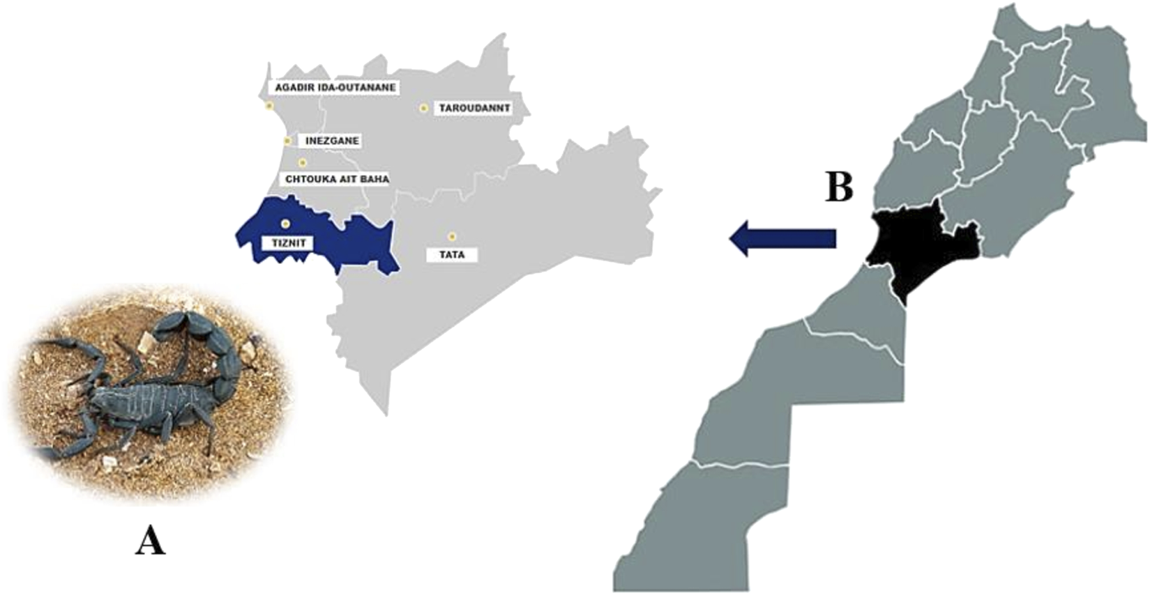
Geographical localization of the collected Am specimens (A). The region of Tiznit in the western part of Morocco (B), is known for its high-risk of scorpion envenomation.
2.2 Venom preparation
2.2.1 Venom solubilization
A quantity of 1 mg of Am venom was solubilized in 1 mL of solution A (0.1% trifluoroacetic acid (TFA)), and the mixture was centrifuged at a speed of 3,500 rpm for 5–10 min. Protein concentration was measured directly using a NanoDrop™ 2000 spectrophotometer (Thermo Fisher Scientific, Waltham, MA, United States of America) at an absorbance wavelength of 280 nm. An extinction coefficient of 10 (ε1%) was applied to estimate protein concentration. Venom samples were diluted appropriately, and each measurement was conducted in triplicate to ensure accuracy and reproducibility (Desjardins et al., 2009).
2.2.2 Removal of salts and large molecules
The removal of salts and large molecules was achieved through solid-phase extraction (SPE), a technique used for the extraction, purification, and enrichment of venoms prior to analysis (Li et al., 2006). This method involves adsorbing the target compounds onto a stationary phase within a SEP-Pak cartridge, followed by their elution. Washing steps are employed to remove interfering substances. The SPE process comprises four main steps. First, the stationary phase was conditioned. Cartridges were mounted on 10 mL syringes and connected to a manifold, a vacuum chamber equipped with a peristaltic pump. The stationary phase was conditioned by adding 10 mL of methanol to clean and wet the phase, followed by 10 mL of solution A (0.1% TFA) to activate the functional groups on the surface. Next, the phase was loaded with 1 mg of venom, allowing compounds with a strong affinity for the stationary phase to be retained. This is followed by a washing step to eliminate molecules weakly retained by the stationary phase by adding 4 mL of solution A (0.1% TFA). Finally, the compounds of interest were eluted by percolating 3 mL of the elution solution (70% Solution B + 30% Solution A), breaking the interactions between the target compounds in the venom and the stationary phase. The yield of recovered proteins was calculated after estimating their concentration by measuring absorbance at 280 nm.
2.2.3 Vacuum concentration using the SpeedVac
Following the solid-phase extraction process, the proteins obtained from the different venoms were initially frozen at −80 °C overnight to ensure their stability. Subsequently, they underwent concentration using the SpeedVac vacuum centrifugal concentrator. This step involved the removal of excess solvent under reduced pressure, allowing for the concentration of proteins. Once concentrated, the proteins were stored at −20 °C until further use.
2.3 Fractionation of the venom by reverse-phase high-performance liquid chromatography (RP-HPLC)
A total quantity of 30 mg of A. mauritanicus venom (1 mg for each run) was subjected to solid-phase extraction (SPE) and fractionated using the Alliance 2795 RP-HPLC system (Waters Corporation, Milford, MA, United States of America). Separation was achieved over 120 min on a Phenomenex C18 analytical column (250 mm, 4.6 mm, 5 µm) with a flow rate of 0.8 mL/min and a linear gradient of solvent B: from 2% to 70% over 113 min, followed by 90% for 6 min. Eluted proteins were detected at a wavelength of 280 nm, and the separated fractions were automatically collected into a 96-well plate. Fractions from each RP-HPLC run were pooled, dried, and stored at −80 °C until use.
2.4 Intact protein LC-MS
The identification of the average molecular masses of all purified fractions was performed on the triple quadrupole ESI-MS mass spectrometer (Micromass Quattro micro triple quadrupole). The fractions were dissolved in 100 µL of nebulization solvent (H2O/ACN/HCOOH, 49.8:50:0.2), and 10 µL of each was directly infused into the instrument using a Hamilton syringe. Ionization was conducted in positive mode in the ESI source, and the generated ions were separated in the Q-q-Q. MS scans with a mass-to-charge ratio (m/z) ranging from 500 to 1,500 Da were recorded. MassLynx 4.0 software (Waters-Micromass) was utilized for spectrum processing and molecular mass identification.
2.5 Identification and sequencing of peptides by mass spectrometry (nano-LC-MS/MS)
2.5.1 Enzymatic digestion
2.5.1.1 Reduction/alkylation
The fractions of interest were dissolved in 10 µL of ACN (30%) and reduced by 100 µL of the DTT solution (10 mM)/ammonium bicarbonate (50 mM, pH 8.3). The mixture was sonicated for 3 min, placed under a nitrogen atmosphere, and incubated at 60 °C for 2 h. Free sulfhydryl groups were blocked by iodoacetamide (IAA) (55 mM)/ammonium bicarbonate (50 mM, pH 8.3). Subsequently, the mixture was incubated for 20 min at room temperature and protected from light. This step concluded with the addition of 10 mM DTT to eliminate excess IAA and prevent overalkylation, followed by incubation for 1 hour at room temperature.
2.5.1.2 Digestion
We used the enzymes trypsin and Lys-C individually for digestion: 1 µg of each enzyme, dissolved in 50 mM ammonium bicarbonate (pH 8.3), was added to each fraction. The samples underwent overnight incubation at 37 °C to allow for complete enzymatic digestion. To halt the enzymatic reaction, 10 µL of 5% formic acid was added to each sample. Subsequently, the entire mixture was evaporated using the SpeedVac concentrator to remove excess solvent and concentrate the peptides for further analysis.
2.5.2 Quadrupole time-of-flight (Q-TOF) LC-MS/MS
The digested fractions were resuspended in 10 µL of 3% ACN/0.1% FA and then analyzed using the nano-LC1200 system coupled to the Q-TOF 6520 mass spectrometer (Agilent Technologies).
2.5.2.1 Q-TOF data acquisition
The analysis was configured in data-dependent acquisition mode, where peptides were ionized in nano-ESI in positive mode with a voltage of 1850 V. Full autoMS1 scans with a mass-to-charge ratio (m/z) range from 200 to 1,700 and autoMS2 scans from 59 to 1,700 m/z were recorded. In each cycle, a maximum of 5 precursor ions sorted by their charge state (excluding singly charged precursor ions) were isolated and fragmented in the collision-induced dissociation (CID) cell. The collision cell energy was automatically adjusted based on the m/z.
2.5.2.2 Q-TOF data processing
The generated data were processed using Peaks 7.5 software (Bioinformatics Solutions Inc., Waterloo, Canada). Peptides were identified by sequence homology using the UniProt database (https://www.uniprot.org) or by de novo sequencing for certain peptides. The search parameters were set as follows: precursor ion mass tolerance of 50 ppm and fragment ion mass tolerance of 0.3 Da. Enzyme specificity was set to trypsin for fractions digested with trypsin and Lys-C for those digested with Lys-C. For variable post-translational modifications, oxidation (M) (+15.9949 Da), carbamidomethylation (C) (+57.0214 Da), pyro-glu of Q and E, dehydration, and amidation were considered, while no fixed modifications were selected.
2.5.3 Q-exactive LC-MS/MS
The analysis of the digested fractions was also subjected to analysis in the Orbitrap Q-Exactive Plus mass spectrometer (Thermo Fisher Scientific, Bremen) coupled with an UltiMate™ 3000 RSLC Nano HPLC system.
2.5.3.1 Nano-HPLC
Fractionation was performed using the same parameters described in the previous paragraphs, except for an analytical column (PepMap RSLC C18, 75 μm × 25 cm, Thermo Scientific) and a flow rate of 300 nL/min.
2.5.3.2 Q-exactive data acquisition
The analysis was configured in data-dependent acquisition mode, where peptides were ionized in positive mode with a spray voltage of 1.6 kV and a capillary temperature of 180 °C. MS spectra were acquired at a resolution of 60,000 with a mass-to-charge ratio (m/z) ranging from 300 to 1,500. The 5 most abundant precursor ions were selected for higher-energy collisional dissociation (HCD) fragmentation with a collision energy of 27. Singly charged ions and those with a charge state >7 were excluded, and MS/MS spectra were acquired at a resolution of 60,000 with a m/z range from 59 to 1,700.
2.5.3.3 Q-exactive data processing
The processing of MS/MS data and peptide identification were performed following the same protocol described in the analysis by Q-TOF LC/MS.
2.6 The enzyme-linked immunosorbent assay (ELISA)
The purified fractions of the venom from the scorpion A. mauritanicus, were assessed for their potential inhibitory effect on the binding between the receptor-binding domain (RBD) of the virus and hACE2. This evaluation was performed using the COVID-19 Spike-ACE2 Binding Assay Kit, generously provided by Atheris Laboratories (3 kits) (CoV-SACE2-1, RayBiotech Inc.), following the protocol outlined by the manufacturer. This type of assay is widely used to screen potential viral entry inhibitors. Previous studies have demonstrated that this type of approach can identify small molecules capable of effectively blocking the RBD–ACE2 interaction and thus preventing SARS-CoV-2 entry into cells (Zhang et al., 2022).
In the first step, the purified fractions were tested at two concentrations: 20 mg/mL and 40 mg/mL dry weight. These concentrations were selected based on several considerations. First, preliminary assessments of solubility ensured that these concentrations were compatible with the assay. Second, the low abundance of potential bioactive peptides in the complex venom fractions required relatively higher concentrations to detect inhibitory activity. Third, lower concentrations had been tested in preliminary experiments, but they did not produce significant inhibition of RBD–ACE2 binding. Finally, the observed dose-dependent inhibition at these concentrations indicates that the effects are specific and not due to nonspecific interactions. This approach is consistent with previous exploratory screenings of venom-derived peptides, which commonly use higher concentrations to identify fractions with potential activity (Chen et al., 2012). All concentrations were assessed in triplicates. Next, the analyzed fractions were mixed with recombinant hACE2 protein, while control samples received PBS instead of venom fractions. The mixture was then added to an ELISA plate pre-coated with SARS-CoV-2 RBD protein and incubated for 2 h at room temperature with shaking. After incubation, unbound ACE2 was removed by washing the plate.
For detection, binding was assessed using the reaction between an HRP-conjugated anti-ACE2 antibody and 3,3′,5,5′-tetramethylbenzidine (TMB). Absorbance was measured at 450 nm using a Mindray MW-12A microplate reader. Finally, Statistical analysis was performed using a one-way ANOVA followed by Dunnett’s post hoc test, comparing each fraction at each concentration with the negative control (PBS). Levels of significance are indicated as “ns” (not significant) (p < 0.01), and (p < 0.001).
2.7 In Vivo acute toxicity evaluation in mice
To assess the neurotoxic potential of the most bioactive venom fractions identified through ELISA-based inhibition of the ACE2–Spike protein interaction, intracerebroventricular (ICV) injections were performed in adult male Swiss mice (18–22 g). The selected fractions which demonstrated the highest inhibitory activity in ELISA assays were reconstituted in sterile phosphate-buffered saline (PBS) and injected in volumes not exceeding 10 µL per mouse.
Groups of 3–5 mice per fraction were used, with each group receiving escalating doses to evaluate the onset of clinical neurotoxic signs (e.g., tremors, ataxia, seizures, respiratory distress), and potential lethality. For lethal fractions, the median lethal dose (LD50) was calculated using the Reed and Muench method. Non-lethal doses and fractions were observed for sublethal neurotoxic signs and behavioral abnormalities during a 4-h acute phase and over 72 h post-injection. These data contribute to defining a safe dose window for future therapeutic development of the concerned fractions.
3 Results
3.1 Protein estimation
The results showed a protein yield of 0.98 mg/mL for A. mauritanicus venom, as estimated using the NanoDrop spectrophotometer.
3.2 Venom fractionation by reverse-phase High-Performance Liquid Chromatography (RP-HPLC)
The chromatogram resulting from the fractionation of A. mauritanicus venom by RP-HPLC is illustrated in Figure 2. This profile represents a partial image of the various constituents of A. mauritanicus venom, comprising 80 different eluted fractions. This array of fractions provides insight into the richness and complexity of this venom. The majority of fractions were eluted with retention times ranging from 3.75 min to 70 min. The most intense fractions were eluted between 26 and 66 min, while the majority of minor peaks were observed within a time interval of 70–110 min. Among the most intense fractions, we find F25 (RT = 37 min), F31 (RT = 45.5 min), F32 (RT = 47 min), F37 (RT = 52 min), and F39 (RT = 55.5 min).
FIGURE 2
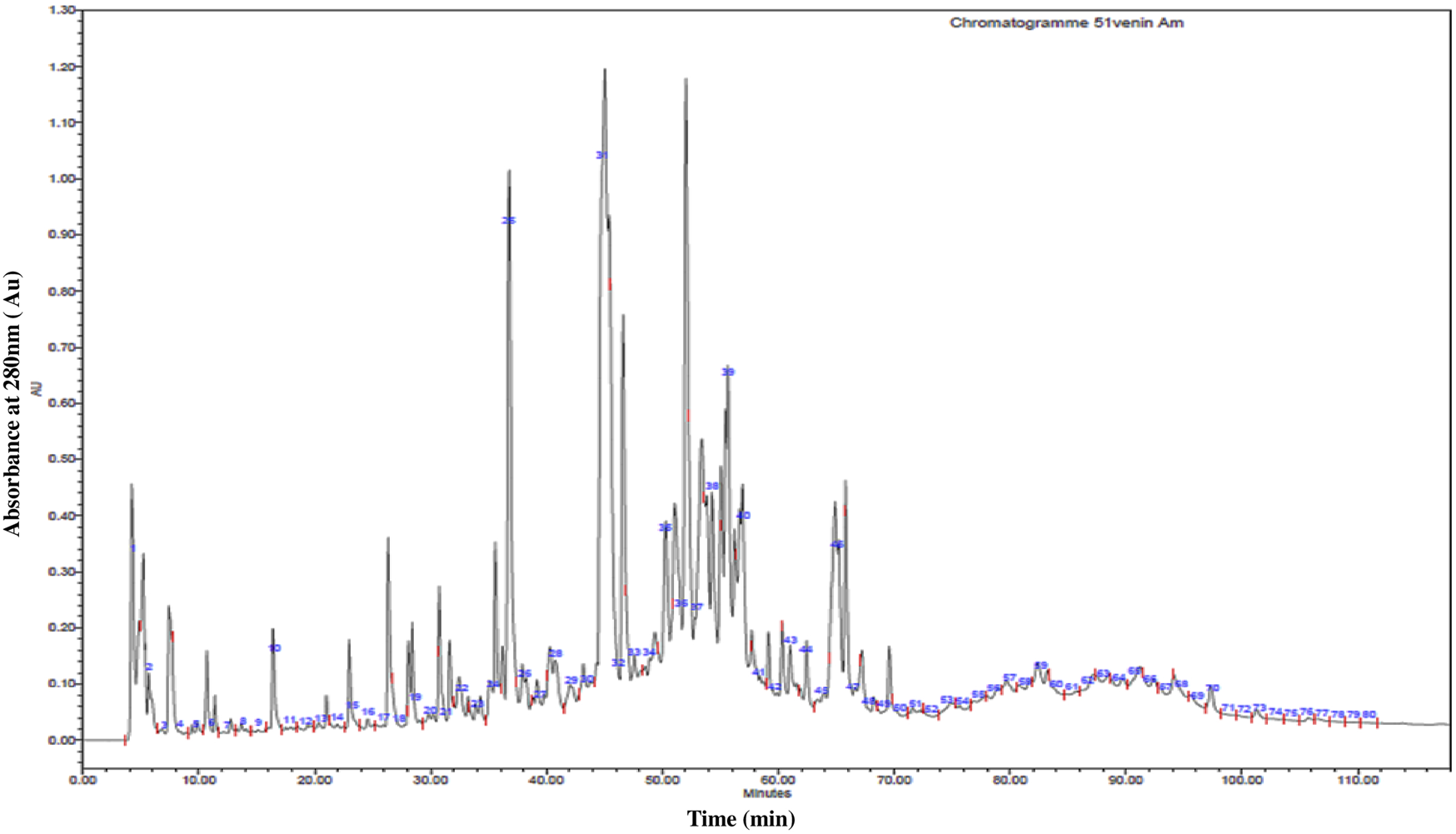
High-Performance Liquid Chromatography (RP-HPLC) profile of 1 mg of A. mauritanicus venom protein conducted with a linear gradient from solvent A (0.1% TFA in water) to 90% solvent B (0.1% TFA in acetonitrile) at a flow rate of 0.8 mL/min, and run for 120 min.
3.3 Identification of average molecular masses by mass spectrometry
The various fractions obtained through RP-HPLC underwent analysis using the triple quadrupole mass spectrometer “ESI-MS” to generate average molecular masses (Table 2). Data processing using MassLynx4 software identified a total of 507 molecular masses ranging from 200.18 Da to 10431 Da. Regarding the toxic profile, fractions of A. mauritanicus venom are rich in molecular masses ranging from 2001 to 5000 Da. These masses, corresponding to neurotoxins targeting potassium, chloride, or calcium channels, are the most abundant, comprising 51.29% of the total. Conversely, masses beyond 5001 Da, corresponding to neurotoxins targeting sodium channels, represent 22.86% of the total.
TABLE 2
| Fractions | Masses |
|---|---|
| 1 | 200.18, 228.43, 270.88, 306.94, 411.69, 443.55, 573.66, 709.69 |
| 2 | 228.36, 246.52, 280.75, 365.23, 573.66 |
| 3 | 212.25, 238.5, 254.66, 266.78, 290.93, 306.94, 421.46, 437.55, 1303.47 |
| 4 | 254.6, 418.73, 497.71, 536.93, 557.9, 574.92 |
| 5 | 207.19, 385.34 |
| 6 | 323.01, 385.36, 595.96 |
| 7 | 207.19, 385.37, 400.66 |
| 8 | 385.37, 400.66 |
| 9 | 207.19, 323.03, 385.37 |
| 10 | 290.86, 306.93, 323.0 |
| 11 | 306.94, 385.35 |
| 12 | 306.95, 368.5, 371.52, 385.37 |
| 13 | 207.18,222.36, 254.58, 254.58, 385.34,492.86 |
| 14 | 222.37, 306.95, 254.6,207.19,290.89,363.82,385.35,414.98 |
| 15 | 222.37, 254.59, 290.87,323.01,385.35,872.25, 254.57, 202 |
| 16 | 208.34, 222.36,254.58, 290.87, 323.01, 385.35, 744.34 |
| 17 | 208.38, 222.37, 254.58, 290.87, 323.01, 385.34 |
| 18 | 222.36, 254.58, 290.88, 323 |
| 19 | 3185, 3214, 3180.12, 3206.3, 2950.72, 2954, 6032, 10212, 10211, 3101.29 |
| 20 | 7559, 3214, 10212, 10211 |
| 21 | 3185, 3206.3, 8355 |
| 22 | 3185, 3214, 3180.12, 3206.3, 2954, 2874.46 |
| 23 | 2934.29, 2950.43, 2988.23, 3975.76, 3993.02 |
| 24 | 550.35, 2934.53, 2963.86, 2988.35, 3005.20, 3416.14, 3950.46, 4054.06, 4054.43, 4260.07 |
| 25 | 6145, 6175, 6512, 4121, 6161, 8218, 4376.27, 2950.72, 2935.29, 2874.46, 6032, 10212, 10211 |
| 26 | 514.35, 3197.9, 3850.96, 6913.93 |
| 27 | 1539.57, 1698.34, 2073.57, 3209.20, 4189.08, 2656.41 |
| 28 | 8218, 2950.72, 2942.58, 6032, 6307, 10431, 10212, 10192, 10211, 7177, 3607.3, 3673, 7176, 5398.1, 8355 |
| 29 | 3875, 3823.5, 6291, 6277, 3751.48, 4156, 4072, 4184.08, 3980.8, 6523, 3962, 4097, 4107, 4021, 6393, 6462, 4121.4, 4376.27, 10431, 10144, 10148, 7177, 7176, 5398.1, 8355 |
| 30 | 3627.65, 3665.52, 3682.19, 3719.31, 4206.79, 5224.74, 6591.18 |
| 31 | 8702, 8059, 9061, 8218, 4156, 4072, 4184.08, 3980.8, 6523, 3962, 4097, 4107, 4021, 6393, 6462, 4121.4, 4376.27, 10431, 10211, 3607.8, 3747.48, 3673, 5398.1 |
| 32 | 3607.88, 3627.42, 3665.40, 3681.31 |
| 33 | 8702, 9061, 8218, 10431, 10212, 10192, 10211, 3607.8, 3673, 1931.94, 1880.93, 5398.1 |
| 34 | 9571, 9496, 8702, 7301,7268, 9649,9366, 9426, 4130, 9412, 7212, 7334.93, 7373, 7226, 7465, 9511, 9524, 7253, 9174, 6872, 8059, 7301, 7733.59, 7468, 9061, 7539, 9283, 9398, 9334, 6918, 9211, 9320, 6882, 7178.27, 8218, 4156, 4072, 4184.08, 3980.8, 6523, 3962, 4097, 4107, 4021, 6393, 6462, 4121.4, 4376.27, 10431, 10144, 4005, 3607.8, 3673, 5398.1 |
| 35 | 9425, 9571, 9496,9068, 7343, 8702, 7301,7268,7194, 7211.3, 9649,9366, 9426, 4130, 8756, 9412, 7021, 9319, 9308, 7212, 7334.93, 7485, 7964, 7267, 9511, 6937, 9442, 9174, 7170.47, 6872, 8059,7040.5,7057, 7301, 7001.82, 7384, 7226, 9061, 6882, 9283, 9398, 7477, 7440, 9271, 7186, 9330, 9334, 6918, 9211, 9320, 6882, 10431, 10212, 10211, 7177, 4005, 3607.8, 3835, 3673, 7913, 7985, 7773 |
| 36 | 9571, 9496, 7268, 7194, 7211.3, 9649, 8756, 7021, 9319, 9308, 7240, 7267, 6872, 7040.5, 7001.82, 7382, 9061, 6882, 6845, 9312, 9330, 9296, 10431, 10212, 10211, 10144, 4005, 3607.8, 3673 |
| 37 | 6872, 7040.5, 7057, 6836.25, 6791.78, 6882, 10431, 10211, 10144, 4005, 3673 |
| 38 | 8702, 6872, 7040.5, 7001.82, 9061, 6882, 9351, 9019, 9312, 9330, 9936, 6918, 6652.15, 6592.5, 7641, 9296, 10431, 10144, 4005, 3607.8, 3673 |
| 39 | 7301, 6872, 7040.5, 9351, 9019, 9901, 9959, 9936, 6652.15, 7641, 7620.3, 10431, 4005, 3607.8, 3673, 7913, 7985, 7773 |
| 40 | 7020.57, 7293.31, 7383.47, 7662.26, 7677.99, 7797.82, 7948.83 |
| 41 | 7301, 6872, 7998, 7786, 8149.39, 9901, 9959, 9936, 7641, 10212, 10211, 4005, 3673 |
| 42 | 2542.55, 2762.53, 3172.12, 4419.48, 5194.61, 5577.46, 5857.4, 5884.81, 7045.13, 7061.68, 7420.22 |
| 43 | 3664.64, 3809.42, 7028.67 |
| 44 | 6872, 7040.5, 6882, 6845, 6836.25, 6791.78, 6918, 6892.4, 10431, 10212, 10211, 10144, 4005, 3673 |
| 45 | 6872, 7040.5, 6882, 6836.25, 6791.78, 10212, 10211, 4005, 3673, 7913, 7985, 7773 |
| 46 | 8702, 6872, 4005, 3673, 7913, 7985, 7773 |
| 47 | 3035.07, 6933.8 |
| 48 | 7785, 9574, 7721, 10212, 10211 |
| 49 | 3809.15, 3824.6, 3863.24, 7785 |
| 50 | 1525.12, 1851.69,2541.32,3050.45,3702.24, 5729.8, 6479.81 |
| 51 | 1348.87, 1419.01, 1547.58, 1842.14, 1982.35, 1998.64, 2135.36, 3538.45, 3910.64 |
| 52 | 1722.18, 2293.99, 2311.55, 2335.57, 2662.75, 3562.63, 3577.40, 3665.69, 4129.31 4698.28, 4996.71, 5900.00, 7160.87 |
| 53 | 4055.48, 4399.39, 4473.21, 5838.77, 5899.73, 6934.73, 7109.03 |
| 54 | 2839, 2968 |
| 55 | 1614.22, 1698.15, 2608.39, 3061.15 |
| 56 | 1542.38, 1849.48, 2013.42, 2041.26, 2049.06, 2571.94, 2738.67, 3524.11, 4099.11, 4310.19, 5769.12 |
| 57 | 1614.44, 1784.97, 2825.59, 3391.29, 3684.08, 4068.98 |
| 58 | 1889.19, 2003.54, 2066.50, 2109.32, 2442.24, 2682.6, 3226.6, 3356.51, 3608.46, 3642.97, 4600.16, 4431.93 |
| 59 | 2325.28, 2785.35, 2921.24, 3259.93, 4068.92 |
| 60 | 1956.09, 3505.12, 8682.28 |
| 61 | 2080.52, 2152.48 |
| 62 | 1753.45, 2200.34, 2631.63, 2932.89, 3760.41, 5118.16, 5132.78, 5263.27, 5670.88, 6140.99, 6396.99, 6700.58, 8955.78 |
| 63 | 574.07, 1076.2, 1303.91, 1305.99, 1336.37, 1545.78, 3482.77, 2831.11, 2921.24, 3579.87, 5725.43, 5303.84, 7000.04, 7281.16, 8274.75 |
| 64 | 1902.47, 2152.26, 2814.62, 2984.1, 3341.34,3482.27, 3579.86, 3912.45, 5706.40, |
| 65 | 1193.67, 1614.1, 1721.96, 2336.51, 2492.56, 2717.42, 2885.76, 3260.84, 4829.64, 5268.65, 6673.50, 7508.48 |
| 66 | 1614.16, 1908.86, 1956.53, 2511.51, 2575.95, 2591.28, 2920.83, 2982.97, 3260.44, 3580.64, 5370.36 |
| 67 | 1722.43, 1956.40, 4707.66, 6414.55 |
| 68 | 1698.4, 1794.63, 2336.4, 2449.39, 2474.35, 3371.23, 3765.74, 7756.25, 8407.82 |
| 69 | 2152.01, 2336.05, 3913.6, 3963.25 |
| 70 | 1794.32, 2831.11, 2336.72, 4564.66, |
| 71 | 1974.26, 2086.2, 2258.68, 2364.46, 2482.35, 2961.39, 3152.1, 3310.08, 3377.73, 3556.74, 3948.52, 4172.39, 4742.32, 5066.1, 5215.49, 5792.35, 6258.59, 6909.91, 7113.48, 8299.06 |
| 72 | 1321.12, 1757.45, 2429.18, 2636.17, 2636.17, 2735.7, 3185.77, 3302.81, 3514.89, 3647.6, 4247.69, 4559.5, 5272.34, 5309.61, 5471.4, 5668.08, 6151.06, 6371.53, 6383.2, 6383.3, 7433.46 |
| 73 | 1675.16, 2093.88, 3062.85, 4187.75, 5324.69, 7328.57 |
| 74 | 2144.37, 2807.84, 3216.55, 3855.34, 4679.74, 4819.18, 5360.92, 5615.69, 5783.02, 6433.11, 6551.63, 6746.85, 7505.29 |
| 75 | 3424.7, 4156.27, 5707.84, 6849.41, 7990.98, 8312.55 |
| 76 | 1788.4, 2683.2, 3667.49, 4471.99, 6261.01 |
| 77 | 1413.66, 1589.97, 2264.38, 2866.38, 2993.07, 3242.19, 3662.96, 3817.42 |
| 78 | 1833.96, 1963.61, 2125.02, 2184.86, 2336.66, 2361.12, 2415.66, 2443.97, 2537.03, 2691.23, 2715.33, 3102.8, 3217.04, 3591.15, 5910.26 |
| 79 | 1413.82, 1614.03, 2301.93, 2654.35, 2810.97, 2830.99, 2870.42, 2891.17, 3542.81, 3662.84, 4008.24, 4563.97, 5062.41, 5881.75, 8695.26 |
| 80 | 2952.67, 4173.81, 6261.99, 7305.65 |
List of the different average masses identified in the various fractions of A. mauritanicus venom.
Interestingly, 86 peptides corresponding to NaScTxs were characterized, 64 of which were identified as NaSctx alpha subfamily (α-NaScTxs) with a sequence coverage ranging from 9% for the peptide similar to Lipolysis-activating peptide 1-alpha chain (D9U2A4) to 86% for the peptides who present homology with the alpha-toxin Lqq4 (P01489), whereas, 23 peptides corresponding to as NaSctx beta subfamily (β-NaScTxs) with a sequence coverage from to 8% (Beta-insect depressant toxin BmKIT4; Q17230) to 39% (Insect toxin AaHIT5; P81504) (Table 3).
TABLE 3
| Accession number | Name | Sequence | Coverage (%) | Fractions | MW |
|---|---|---|---|---|---|
| NaScTxs | |||||
| α-NaScTxs | |||||
| Q8I0K7 | Depressant scorpion toxin BmKIM OS = Mesobuthus martensii GN = KIM2 PE = 2 SV = 1 | MKLFLLLVFFASMLIDGLVNADGYIRGSNGCKISCLWGNEGCNKECKGFGAYYGYCWTWGLACWCEGLPDDKTWKSESNTCGGKK | 6 | F35 | 9,425 |
| P17728 | Alpha-insect toxin LqhaIT OS = Leiurus quinquestriatus hebraeus PE = 1 SV = 2 | MNHLVMISLALLLLLGVESVRDAYIAKNYNCVYECFRDAYCNELCTKNGASSGYCQWAGKYGNACWCYALPDNVPIRVPGKCHRK | 40 | F34; F35; F36 | 9,571 |
| P45697 | Alpha-like toxin BmK-M1 OS = M. martensii | MNYLVMISFALLLMTGVESVRDAYIAKPHNCVYECARNEYCNDLCTKNGAKSGYCQWVGKYGNGCWCIELPDNVPIRVPGKCHR | 48 | F34; F35; F36 | 9,496 |
| P01480 | Alpha-mammal toxin Aah3 OS = A. australis | MNYLVMISLALLLMTGVESVRDGYIVDSKNCVYHCVPPCDGLCKKNGAKSGSCGFLIPSGLACWCVALPDNVPIKDPSYKCHSR | 12 | F35 | 9068 |
| P09981 | Alpha-mammal toxin BeM9 OS = Mesobuthus eupeus | ARDAYIAKPHNCVYECYNPKGSYCNDLCTENGAESGYCQILGKYGNACWCIQLPDNVPIRIPGKCH | 12 | F35 | 7,343 |
| D5HR50 | Alpha-toxin Ac1 (Fragment) OS = A. crassicauda | YIVMISLALVVMIGVESVRDGYIVYPNNCVYHCIPACDGLCKKNGGTSGSCSFLIGSGIACWCKDLPDNVPIKDPSQKCTR | 16 | F31; F33; F34; F35; F38; F46 | 8,702 |
| P01482 | Alpha-toxin Amm5 OS = A. mauritanicus | LKDGYIIDDLNCTFFCGRNAYCDDECKKKGGESGYCQWASPYGNACWCYKLPDRVSIKEKGRCN | 48 | F34; F35; F39; F41 | 7,301 |
| P01488 | Alpha-toxin Bot1 OS = Buthus occitanus tunetanus | GRDAYIAQPENCVYECAQNSYCNDLCTKNGATSGYCQWLGKYGNACWCKDLPDNVPIRIPGKCHF | 32 | F34; F35; F36 | 7,268 |
| P01489 | Alpha-toxin Lqq4 OS = L. quinquestriatus | GVRDAYIADDKNCVYTCGSNSYCNTECTKNGAESGYCQWLGKYGNACWCIKLPDKVPIRIPGKCR | 86 | F35; F36 | 7,194 |
| P83644 | Toxin Lqh4 OS = L. quinquestriatus hebraeus | GVRDAYIADDKNCVYTCGANSYCNTECTKNGAESGYCQWFGKYGNACWCIKLPDKVPIRIPGKCR | 66 | F35; F36 | 7,211.3 |
| Q9GQW3 | Toxin BmKaIT1 OS = M. martensii | MNYLVMISFAFLLMTGVESVRDAYIAQNYNCVYHCARDAYCNELCTKNGAKSGSCPYLGEHKFACYCKDLPDNVPIRVPGKCHRR | 39 | F34; F35; F36 | 9,649 |
| Q9GYX2 | Toxin BmKa1 OS = M. martensii PE = 2 SV = 1 | MNYLVFFSLALLLMTGVGSVRDGYIADDKNCPYFCGRNAYCDDECKKNGAESGYCQWAGVYGNACWCYKLPDKVPIRVPGKCNGG | 25 | F34; F35 | 9,366 |
| Q9GUA7 | Toxin BmKa3 OS = M. martensii PE = 2 SV = 1 | MNYLVFFSLALLLMTGVESVRDGYIADDKNCAYFCGRNAYCDDECKKKGAESGYCQWAGVYGNACWCYKLPDKVPIRVPGKCNGG | 25 | F34; F35 | 9,426 |
| Q2YHM1 | Neurotoxin 8-related gene product 1/2/3 OS = A. mauritanicus PE = 1 SV = 1 | VRDAYIAQNYNCVYTCFKNDYCNDICTKNGAXXGYC | 78 | F34; F35 | 4,130 |
| P45698 | Neurotoxin BmK-M9 OS = M. martensii PE = 1 SV = 1 | MISFALLLMTGVESVRDAYIAKPENCVYHCATNEGCNKLCTDNGAESGYCQWGGRYGNACWCIKLPDRVPIRVPGKCHR | 27 | F35; F36 | 8,756 |
| Q95P69 | Toxin BmKT OS = M. martensii PE = 2 SV = 1 | MNYLVFFSLALLLMTGVESVRDGYIADDKNCAYFCGRNAYCDDECKKNGAESGYCQWAGVYGNACWCYKLPDKVPIRVPGKCNGG | 25 | F34; F35 | 9,412 |
| P58328 | Alpha-like toxin BmK-M4 OS = M. martensii | VRDAYIAKPENCVYHCAGNEGCNKLCTDNGAESGYCQWGGRYGNACWCIKLPDDVPIRVPGKCH | 30 | F35; F36 | 7,021 |
| Q9NJC8 | Toxin BmKaTx13 OS = M. martensii | MNYLVMISFALLLMKGVESVRDAYIAKPENCVYHCAGNEGCNKLCTDNGAESGYCQWGGRYGNACWCIKLPDDVPIRVPGKCHR | 23 | F35; F36 | 9,319 |
| Q9N682 | Neurotoxin BmK-M11 OS = M. martensii PE = 3 SV = 1 | MNYLVMISFALLLMTGVESVRDAYIAKPENCVYHCATNEGCNKLCTDNGAESGYCQWGGKYGNACWCIKLPDDVPIRVPGKCHR | 22 | F35; F36 | 9,308 |
| D9U2A4 | Lipolysis-activating peptide 1-alpha chain OS = Lychas mucronatus PE = 2 SV = 1 | MNITLFCSVFILISLAGLSVSDDVPGNYPMSLYGNKYSCGVLGENEYCRKICKSHGVSYGYCFNSRCWCEYLEDKDVDFWAAHKNHCKNDKLYPPKK | 9 | F38; F39 | 11,094 |
| P01487 | Alpha-insect toxin Lqq3 OS = L. quinquestriatus PE = 1 SV = 2 | VRDAYIAKNYNCVYECFRDSYCNDLCTKNGASSGYCQWAGKYGNACWCYALPDNVPIRVPGKCH | 28 | F36 | 7,240 |
| P58488 | Alpha-like toxin BmK-M2 OS = M. martensii PE = 1 SV = 1 | VRDAYIAKPHNCVYECARNEYCNNLCTKNGAKSGYCQWSGKYGNGCWCIELPDNVPIRVPGKCH | 36 | F34; F35 | 7,212 |
| P86406 | Neurotoxin MeuNaTx-6 OS = M. eupeus PE = 1 SV = 1 | MMKIIIFLIVSSLVLIGVKTDNGYLLDKYTGCKVWCVINNESCNSECKIRRGNYGYCYFWKLACYCEGAPKSELWHYETNKCNGRM | 15 | F48; F49 | 7,785 |
| P55902 | Alpha-insect toxin BotIT1 OS = B. occitanus tunetanus | VRDAYIAQNYNCVYFCMKDDYCNDLCTKNGASSGYCQWAGKYGNACWCYALPDNVPIRIPGKCHS | 75 | F34; F35 | 7,334.93 |
| P01490 | Alpha-toxin BeM10 OS = M. eupeus | VRDGYIADDKDCAYFCGRNAYCDEECKKGAESGKCWYAGQYGNACWCYKLPDWVPIKQKVSGKCN | 14 | F34 | 7,373 |
| P0DJH8 | Alpha-toxin Bu1 OS = Buthacus macrocentrus | GVRDAYIADDKNCVYTCAKNSYCNTECTKNGAESGYCQWLGKYGNGCWCIKLPDKVPIRIPGRCRGR | 75 | F35 | 7,485 |
| P82815 | Bukatoxin OS = M. martensii | VRDGYIADDKNCAYFCGRNAYCDEECIINGAESGYCQQAGVYGNACWCYKLPDKVPIRVSGECQQ | 14 | F34 | 7,226 |
| P15224 | Toxin Os1 OS = Orthochirus scrobiculosus | ERDGYIVQLHNCVYHCGLNPYCNGLCTKNGATSGSYCQWMTKWGNACYCYALPDKVPIKWLDPKCY | 41 | F20 | 7,559 |
| P60256 | Toxin Boma6b OS = Buthus occitanus mardochei | VRDAYIAQNYNCVYDCARDAYCNDLCTKNGAKSGYCEWFGPHGDACWCIDLPNNVPIKVEGKCHRK | 15 | F34 | 7,465 |
| P60258 | Toxin Boma6d OS = B. occitanus mardochei | VRDAYIAQNYNCVYTCFKDAHCNDLCTKNGASSGYCQWAGKYGNACWCYALPDNVPIRIPGKCHRK | 26 | F35 | 7,964 |
| P60259 | Toxin Boma6e OS = B. occitanus mardochei | VRDAYIAQNYNCVYACARDAYCNDLCTKNGARSGLFATFGPHGDACWCIALPNNVPLKVQGKCHRK | 32 | F35; F36 | 7,267 |
| Q9GQV6 | Toxin BmKaTx16 OS = M. martensii | MNYLVMISFALLLMTGVESVRDAYIAKPHNCVYECARNEYCNDLCTKNGAKSGYCQWVGKYGNGCWCKELPDNVPIRVPGKCHR | 48 | F34; F35 | 9,511 |
| M1JBC0 | Sodium channel alpha-toxin Acra4 OS = A. crassicauda PE = 1 SV = 1 | VRDGYIVDDKNCVYHCIPPCDGLCKKNGGKSGSCSFLVPSGLACWCKALPDNVPIKDPSYKCHKR | 46 | F35 | 6,937 |
| Q9NJC7 | BmK AGP-SYPU2 OS = M. martensii PE = 1 SV = 1 | MNYMVIISLALLVMTGVESVKDGYIADDRNCPYFCGRNAYCDGECKKNRAESGYCQWASKYGNACWCYKLPDDARIMKPGRCNGG | 11 | F34 | 9,524 |
| G4V3T9 | Neurotoxin BmK AGAP-SYPU2 (Fragment) OS = M. martensii PE = 1 SV = 1 | VKDGYIVDDKNCAYFCGRNAYCDDECEKNGAESGYCQWAGVYGNACWCYKLPDKVPIRVPGRCNG | 14 | F34 | 7,253 |
| P86404 | Neurotoxin MeuNaTx-4 OS = M. eupeus PE = 1 SV = 1 | MNYLILISFALLVITGVESARDAYIAKPHNCVYECFDAFSSYCNGVCTKNGAKSGYCQILGTYGNGCWCIVLPDNVPIRIPGKCHR | 38 | F35 | 9,442 |
| Q17254 | Alpha-insect toxin Bot14 OS = B. occitanus tunetanus PE = 2 SV = 1 | MSSLMISTAMKGKAPYRQVRDGYIAQPHNCAYHCLKISSGCDTLCKENGATSGHCGHKSGHGSACWCKDLPDKVGIIVHGEKCHR | 19 | F34; F35 | 9,174 |
| P86405 | Neurotoxin MeuNaTx-5 OS = M. eupeus PE = 1 SV = 1 | MNYLILISFALLVITGVESARDAYIAKPHNCVYECFDAFSSYCNGVCTKNGAKSGYCQILGTYGNGCWCIALPDNVPIRIPGKCHR | 38 | F35 | 7,170.47 |
| P13488 | Alpha-like toxin Bom3 OS = B. occitanus mardochei | GRDGYIAQPENCVYHCFPGSSGCDTLCKEKGATSGHCGFLPGSGVACWCDNLPNKVPIVVGGEKCH | 77 | F34; F35; F36; F37; F38; F39; F41; F44; F45; F46 | 6,872 |
| P01485 | Alpha-mammal toxin Bot3 (Fragment) OS = B. occitanus tunetanus | LVMAGVESVKDGYIVDDRNCTYFCGRNAYCNEECTKLKGESGYCQWASPYGNACYCYKVPDHVRTKGPGRCN | 62 | F31; F34; F35 | 8,059 |
| O61705 | Neurotoxin BmK-M10 OS = M. martensii PE = 1 SV = 1 | MNYLIMFSLALLLVIGVESGRDGYIVDSKNCVYHCYPPCDGLCKKNGAKSGSCGFLVPSGLACWCNDLPENVPIKDPSDDCHKR | 43 | F35; F36; F37; F38; F39; F44; F45 | 7,040.5 |
| P04099 | Alpha-toxin Bot9 OS = B. occitanus tunetanus | AEIKVRDGYIVYPNNCVYHCGLNPYCNDLCTKNGAKSGYCQWLTKWGNACYCYALPEKVPIKDPSYKCYS | 26 | F41 | 7,998 |
| P56678 | Alpha-like toxin Lqh3 OS = L. quinquestriatus hebraeus PE = 1 SV = 1 | VRDGYIAQPENCVYHCFPGSSGCDTLCKEKGGTSGHCGFKVGHGLACWCNALPDNVGIIVEGEKCHS | 43 | F35; F37 | 7,057 |
| P01481 | Alpha-mammal toxin Lqq5 OS = L. quinquestriatus PE = 1 SV = 1 | LKDGYIVDDKNCTFFCGRNAYCNDECKKKGGESGYCQWASPYGNACWCYKLPDRVSIKEKGRCN | 14 | F34; F35 | 7,301 |
| Q4TUA4 | Alpha-toxin 4 OS = M. martensii PE = 1 SV = 1 | MNYLVFFSLALLLMTGVESVRDGYIADDKNCAYFCGRNAYCDDECKKKGAESGYCQWAGVYGNACWCYKLPDKVPIRVPGRCNGG | 11 | F34 | 7,733.59 |
| P0C910 | Alpha-toxin Amm3 OS = A. mauritanicus PE = 1 SV = 1 | GRDGYIVDTKNCVYHCYPPCDGLCKKNQAKSGSCGFLYPSGLACWCVALPENVPIKDPNDDCHK | 53 | F35; F36; F38 | 7,001.82 |
| Q7YXD3 | Alpha-toxin Amm8 OS = A. mauritanicus PE = 1 SV = 1 | MNYLVMISLALLFMTGVESLKDGYIVNDINCTYFCGRNAYCNELCIKLKGESGYCQWASPYGNSCYCYKLPDHVRTKGPGRCNDR | 59 | F35; F 36 | 7,382 |
| P01486 | Alpha-toxin Bot11 OS = B. occitanus tunetanus PE = 1 SV = 1 | LKDGYIVDDRNCTYFCGTNAYCNEECVKLKGESGYCQWVGRYGNACWCYKLPDHVRTVQAGRCRS | 14 | F34 | 7,468 |
| P59360 | Neurotoxin BmK-II OS = M. martensii PE = 1 SV = 1 | VRDAYIAKPHNCVYECARNEYCNDLCTKDGAKSGYCQWVGKYGNGCWCIELPDNVPIRIPGNCH | 47 | F35 | 7,226 |
| Q7Z0H4 | Neurotoxin BmP08 OS = M. martensii PE = 1 SV = 1 | MKIFFAVLVILVLFSMLIWTAYGTPYPVNCKTDRDCVMCGLGISCKNGYCQGCTR | 15 | F25 | 6,145 |
| Q7Z0F1 | Neurotoxin X-29S OS = M. martensii PE = 3 SV = 1 | MKIFFAVLVILVLFSMLIWTAYGTPYPVNCKTDRDCVMCGLGISCKNGYCQSCTR | 15 | F25 | 6,175 |
| P01479 | Neurotoxin-1'' OS = A. australis PE = 1 SV = 3 | MNYLVMISLALLLMIGVESKRDGYIVYPNNCVYHCVPPCDGLCKKNGGSSGSCSFLVPSGLACWCKDLPDNVPIKDTSRKCTR | 29 | F31; F33; F34; F35; F36; F38 | 9,061 |
| M1JMR8 | Sodium channel alpha-toxin Acra8 OS = A. crassicauda PE = 3 SV = 1 | VRDGYIVDDKNCTFFCGRNAYCNDECKKKGGESGYCQWASPYGNACWCYKLPDRVPIKEKGRCNGR | 14 | F34 | 7,539 |
| P45658 | Toxin Aah4 OS = A. australis PE = 1 SV = 2 | MNYLIMFSLALLLVIGVESGRDGYIVDSKNCVYHCYPPCDGLCKKNGAKSGSCGFLVPSGLACWCNDLPENVPIKDPSDDCHKR | 46 | F35; F36; F37; F38; F44; F45 | 6,882 |
| P21150 | Toxin AaHIT4 OS = A australis PE = 1 SV = 1 | EHGYLLNKYTGCKVWCVINNEECGYLCNKRRGGYYGYCYFWKLACYCQGARKSELWNYKTNKCDL | 25 | F41 | 7,786 |
| Q86SE0 | Toxin Aam2 OS = A. amoreuxi PE = 1 SV = 1 | MNYLITISLALLLMTGVASGVRDGYIADAGNCGYTCVANDYCNTECTKNGAESGYCQWFGRYGNACWCIKLPDKVPIKVPGKCNGR | 38 | F34; F35 | 9,283 |
| Q9GNG8 | Toxin BmKaTX15 OS = M. martensii PE = 2 SV = 1 | MNYLVFFSLALLVMTGVESVRDGYIADDKNCAYFCGRNAYCDDECKKNGAESGYCQWAGVYGNACWCYKLPDKVPIRVPGKCNGG | 25 | F34; F35 | 9,398 |
| P60255 | Toxin Boma6a OS = B. occitanus mardochei PE = 3 SV = 1 | VRDAYIAQNYNCVYDCARDAYCNDLCTKNGAKSGYCEWFGPHGDACWCIDLPNNVPIKVEGKCHRK | 32 | F35 | 7,477 |
| P60257 | Toxin Boma6c OS = B. occitanus mardochei PE = 3 SV = 1 | VRDAYIAQNYNCVYTCFKDAHCNDLCTKNGASSGYCQWAGKYGNACWCYALPDNVPIRIPGKCHRK | 59 | F35 | 7,440 |
| Q4LCT3 | Toxin-like peptide AaF1CA1 OS = A australis PE = 2 SV = 1 | MMKLVLFSVIVILFSLIGSIHGADVPGNYPLRPFRYRYGCAVPGDSDYCVRVCRKHGVRYGYCWFFTCWCEYLEDKNIKI | 11 | F38 and F39 | 9,351 |
| Q9BKJ0 | Anti-neuroexcitation peptide 3 OS = M. martensii PE = 2 SV = 2 | MKLSLLLVISASMLIDGLVNADGYIRGSNGCKISCLWGNEGCNKECKGFGAYYGYCWTWGLACWCEGLPDDKTWKSESNTCGGKK | 6 | F35 | 9,271 |
| Q4LCS7 | Toxin-like peptide AaF1CA26 OS = A. australis GN = aaF1CA26 PE = 2 SV = 1 | MMKLMLFSIIVILFSLIGSIHGADVPGNYPLDSSDDTYLCAPLGENPSCIQICRKHGVKYGYCYAFQCWCEYLEDKNVKS | 11 | F38 and F39 | 9,019 |
| β-NaScTxs | |||||
| P80962 | Beta-insect depressant toxin BaIT2 OS = Buthacus arenicola PE = 1 SV = 1 | DGYIRRRDGCKVSCLFGNEGCDKECKAYGGSYGYCWTWGLACWCEGLPDDKTWKSETNTCG | 20 | F36; F44 | 6,845 |
| Q17230 | Beta-insect depressant toxin BmKIT4 OS = M. martensii PE = 2 SV = 2 | DGYIRGSNGCKISCLWGNEGCNKECKGFGAYYGYCWTWGLACWCEGLPDDKTWKSESNTCGRKK | 8 | F35 | 7,186 |
| Q9XY87 | Beta-insect depressant toxin BmKITa OS = M. martensii PE = 1 SV = 1 | MKLFLLLLISASMLIDGLVNADGYIRGSNGCKVSCLWGNEGCNKECRAYGASYGYCWTWGLACWCQGLPDDKTWKSESNTCGGKK | 12 | F36; F38 | 9,312 |
| Q95WX6 | Beta-insect depressant toxin BmKITb OS = M. martensii PE = 1 SV = 1 | MKLFLLLVISASMLIDGLVNADGYIRGSNGCKVSCLWGNEGCNKECKAFGAYYGYCWTWGLACWCQGLPDDKTWKSESNTCGGKK | 12 | F35; F36; F38 | 9,330 |
| P55903 | Beta-insect depressant toxin BotIT4 OS = B. occitanus tunetanus PE = 1 SV = 1 | DGYIRRRDGCKVSCLFGNEGCDKECKAYGGSYGYCWTWGLACWCEGLPDDKTWKSETNTCG | 20 | F37; F44; F45 | 6,836.25 |
| P55904 | Beta-insect depressant toxin BotIT5 OS = B. occitanus tunetanus PE = 1 SV = 1 | DGYIRKRDGCKVSCLFGNEGCDKECKAYGGSYGYCWTWGLACWCEGLPDDKTWKSETNTCG | 20 | F37; F44; F45 | 6,791.78 |
| Q26292 | Beta-insect depressant toxin LqhIT2 OS = L. quinquestriatus hebraeus PE = 1 SV = 1 | MKLLLLLIVSASMLIESLVNADGYIKRRDGCKVACLIGNEGCDKECKAYGGSYGYCWTWGLACWCEGLPDDKTWKSETNTCGGKK | 33 | F34; F35 | 9,334 |
| O77091 | Beta-insect excitatory toxin BmK IT-AP OS = M. martensii GN = IT-AP PE = 1 SV = 1 | MKFFLIFLVIFPIMGVLGKKNGYAVDSSGKVAECLFNNYCNNECTKVYYADKGYCCLLKCYCFGLADDKPVLDIWDSTKNYCDVQIIDLS | 20 | F41 | 8,149.39 |
| P68721 | Beta-insect excitatory toxin LqhIT1a OS = L. quinquestriatus hebraeus PE = 3 SV = 1 | MKFFLLFLVVLPIMGVLGKKNGYAVDSKGKAPECFLSNYCNNECTKVHYADKGYCCLLSCYCFGLNDDKKVLEISGTTKKYCDFTIIN | 10 | F39; F41 | 9,901 |
| P68722 | Beta-insect excitatory toxin LqhIT1b OS = L. quinquestriatus hebraeus PE = 1 SV = 1 | MKFFLLFLVVLPIMGVLGKKNGYAVDSKGKAPECFLSNYCNNECTKVHYADKGYCCLLSCYCFGLNDDKKVLEISDTTKKYCDFTIIN | 10 | F39; F41 | 9,959 |
| P68723 | Beta-insect excitatory toxin LqhIT1c OS = L. quinquestriatus hebraeus PE = 1 SV = 1 | MKFFLLFLVVLPIMGVLGKKNGYAVDSKGKAPECFFSNYCNNECTKVHYAEKGYCCLLSCYCVGLNDDKKVMEISDTRKKICDTTIIN | 18 | F38; F39; F41 | 9,936 |
| P0C5H3 | Beta-mammal/insect toxin Lqhb1 OS = L. quinquestriatus hebraeus PE = 1 SV = 1 | MKIIIFLIVSSLMLIGVKTDNGYLLNKATGCKVWCVINNASCNSECKLRRGNYGYCYFWKLACYCEGAPKSELWAYATNKCNGKL | 12 | F48; F49 | 9,574 |
| Q9UAC8 | Beta-toxin BmKAs1 OS = M. martensii PE = 1 SV = 1 | MKIIIFLIVCSFVLIGVKADNGYLLNKYTGCKIWCVINNESCNSECKLRRGNYGYCYFWKLACYCEGAPKSELWAYETNKCNGKM | 12 | F48; F49 | 7,721 |
| P59863 | Beta-toxin BotIT2 OS = B. occitanus tunetanus PE = 1 SV = 1 | DGYIKGYKGCKITCVINDDYCDTECKAEGGTYGYCWKWGLACWCEDLPDEKRWKSETNTC | 18 | F34; F35; F38; F44 | 6,918 |
| Q4LCT0 | Beta-toxin KAaH1 OS = A. australis PE = 1 SV = 1 | MMKLMLFSIIVILFSLIGSIHGADVPGNYPLDSSDDTYLCAPLGENPFCIKICRKHGVKYGYCYAFQCWCEYLEDKNVKI | 11 | F38; F39 | 6,652.15 |
| Q4LCS9 | Beta-toxin KAaH2 OS = A. australis PE = 1 SV = 1 | MMKLMLFSIIVILFSLIGSIHGADVPGNYPLDSSDDTYLCAPLGENPSCIQICRKHGVKYGYCYAFQCWCEYLEDKNVKI | 11 | F38; F39 | 6,592.5 |
| P68725 | Insect toxin 2-13 OS = L. quinquestriatus hebraeus PE = 1 SV = 1 | MKLLLLLIITASMLIEGLVNADVYIRRHDGCKISCTVNDKYCDNECKSEGGSYGYCYAFGCWCEGLPNDKAWKSETNTCGGKK | 12 | F34; F35 | 9,211 |
| P68726 | Insect toxin 2-53 OS = L. quinquestriatus hebraeus PE = 1 SV = 1 | MKLLLLLIVSASMLIESLVNADGYIKRRDGCKVACLVGNEGCDKECKAYGGSYGYCWTWGLACWCEGLPDDKTWKSETNTCGGKK | 33 | F34; F35 | 9,320 |
| P81504 | Insect toxin AaHIT5 OS = A. australis PE = 1 SV = 1 | DGYIKRHDGCKVTCLINDNYCDTECKREGGSYGYCYSVGFACWCEGLPDDKAWKSETNTCD | 39 | F34; F35 | 6,882 |
| P82812 | Insect toxin BsIT2 OS = Hottentotta tamulus sindicus PE = 1 SV = 1 | DGYIKKSKGCKVSCVINNVYCNSMCKSLGGSYGYCWTYGLACWCEGLPNAKRWKYETKTCK | 8 | F44 | 6,892.4 |
| P80950 | Neurotoxin-like protein STR1 OS = A. australis PE = 1 SV = 1 | ARDGYIVHDGTNCKYSCEFGSEYKYCGPLCEKKKAKTGYCYLFACWCIEVPDEVRVWGEDGFMCWS | 38 | F38; F39; F41 | 7,641 |
| P15228 | Toxin BmKAEP OS = M. martensii PE = 1 SV = 2 | MKLFLLLVISASMLIDGLVNADGYIRGSNGCKVSCLLGNEGCNKECRAYGASYGYCWTWKLACWCQGLPDDKTWKSESNTCGGKK | 12 | F36; F38 | 9,296 |
| P86408 | Neurotoxin MeuNaTx-1 OS = M. eupeus PE = 1 SV = 1 | MNSLVMISLALLVMTGVESVRDGYIADDKNCAYFCGRNAYCDEECKKKGAESGYCQWAGQYGNACWCYKLPDKVPIKVSGKCNGR | 14 | F34 | 7,178.27 |
| E7BLC7 | Toxin Acra3 OS = A. crassicauda PE = 1 SV = 1 | MKIIFLVLMMILSEVYSDRDGYPVHDGTNCKYSCDIREKWEYCTPLCKRRNAKTGYCYAFACWCIGLPDEVKVYGDDGIFCKSG | 17 | F39 | 7,620.3 |
The different NaScTxs identified in the fractions of A. mauritanicus venom.
The analysis of the different fractions of interest allowed the identification of 42 peptides corresponding to KScTxs, with a sequence coverage ranging from 13% (K7XFK5) to 100% (P56215), 33 are those belonging to the alpha family ‘α-KScTxs’, while nine were corresponding to beta family β-KScTxs (Table 4).
TABLE 4
| Accession number | Name | Sequence | Coverage (%) | Fractions | MW (Da) |
|---|---|---|---|---|---|
| KScTxs | |||||
| α-KScTxs | |||||
| P60233 | Potassium channel toxin alpha-KTx 15.1 OS = A. australis PE = 1 SV = 1 | QNETNKKCQGGSCASVCRRVIGVAAGKCINGRCVCYP | 81 | F29 | 3,875 |
| P60208 | Potassium channel toxin alpha-KTx 15.3 OS = A. mauritanicus PE = 1 SV = 1 | QNETNKKCQGGSCASVCRRVIGVAAGKCINGRCVCYP | 84 | F29 | 3,823.5 |
| Q867F4 | Potassium channel toxin alpha-KTx 15.4 OS = A. australis PE = 1 SV = 1 | MKFSSIILLTLLICSMSIFGNCQIETNKKCQGGSCASVCRRVIGVAAGKCINGRCVCYP | 51 | F29 | 6,291 |
| Q86SD8 | Potassium channel toxin alpha-KTx 15.5 OS = A. australis PE = 2 SV = 1 | MKFSSIILLTLLICSMSIFGNCQVETNKKCQGGSCASVCRRVIGVAAGKCINGRCVCYP | 51 | F29 | 6,277 |
| Q5K0E0 | Potassium channel toxin alpha-KTx 15.7 OS = A. amoreuxi PE = 1 SV = 1 | MKFSSIILLTLLICSMSIFGNGQVQTNKKCKGGSCASVCAKEIGVAAGKCINGRCVCYP | 36 | F29 | 3,751.48 |
| B8XH42 | Potassium channel toxin alpha-KTx 16.6 OS = Buthus occitanus israelis PE = 2 SV = 1 | MKILSVLLIALIICSINICSEAGLIDVRCYASRECWEPCRRVTGSAQAKCQNNQCRCY | 19 | F25 | 6,512 |
| P0DL46 | Potassium channel toxin alpha-KTx 16.9 OS = Buthus paris PE = 1 SV = 1 | GLIDVRCYASRECWEPCRKVTGSGQAKCQNNQCRCY | 75 | F25 | 4,121 |
| Q95NJ8 | Potassium channel toxin alpha-KTx 17.1 OS = M. martensii PE = 1 SV = 1 | MKFIIVLILISVLIATIVPVNEAQTQCQSVRDCQQYCLTPDRCSYGTCYCKTTGK | 16 | F25 | 6,161 |
| B8XH44 | Potassium channel toxin alpha-KTx 27.1 OS = B. occitanus israelis PE = 3 SV = 1 | MKFLFLTLFVCCFIAVLVIPSEAQIDINVSCRYGSDCAEPCKRLKCLLPSKCINGKCTCYPSIKIKNCKVQTY | 34 | F25; F28; F34; F31; F 33 | 8,218 |
| P24662 | Potassium channel toxin alpha-KTx 3.1 OS = A. mauritanicus PE = 1 SV = 2 | GVEINVKCSGSPQCLKPCKDAGMRFGKCMNRKCHCTPK | 32 | F29; F31; F34 | 4,156 |
| P0C909 | Potassium channel toxin alpha-KTx 3.11 OS = Odontobuthus doriae PE = 1 SV = 1 | GVPTDVKCRGSPQCIQPCKDAGMRFGKCMNGKCHCTPK | 21 | F29; F31; F34 | 4,072 |
| P0C8R1 | Potassium channel toxin alpha-KTx 3.12 OS = A. amoreuxi PE = 1 SV = 1 | VGINVKCKHSGQCLKPCKDAGMRFGKCMNGKCDCTPK | 21 | F29; F31; F34 | 4,184.08 |
| P86396 | Potassium channel toxin alpha-KTx 3.13 OS = M. eupeus PE = 1 SV = 1 | VGINVKCKHSGQCLKPCKDAGMRFGKCMNGKCDCTPK | 22 | F29; F31; F34 | 3,980.8 |
| K7XFK5 | Potassium channel toxin alpha-KTx 3.16 OS = Mesobuthus gibbosus PE = 2 SV = 1 | MKVFSAVLIILFVCSMIIGISEGKEIPVKCKHSGQCLQPCKDAGMRFGKCMNGKCNCTPK | 13 | F29; F31; F34 | 6,523 |
| C0HJQ6 | Potassium channel toxin alpha-KTx 3.19 OS = M. eupeus PE = 1 SV = 1 | VGINVKCKHSGQCLKPCKDAGMRFGKCINGKCDCTPK | 22 | F29; F31; F34 | 3,962 |
| P31719 | Potassium channel toxin alpha-KTx 5.2 OS = A. mauritanicus PE = 1 SV = 1 | GVPINVSCTGSPQCIKPCKDAGMRFGKCMNRKCHCTPK | 22 | F29; F31; F34 | 4,097 |
| P46112 | Potassium channel toxin alpha-KTx 3.3 OS = L. quinquestriatus hebraeus PE = 1 SV = 1 | GVPINVPCTGSPQCIKPCKDAGMRFGKCMNRKCHCTPK | 21 | F29; F31; F34 | 4,107 |
| P46110 | Potassium channel toxin alpha-KTx 3.4 OS = L. quinquestriatus hebraeus PE = 1 SV = 1 | GVPINVKCTGSPQCLKPCKDAGMRFGKCINGKCHCTPK | 21 | F29; F31; F34 | 4,021 |
| P45696 | Potassium channel toxin alpha-KTx 3.5 OS = A. australis GN = KTX2 PE = 1 SV = 1 | MKVFSAVLIILFVCSMIIGINAVRIPVSCKHSGQCLKPCKDAGMRFGKCMNGKCDCTPK | 14 | F29; F31; F34 | 6,393 |
| Q9NII7 | Potassium channel toxin alpha-KTx 3.6 OS = M. martensii PE = 1 SV = 1 | MKVFFAVLITLFICSMIIGIHGVGINVKCKHSGQCLKPCKDAGMRFGKCINGKCDCTPKG | 13 | F29; F31; F34 | 6,462 |
| P59886 | Potassium channel toxin alpha-KTx 3.8 OS = H. tamulus sindicus PE = 1 SV = 1 | GVPINVKCRGSPQCIQPCRDAGMRFGKCMNGKCHCTPQ | 21 | F29; F31; F34 | 4,121.4 |
| P59290 | Potassium channel toxin alpha-KTx 3.9 OS = B. occitanus tunetanus GN = KTX3 PE = 1 SV = 1 | VGIPVSCKHSGQCIKPCKDAGMRFGKCMNRKCDCTPK | 43 | F25; F29; F31; F34 | 4,376.27 |
| P56215 | Potassium channel toxin alpha-KTx 8.1 OS = Androctonus mauritanicus PE = 1 SV = 1 | VSCEDCPEHCSTQKAQAKCDNDKCVCEPI | 100 | F19; F21; F22 | 3,185 |
| P80671 | Potassium channel toxin alpha-KTx 8.4 OS = L. quinquestriatus hebraeus PE = 1 SV = 1 | VSCEDCPDHCSTQKARAKCDNDKCVCEPK | 79 | F19; F20; F22 | 3,214 |
| P0CC12 | Potassium channel toxin alpha-KTx 8.5 OS = O. doriae PE = 1 SV = 1 | VSCEDCPEHCSTQKARAKCDNDKCVCESV | 48 | F19; F22 | 3,180.12 |
| A0A1L2FZD4 | Potassium channel toxin alpha-KTx 8.8 OS = O. scrobiculosus GN = OSK3 PE = 1 SV = 1 | MCRLYAIILIVLVMNVIMTIIPDSKVEVVSCEDCPEHCSTQKARAKCDNDKCVCEPI | 44 | F19; F21; F22 | 3,206.3 |
| Q9NJP7 | Potassium channel toxin alpha-KTx 9.1 OS = M. martensii PE = 1 SV = 1 | MSRLFTLVLIVLAMNVMMAIISDPVVEAVGCEECPMHCKGKNAKPTCDDGVCNCNV | 48 | F19; F25; F28 | 2,950.72 |
| Q9U8D1 | Potassium channel toxin alpha-KTx 9.2 OS = M. martensii PE = 1 SV = 1 | MSRLFTLVLIVLAMNVMMAIISDPVVEAVGCEECPMHCKGKNANPTCDDGVCNCNV | 50 | F25; F28 | 2,935.29 |
| P80669 | Potassium channel toxin alpha-KTx 9.3 OS = L. quinquestriatus hebraeus PE = 1 SV = 1 | VGCEECPMHCKGKNAKPTCDNGVCNCNV | 96 | F19; F 22 | 2,954 |
| P84744 | Potassium channel toxin alpha-KTx 9.5 OS = B. occitanus tunetanus PE = 1 SV = 1 | VGCEECPMHCKGKHAVPTCDDGVCNCNV | 43 | F28 | 2,942.58 |
| P83406 | Potassium channel toxin alpha-KTx 9.7 OS = Hottentotta judaicus PE = 1 SV = 1 | VGCEECPAHCKGKNAIPTCDDGVCNCNV | 61 | F22; F25 | 2,874.46 |
| B8XH46 | Potassium channel toxin alpha-KTx 9.8 OS = B. occitanus israelis PE = 2 SV = 1 | MSRLFTLVLIVLAMNVMMAIISDPVVEAVGCEECPMHCKGKMAKPTCDDGVCNCNV | 48 | F19; F25; F28 | 6,032 |
| B8XH33 | Potassium channel toxin alpha-KTx 9.9 (Fragment) OS = B. occitanus israelis PE = 2 SV = 2 | KKTSRLFTLVLIVLAMNVMMAIISDPVVEAVGCEECPMHCKGKMAKPTCYDGVCNCNV | 21 | F28 | 6,307 |
| β-KScTxs | |||||
| Q9NJC6 | Potassium channel toxin BmTXK-beta OS = M. martensii PE = 2 SV = 1 | MMKQQFFLFLAVIVMISSVIEAGRGKEIMKNIKEKLTEVKDKMKHSWNKLTSMSEYACPVIEKWCEDHCAAKKAIGKCEDTECKCLKLRK | 31 | F28; F29; F31; F 33; F 34; F 35; F 36; F 37; F 38; F 39; F 44 | 10,431 |
| Q9N661 | Potassium channel toxin BmTXK-beta-2 OS = M. martensii PE = 2 SV = 1 | MQRNLVVLLFLGMVALSSCGLREKHFQKLVKYAVPEGTLRTIIQTAVHKLGKTQFGCPAYQGYCDDHCQDIKKEEGFCHGFKCKCGIPMGF | 56 | F19; F 20; F 25; F28; F 33; F35; F 36; F 41; F44; F45; F48 | 10,212 |
| B8XH40 | Potassium channel toxin BuTXK-beta OS = B. occitanus israelis PE = 2 SV = 1 | MQRNLVVLLLLGMVALSSCGLREKHFQKLVKYAVPESTLRTILQTAVHKLGKTQFGCPAYQGYCDDHCQDIKKEEGFCHGMKCKCGIPMGF | 43 | F28; F 33 | 10,192 |
| A0A059UI30 | Potassium channel toxin Meg-beta-KTx1 OS = M. gibbosus PE = 3 SV = 1 | MQRNLVVLLFLGMVALSSCGLREKHFQKLVKYAVPEGTLRTIIQTAVHKLGKTQFGCPAYQGYCDDHCQDIKKQEGFCHGFKCKCGIPMGF | 38 | F19; F20; F25; F28; F31; F33; F35; F36; F37; F 41; F44; F45; F 48 | 10,211 |
| P0CH57 | Potassium channel toxin MeuTXKbeta3 OS = M. eupeus PE = 1 SV = 1 | MMKQQFFLFLAVIVMISSVIEAGRGREFMSNLKEKLSGVKEKMKNSWNRLTSMSEYACPVIEKWCEDHCQAKNAIGRCENTECKCLSK | 27 | F29; F34; F36; F37; F38; F44 | 10,144 |
| P69939 | Potassium channel toxin AaTXK-beta OS = A. australis PE = 1 SV = 1 | MQRNLVVLLFLGMVALSSCGLREKHVQKLVKYAVPVGTLRTILQTVVHKVGKTQFGCPAYQGYCDDHCQDIKKEEGFCHGFKCKCGIPMGF | 35 | F29 | 10,148 |
| P15230 | Peptide 2 OS = H. tamulus sindicus PE = 1 SV = 1 | VGCEEDPMHCKGKQAKPTCCNGVCNCNV | 56 | F54 | 2,968 |
| P86399 | Neurotoxin lambda-MeuTx OS = M. eupeus PE = 1 SV = 2 | MSTFIVVFLLLTAILCHAEHAIDETARGCNRLNKKCNSDADCCRYGERCISTGVNYYCRPDFGP | 25 | F28; F29; F35 | 7,177 |
| P80670 | Gating modifier of anion channels 2 OS = L. quinquestriatus hebraeus PE = 1 SV = 1 | VSCEDCPDHCSTQKARAKCDN DKCVCEPI |
79 | F19 | 3,191.29 |
The different KScTxs identified in the fractions of A. mauritanicus venom.
Five peptides corresponding to ClScTx were detected with a sequence coverage of 43% (Insectotoxin-I5; P60270) at 100% Neurotoxin P2; P01498) (Table 5).
TABLE 5
| Accession number | Name | Sequence | Coverage (%) | Fractions | MW |
|---|---|---|---|---|---|
| P45639 | Chlorotoxin OS = L. quinquestriatus | MCMPCFTTDHQMARKCDDCCGGKGRGKCYGPQCLCR | 75 | F34; F35; F36; F37; F38; F39; F41; F44; F45; F46 | 4,005 |
| P86436 | Chlorotoxin-like peptide OS = A. australis | MCIPCFTTNPNMAAKCNACCGSRRGSCRGPQCIC | 53 | F28; F29; F31; F33; F34; F 35; F36; F38; F39 | 3,607.8 |
| P60270 | Insectotoxin-I5 OS = M. eupeus | MCMPCFTTDPNMANKCRDCCGGGKKCFGPQCLCNR | 43 | F35 | 3,835 |
| Q9UAD0 | Neurotoxin BmK CT OS = M. martensii | MKFLYGIVFIALFLTVMFATQTDGCGPCFTTDANMARKCRECCGGIGKCFGPQCLCNRI | 64 | F31 | 3,747.48 |
| P01498 | Neurotoxin P2 OS = A. mauritanicus | CGPCFTTDPYTESKCATCCGGRGKCVGPQCLCNRI | 100 | F28; F31; F33; F34; F35; F36; F37; F38; F39; F41; F44; F45; F46 | 3,673 |
The different ClScTxs identified in the fractions of A. mauritanicus venom.
Interestingly, among the identified neurotoxins, one peptide shares a similarity of 25% with the toxin BmCa-1 (Q8I6X9). A CaScTx was identified for the first time in Mesobuthus martensii (Table 6).
TABLE 6
| Accession number | Name | Sequence | Coverage (%) | Fractions | MW |
|---|---|---|---|---|---|
| Q8I6X9 | Toxin BmCa-1 OS = M. martensii | MNTFVVVFLLLTAILCHAEHALDETARGCNRLNKKCNSDGDCCRYGERCISTGVNYYCRPDFGP | 25 | F28; F29 | 7,176 |
The CaScTx identified in the fractions of A. mauritanicus venom.
Moreover, other than neurotoxins, we identified other peptides generally with a low sequence coverage (
Table 7) corresponding to:
- AMPs: (Venom antimicrobial peptide (E4VP07); Antimicrobial peptide 1 (G8YYA5) and Antimicrobial peptide 2 (G8YYA6);
- Amphipathic peptides (Amphipathic peptide Tx348 (B8XH50); Mauriporin (N0EAL3) and Bradykinin-potentiating peptide NDBP6 (D9U2B5).
TABLE 7
| Accession number | Name | Sequence | Coverage (%) | Fractions | MW |
|---|---|---|---|---|---|
| Antimicrobial peptides (AMPs) | |||||
| Q9GQW4 | Peptide BmKn1 OS = M. martensii | MKSQTFFLLFLVVLLLAISQSEAFIGAVAGLLSKIFGKRSMRDMDTMKYLYDPSLSAADLKTLQKLMENY | 20 | F35; F39; F45; F46 | 7,913 |
| Q6JQN2 | Peptide BmKn2 OS = M. martensii | MKSQTFFLLFLVVLLLAISQSEAFIGAIANLLSKIFGKRSMRDMDTMKYLYDPSLSAADLKTLQKLMENY | 19 | F35; F39; F45; F46 | 7,985 |
| E4VP07 | Venom antimicrobial peptide-6 OS = M. eupeus | MKSQTFFLLFLVVFLLAITQSEAIFGAIAGLLKNIFGKRSLRDMDTMKYLYDPSLSAADLKTLQKLMENY | 19 | F35; F39; F45; F46 | 7,985 |
| G8YYA5 | Antimicrobial peptide 1 OS = A. amoreuxi | MEIKYLLTVFLVLLIGSDYCQAFLFSLIPHAIGGLISAFKGRRKRDLDGQIDRSRNFRKRDAELEELLSKLPIY | 16 | F33 | 1,931.94 |
| G8YYA6 | Antimicrobial peptide 2 OS = A. amoreuxi | MEIKYLLTVFLVLLIVSDHCQAFPFSLIPHAIGGLISAIKGRRKRDLDGQIDRSRNFRKRDAELEELLSKLPIY | 16 | F33 | 1,880.93 |
| Amphipathic peptides | |||||
| B8XH50 | Amphipathic peptide Tx348 OS = B. occitanus israelis | MKSQAFFLLFLVVLLLATTQSEAFIMDLLGKIFGRRSMRNMDTMKYLYDPSLSAADLKTLQKLMENY | 19 | F35; F39; F45; F46 | 7,773 |
| N0EAL3 | Mauriporin OS = A. mauritanicus PE = 1 SV = 1 | MNKKTLLVIFFITMLIVDEVNSFKIGGFIKKLWRSKLAKKLRAKGRELLKDYANRVINGGPEEEAAVPAERRR | 38 | F28; F29; F31; F33; F34 | 5,398.1 |
| D9U2B5 | Bradykinin-potentiating peptide NDBP6 OS = L. mucronatus PE = 1 SV = 1 | MNKKTLLVIFFVTMLIVDEVNSFRFGSFLKKVWKSKLAKKLRSKGKQLLKDYANRVLNGPEEEAAAPAERRR | 18 | F 21; F28; F29 |
8,355 |
Other peptides identified in the fractions of A. mauritanicus venom.
To provide a global overview, the proportional distribution of these toxin families presents in A. Mauritanicus venom is illustrated in Figure 3 highlighting the predominance of NaScTxs, followed by KScTxs, while CaScTxs, ClScTxs and other peptides were less abundant.
FIGURE 3
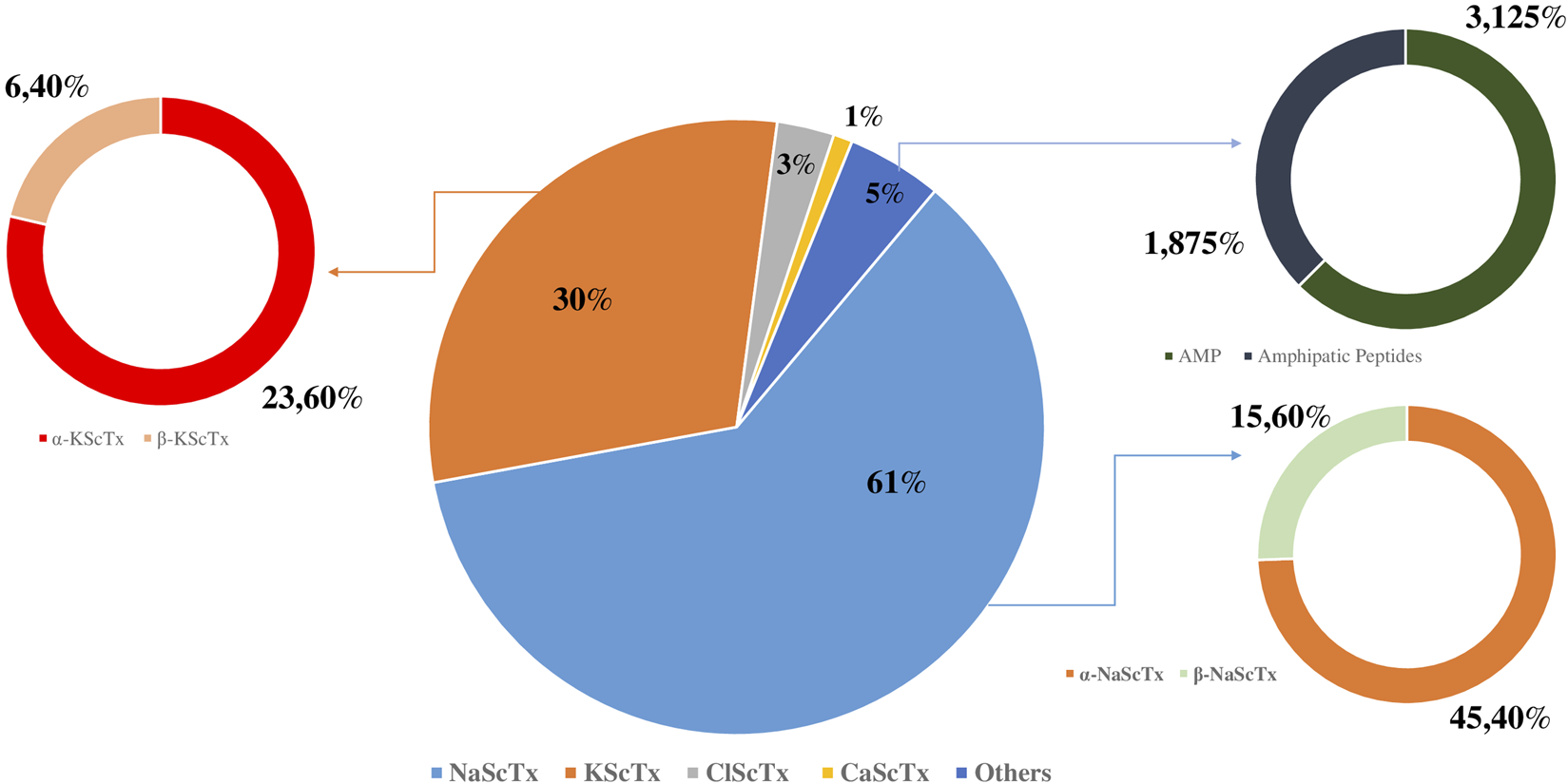
Proportional composition of peptides in Androctonus mauritanicus venom.
3.4 ELISA
The enzyme-linked immunosorbent assay was employed to evaluate the inhibitory effects of various fractions on the interaction between ACE2 and the receptor-binding domain (RBD) of the SARS-CoV-2 Spike protein. As summarized in Table 8 and illustrated in Figure 4, fractions F29 and F34 demonstrated the strongest inhibitory activity, with 79.7% and 73.9% inhibition at 40 μg/mL, respectively. Both fractions exhibited distinct retention times (42 min; 49 min) and had estimated IC50 values below 20 μg/mL, indicating high potency even at low concentrations.
TABLE 8
| Fraction | Retention time (min) | Concentration (µg/mL) | OD450 | Inhibition (%) | Estimated IC50 (µg/mL) |
|---|---|---|---|---|---|
| F29 | 42 | 20 | 0.50 | 71.01 | <20 |
| F29 | 42 | 40 | 0.35 | 79.71 | <20 |
| F31 | 45 | 20 | 1.45 | 15.94 | >40 |
| F31 | 45 | 40 | 1.31 | 23.91 | >40 |
| F34 | 49 | 20 | 0.65 | 62.32 | <20 |
| F34 | 49 | 40 | 0.45 | 73.91 | <20 |
| F35 | 51 | 20 | 1.33 | 23.19 | >40 |
| F35 | 51 | 40 | 1.24 | 28.12 | >40 |
| F36 | 52 | 20 | 1.45 | 15.94 | >40 |
| F36 | 52 | 40 | 1.28 | 26.09 | >40 |
Inhibition of ACE2-RBD interaction by fractions at different concentrations.
FIGURE 4
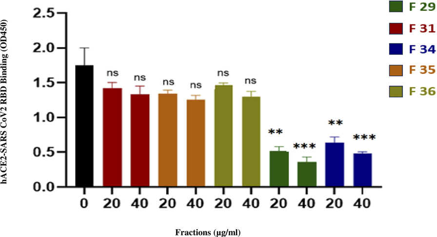
Inhibition potency of fractions on the interaction between ACE2 and SARS-CoV-2 spike (RBD) protein.
Fractions F31, F35, and F36 displayed lower inhibition (15.9%–28.1% at 40 μg/mL) and correspondingly higher IC50 values (>40 μg/mL), suggesting a moderate effect on ACE2-RBD interaction. The negative control showed no inhibition, confirming the specificity of the assay. These results highlight the differences in inhibitory potency among the tested fractions and suggest that F29 and F34 are promising candidates for further therapeutic exploration.
3.5 In Vivo neurotoxicity evaluation of A. mauritanicus venom fractions
The neurotoxic potential of A. mauritanicus crude venom and selected RP-HPLC fractions was evaluated in vivo using intracerebroventricular (ICV) injections in Swiss mice. Clinical signs, mortality within 2 h post-injection, and LD50 values were recorded where applicable. The results are summarized in Table 9.
TABLE 9
| Venom/Fraction | Toxicity | Mortality after 2 h | LD50 (µg/g) |
|---|---|---|---|
| Crude venom | +++ | Yes | 2.40 |
| Fraction 29 (F29) | – | No | |
| Fraction 34 (F34) | – | No | – |
| Fraction 31 (F31) | + | Yes | 1.21 |
| Fraction 35 (F35) | – | No | – |
| Fraction 36 (F36) | – | No | – |
| F29 + F34 | – | No | – |
Neurotoxicity profile of crude venom and selected RP-HPLC fractions of A. mauritanicus.
Fractions 29 and 34, despite demonstrating high Spike–ACE2 inhibition in ELISA, did not induce any observable toxicity or mortality, even when combined, indicating excellent safety profiles in vivo. In contrast, fraction 31 showed mild neurotoxicity with delayed onset of symptoms and resulted in mortality at higher doses, with an estimated LD50 of 1.21 μg/g. Fractions 35 and 36 were inactive in both antiviral and neurotoxicity assays, showing no behavioral effects or lethality.
4 Discussion
The venom of A. mauritanicus is a complex mixture containing peptides, enzymes, and small proteins, many of which exhibit neurotoxic properties (Watt and Simard, 1984). These molecules target ion channels, such as sodium, potassium, and calcium channels, interfering with the nervous system of prey or potential predators (Quintero-Hernández et al., 2013). However, beyond these toxic effects, the venom contains bioactive peptides that possess antimicrobial, antifungal, and antiviral activities (Hong et al., 2014; Harrison et al., 2014; Guilhelmelli et al., 2016). The molecular diversity of these peptides makes A. mauritanicus venom a promising candidate for bioprospecting, particularly in the search for new drugs (Xia et al., 2023; Hilal et al., 2023b). In the context of SARS-CoV-2, which causes COVID-19, peptides from A. mauritanicus venom could offer a novel antiviral strategy. Proteins like the SARS-CoV-2 spike protein (S protein) are crucial for viral entry, making them attractive targets for inhibition. The discovery and characterization of venom peptides capable of binding to or blocking the S protein could provide valuable leads for the development of new antiviral drugs (Ghazal et al., 2024).
This study aimed to fractionate A. mauritanicus venom using Reverse Phase High-Performance Liquid Chromatography (RP-HPLC) and characterize the resulting fractions via various mass spectrometry platforms, including triple quadrupole ESI-MS, Q-TOF LC/MS, and Q-Exactive LC/MS. The primary objective was to identify novel neurotoxins and isolate those responsible for severe symptoms, as well as those with potential therapeutic or biotechnological applications. The RP-HPLC fractionation of A. mauritanicus venom yielded 80 fractions (Figure 2). A comparable number of fractions were identified in the venom of Opisthacanthus cayaporum, while fewer fractions were obtained from other species: Buthacus macrocentrus (70 fractions), Tityus pachyurus (57 fractions), M. martensii (29 fractions), and Heterometrus longimanus (19 fractions) (Schwartz et al., 2008; Caliskan et al., 2012; Barona et al., 2006; Xu et al., 2014; Bringans et al., 2008). Each fraction was analyzed using direct infusion into the ESI-MS triple quadrupole spectrometer to determine average molecular masses, providing insights into the proteome of each fraction (Volmer et al., 2007). The analysis yielded 507 distinct molecular masses (Table 2), surpassing the number obtained from whole-venom analysis, highlighting the value of RP-HPLC for enhancing molecular resolution (Kumar and Kumar, 2012).
Fractions of interest were further analyzed using Q-TOF LC/MS to generate monoisotopic masses. The results confirmed significant heterogeneity in the composition of individual fractions. Differences in the number of masses detected by ESI-MS and Q-TOF LC/MS reflected the enhanced sensitivity of Q-TOF LC/MS for monoisotopic mass detection, coupled with nano-HPLC separation. The molecular diversity of the fractions was evident, with each containing multiple bioactive peptides with diverse biological properties. Fractions F19, F20, F21, F22, F25, F28, and F29 were found to be enriched in peptides corresponding to neurotoxins such as KScTxs, ClScTxs, and CaScTxs (Table 3–6). Notably, NaScTxs were detected only from fraction F31 onward, albeit with limited sequence coverage.
In total, 86 NaScTx-related peptides were identified, with sequence coverage ranging from 9% (Lipolysis-activating peptide 1-alpha chain, D9U2A4) to 86% (Alpha-toxin Lqq4, P01489). Additionally, 42 KScTx peptides were detected, with sequence coverage spanning from 13% (Alpha-KTx 3.16, K7XFK5) to 100% (Alpha-KTx 8.1, P56215). Among the ClScTxs, five peptides were identified with sequence coverage between 43% (Insectotoxin-I5, P60270) and 100% (Neurotoxin P2, P01498). A single CaScTx-related peptide was detected, exhibiting 25% sequence similarity to Toxin BmCa-1 (Q8I6X9).
Interestingly, 55 NaScTx peptides were identified for the first time, with 31 matching those reported in previous studies on
A. mauritanicusvenom. Fragments of some of the most toxic NaScTxs were detected, including:
- Alpha-toxin Amm 5 in fractions F34, F35, F39, and F41, with the highest sequence coverage (48%) observed in fraction F41.
- Alpha-toxin Amm 3 in fractions F35, F36, and F38, with maximum coverage (52%) in fractions F35 and F36.
- Alpha-mammal toxin Lqq5 in fractions F34 and F35, with 14% sequence coverage.
These neurotoxins were exclusively detected by Q-Exactive LC/MS and not by Q-TOF LC/MS, likely due to the superior sensitivity of the Orbitrap analyzer used in Q-Exactive (Fedorova et al., 2013). This instrument’s High-Energy Collisional Dissociation (HCD) chamber provides higher collision energy than the Collision-Induced Dissociation (CID) mechanism used in Q-TOF LC/MS, enabling improved peptide fragmentation. However, both CID and HCD were limited in efficiently fragmenting long toxins, resulting in low sequence coverage for intact neurotoxins.
Regarding KScTx, 32 peptides were identified, 10 of which had previously been detected. The analysis revealed that α-KScTxs exhibited notable molecular diversity, with seven subfamilies identified: Alpha-KTx 03, 08, 09, 15, 16, 17, and 27. Some KScTx peptides displayed sequence homology with:
- Gating modifiers of anion channels (P80670), potent inhibitors of chloride channel ClC-2/CLCN2.
- Alpha-KTx 9.1 homolog (Q9NJP7), a specific inhibitor of small-conductance potassium channels (KCa2).
- Alpha-KTx 9.7 homolog (P83406), an activator of calcium channels that reversibly modulates the ryanodine receptor 1 (RYR1).
Only α-KScTxs and β-KScTxs were identified in this study, while other scorpion venoms, such as that of Centruroides hirsutipalpus, are known to contain additional KScTx families (γ-, δ-, and ε-) (Table 4) (Valdez-Velázquez et al., 2020).
Two chloride channel-targeting neurotoxins were also identified:
- A chlorotoxin-like peptide with a molecular mass of 3624.28 Da in fraction F33% and 53% sequence coverage.
- Chlorotoxin with a monoisotopic mass of 3806.45 Da in fraction F44% and 42% sequence coverage.
For CaScTxs, only one peptide was detected, with 25% sequence homology to BmCa-1 (Q8I6X9) (Table 6). This result aligns with previous findings that CaScTxs are relatively rare in scorpion venoms (Olamendi-Portugal et al., 2002; Shahbazzadeh et al., 2007; Fajloun et al., 2000) and further underscore the extensive molecular diversity of A. mauritanicus venom, particularly in neurotoxins targeting ion channels. This diversity is reflected not only in the polymorphism of the identified families but also in the variety of membrane receptors and ion channels they modulate. The identification of novel peptides, especially those targeting ion channels, highlights the unexplored biotechnological potential of this venom and opens new avenues for therapeutic development (Díaz-García and Varela, 2020).
Arachnid venoms, employed as tools for both defense and predation, serve to kill or immobilize prey for feeding or to neutralize competitors and potential predators. These venoms exhibit remarkable molecular diversity and complexity, with the expression of proteins and peptides governed by intricate gene regulation mechanisms that are still under investigation (Suranse et al., 2018; Marchi et al., 2022).
Scorpion venoms have been extensively studied, primarily due to their clinical effects on humans, which can sometimes lead to fatal outcomes (Chippaux and Goyffon, 2008). Paradoxically, deeper insights into the mechanisms of action of venom components have paved the way for biotechnological applications, with many research efforts focused on developing novel therapeutics based on the structure and function of these molecules (Rates et al., 2011). Despite its potential, A. mauritanicus venom remains an underexplored source of novel proteins that could contribute to biotechnological advancements.
The rapid expansion of identified scorpion venom compounds has revealed several promising drug candidates to address emerging global medical challenges (Hmed et al., 2013). Biologically active peptides from scorpion venoms are broadly classified into disulfide-bridged peptides (DBPs) (Lavergne et al., 2015) and non-disulfide-bridged peptides (NDBPs) (Almaaytah and Albalas, 2014; Zeng et al., 2005). Notably, DBPs represent the major components responsible for the neurotoxic symptoms observed in cases of scorpion envenomation (Mata et al., 2017).
Scorpion venom presents promising therapeutic potential for the treatment of infectious diseases. Several antiviral molecules, including neurotoxins and DBPs, have been identified from these venoms (Table 7) (Li et al., 2011). DBPs typically consist of approximately 30 amino acids, with three or four disulfide bridges arranged in a cysteine-stabilized motif (CS-), where a loop between two strands mimics the CDR2 loop of the CD4 receptor (Mata et al., 2017). DBPs can bind to the gp120 glycoprotein of HIV through molecular mimicry of CD4+ receptors on host cells. This interaction disrupts the gp120-CD4 binding, thereby preventing viral entry into host cells (Quinlan et al., 2014). Furthermore, scorpion potassium channel toxins, such as charybdotoxin (ChTx) and scyllatoxin, exhibit similar activity in blocking the gp120-CD4 interaction. Interestingly, mucroporin and its derivative mucroporin-M1, both with enhanced positive charge, interact directly with viral envelopes and have demonstrated antiviral effects against SARS-CoV and H5N1 viruses (Li et al., 2011).
Consistent with the antiviral potential of scorpion venoms, the non-disulfide-bridged peptide (NDBP) Ctry2459, isolated from the venom gland of Chaerilus tryznai, was shown to inhibit initial HCV infection in Huh7.5.1 cells by directly inactivating infectious viral particles (Mata et al., 2017). However, the 13-amino-acid peptide displayed limited bioavailability and was unable to suppress established infection. To overcome this limitation, histidine-rich analogs derived from the Ctry2459 scaffold—Ctry2459-H2 and Ctry2459-H3—were engineered to improve helicity, amphiphilicity, and endosomal escape. These modified peptides exhibited enhanced antiviral activity, reducing intracellular viral RNA by 40% and 70%, respectively, whereas the parental peptide mainly affected viral infectivity without significantly lowering intracellular viral levels (El Hidan et al., 2021). In comparison, crude venoms from S. maurus palmatus and A. australis have also demonstrated antiviral activity against HCV, with IC50 values of 6.3 ± 1.6 and 88.3 ± 5.8 μg/mL, respectively. Notably, the venom of Scorpio maurus palmatus reduces viral infectivity via a virucidal mechanism targeting the entry step, without affecting intracellular viral replication, and its activity is resistant to metalloprotease inhibition or heat treatment at 60 °C. In contrast, the Ctry2459-derived peptides represent a novel class of NDBPs capable not only of virucidal action but also of reducing intracellular viral RNA, highlighting their potential as more versatile antiviral agents compared with previously reported scorpion venom-derived compounds such as those from A. australis (El-Bitar et al., 2015; Mata et al., 2017).
The COVID-19 pandemic has emphasized the importance of vaccination in controlling the spread of SARS-CoV-2. However, the challenges posed by delays in vaccine production and distribution, along with the emergence of viral variants partially resistant to natural or vaccine-induced immune responses, highlight the need for additional preventive mauritanicus and therapeutic strategies. Combining vaccination with other measures such as diagnostics, protective protocols, and novel treatments remains essential. Developing new therapeutic agents, particularly those that inhibit viral entry, could offer significant benefits in reducing transmission and mitigating severe cases of infection. Recent research has turned to bioactive molecules from venomous animals, such as scorpions, to explore their potential as antiviral agents. Venom-derived molecules, known for their diverse pharmacological properties, have emerged as valuable candidates for therapeutic development.
This research focused on peptides isolated from Moroccan scorpion A. mauritanicus, to identify potential antiviral agents targeting SARS-CoV-2. The spike (S) protein of the virus, which plays a crucial role in its entry into host cells by binding to the ACE2 receptor (Lan et al., 2020). Our fraction’s antiviral potential was validated through ELISA analysis, uncovering five promising candidates with two demonstrating strong binding affinity (Figure 4). These fractions suggest a potential mechanism of viral entry inhibition, the peptides present in fractions F29 and F34 may directly bind to the receptor-binding domain (RBD) of the Spike protein, thereby preventing its interaction with the human ACE2 receptor. This specific inhibitio provides a plausible mechanistic hypothesis that nevertheless requires further functional validation.
The identification of 507 distinct molecular masses in A. mauritanicus venom, including 55 novel NaScTxs and ion channel-targeting peptides, significantly expands the known pharmacopeia of scorpion-derived bioactive compounds. These findings are particularly relevant given the urgent need for novel antiviral strategies, as exemplified by the COVID-19 pandemic. The discovery of fractions (F19–F29) with high-affinity binding to SARS-CoV-2 spike protein suggests a potential mechanism for viral entry inhibition, mirroring the gp120-CD4 blockade observed in HIV by other scorpion DBPs (Quinlan et al., 2014). This work provides the first evidence that A. mauritanicus peptides may interfere with ACE2-S protein interactions, offering a template for developing peptide-based antivirals against emerging coronaviruses. Furthermore, the heterogeneous neurotoxin profiles (e.g., α-toxins Amm3/5) highlight untapped opportunities for ion channel research, with potential applications in pain management and neurological disorders.
The study’s major strength lies in its multi-platform mass spectrometry approach, which enabled detection of low-abundance peptides (e.g., CaScTxs) through Q-Exactive LC/MS’s superior HCD sensitivity (Fedorova et al., 2013). However, key limitations must be acknowledged; A. mauritanicus venom procurement remains challenging due to the species’ endangered status in Morocco and low venom yields (∼0.5 mg per milking) (Oukkache et al., 2013), restricting large-scale studies. Limited fragmentation efficiency for long toxins during sequence coverage (e.g., 9% coverage for Lipolysis-activating peptide) underscores the need for hybrid techniques like ETD-MS/MS in future work. While ELISA confirmed S-protein binding, it is important to note that the RBD–ACE2 binding assay has been validated in previous studies to predict antiviral efficacy. For instance, it was demonstrated that compounds such as zafirlukast, identified by their ability to inhibit S1RBD–ACE2 binding, effectively blocked the entry of SARS-CoV-2 pseudovirus into cells (Zhang et al., 2022). These findings support the idea that inhibition of the ACE2–RBD interaction is a critical step that can lead to antiviral effects. Nevertheless, further assays using viral or pseudoviral infection models are recommended to fully confirm the antiviral potential of the identified venom fractions. In vivo efficacy and toxicity profiles of lead peptides (e.g., F19–F22) also remain to be characterized.
Our research highlights the safety profiles in vivo of fractions 29 and 34 as promising antiviral candidates. The absence of neurotoxic symptoms and lethality suggests that these fractions can effectively inhibit viral entry without inducing adverse neurological effects, a critical consideration for therapeutic development (Table 9). Nevertheless, further in vivo studies are required to evaluate their systemic efficacy, pharmacokinetics, and long-term safety in relevant infection models. Such investigations will be essential to fully characterize their therapeutic potential and ensure translational relevance.
This study establishes A. mauritanicus venom as a rich source of both neurotoxins and antiviral candidates, but translational success will require interdisciplinary collaboration between toxinology, virology, and drug delivery fields. Rational design of truncated analogs (e.g., mucroporin-M1 derivatives (Mata et al., 2017) could enhance ACE2-binding affinity while reducing neurotoxicity. Evaluating fractions against viral variants (e.g., Omicron sublineages) and other enveloped viruses (e.g., influenza) given conserved targeting of host receptors. Nanocarrier encapsulation could address peptide stability issues, as demonstrated for chlorotoxin glioma therapies (Costa et al., 2013). Recombinant expression of high-priority toxins (e.g., α-KTx 9.1 homologs) in E. coli or yeast to circumvent venom supply constraints (King and Hardy, 2013). This interdisciplinary enrichment of our results and the development of this research is encouraged.
5 Conclusion and perspectives
This work not only demonstrates the molecular diversity and therapeutic potential of scorpion venoms but also highlights the importance of innovative approaches in addressing emerging viral threats. As global efforts continue to develop effective COVID-19 treatments, venom-derived peptides could play a pivotal role in complementing existing strategies and mitigating future outbreaks.
This study focused on mapping the proteome of A. mauritanicus venom, the species most frequently associated with severe envenomation in Morocco. The aim was to investigate the molecular diversity and toxic components of the venom to develop an effective antivenom, while also identifying novel molecules with potential for therapeutic or biotechnological applications. Analysis of the venom after fractionation revealed its complex nature, comprising a broad range of components, with neurotoxins primarily targeting sodium (NaScTxs) and potassium channels (KscTxs). It was found that the venom is particularly rich in NaScTxs, which act on mammalian sodium channels, explaining its role in the most fatal scorpion envenomations reported in the region.
Moreover, the venom also contains neurotoxins that affect chloride channels (ClScTxs), further demonstrating the vast diversity of its toxic arsenal. This diversity is reflected both in the molecular variability within toxin families and in the variety of targeted receptors and ion channels, some of which may offer promising avenues for pharmaceutical development. The findings presented in this paper not only shed light on the molecular diversity of A. mauritanicus venom but also contribute to the growing field of venom-based therapeutics. The identification of peptides with inhibitory effects on SARS-CoV-2 provides a foundation for developing new antiviral drugs, offering a complementary strategy to vaccination and other treatments. However, claims regarding potential therapeutic or industrial applications must be interpreted cautiously. The antiviral activity reported here is based on in vitro ELISA assays targeting the Spike–ACE2 interaction, and further validation using pseudoviral or authentic SARS-CoV-2 infection models is necessary to confirm efficacy, elucidate mechanisms of action, and assess in vivo safety. These findings therefore highlight promising candidates for future studies rather than immediate therapeutic use, emphasizing the need for additional research to explore their full potential.
Statements
Data availability statement
Raw data are available via ProteomeXchange (PRIDE) with identifier PXD069182. Available at: https://proteomecentral.proteomexchange.org/cgi/GetDataset?ID=PXD069182.
Ethics statement
The experimental procedures involving animals were carried out in compliance with internationally accepted ethical standards for laboratory animal care and use, following the recommendations of the World Health Organization (WHO) and the European Directive 2010/63/EU. The study was conducted in accordance with the local legislation and institutional requirements.
Author contributions
RC: Conceptualization, Data curation, Formal Analysis, Investigation, Methodology, Software, Visualization, Writing – original draft, Writing – review and editing. JG: Conceptualization, Data curation, Formal Analysis, Investigation, Methodology, Validation, Visualization, Writing – review and editing. HH: Conceptualization, Formal Analysis, Investigation, Methodology, Writing – review and editing. AL: Conceptualization, Data curation, Formal Analysis, Investigation, Methodology, Visualization, Writing – review and editing. SC: Data curation, Investigation, Methodology, Writing – review and editing. HA: Conceptualization, Data curation, Formal Analysis, Investigation, Supervision, Visualization, Writing – review and editing. RB: Conceptualization, Formal Analysis, Investigation, Methodology, Validation, Visualization, Writing – review and editing. NB: Conceptualization, Data curation, Investigation, Methodology, Visualization, Writing – review and editing. RS: Conceptualization, Data curation, Formal Analysis, Investigation, Software, Supervision, Validation, Visualization, Writing – review and editing. RE: Conceptualization, Data curation, Formal Analysis, Funding acquisition, Investigation, Methodology, Project administration, Resources, Software, Validation, Visualization, Writing – review and editing. NO: Conceptualization, Data curation, Formal Analysis, Investigation, Methodology, Project administration, Resources, Software, Supervision, Validation, Visualization, Writing – original draft, Writing – review and editing.
Funding
The author(s) declare that financial support was received for the research and/or publication of this article. This work was conducted as part of the M-Cov project, a collaborative initiative between the Institut Pasteur and UM6P of Benguerir. The project aims to harness bioactive compounds from venoms to develop novel therapeutics against SARS-CoV-2. The publication of these findings was made possible through the support provided by this initiative.
Acknowledgments
We thank the staff of the Experimental Center of Tit Mellil, Institut Pasteur of Morocco, for providing the laboratory animals, as well as the facilities to support this research.
Conflict of interest
The authors declare that the research was conducted in the absence of any commercial or financial relationships that could be construed as a potential conflict of interest.
Generative AI statement
The author(s) declare that no Generative AI was used in the creation of this manuscript.
Any alternative text (alt text) provided alongside figures in this article has been generated by Frontiers with the support of artificial intelligence and reasonable efforts have been made to ensure accuracy, including review by the authors wherever possible. If you identify any issues, please contact us.
Publisher’s note
All claims expressed in this article are solely those of the authors and do not necessarily represent those of their affiliated organizations, or those of the publisher, the editors and the reviewers. Any product that may be evaluated in this article, or claim that may be made by its manufacturer, is not guaranteed or endorsed by the publisher.
References
1
Abbas N. Gaudioso-Tyzra C. Bonnet C. Gabriac M. Amsalem M. Lonigro A. et al (2013). The scorpion toxin Amm VIII induces pain hypersensitivity through gain-of-function of TTX-sensitive Na+ channels. Pain154 (8), 1204–1215. 10.1016/j.pain.2013.03.037
2
Al-Asmari A. K. Riyasdeen A. Abbasmanthiri R. Arshaduddin M. Al-Harthi F. A. (2016). Scorpion (A. bicolor) venom exhibits cytotoxicity and induces cell cycle arrest and apoptosis in breast and colorectal cancer cell lines. Indian J. Pharmacol.48 (5), 537–543. 10.4103/0253-7613.190742
3
Almaaytah A. Albalas Q. (2014). Scorpion venom peptides with no disulfide bridges: a review. Peptides51, 35–45. 10.1016/j.peptides.2013.10.021
4
Almaaytah A. Zhou M. Wang L. Chen T. Walker B. Shaw C. (2012). Antimicrobial/cytolytic peptides from the venom of the North African scorpion, Androctonus amoreuxi: biochemical and functional characterization of natural peptides and a single site-substituted analog. Peptides35 (2), 291–299. 10.1016/j.peptides.2012.03.016
5
Almaaytah A. Tarazi S. Mhaidat N. Al-Balas Q. Mukattash T. L. (2013). Mauriporin, a novel cationic α-Helical peptide with selective cytotoxic activity against prostate cancer cell lines from the venom of the scorpion Androctonus mauritanicus. Int. J. Pept. Res. Ther.19, 281–293. 10.1007/s10989-013-9350-3
6
Barona J. Batista C. V. Zamudio F. Z. Gomez-Lagunas F. Wanke E. Otero R. et al (2006). Proteomic analysis of the venom and characterization of toxins specific for Na+-and K+-channels from the Colombian scorpion Tityus pachyurus. Biochimica et Biophysica Acta (BBA)-Proteins and Proteomics1764 (1), 76–84. 10.1016/j.bbapap.2005.08.010
7
BenAissa R. Othman H. Villard C. Peigneur S. Mlayah-Bellalouna S. Abdelkafi-Koubaa Z. et al (2020). AaHIV a sodium channel scorpion toxin inhibits the proliferation of DU145 prostate cancer cells. Biochem. Biophysical Res. Commun.521 (2), 340–346. 10.1016/j.bbrc.2019.10.115
8
Bringans S. Eriksen S. Kendrick T. Gopalakrishnakone P. Livk A. Lock R. et al (2008). Proteomic analysis of the venom of Heterometrus longimanus (Asian black scorpion). Proteomics8 (5), 1081–1096. 10.1002/pmic.200700948
9
Caliskan F. I. G. E. N. Quintero-Hernández V. Restano-Cassulini R. Batista C. V. F. Zamudio F. Z. Coronas F. I. et al (2012). Turkish scorpion B. macrocentrus: general characterization of the venom and description of Bu1, a potent mammalian Na+-channel α-toxin. Toxicon59 (3), 408–415. 10.1016/j.toxicon.2011.12.013
10
Calvete J. J. (2017). Venomics: integrative venom proteomics and beyond. Biochem. J.474 (5), 611–634. 10.1042/BCJ20160577
11
Chen Y. Cao L. Zhong M. Zhang Y. Han C. Li Q. et al (2012). Anti-HIV-1 activity of a new scorpion venom peptide derivative Kn2-7. PloS one7 (4), e34947. 10.1371/journal.pone.0034947
12
Cheng Y. Zhao J. Qiao W. Chen K. (2014). Recent advances in diagnosis and treatment of gliomas using chlorotoxin-based bioconjugates. Am. J. Nucl. Med. Mol. Imaging4 (5), 385–405.
13
Chippaux J. P. Goyffon M. (2008). Epidemiology of scorpionism: a global appraisal. Acta Trop.107 (2), 71–79. 10.1016/j.actatropica.2008.05.021
14
Costa P. M. Cardoso A. L. Mendonça L. S. Serani A. Custódia C. Conceição M. et al (2013). Tumor-targeted chlorotoxin-coupled nanoparticles for nucleic acid delivery to glioblastoma cells: a promising system for glioblastoma treatment. Mol. Ther. Nucleic Acids.2 (6), e100. Available online at: https://www.ncbi.nlm.nih.gov/pmc/articles/PMC3696908/.
15
Desjardins P. Hansen J. B. Allen M. (2009). Microvolume protein concentration determination using the NanoDrop 2000c spectrophotometer. J. Vis. Exp. JoVE (33), 1610. 10.3791/1610
16
Díaz-García A. Varela D. (2020). Voltage-gated K+/Na+ channels and scorpion venom toxins in cancer. Front. Pharmacol.11, 913. 10.3389/fphar.2020.00913
17
Du Q. Hou X. Wang L. Zhang Y. Xi X. Wang H. et al (2015). AaeAP1 and AaeAP2: novel antimicrobial peptides from the venom of the scorpion, Androctonus aeneas: structural characterisation, molecular cloning of biosynthetic precursor-encoding cDNAs and engineering of analogues with enhanced antimicrobial and anticancer activities. Toxins7 (2), 219–237. 10.3390/toxins7020219
18
Ehret-Sabatier L. Loew D. Goyffon M. Fehlbaum P. Hoffmann J. A. van Dorsselaer A. et al (1996). Characterization of novel cysteine-rich antimicrobial peptides from scorpion blood. J. Biol. Chem.271 (47), 29537–29544. 10.1074/jbc.271.47.29537
19
El Hidan M. A. Laaradia M. A. El Hiba O. Draoui A. Aimrane A. Kahime K. (2021). Scorpion-derived antiviral peptides with a special focus on medically important viruses: an update. BioMed Res. Int.2021, 9998420. 10.1155/2021/9998420
20
El Oufir R. (2019). Piqures et Envenimations Scorpioniques (PES); Rapports general et specifiques. Rev. Toxicol. Maroc. Cent. Anti Poison Du. Maroc. (CAPM) Rabat, Moroc.43, 11.
21
El-Bitar A. M. Sarhan M. M. Aoki C. Takahara Y. Komoto M. Deng L. et al (2015). Virocidal activity of Egyptian scorpion venoms against hepatitis C virus. Virology J.12, 47. 10.1186/s12985-015-0276-6
22
Fajloun Z. Kharrat R. Chen L. Lecomte C. Di Luccio E. Bichet D. et al (2000). Chemical synthesis and characterization of maurocalcine, a scorpion toxin that activates Ca2+ release channel/ryanodine receptors. FEBS Lett.469 (2-3), 179–185. 10.1016/s0014-5793(00)01239-4
23
Fedorova G. Randak T. Lindberg R. H. Grabic R. (2013). Comparison of the quantitative performance of a Q-exactive high‐resolution mass spectrometer with that of a triple quadrupole tandem mass spectrometer for the analysis of illicit drugs in wastewater. Rapid Commun. Mass Spectrom.27 (15), 1751–1762. 10.1002/rcm.6628
24
Ghazal A. Clarke D. Abdel-Rahman M. A. Ribeiro A. Collie-Duguid E. Pattinson C. et al (2024). Venomous gland transcriptome and venom proteomic analysis of the scorpion Androctonus amoreuxi reveal new peptides with anti-SARS-CoV-2 activity. Peptides173, 171139. 10.1016/j.peptides.2023.171139
25
Guilhelmelli F. Vilela N. Smidt K. S. De Oliveira M. A. da Cunha Morales Álvares A. Rigonatto M. C. et al (2016). Activity of scorpion venom-derived antifungal peptides against planktonic cells of Candida spp. and Cryptococcus neoformans and Candida albicans biofilms. Front. Microbiol.7, 1844. 10.3389/fmicb.2016.01844
26
Harrison P. L. Abdel-Rahman M. A. Miller K. Strong P. N. (2014). Antimicrobial peptides from scorpion venoms. Toxicon88, 115–137. 10.1016/j.toxicon.2014.06.006
27
Hilal I. Khourcha S. Safi A. Hmyene A. Asnawi S. Othman I. et al (2023a). Comparative proteomic analysis of the venoms from the Most dangerous scorpions in Morocco: androctonus mauritanicus and Buthus occitanus. Life13 (5), 1133. 10.3390/life13051133
28
Hilal I. Khourcha S. Safi A. Hmyene A. Stöcklin R. Oukkache N. (2023b). Exploring the inter- and intra-specific variability of androctonus scorpion venoms. Biol. Life Sci. Forum. 24, 12. 10.3390/iect2023-14797
29
Hmed B. Serria H. T. Mounir Z. K. (2013). Scorpion peptides: potential use for new drug development. J. Toxicol.2013 (1), 958797. 10.1155/2013/958797
30
Hong W. Li T. Song Y. Zhang R. Zeng Z. Han S. et al (2014). Inhibitory activity and mechanism of two scorpion venom peptides against herpes simplex virus type 1. Antivir. Res.102, 1–10. 10.1016/j.antiviral.2013.11.013
31
Iketani S. Ho D. D. (2024). SARS-CoV-2 resistance to monoclonal antibodies and small-molecule drugs. Cell Chem. Biol.31 (4), 632–657. 10.1016/j.chembiol.2024.03.008
32
Kim E. Hwang D. H. Mohan Prakash R. L. Asirvatham R. D. Lee H. Heo Y. et al (2025). Animal venom in modern medicine: a review of therapeutic applications. Toxins17 (8), 371. 10.3390/toxins17080371
33
King G. F. Hardy M. C. (2013). Recombinant expression of margatoxin and agitoxin-2 in pichia pastoris: an efficient method for production of KV1.3 channel blockers. PLoS One8 (1), e53638. Available online at: https://www.ncbi.nlm.nih.gov/pmc/articles/PMC3530466/.
34
Kumar S. D. Kumar D. H. (2012). Importance of RP-HPLC in analytical method development: a review. Int. J. Pharm. Sci. Res.3 (12), 4626. 10.13040/IJPSR.0975-8232.3(12).4626-33
35
Lan J. Ge J. Yu J. Shan S. Zhou H. Fan S. et al (2020). Structure of the SARS-CoV-2 spike receptor-binding domain bound to the ACE2 receptor. Nature581 (7807), 215–220. 10.1038/s41586-020-2180-5
36
Lavergne V. Alewood P. F. Mobli M. King G. F. (2015). The structural universe of disulfide-rich venom peptides.
37
Li K. M. Rivory L. P. Clarke S. J. (2006). Solid-phase extraction (SPE) techniques for sample preparation in clinical and pharmaceutical analysis: a brief overview. Curr. Pharm. Anal.2 (2), 95–102. 10.2174/157341206776819346
38
Li Q. Zhao Z. Zhou D. Chen Y. Hong W. Cao L. et al (2011). Virucidal activity of a scorpion venom peptide variant mucroporin-M1 against measles, SARS-CoV and influenza H5N1 viruses. Peptides32 (7), 1518–1525. 10.1016/j.peptides.2011.05.015
39
Li Z. Hu P. Wu W. Wang Y. (2019). Peptides with therapeutic potential in the venom of the scorpion Buthus martensii Karsch. Peptides115, 43–50. 10.1016/j.peptides.2019.02.009
40
Liu G. Yang F. Li F. Li Z. Lang Y. Shen B. et al (2018). Therapeutic potential of a scorpion venom-derived antimicrobial peptide and its homologs against antibiotic-resistant gram-positive bacteria. Front. Microbiol.9 (1159), 1159. 10.3389/fmicb.2018.01159
41
Mahendran T. R. Cynthia B. Thevendran R. Maheswaran S. (2024). Prospects of innovative therapeutics in combating the COVID-19 pandemic. Mol. Biotechnol.67, 2598–2606. 10.1007/s12033-024-01240-4
42
Marchi F. C. Mendes-Silva E. Rodrigues-Ribeiro L. Bolais-Ramos L. G. Verano-Braga T. (2022). Toxinology in the proteomics era: a review on arachnid venom proteomics. J. Venom. Animals Toxins Incl. Trop. Dis.28, 20210034. 10.1590/1678-9199-JVATITD-2021-0034
43
Mata É. C. G. D. Mourão C. B. F. Rangel M. Schwartz E. F. (2017). Antiviral activity of animal venom peptides and related compounds. J. Venom. Animals Toxins Incl. Trop. Dis.23, 3. 10.1186/s40409-016-0089-0
44
Murdocca M. Romeo I. Citro G. Latini A. Centofanti F. Bugatti A. et al (2024). A dynamic and effective peptide-based strategy for promptly addressing emerging SARS-CoV-2 variants of concern. Pharm. Basel, Switz.17 (7), 891. 10.3390/ph17070891
45
Olamendi-Portugal T. García B. I. López-González I. Van Der Walt J. Dyason K. Ulens C. et al (2002). Two new scorpion toxins that target voltage-gated Ca2+ and Na+ channels. Biochem. Biophysical Res. Commun.299 (4), 562–568. 10.1016/s0006-291x(02)02706-7
46
Oldrati V. Arrell M. Violette A. Perret F. Sprüngli X. Wolfender J. L. et al (2016). Advances in venomics. Mol. Biosyst.12 (12), 3530–3543. 10.1039/c6mb00516k
47
Ortiz E. Gurrola G. B. Schwartz E. F. Possani L. D. (2015). Scorpion venom components as potential candidates for drug development. Toxicon Official J. Int. Soc. Toxinology93, 125–135. 10.1016/j.toxicon.2014.11.233
48
Quinlan B. D. Joshi V. R. Gardner M. R. Ebrahimi K. H. Farzan M. (2014). A double-mimetic peptide efficiently neutralizes HIV-1 by bridging the CD4 and coreceptor-binding sites of gp120. J. Virology88 (6), 3353–3358. 10.1128/JVI.03800-13
49
Quintero-Hernández V. Jiménez-Vargas J. M. Gurrola G. B. Valdivia H. H. Possani L. (2013). Scorpion venom components that affect ion-channels function. Toxicon76, 328–342. 10.1016/j.toxicon.2013.07.012
50
Oukkache N. Chgoury F. Lalaoui M. Cano A. A. Ghalim N. (2013). Comparison between two methods of scorpion venom milking in Morocco. J. Venom. Anim. Toxins. Incl. Trop. Dis.19 (1), 5. 10.1186/1678-9199-19-5
51
Rates B. Verano-Braga T. Moreira Santos D. Pedrosa Nunes K. MC Pimenta A. Elena De Lima M. (2011). From the stretcher to the pharmacy's shelf: drug leads from medically important Brazilian venomous arachnid species. Inflamm. Allergy-Drug Targets (Formerly Current Drug Targets-Inflammation & Allergy) (Discontinued)10 (5), 411–419. 10.2174/187152811797200614
52
Rjeibi I. Mabrouk K. Mosrati H. Berenguer C. Mejdoub H. Villard C. et al (2011). Purification, synthesis and characterization of AaCtx, the first chlorotoxin-like peptide from Androctonus australis scorpion venom. Peptides32, 656–663. 10.1016/j.peptides.2011.01.015
53
Schwartz E. F. Camargos T. S. Zamudio F. Z. Silva L. P. Bloch Jr C. Caixeta F. et al (2008). Mass spectrometry analysis, amino acid sequence and biological activity of venom components from the Brazilian scorpion Opisthacanthus cayaporum. Toxicon51 (8), 1499–1508. 10.1016/j.toxicon.2008.03.029
54
Shahbazzadeh D. Srairi-Abid N. Feng W. Ram N. Borchani L. Ronjat M. et al (2007). Hemicalcin, a new toxin from the Iranian scorpion Hemiscorpius lepturus which is active on ryanodine-sensitive Ca2+ channels. Biochem. J.404 (1), 89–96. 10.1042/BJ20061404
55
Suranse V. Srikanthan A. Sunagar K. (2018). Animal venoms: origin, diversity and evolution. eLS, 1–20. 10.1002/9780470015902.a0000939.pub2
56
Uzair B. Bint-E-Irshad S. Khan B. A. Azad B. Mahmood T. Rehman M. U. et al (2018). Scorpion venom peptides as a potential source for human drug candidates. Protein peptide Lett.25 (7), 702–708. 10.2174/0929866525666180614114307
57
V V. Achar R. R. M U H. N A. T Y. S. Kameshwar V. H. et al (2021). Venom peptides - a comprehensive translational perspective in pain management. Curr. Res. Toxicol.2, 329–340. 10.1016/j.crtox.2021.09.001
58
Valdez-Velázquez L. L. Cid-Uribe J. Romero-Gutierrez M. T. Olamendi-Portugal T. Jimenez-Vargas J. M. Possani L. D. (2020). Transcriptomic and proteomic analyses of the venom and venom glands of Centruroides hirsutipalpus, a dangerous scorpion from Mexico. Toxicon179, 21–32. 10.1016/j.toxicon.2020.02.021
59
Volmer D. A. Sleno L. Bateman K. Sturino C. Oballa R. Mauriala T. et al (2007). Comparison of MALDI to ESI on a triple quadrupole platform for pharmacokinetic analyses. Anal. Chem.79 (23), 9000–9006. 10.1021/ac7016234
60
Watt D. D. Simard J. M. (1984). Neurotoxic proteins in scorpion venom. J. Toxicol. Toxin Rev.3 (2-3), 181–221. 10.3109/15569548409097925
61
Xia Z. He D. Wu Y. Kwok H. F. Cao Z. (2023). Scorpion venom peptides: molecular diversity, structural characteristics, and therapeutic use from channelopathies to viral infections and cancers. Pharmacol. Res.197, 106978. 10.1016/j.phrs.2023.106978
62
Xia Z. Xie L. Li B. Lv X. Zhang H. Cao Z. (2024). Antimicrobial potential of scorpion-venom-derived peptides. Molecules29 (21), 5080. 10.3390/molecules29215080
63
Xu X. Duan Z. Di Z. He Y. Li J. Li Z. et al (2014). Proteomic analysis of the venom from the scorpion Mesobuthus martensii. J. Proteomics106, 162–180. 10.1016/j.jprot.2014.04.032
64
Yaqoob R. Tahir H. M. Arshad M. Naseem S. Ahsan M. M. (2016). Optimization of the conditions for maximum recovery of venom from scorpions by electrical stimulation. Pak. J. Zoology48 (1).
65
Zargan J. Sajad M. Umar S. Naime M. Ali S. Khan H. A. (2011). Scorpion (Androctonus crassi-cauda) venom limits growth of transformed cells (SH-SY5Y and MCF-7) by cytotoxicity and cell cycle arrest. Experimental Mol. Pathology91 (1), 447–454. 10.1016/j.yexmp.2011.04.008
66
Zeng X. C. Corzo G. Hahin R. (2005). Scorpion venom peptides without disulfide bridges. IUBMB Life57 (1), 13–21. 10.1080/15216540500058899
67
Zerouti K. Khemili D. Laraba-Djebari F. Djelila H.-T. (2019). Nontoxic fraction of scorpion venom reduces bacterial growth and inflammatory response in a mouse model of infection. Toxin Rev.40, 310–324. 10.1080/15569543.2019.1614064
68
Zhang S. Gao C. Das T. Luo S. Tang H. Yao X. et al (2022). The spike-ACE2 binding assay: an in vitro platform for evaluating vaccination efficacy and for screening SARS-CoV-2 inhibitors and neutralizing antibodies. J. Immunol. Methods503, 113244. 10.1016/j.jim.2022.113244
Summary
Keywords
Androctonus mauritanicus , scorpion venom, proteomics, mass spectrometry, antiviral peptides, SARS-CoV-2
Citation
Chahir R, Galan J, Hboub H, Lahlou AS, Chakir S, Aassila H, Ben Mrid R, Bouchmaa N, Stöcklin R, El Fatimy R and Oukkache N (2025) Characterization of Androctonus mauritanicus venom and in vitro screening of SARS-CoV-2 entry inhibitors candidates. Front. Pharmacol. 16:1678606. doi: 10.3389/fphar.2025.1678606
Received
03 August 2025
Accepted
18 September 2025
Published
03 November 2025
Volume
16 - 2025
Edited by
Chang Liu, University of Rhode Island, United States
Reviewed by
Krzysztof Brzezinski, Polish Academy of Sciences, Poland
Huifang Li, University of Rhode Island, United States
Rui Qi, Yoh Services LLC, United States
Updates
Copyright
© 2025 Chahir, Galan, Hboub, Lahlou, Chakir, Aassila, Ben Mrid, Bouchmaa, Stöcklin, El Fatimy and Oukkache.
This is an open-access article distributed under the terms of the Creative Commons Attribution License (CC BY). The use, distribution or reproduction in other forums is permitted, provided the original author(s) and the copyright owner(s) are credited and that the original publication in this journal is cited, in accordance with accepted academic practice. No use, distribution or reproduction is permitted which does not comply with these terms.
*Correspondence: Reda Chahir, reda.chahir.doc@uhp.ac.ma; Jacob Galan, jacob.galan@utrgv.edu; Naoual Oukkache, naoual.oukkache@pasteur.ma
ORCID ID:Hinde Aassila, 0000-0002-1372-4986
Disclaimer
All claims expressed in this article are solely those of the authors and do not necessarily represent those of their affiliated organizations, or those of the publisher, the editors and the reviewers. Any product that may be evaluated in this article or claim that may be made by its manufacturer is not guaranteed or endorsed by the publisher.