 YaFeng Zheng
 XiaoNa Ye1
 Dandan Meng
- 1Department of Preventive Treatment of Disease, The Second Affiliated Hospital of Shandong University of Traditional Chinese Medicine, Jinan, Shandong, China
- 2Department of Traditional Chinese Medicine, Shandong Provincial Maternal and Child Health Care Hospital Affiliated to Qingdao University, Jinan, Shandong, China
- 3School of Continuing Education, Shandong University of Traditional Chinese Medicine, Jinan, Shandong, China
- 4College of Traditional Chinese Medicine, Shandong University of Traditional Chinese Medicine, Jinan, Shandong, China
Diabetic nephropathy (DN) is one of the most common and disabling chronic complications of diabetes, with a complex pathogenesis involving genetic susceptibility, inflammatory responses, oxidative stress, and other pathological processes. Current therapeutic approaches can partially control hyperglycemia and slow the decline of renal function, but remain insufficient to reverse established structural damage to the kidneys. This underscores the urgent need for novel, safe, and multi-targeted intervention strategies. In recent years, natural bioactive compounds have attracted considerable attention for their potential in preventing and treating chronic diseases. Quercetin, a natural flavonoid widely distributed in plants, exhibits multiple biological activities—including anti-inflammatory, antioxidant, anti-fibrotic, and cell death–modulating effects—and has shown significant promise in DN therapy. This review provides a comprehensive overview of the major pathogenic mechanisms of DN and recent advances in understanding the regulatory effects of quercetin on key pathological processes. We highlight its potential mechanisms of action, including suppression of inflammation and oxidative stress, inhibition of TGF-β1–mediated renal fibrosis, protection of podocyte function, and induction of ferroptosis, and discuss the possible synergistic interactions among these effects in modulating the DN disease network. In addition, we evaluate the current status and limitations of preclinical research on quercetin, and explore feasible strategies—such as nanoparticle-based delivery systems and structural modification—to enhance its bioavailability and tissue targeting. Finally, we propose future research directions for quercetin-based interventions in DN, aiming to provide a theoretical foundation and novel insights for its clinical translation.
1 Introduction
Diabetic nephropathy (DN) is a common chronic microvascular complication of diabetes, characterized by pathological features such as glomerulosclerosis, tubulointerstitial fibrosis, and progressive renal insufficiency (Yang and Liu, 2022). The development of DN is primarily associated with structural and functional damage to the kidneys caused by prolonged hyperglycemia, with persistent proteinuria and a gradual decline in renal function being the main clinical manifestations (Samsu, 2021; Ricciardi and Gnudi, 2021). In addition, alterations in renal hemodynamics exacerbate swelling and sclerosis of the glomeruli and renal tubules, further impairing filtration capacity (Tanabe et al., 2020).
Epidemiological studies have demonstrated that DN is a leading cause of chronic kidney failure and end-stage renal disease (ESRD) worldwide (Zou et al., 2022), and is also an important contributor to increased all-cause mortality (Sabanayagam et al., 2019). Compared with other immune-mediated kidney diseases, DN is associated with a higher mortality rate. In recent years, the global prevalence of diabetes has been steadily rising. By 2021, the number of people with diabetes had reached 537 million worldwide, and it is projected to increase to 783 million by 2045, with an alarming trend toward younger onset (Sun et al., 2022; Yu et al., 2024). This growing prevalence not only poses a serious threat to public health but also imposes a substantial socioeconomic and healthcare burden.
The pathogenesis of DN is complex and may be associated with multiple factors, including disturbances in glucose metabolism, oxidative stress, inflammation, and autophagy (Raina et al., 2021; Naaman and Bakris, 2023). Current conventional therapeutic approaches primarily focus on symptomatic management, such as blood pressure control, reduction of proteinuria, and glycemic regulation, often combined with novel hypoglycemic agents. However, these strategies have limited efficacy in preventing disease progression to ESRD, and some pharmacological agents are associated with undesirable adverse effects. Consequently, elucidating the molecular mechanisms underlying DN and identifying safe and effective anti-DN agents remain of critical importance (Kopel et al., 2019). Complementary and alternative medicine (CAM), owing to its abundance of natural bioactive constituents, has emerged as a potential therapeutic strategy. In recent years, natural products have demonstrated unique advantages in DN management, including the ability to slow disease progression, alleviate clinical symptoms, and minimize adverse effects (Yu et al., 2021; Xie et al., 2021).
Quercetin, a naturally occurring flavonoid, is derived from natural sources such as Ginkgo biloba and rutin, with the molecular formula C15H10O7. It is also known as 3,3′,4′,5,7-pentahydroxyflavone and is one of the most abundant natural compounds found in plants and fruits. Quercetin exhibits a broad spectrum of pharmacological activities, including antioxidant, anti-inflammatory, hypoglycemic, hypolipidemic, anti–Alzheimer’s disease, anticancer, and antimicrobial effects (Ebrahimpour et al., 2020; Khursheed et al., 2020; Patel et al., 2018; Yang D. et al., 2020; Zou et al., 2021), suggesting its wide therapeutic potential across multiple systems. Emerging evidence indicates that quercetin exerts renoprotective effects in DN through diverse mechanisms, such as attenuation of oxidative stress and inflammation, inhibition of fibrosis, regulation of apoptosis and autophagy, modulation of glucose and lipid metabolism, and induction of ferroptosis (Liu et al., 2024; Jiang et al., 2019; Abdou and Abd elkader, 2022; Lu et al., 2015). Therefore, this review aims to provide a systematic summary of the current research progress on quercetin in the treatment of DN. We first outline the pathogenic mechanisms of DN, followed by an in-depth discussion of the potential molecular pathways through which quercetin exerts its effects, with particular emphasis on key signaling pathways such as Nrf2, NF-κB, TGF-β1/Smad, AMPK/mTOR, and SIRT1. Experimental evidence from both in vitro and in vivo studies will be reviewed to support these mechanisms. Furthermore, we discuss the existing limitations in current research and propose future directions to improve quercetin’s bioavailability and promote its clinical translation. By elucidating the mechanistic basis of quercetin’s therapeutic effects in DN, this review seeks to provide a scientific foundation for its development as an adjunct or alternative therapeutic strategy for DN management.
2 Pathogenesis of diabetic nephropathy
The pathogenesis of DN is highly complex and remains incompletely understood. Current evidence suggests that DN is the result of multiple interacting factors and involves intricate and multifaceted molecular mechanisms. These mechanisms engage numerous signaling pathways, mediators, and interrelated metabolic and hemodynamic variables (Pérez-Morales et al., 2019). Existing studies indicate that the development and progression of DN are closely associated with genetic predisposition, disturbances in glucose and lipid metabolism, hemodynamic abnormalities, oxidative stress, inflammation, and dysregulation of autophagy (Raina et al., 2021). The cumulative effects of these factors over time lead to progressive structural and functional damage to the kidneys. A thorough understanding of the pathogenic mechanisms of DN can help identify key etiological factors, thereby facilitating targeted preventive measures, reducing disease incidence, and ultimately lowering DN-related morbidity and mortality. The pathogenic mechanisms of DN are illustrated in Figure 1.
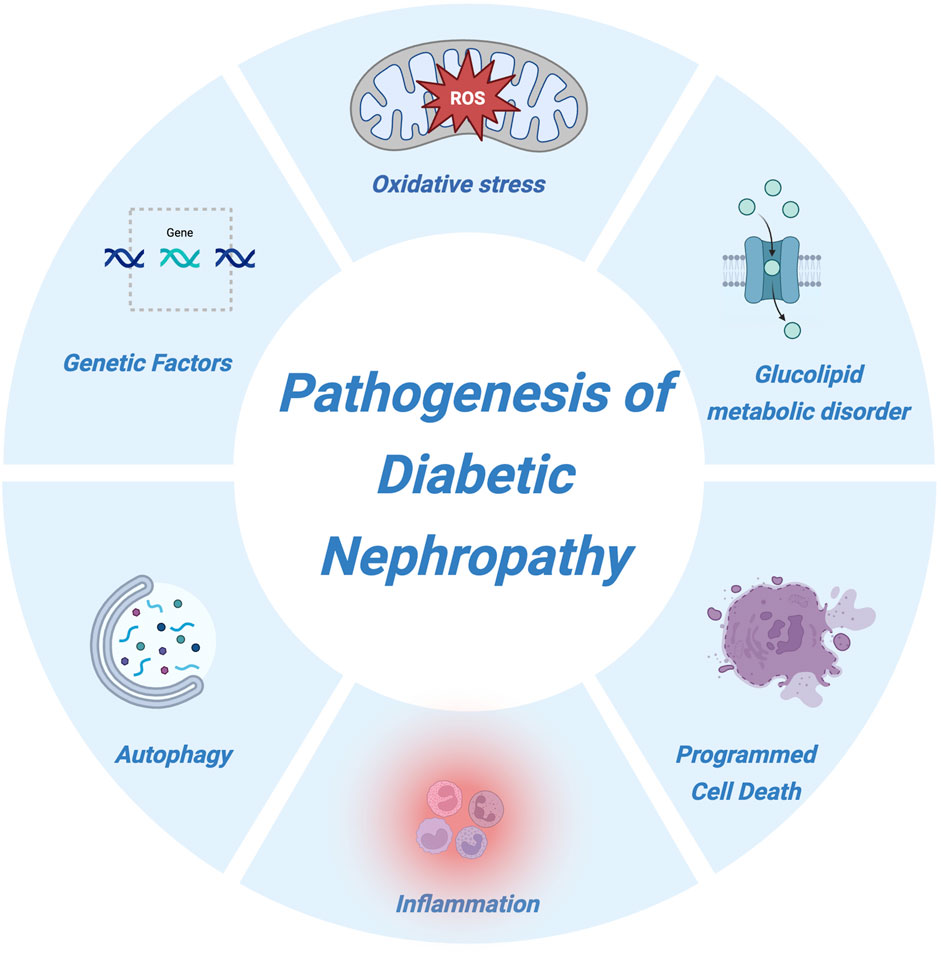
Figure 1. Pathogenesis of diabetic nephropathy. This schematic illustration summarizes the major pathogenic mechanisms involved in the development and progression of diabetic nephropathy. Key contributors include genetic susceptibility, oxidative stress, glucolipid metabolic disorders, inflammation, autophagy dysfunction, and various forms of programmed cell death. These interconnected pathways collectively drive renal injury and accelerate disease progression.
2.1 Genetic factors
Although hyperglycemia is the primary trigger for the onset of DN, not all patients with diabetes progress to this complication. Epidemiological data indicate that approximately 30%–40% of individuals with diabetes will eventually develop DN (Sun et al., 2022), suggesting that genetic factors play a pivotal role in its pathogenesis. The incidence of DN varies significantly among different ethnic groups and families, further supporting the concept of genetic susceptibility (Conserva et al., 2016).
Multiple genes have been implicated in the development of DN, including CARS, PIK3C2G, INPPL1, TBC1D4, NRXN3, and NLRP3, which have been identified as DN-associated genes in various studies (Pezzolesi and Krolewski, 2013; Saeed, 2018). In addition, single nucleotide polymorphism (SNP) rs4880 has been reported to be associated with diabetes and certain diabetic complications, such as cardiovascular disease, nephropathy, and retinopathy (Yahya et al., 2019). With the advancement of epigenetic research, novel regulatory mechanisms have also been linked to DN, including the involvement of non-coding RNAs, post-translational chromatin modifications, and DNA methylation (Mcknight et al., 2015). These findings indicate that genetic and epigenetic factors jointly contribute to individual susceptibility and disease progression in DN.
2.2 Dysregulation of glucose and lipid metabolism
Disorders of glucose and lipid metabolism play a crucial role in the pathogenesis of DN, not only exacerbating glomerular hyperfiltration induced by hyperglycemia but also promoting inflammation, oxidative stress, lipotoxicity, and structural as well as functional renal impairment through multiple metabolic stress pathways. Hyperglycemia is the primary pathogenic factor in DN.
Both hyperglycemia and oxidative stress can lead to abnormal accumulation of extracellular matrix (ECM)–related factors in renal tubular cells, resulting in glomerulosclerosis and tubulointerstitial fibrosis. In the early stages, DN lacks specific clinical manifestations; however, as the disease progresses, patients may develop alterations in glomerular filtration rate, hypertension, and proteinuria, ultimately progressing to ESRD (Sun et al., 2021; Li et al., 2018). Previous studies have reported that more than 80% of biopsy-confirmed DN patients exhibit disturbances in glucose and lipid metabolism (Russo et al., 2020). Persistent hyperglycemia may impair vascular endothelial function, leading to abnormal blood supply to target organs. Moreover, chronic hyperglycemia and glucose fluctuations contribute to excessive lipid accumulation, reduced lipid catabolism, and elevated expression of lipid metabolism–related biomarkers, collectively accelerating renal injury.
Lipids play essential roles in intracellular signal transduction, immune regulation, material transport, and the maintenance of cellular metabolism. Dyslipidemia is one of the hallmarks of diabetes, characterized by abnormal plasma lipid profiles and ectopic lipid accumulation in various tissues and organs, including the kidneys (Thongnak et al., 2020). Such metabolic disturbances may lead to impaired cholesterol metabolism, increased lipid uptake or synthesis, altered fatty acid oxidation, lipid droplet accumulation, and imbalances in bioactive sphingolipids—such as ceramide, ceramide-1-phosphate, and sphingosine-1-phosphate. These changes contribute to renal lipid deposition, glomerulosclerosis, and mesangial expansion, thereby exacerbating proteinuria and the progression of tubulointerstitial fibrosis. In addition, direct podocyte injury may occur, leading to irreversible impairment of renal function (Mitrofanova et al., 2021).
Non-esterified fatty acids (NEFAs) serve as a major energy source for proximal tubular epithelial cells. Fatty acid uptake occurs via endocytosis or through transporters such as CD36 and fatty acid–binding proteins. Excess circulating free fatty acids can downregulate insulin transcription, induce β-cell apoptosis, reduce insulin secretion, and ultimately lead to insulin resistance (Noels et al., 2021; Opazo-Ríos et al., 2020). Recent studies have suggested that, beyond traditional lipid parameters, apolipoprotein A1 (ApoA1), apolipoprotein B (ApoB), and lipid ratios are particularly relevant in patients with type 2 diabetes (Gao et al., 2023). In particular, the low-density lipoprotein (LDL)/ApoB ratio shows a strong correlation with DN, estimated glomerular filtration rate (eGFR), and the urine albumin-to-creatinine ratio (UACR), with lower ratios indicating a higher risk of DN. Furthermore, serum lipoprotein(a) [Lp(a)] and high-density lipoprotein (HDL) levels have also been investigated in relation to DN. Patients with DN exhibit significantly elevated serum Lp(a) levels and reduced HDL levels compared with diabetic patients without nephropathy. Elevated Lp(a) has been identified as a risk factor for DN, whereas HDL appears to exert a protective effect (Mitrofanova et al., 2023).
Collectively, these findings indicate that abnormalities in glucose and lipid metabolism can impair endothelial function, disrupt renal blood supply, and lead to excessive lipid deposition, thereby inducing both structural and functional alterations in renal vasculature and accelerating DN progression. Therefore, regulating glucose and lipid metabolism represents an important therapeutic strategy for the clinical management of DN and for delaying disease progression (Koch et al., 2020).
2.3 Oxidative stress
The dynamic balance between cellular oxidation and reduction is fundamental for maintaining cell growth, development, and activity, and is essential for overall health. Oxidative stress refers to a state in which, upon exposure to various harmful stimuli, the body excessively generates reactive oxygen species (ROS) and reactive nitrogen species (RNS), while losing the ability to effectively eliminate these free radicals. This imbalance between oxidant production and antioxidant defenses disrupts tissue and cellular homeostasis, leading to structural and functional impairment (Sies, 2015; Chen et al., 2022). Clinically, oxidative stress is recognized as a major contributor to aging and the development of numerous diseases, underscoring the importance of research on oxidative and antioxidant mechanisms (Ramos-Riera et al., 2023). Under physiological conditions, the oxidative and antioxidant systems remain in dynamic equilibrium to meet normal metabolic demands. However, in patients with diabetes, chronic hyperglycemia exerts deleterious effects on mitochondria, leading to mitochondrial injury and excessive production of ROS and RNS. This disruption of redox balance triggers oxidative stress, thereby contributing to the onset and progression of various diabetic complications, including DN (Zhang Z. et al., 2023).
Oxidative stress represents a converging outcome of multiple pathogenic pathways involved in DN. Hyperglycemia-induced overproduction of ROS is considered a central event in the disease’s pathogenesis. The damage mediated by oxidative stress can occur either directly or indirectly. Directly, ROS injure podocytes, mesangial cells, and endothelial cells, leading to proteinuria and tubulointerstitial fibrosis (Duni et al., 2019). The glomerulus is particularly susceptible to oxidative damage. Hyperglycemia contributes to damage of DNA, lipids, and proteins, and the extent of such injury is closely associated with ROS-induced oxidative stress (Luc et al., 2019).
Oxidative stress is also linked to metabolic and hemodynamic alterations in the kidney, both of which exert synergistic pathogenic effects. Chronic hyperglycemia–induced oxidative stress can elevate angiotensin II (Ang II) levels, activate protein kinase C (PKC), and increase transforming growth factor-β (TGF-β) expression, all of which are recognized as potent pro-oxidative stimuli (Winiarska et al., 2021). Moreover, TGF-β has been shown to participate in NADPH oxidase (NOX)-mediated ROS generation (Wang L. et al., 2021). Sustained overexpression and activation of TGF-β driven by elevated ROS promote excessive ECM remodeling in mesangial cells and accelerate tubulointerstitial fibrosis. Typically, hyperglycemia-induced oxidative stress is thought to enhance pro-inflammatory protein levels by promoting macrophage infiltration. Activated macrophages secrete inflammatory cytokines, thereby driving both local and systemic inflammation. Consequently, oxidative stress is closely interconnected with inflammation and renin–angiotensin–aldosterone system (RAAS) activation, forming a vicious cycle that exacerbates renal injury in DN.
2.4 Inflammatory response
Inflammation plays a critical role in the pathogenesis of DN (Moreno et al., 2018; Qiu and Tang, 2016). Rather than being a mere secondary consequence of tissue injury, inflammation represents a key pathophysiological driver of disease onset and progression (Jung and Moon, 2021). Recent studies have highlighted the pivotal role of inflammatory processes in initiating and accelerating DN. Evidence supports the involvement of interleukin (IL)-1, IL-6, and IL-18 in the development and progression of DN (Pichler et al., 2017). Leukocytes, monocytes, and macrophages actively participate in the pathogenic processes of DN, and elevated levels of inflammatory biomarkers are associated with an increased risk of disease. Furthermore, the degree of inflammatory cell infiltration within the kidney strongly correlates with DN severity. In animal models of DN, inhibition of inflammatory cell recruitment to the kidney has been shown to exert renoprotective effects. These findings suggest that inflammation is not only a marker but also a major causal factor in the initiation and progression of DN.
Pro-inflammatory and pro-fibrotic cytokines synthesized and secreted by these infiltrating cells within the local microenvironment can directly disrupt renal architecture, subsequently triggering the epithelial–mesenchymal transition (EMT) process (Liu, 2011), which leads to ECM accumulation. In addition to producing pro-inflammatory cytokines, renal cells in both diabetic animal models and patients exhibit upregulated expression of chemokines and adhesion molecules. These molecules act as key mediators of renal injury by attracting circulating leukocytes and facilitating their transmigration into renal tissue. The infiltrating immune cells serve as an additional source of cytokines and other mediators, thereby contributing to the progression of renal injury and sustaining the inflammatory response. Under hyperglycemic conditions, monocyte chemoattractant protein-1 (MCP-1) secretion is markedly increased in renal tissue, promoting the recruitment of various inflammatory cells. These recruited cells, in turn, secrete large amounts of pro-inflammatory cytokines such as IL-1 and IL-18, creating a vicious cycle of inflammatory injury. Moreover, these inflammatory cells and cytokines can increase urinary albumin excretion and impair renal function (Pérez-Morales et al., 2019).
Taken together, these findings demonstrate that inflammation actively drives the onset and progression of DN, highlighting the damaging effects of hyperglycemia-induced inflammatory responses on the kidney.
2.5 Autophagy
Under physiological conditions, autophagy is a protective cellular process that maintains intracellular homeostasis and regulates cell death, playing a critical role in sustaining organismal stability. Both excessive activation and inhibition of autophagy can be detrimental, potentially leading to tissue damage and disease onset (Yu et al., 2023). Studies have shown that in DN model mice, the autophagic activity of intrinsic renal cells—such as podocytes and mesangial cells—is significantly reduced (Kitada et al., 2017; Liu et al., 2018). This decline impairs the timely clearance of damaged proteins and organelles, resulting in renal cell injury, thickening of the glomerular basement membrane (GBM), excessive ECM production, and ultimately promotion of renal fibrosis (Xu et al., 2020). These findings suggest that maintaining normal autophagic activity may represent a promising therapeutic target for DN.
Experimental evidence indicates that modulation of autophagy through upregulation of AMP-activated protein kinase (AMPK) or downregulation of mammalian target of rapamycin (mTOR) can restore autophagic function in DN model mice, increase autophagosome formation, and thereby alleviate renal injury (Sheng et al., 2022; Liu H. et al., 2021). Furthermore, other studies have demonstrated that certain therapeutic agents for DN exert their hypoglycemic effects via regulation of the phosphatidylinositol 3-kinase (PI3K)/protein kinase B (Akt) signaling pathway (Wang et al., 2025; Wu et al., 2025).
Recent studies have revealed that certain non-coding RNAs (ncRNAs) can also influence the development and progression of DN by modulating autophagic activity. Long non-coding RNAs (lncRNAs) constitute the majority of ncRNAs in the genome. For example, lncRNA H19 is highly expressed under hyperglycemic conditions and can suppress podocyte autophagy by downregulating DIRAS3 and subsequently activating the mTOR pathway (Wu and Huang, 2023). Chen et al. (2019) reported that inhibition of the PI3K/Akt/mTOR axis, accompanied by downregulation of miR-142-5p, could activate autophagy and mitigate renal injury.
Taken together, these findings suggest that dysregulated autophagy may play an important role in the onset and progression of DN. Designing precise intervention strategies targeting autophagy regulatory networks—such as inhibiting excessive autophagy in type 1 diabetes mellitus (T1DM) or enhancing lysosomal function in type 2 diabetes mellitus (T2DM)—may provide novel therapeutic approaches to delay DN progression.
3 Mechanisms of quercetin in the treatment of DN
3.1 Antioxidative stress
Oxidative stress is one of the key mechanisms driving DN progression. It results from excessive accumulation of ROS induced by hyperglycemia through mechanisms such as mitochondrial electron transport chain dysfunction, glucose auto-oxidation, and advanced glycation end-product (AGE) formation. These processes impair the antioxidant defense system, activate inflammatory pathways, and establish a vicious cycle of “oxidative stress–inflammation,” ultimately leading to structural damage in the glomeruli and tubulointerstitium. Prolonged hyperglycemia disrupts the dynamic balance between ROS production and clearance, triggering oxidative stress, which is recognized as an important pathological process in DN (Arellano-Buendía et al., 2014). Under oxidative stress conditions, excessive ROS production is closely linked to DN onset and progression. Elevated ROS can increase glomerular capillary permeability, activate cytokines, impair glomerular filtration function, and induce proteinuria. Therefore, ameliorating oxidative stress represents a promising therapeutic strategy for the prevention and management of DN (Zhou et al., 2019; Du et al., 2018).
Superoxide dismutase (SOD) is a critical ROS scavenger and a key biomarker of oxidative stress, protecting cells from ROS-induced damage. Malondialdehyde (MDA), a major product of lipid peroxidation induced by ROS, can indirectly reflect the level of free radicals and the extent of lipid peroxidation, providing an estimate of the redox balance in plasma (Hofni et al., 2014). In an experimental study using a DN model in C57BL/6J mice, Gomes et al. (2015) demonstrated that low-dose quercetin administration exerted antioxidant, anti-apoptotic, lipid-lowering, and renoprotective effects. These findings suggest that the antioxidative properties of quercetin contribute significantly to its therapeutic potential in DN.
Recent studies have demonstrated that dysregulation of microRNAs is closely associated with renal diseases. It has been reported that overexpression of miR-370 and miR-217 contributes to high glucose (HG)-induced podocyte injury, thereby promoting the development of DN (Xian et al., 2018; Sun et al., 2017). Building on this evidence, Wan et al. (2022) investigated the role of miR-485-5p in DN. Their study revealed that quercetin suppressed HG-induced proliferation, inflammation, and oxidative stress in human mesangial cells (HMCs). Moreover, serum samples from DN patients showed downregulation of miR-485-5p and upregulation of Yes-associated protein 1 (YAP1). Mechanistically, quercetin exerted its protective effects by modulating the miR-485-5p/YAP1 axis to attenuate HG-induced oxidative stress, suggesting a novel therapeutic strategy for DN.
Transforming growth factor-β1 (TGF-β1) upregulation occurs in nearly all kidney diseases and serves as a central mediator of podocyte injury through both Smad-dependent and Smad-independent pathways (Herman-Edelstein et al., 2013). Smad proteins are transcription factors that mediate TGF-β signaling by forming complexes with one another. The generation of ROS has been shown to activate the TGF-β1/Smad signaling pathway and induce podocyte injury (Chang et al., 2016; Yu et al., 2010). Therefore, inhibition of oxidative stress and TGF-β1/Smad pathway–mediated podocyte damage may represent a potential therapeutic target for DN.
In an in vivo study, Gao et al. (2018) reported that quercetin ameliorated podocyte injury in DN rats by reducing the expression of nephrin, an effect potentially associated with the suppression of oxidative stress and the TGF-β1/Smad signaling pathway. Specifically, quercetin increased SOD and glutathione (GSH) levels while reducing MDA levels. Moreover, quercetin inhibited TGF-β1–induced phosphorylation of Smad2 and Smad3 in the kidneys of DN rats. Interestingly, quercetin treatment also markedly upregulated Smad7, a negative regulator of TGF-β1 signaling. Collectively, these findings indicate that quercetin protects podocytes in DN by attenuating oxidative stress and inhibiting the TGF-β1/Smad pathway. This provides promising new evidence supporting the potential application of quercetin in maintaining podocyte integrity and preventing proteinuria. The mechanisms by which quercetin alleviates oxidative stress in the treatment of DN.
3.2 Anti-inflammatory
Inflammation is one of the central drivers of DN progression. Hyperglycemic stimulation can induce the production of large amounts of pro-inflammatory cytokines in renal tissue, activate immune cells, and ultimately lead to glomerulosclerosis and tubulointerstitial fibrosis. Infiltration of inflammatory cells such as lymphocytes, neutrophils, and macrophages has been shown to contribute to renal injury in DN (Wang et al., 2021b). Quercetin, as a natural flavonoid, has demonstrated clear anti-inflammatory properties in multiple studies (Tang et al., 2019; Li et al., 2020). Clinical and experimental evidence indicates that interleukin-6 (IL-6) and tumor necrosis factor-alpha (TNF-α) are significantly associated with proteinuria in DN patients. Upregulation of TNF-α can enhance local production of ROS, exacerbate oxidative stress–induced renal injury, and trigger the release of other inflammatory cytokines, including IL-6. This cascade promotes inflammatory cell infiltration and endothelial dysfunction (Sathibabu Uddandrao et al., 2019).
Abdou and Abd elkader (2022) evaluated the antidiabetic, antioxidant, and anti-inflammatory effects of quercetin extracted from Trifolium alexandrinum extract (TAE) in a streptozotocin (STZ)-induced DN model. Compared with the untreated STZ-diabetic group, quercetin treatment significantly reduced renal levels of TGF-β, TNF-α, and IL-6, improved renal function, and alleviated inflammatory damage. These findings suggest that the renoprotective effects of quercetin may be mediated, at least in part, by its anti-inflammatory properties. Both quercetin and TAE-derived formulations hold promise as potential therapeutic agents for DN with translational clinical value.
Yes-associated protein (YAP), also known as YAP1, is a transcriptional coactivator that functions as a critical molecular switch and downstream effector in the Hippo signaling pathway (Yagi et al., 1999; Ramos and Camargo, 2012). Previous studies have demonstrated that YAP1 is closely associated with the onset and progression of DN (Huang et al., 2018; Ma et al., 2019; Yang T. et al., 2020). In an experimental study, Wan et al. (2022) investigated the effects of quercetin on cell proliferation, inflammatory cytokines (TNF-α, IL-1β, and IL-6), and YAP1-related expression. They found that quercetin suppressed HG-induced proliferation and inflammatory responses in HMCs. Enzyme-linked immunosorbent assay (ELISA) results showed that the protective effects of quercetin on stress-induced inflammatory cytokines (TNF-α, IL-1β, and IL-6) were largely abolished by a miR-485-5p inhibitor. Mechanistically, miR-485-5p directly binds to YAP1 and inhibits its expression. These findings indicate that quercetin exerts anti-inflammatory effects against HG-induced injury by regulating the miR-485-5p/YAP1 axis.
Yin Yang 1 (YY1) is a nuclear transcription factor that plays a pivotal role in regulating cellular metabolism (Zhang et al., 2017). Recent studies have demonstrated that upregulation of YY1 contributes to renal fibrosis in DN (Du et al., 2021; Yang et al., 2023a). Increasing evidence further suggests that YY1 functions as a novel pro-inflammatory mediator involved in various inflammation-related diseases (Mu et al., 2020; Lin et al., 2021). Based on this, Yang et al. (2023b) investigated the critical anti-inflammatory role of quercetin in DN-associated tubulointerstitial inflammation. They found that in diabetic models (db/db mice and HG–treated HK-2 cells), YY1 expression was markedly elevated, which promoted activation of the IL-6/STAT3 inflammatory pathway and exacerbated tubulointerstitial inflammation. Quercetin was shown to bind directly to YY1, reducing its protein expression. Mechanistically, YY1 binds to the promoter region of IL-6, directly suppressing its transcription. By directly inhibiting the inflammatory transcription factor YY1, quercetin downregulated the IL-6/STAT3 pathway, thereby significantly alleviating tubulointerstitial inflammation in DN models. These findings reveal a novel mechanism by which quercetin exerts therapeutic effects against DN and provide a mechanistic basis for developing quercetin as a targeted anti-DN agent.
Macrophages are a crucial component of the innate immune system, playing essential roles in immune regulation, inflammatory responses, and tissue repair (Locati et al., 2020; Park et al., 2022). Recent evidence indicates that imbalanced macrophage polarization is increasingly recognized as a major driver of inflammatory responses and renal fibrosis in DN (Landis et al., 2018; Klessens et al., 2017). In the early stages of DN, pro-inflammatory M1 macrophages exacerbate renal tubular epithelial cell injury and initiate renal inflammation through the secretion of pro-inflammatory cytokines. In contrast, anti-inflammatory M2 macrophages promote tissue repair and resolution of inflammation by secreting anti-inflammatory mediators such as arginase-1 (Arg-1) and interleukin-10 (IL-10) (Gu et al., 2024). During DN progression, excessive activation of M1 macrophages sustains chronic renal inflammation and fibrosis (Calle and Hotter, 2020). A recent study (Abudoureyimu et al., 2025) demonstrated that quercetin markedly reduced the expression of NLRC5, NLRP3, TNF-α, IL-6, and IL-1β in HG-stimulated RAW 264.7 macrophages in vitro, while increasing the expression of M2-associated markers including CD206, Arg-1, and IL-10. In DN mice, quercetin treatment improved renal histopathological injury and fibrosis, significantly downregulated NLRC5, NLRP3, Col1a1, and α-SMA expression, and decreased renal levels of TNF-α, IL-6, and IL-1β.
These findings suggest that quercetin ameliorates DN by targeting the NLRC5/NLRP3 pathway to suppress M1 macrophage polarization while promoting an anti-inflammatory M2 phenotype. This highlights a novel molecular target for DN treatment and underscores quercetin’s potential as a promising therapeutic candidate for DN. The mechanisms by which quercetin exerts anti-inflammatory effects in the treatment of DN are illustrated in Figure 2.
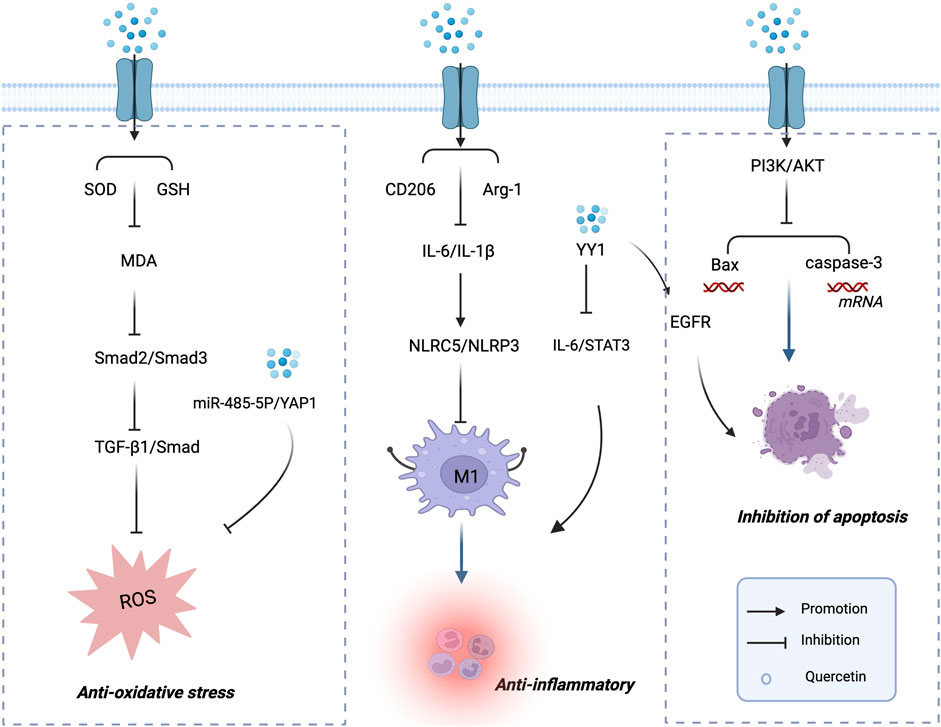
Figure 2. Mechanisms of Quercetin in the Treatment of DN. Mechanisms of quercetin in the treatment of diabetic nephropathy. This schematic illustration depicts the major mechanisms by which quercetin exerts renoprotective effects in diabetic nephropathy. Quercetin alleviates oxidative stress by enhancing SOD and GSH activity and reducing MDA, thereby inhibiting the Smad2/3 and TGF-β1/Smad signaling pathways. It suppresses inflammatory responses by modulating CD206, Arg-1, IL-6, and IL-1β, leading to the regulation of NLRC5/NLRP3 inflammasome activation and IL-6/Stat3 signaling. In addition, quercetin inhibits apoptosis via the PI3K/AKT pathway, downregulating Bax and caspase-3 expression while enhancing EGFR signaling. These multi-targeted actions highlight the therapeutic potential of quercetin in preventing renal injury and slowing the progression of diabetic nephropathy.
3.3 Inhibition of podocyte apoptosis
Apoptosis plays a crucial role in maintaining cellular homeostasis throughout growth and development, and its dysregulation is a recognized risk factor for multiple diseases (Godse et al., 2017). Numerous studies have demonstrated that suppressing tubular epithelial cell apoptosis can effectively ameliorate DN (Wang et al., 2022; Xing et al., 2021), further supporting the therapeutic relevance of targeting apoptosis in DN prevention and treatment. Liu et al. (2024), through both in vivo and in vitro experiments, found that in the diabetic mellitus (DM) group, mRNA and protein levels of BAX, as well as cleaved caspase-3 (c-Caspase3), were significantly elevated, with a concomitant increase in the BAX/BCL-2 ratio—indicating enhanced pro-apoptotic signaling. Conversely, quercetin intervention markedly inhibited diabetes-associated renal cell apoptosis, evidenced by reduced Bax and Caspase-3 mRNA levels, decreased BAX and c-Caspase3 protein expression, and a lowered BAX/BCL-2 ratio. Mechanistically, these effects were mediated via activation of the PI3K/AKT pathway, which suppressed tubular epithelial cell apoptosis. Collectively, these findings suggest that quercetin prevents DN progression by attenuating tubular epithelial cell apoptosis through PI3K/AKT signaling modulation, highlighting its potential as a promising candidate for DN prevention.
Podocyte apoptosis is one of the key mechanisms underlying the transition from compensatory hypertrophy to detachment during podocyte injury. This process is primarily characterized by the formation of apoptotic bodies and is accompanied by upregulated expression of pro-apoptotic proteins such as Bax and cleaved caspase-3. Previous studies have shown that pretreatment with Abelmoschus manihot total flavonoids (TFA) markedly reduces the number of apoptotic podocytes in DN rats, decreases the expression of pro-apoptotic proteins caspase-3 and caspase-8, thereby attenuating proteinuria and improving renal function (Zhou et al., 2012). These findings suggest that inhibiting podocyte apoptosis is a critical step in alleviating podocyte injury and reducing proteinuria. Liu Y. et al. (2021) investigated the relationship between the epidermal growth factor receptor (EGFR) signaling pathway and the protective effects of quercetin against podocyte apoptosis. They found that in HG-induced podocytes and diabetic mice, the expression of Bax and c-Caspase3 was significantly upregulated, whereas Bcl-2 expression was markedly downregulated. Quercetin treatment reversed these alterations, indicating that quercetin attenuates podocyte apoptosis under diabetic conditions.
Previous research has demonstrated that inhibition of the EGFR pathway can ameliorate renal injury by reducing inflammation, oxidative stress, apoptosis, and fibrosis both in vivo and in vitro (Skibba et al., 2016). Consistent with these findings, Liu et al. reported that quercetin suppresses Bax and caspase-3 expression via inhibition of EGFR signaling. Collectively, these results highlight quercetin as a natural compound with significant therapeutic potential in DN, exerting its renoprotective effects, at least in part, through suppression of the EGFR-mediated apoptotic pathway.
Network pharmacology, as an auxiliary approach, enables the elucidation of drug mechanisms from a holistic perspective, breaking through the traditional “one drug–one target” paradigm and achieving multi-target synergistic effects (Zhang et al., 2019). Ma et al. (2022) employed network pharmacology, microarray data analysis, and molecular docking to investigate the protective mechanism of quercetin against podocyte injury in order to attenuate DN. Using this integrative strategy, they identified three key targets—TNF, VEGFA, and AKT1—and the advanced glycation end products–receptor for AGE (AGE–RAGE) signaling pathway as the primary mechanism implicated in diabetic complications.
Molecular docking models demonstrated that QUE exhibited strong binding affinities with these key targets. Further experiments revealed that QUE exerts its protective effects on DN-associated podocyte injury through multiple mechanisms: (i) downregulating the pro-inflammatory cytokine TNF, (ii) inhibiting apoptosis via stimulation of AKT1 phosphorylation, and (iii) suppressing AGE-induced oxidative stress by modulating the AGE–RAGE signaling pathway. These findings provide a scientific basis for the development of QUE as a potential natural therapeutic agent for DN. The mechanisms by which quercetin protects podocyte structure and function in diabetic nephropathy are illustrated in Figure 2.
3.4 Anti-fibrotic
The late-stage pathological changes of DN are characterized by glomerulosclerosis and tubulointerstitial fibrosis, with the progression of fibrosis being a critical factor leading to irreversible loss of renal function. Current evidence indicates that renal fibrosis is a major driver of DN progression toward ESRD (Zheng et al., 2021). Among the various mechanisms involved, EMT represents a pivotal step in the development and progression of renal fibrosis. EMT refers to the process whereby epithelial cells gradually lose their characteristic epithelial phenotype and acquire features of fibroblasts. This transition is typified by the downregulation of epithelial markers such as E-cadherin, concomitant with the upregulation of EMT-associated markers including collagen type III alpha 1 chain (Col3α1) and vimentin (Zhang HF. et al., 2023; Chen et al., 2024).
Lu et al. (2015) demonstrated, in both in vitro and in vivo experiments, that HG significantly induces EMT in HK-2 and NRK-52E tubular epithelial cells, accompanied by activation of the mTORC1/p70S6K signaling pathway, including phosphorylation of mTOR and p70S6K. Quercetin treatment reversed these changes by increasing E-cadherin expression while decreasing α-SMA and vimentin levels. Furthermore, quercetin suppressed mTOR and p70S6K phosphorylation, thereby blocking EMT signaling. In STZ-induced diabetic rats, quercetin markedly inhibited mTORC1/p70S6K activation in the renal cortex and alleviated histological signs of fibrosis. Mechanistically, quercetin appears to exert its anti-fibrotic effects by targeting the mTORC1/p70S6K pathway, effectively suppressing EMT in tubular epithelial cells, and preventing the upregulation of EMT transcription factors such as Snail and Twist. This, in turn, significantly mitigates DN-associated tubulointerstitial fibrosis and functional impairment.
In addition, studies have demonstrated that quercetin modulates the TGF-β1/Smad signaling pathway, which is recognized as a central molecular axis in the development and progression of renal fibrosis. In diabetic models, quercetin markedly downregulates the expression of TGF-β1 and the phosphorylation of Smad2/3, while restoring the expression of the inhibitory regulator Smad7 (Gao et al., 2018; Lai et al., 2012). Through these actions, quercetin effectively blocks TGF-β1–mediated ECM synthesis and EMT. Furthermore, quercetin activates the Nrf2/HO-1 antioxidant signaling pathway, thereby reducing ROS-induced TGF-β1 overexpression and indirectly suppressing the activation of fibrogenic signaling (Feng et al., 2023).
Glomerulosclerosis and tubulointerstitial fibrosis are two major pathological hallmarks of DN (Mori et al., 2021; Ruiz-Ortega et al., 2020), both of which are closely associated with renal inflammation. Yang et al. (2023b) reported that the expression of Yin Yang 1 (YY1) was markedly upregulated in HG-treated HK-2 tubular epithelial cells and in the kidneys of db/db diabetic mice, accompanied by increased α-SMA expression and activation of the EMT process, thereby aggravating tubulointerstitial fibrosis. Quercetin treatment directly bound to YY1, downregulating its expression and subsequently reducing the levels of fibrosis markers such as α-SMA and vimentin. In addition, quercetin suppressed the upstream activation of YY1 by blocking the mTORC1/p70S6K signaling pathway, thereby attenuating EMT and tubulointerstitial fibrosis.
In in vivo experiments, quercetin administration in diabetic mice markedly increased the expression of E-cadherin and ZO-1, while reducing the expression of α-SMA, vimentin, Snail, and Twist in the renal cortex. Further mechanistic investigations revealed that quercetin effectively inhibited YY1-mediated EMT and ECM protein production by suppressing the mTORC1/p70S6K–YY1 pathway, thus significantly alleviating DN-associated tubulointerstitial fibrosis. This mechanistic evidence provides a clear molecular basis for quercetin’s anti-fibrotic activity and suggests a potential pharmacological strategy for targeting DN. Collectively, these findings offer valuable insights into the therapeutic potential of quercetin in clinical DN management. The mechanisms by which quercetin inhibits renal fibrosis in the treatment of DN are illustrated in Figure 3.
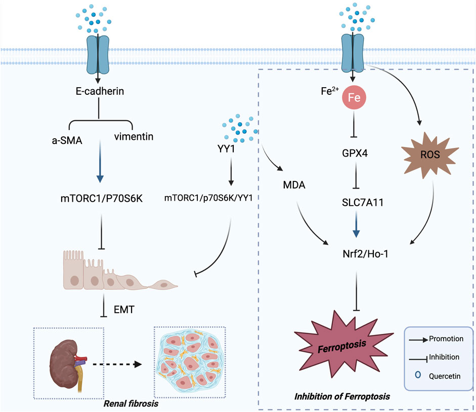
Figure 3. Mechanisms of Quercetin in the Treatment of DN. Mechanisms of quercetin in attenuating renal fibrosis and ferroptosis in diabetic nephropathy. This schematic illustration highlights the dual mechanisms by which quercetin exerts renoprotective effects in diabetic nephropathy. On the left, quercetin suppresses renal fibrosis by downregulating α-SMA and vimentin, inhibiting the mTORC1/p70S6K and YY1 signaling pathways, and preventing EMT. On the right, quercetin attenuates ferroptosis by regulating Fe2+ homeostasis, enhancing GPX4 and SLC7A11 expression, and activating the Nrf2/HO-1 signaling pathway, thereby reducing lipid peroxidation and ROS accumulation. Collectively, these actions contribute to the inhibition of renal injury and disease progression in diabetic nephropathy.
3.5 Induction of ferroptosis
Ferroptosis is a recently identified form of regulated cell death characterized by iron-dependent lipid peroxidation, involving a dynamic imbalance among iron metabolism, lipid peroxidation, and antioxidant defense systems (Dixon et al., 2012). An increasing body of evidence suggests that ferroptosis plays a role in a variety of human diseases, including those affecting the kidney (Tonnus et al., 2021; Li et al., 2021). Recent studies have highlighted its contribution to tubular cell injury observed in DN (Kim et al., 2021). Therefore, inhibition of ferroptosis has emerged as a potential therapeutic strategy for the management of DN. Quercetin, a natural flavonoid compound, exerts multiple biological activities, including potent antioxidant effects. Its antioxidative properties are mediated through several mechanisms, such as increasing GSH levels, enhancing antioxidant signaling pathways, and mitigating reactive ROS induced oxidative damage (Feng et al., 2023; Xu et al., 2019). Notably, recent research indicates that quercetin can chelate iron, which may contribute to its renoprotective effects in both acute kidney injury and DN in db/db mouse models (Feng et al., 2023; Wang et al., 2021c). These findings suggest that quercetin’s ability to modulate ferroptosis could represent an important mechanism underlying its therapeutic potential in DN.
Transcription factor nuclear factor erythroid 2–related factor 2 (Nrf2) is a master regulator of cellular antioxidant responses and plays a pivotal role in controlling ROS levels and preventing ferroptosis (Dodson et al., 2019). Zhang et al. (2024) investigated the effects of quercetin on HG-induced renal tubular epithelial cell injury and its regulation of Nrf2 in a DN rat model, with a focus on the underlying molecular mechanisms. Their results showed that elevated glucose levels promoted the progression of ferroptosis in the kidneys of DN rats. Furthermore, they observed that Nrf2 expression was reduced in DN renal tissues compared with controls, whereas treatment with quercetin or ferrostatin-1 (Fer-1, a ferroptosis inhibitor) restored Nrf2 expression. These findings indicate that quercetin protects against diabetic renal injury by activating the Nrf2 signaling pathway to inhibit ferroptosis. Overall, this study highlights the therapeutic potential of quercetin in DN and provides novel insights into its renoprotective mechanisms.
Feng et al. (2023) conducted both in vivo and in vitro experiments and observed increased intracellular iron levels, decreased expression of glutathione peroxidase 4 (GPX4) and solute carrier family 7 member 11 (SLC7A11), as well as elevated MDA and ROS levels in DN models, indicating a significant involvement of ferroptosis in disease progression. Administration of quercetin or a ferroptosis inhibitor significantly reversed these abnormalities. In vitro, quercetin upregulated Nrf2 and HO-1, thereby activating the Nrf2/HO-1 pathway (Feng et al., 2023). Activated Nrf2 coordinates antioxidant and iron-handling programs—acting together with HO-1, ferritin (FTH1), and the iron exporter ferroportin (SLC40A1)—to restrict the labile iron pool and suppress lipid peroxidation (Dodson et al., 2019). Notably, Nrf2 directly binds antioxidant response elements (AREs) within the SLC7A11 promoter and, in specific cellular contexts, has also been shown to bind the GPX4 promoter, enhancing its transcriptional activity and thereby maintaining glutathione homeostasis and promoting the clearance of lipid peroxides (Feng et al., 2021; Gu et al., 2023). Consequently, quercetin suppresses tubular epithelial ferroptosis through coordinated activation of the Nrf2–HO-1–GPX4/SLC7A11 axis, elucidating its nephroprotective mechanism in DN and supporting its promise as a ferroptosis-targeting therapeutic candidate (Li et al., 2021). The mechanisms by which quercetin regulates ferroptosis in the context of diabetic nephropathy are illustrated in Figure 3, and the overall mechanisms of quercetin in the treatment of diabetic nephropathy are summarized in Table 1.
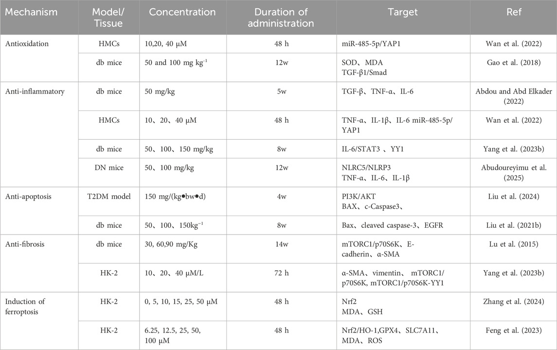
Table 1. Mechanisms of quercetin in the treatment of DN.
Although the present review focuses on inflammation, oxidative stress, and ferroptosis, accumulating evidence indicates that dietary phenolics—including quercetin—exert hypoglycemic and lipid-regulating effects that can secondarily mitigate renal lipid deposition in DN (Sarkar et al., 2021; DE Paulo Farias et al., 2021). For completeness, we cite a broader review on functional-food phenolics supporting these metabolic actions (Golovinskaia and Wang, 2023), while a systematic appraisal of metabolic endpoints remains beyond the prespecified scope of this manuscript.
3.6 Consistency of Quercetin’s efficacy across different DN models
Given that different DN models capture distinct facets of the human disease, we compared quercetin’s efficacy across STZ-induced rats, db/db mice, and HFD + STZ models to assess cross-model consistency. Overall, quercetin consistently suppressed TGF-β1/Smad signaling, attenuated EMT/ECM accumulation, and activated Nrf2/HO-1, as evidenced in STZ-induced and db/db models (Gao et al., 2018; Lai et al., 2012; Feng et al., 2023). In addition, inhibition of NF-κB–mediated inflammation has been observed in multiple models, suggesting shared upstream regulatory effects (Yang et al., 2023b).
Comparative analyses across models further show convergent renal protection, including improvements in renal histopathology and reductions in serum creatinine and urea nitrogen (Gao et al., 2018; Lai et al., 2012). Nonetheless, certain discrepancies exist: quercetin’s modulation of AMPK or PI3K/Akt appears model-dependent, likely reflecting differences in insulin-resistance severity, metabolic background, and dosing regimens (Yang et al., 2023b). Taken together, the available evidence indicates that quercetin exhibits a stable and reproducible therapeutic effect across diverse DN models, supporting its potential as a broadly applicable candidate for DN intervention.
3.7 Dose–response relationship and safety
Across DN models, quercetin tends to show a dose–dependent pattern, whereby higher doses are associated with greater suppression of TGF-β1/Smad signaling, reduced EMT/ECM accumulation, and enhanced Nrf2/HO-1 activation (Table 1). These findings point to a therapeutic window in which efficacy can be improved without compromising safety. Considering between-study differences in model background, dosing route and duration, and formulation, future preclinical studies should implement standardized dosing frameworks and uniform efficacy/safety endpoints to more precisely define exposure–response relationships and guide clinical dose selection.
4 Current clinical application status and challenges
Antioxidants can mitigate oxidative stress by either inhibiting enzymes responsible for ROS production or chelating trace elements involved in free radical generation, thereby reducing the risk of oxidative damage and disease development. Quercetin, a naturally occurring flavonoid antioxidant, effectively scavenges free radicals, modulates immune responses through multiple signaling pathways, regulates enzymatic activity, alters cell cycle progression, and influences gene expression. Through these mechanisms, Quercetin exerts protective effects at all stages of pathogenesis by inhibiting, reversing, or delaying disease progression.
Quercetin exhibits a broad spectrum of biological activities and regulates numerous intra- and extracellular signaling pathways implicated in disease progression. Despite its pharmacological advantages, its instability in physiological media, short biological half-life, and extremely low oral bioavailability remain major barriers to clinical translation (Almeida et al., 2018). Increasing evidence indicates that synthesizing novel Quercetin derivatives can improve its poor solubility and bioavailability. Structural modifications—such as hydroxyl etherification or esterification, carbonyl substitution, and functionalization of the A- and B-rings—have yielded derivatives with enhanced solubility, stability, and bioavailability, as well as improved antioxidant, anti-inflammatory, and anticancer activities (Massi et al., 2017; Sharma et al., 2018; Rajaram et al., 2019).
Given these challenges, recent research has shifted toward multidimensional optimization strategies that combine chemical modification, metal complex formation, and advanced drug delivery systems to overcome Quercetin’s pharmacokinetic limitations and maximize its therapeutic potential, particularly in the treatment of DN.
4.1 Structural modification and delivery strategies
Quercetin, as a multitarget and multipathway natural antioxidant, has shown promising pharmacological activity in various chronic diseases, especially DN. However, its instability in physiological environments, short half-life, and poor oral bioavailability significantly limit its clinical utility (Almeida et al., 2018). To address these issues, three main approaches have been investigated: (1) chemical structural modification, (2) metal complex formation, and (3) advanced drug delivery systems.
4.1.1 Chemical structural modification
Chemical modifications targeting hydroxyl and carbonyl groups, as well as the A- and B-ring structures of Quercetin, have been employed to improve its solubility, stability, and biodistribution. Typical strategies include hydroxyl etherification and esterification, carbonyl substitution, and aromatic ring functionalization. These modifications have not only significantly enhanced water solubility and oral bioavailability but also, in some derivatives, augmented antioxidant, anti-inflammatory, and antitumor activities (Massi et al., 2017; Sharma et al., 2018; Rajaram et al., 2019). Such molecular tailoring provides a rational foundation for optimizing Quercetin’s pharmacodynamics in chronic disease therapy.
4.1.2 Metal complex strategy
Metal ions and their complexes have broad applications in drug development and clinical practice due to their capacity to improve stability, increase target specificity, and modulate redox homeostasis. Refat et al. (2021) investigated the antidiabetic activity of a quercetin–zinc(II) complex (13a) in a STZ-induced diabetic rat model. Their findings revealed that combination therapy with 13a and mesenchymal stem cells (MSCs) markedly enhanced insulin secretion, suppressed inflammatory responses, improved pancreatic injury and glucose metabolism abnormalities, and exhibited superior efficacy compared with either treatment alone. Additionally, this regimen effectively reduced hyperglycemia and genotoxicity, highlighting the potential of Quercetin–metal complexes for managing diabetes and its complications.
4.1.3 Advanced drug delivery systems
Nanotechnology offers an innovative solution to overcome Quercetin’s poor oral bioavailability. An ideal delivery system should protect Quercetin from degradation in the upper gastrointestinal tract, achieve targeted release in the colon, provide sustained release, and significantly improve in vivo stability (Liu et al., 2025). Nanoparticles—owing to their high surface area, tunable targeting capabilities, protection against enzymatic degradation, and single-dose controlled-release properties—have emerged as a highly promising platform for Quercetin delivery (Sarkar et al., 2002; Rizvi and Saleh, 2018). Barbosa et al. (2019) developed polymeric Quercetin nanoparticles via polyelectrolyte self-assembly using fucoidan and chitosan as carriers. This nanosystem demonstrated excellent antioxidant activity and sustained-release characteristics in simulated gastrointestinal environments, particularly under 3F/1C and 5F/1C conditions, showing remarkable tolerance to pH fluctuations and effectively preventing Quercetin degradation. Nonetheless, research on Quercetin nanodelivery specifically for DN remains limited, and further comprehensive studies on pharmacokinetics, biodistribution, and mechanisms of action are warranted.
In conclusion, chemical modification, metal complex formation, and advanced drug delivery technologies provide complementary strategies to optimize Quercetin for clinical application in DN. Future efforts should integrate mechanistic insights with targeted delivery and intelligent release platforms to accelerate the translation of Quercetin from laboratory research to clinical practice.
5 Discussion and perspectives
DN is a multifactorial and complex disease involving diverse pathological processes, including metabolic disorders, oxidative stress, inflammatory responses, apoptosis, and fibrosis. Current clinical treatments can delay disease progression to some extent but remain insufficient to reverse established structural and functional damage to the kidneys. Therefore, identifying therapeutic agents with broad targets, low toxicity, and suitability for long-term intervention remains a critical priority in the prevention and treatment of DN.
Quercetin, a naturally occurring flavonoid, exhibits multi-target regulatory properties and a favorable safety profile, demonstrating considerable potential in multi-pathway intervention for DN. As shown in Figure 4, quercetin exerts renoprotective effects through anti-oxidative stress, anti-inflammatory, anti-fibrotic, and anti-apoptotic/ferroptotic mechanisms. A growing body of experimental evidence indicates that quercetin not only mitigates renal injury through its antioxidant and anti-inflammatory activities but also suppresses TGF-β1–mediated fibrosis, preserves podocyte function, and, under certain conditions, induces ferroptosis, thereby impeding disease progression. Notably, these mechanisms may act in a synergistic manner, suggesting that quercetin possesses an integrative capacity to modulate the disease network in DN.
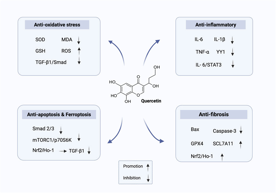
Figure 4. Mechanisms of quercetin in the treatment of DN.
Despite abundant preclinical evidence supporting its therapeutic potential, the clinical translation of quercetin remains challenging. First, its low oral bioavailability and limited absorption hinder the maintenance of effective concentrations in target tissues. Second, variations in efficacy across different experimental models and dosing regimens highlight the need to determine optimal therapeutic doses and administration strategies. Third, there is a lack of high-quality, long-term randomized controlled trials to confirm its safety and efficacy. Future research should focus on: (i) enhancing the solubility and tissue targeting of quercetin through strategies such as nanoparticle-based delivery systems, structural modification, and co-delivery with other agents; (ii) applying multi-omics technologies and network pharmacology to elucidate its multi-target, multi-pathway mechanisms and the interplay between them; (iii) conducting multicenter, large-sample clinical trials to assess its efficacy and safety across different stages of DN, both as monotherapy and in combination with conventional treatments; and (iv) exploring synergistic effects between quercetin and other natural compounds or existing drugs to develop more effective combination therapies.
In summary, quercetin holds broad application prospects in DN therapy. Its multi-target, multi-pathway regulatory features provide novel insights for the integrated management of complex diseases. With continued advancements in drug delivery technologies and precision medicine, quercetin is expected to achieve successful translation from bench to bedside, offering safer and more effective therapeutic options for patients with DN.
Author contributions
YZ: Validation, Conceptualization, Supervision, Writing – review and editing, Writing – original draft. XY: Supervision, Conceptualization, Writing – original draft, Validation. XH: Validation, Writing – original draft, Supervision, Investigation. WG: Data curation, Investigation, Writing – review and editing. LZ: Methodology, Investigation, Writing – review and editing. DM: Writing – review and editing, Conceptualization, Writing – original draft, Validation.
Funding
The author(s) declare that financial support was received for the research and/or publication of this article. This study was supported by the Shandong Provincial Medical and Health Science and Technology Youth Project (Project No. 202303031041): “Research on the Synergistic Mechanism of Paneth Cell Regulation and Macrophage M1/M2 Activation in Resisting ACLF Liver Injury.” In addition, this work was supported by the Qilu Traditional Chinese Medicine Preventive Medicine Advantage Specialty Construction Project.
Acknowledgements
We thank all authors for their contributions to this study. All illustrations were created with BioRender.com. We gratefully acknowledge BioRender for its support.
Conflict of interest
The authors declare that the research was conducted in the absence of any commercial or financial relationships that could be construed as a potential conflict of interest.
Generative AI statement
The author(s) declare that no Generative AI was used in the creation of this manuscript.
Any alternative text (alt text) provided alongside figures in this article has been generated by Frontiers with the support of artificial intelligence and reasonable efforts have been made to ensure accuracy, including review by the authors wherever possible. If you identify any issues, please contact us.
Publisher’s note
All claims expressed in this article are solely those of the authors and do not necessarily represent those of their affiliated organizations, or those of the publisher, the editors and the reviewers. Any product that may be evaluated in this article, or claim that may be made by its manufacturer, is not guaranteed or endorsed by the publisher.
References
Abdou, H. M., and Abd Elkader, H. T. A. E. (2022). The potential therapeutic effects of trifolium alexandrinum extract, hesperetin and quercetin against diabetic nephropathy via attenuation of oxidative stress, inflammation, GSK-3β and apoptosis in Male rats. Chemico-Biological Interact. 352, 109781. doi:10.1016/j.cbi.2021.109781
Abudoureyimu, A., Chen, C., Hu, Y., Nuermaimaiti, D., and Liu, T. (2025). Quercetin alleviates diabetic nephropathy by inhibiting M1 macrophage polarization via targeting NLRC5/NLRP3 pathway. Cell. Immunol. 414, 104997. doi:10.1016/j.cellimm.2025.104997
Almeida, A. F., Borge, G. I. A., Piskula, M., Tudose, A., Tudoreanu, L., Valentová, K., et al. (2018). Bioavailability of quercetin in humans with a focus on interindividual variation. Compr. Rev. Food Sci. Food Saf. 17 (3), 714–731. doi:10.1111/1541-4337.12342
Arellano-Buendía, A. S., García-Arroyo, F. E., Cristóbal-García, M., Loredo-Mendoza, M. L., Tapia-Rodríguez, E., Sánchez-Lozada, L. G., et al. (2014). Urinary excretion of neutrophil gelatinase-associated lipocalin in diabetic rats. Oxidative Med. Cell. Longev. 2014, 961326. doi:10.1155/2014/961326
Barbosa, A. I., Costa Lima, S. A., and Reis, S. (2019). Application of pH-Responsive fucoidan/chitosan nanoparticles to improve oral Quercetin delivery. Mol. 24 (2), 346. doi:10.3390/molecules24020346
Calle, P., and Hotter, G. (2020). Macrophage phenotype and fibrosis in diabetic nephropathy. Int. J. Mol. Sci. 21 (8), 2806. doi:10.3390/ijms21082806
Chang, A. S., Hathaway, C. K., Smithies, O., and Kakoki, M. (2016). Transforming growth factor-β1 and diabetic nephropathy. Am. J. Physiology. Ren. Physiology 310 (8), F689–F696. doi:10.1152/ajprenal.00502.2015
Chen, J., Cui, Y., Zhang, N., Yao, X., Wang, Z., and Yang, L. (2019). Oleanolic acid attenuated diabetic mesangial cell injury by activation of autophagy via miRNA-142-5p/PTEN signaling. Cytotechnology 71 (5), 925–933. doi:10.1007/s10616-019-00335-0
Chen, G. H., Song, C. C., Pantopoulos, K., Wei, X. L., Zheng, H., and Luo, Z. (2022). Mitochondrial oxidative stress mediated Fe-induced ferroptosis via the NRF2-ARE pathway. Free Radic. Biol. and Med. 180, 95–107. doi:10.1016/j.freeradbiomed.2022.01.012
Chen, H., Zhang, H., Li, A. M., Liu, Y. T., Liu, Y., Zhang, W., et al. (2024). VDR regulates mitochondrial function as a protective mechanism against renal tubular cell injury in diabetic rats. Redox Biol. 70, 103062. doi:10.1016/j.redox.2024.103062
Conserva, F., Gesualdo, L., and Papale, M. (2016). A systems biology overview on human diabetic nephropathy: from genetic susceptibility to post-transcriptional and post-translational modifications. J. Diabetes Res. 2016, 7934504. doi:10.1155/2016/7934504
DE Paulo Farias, D., DE Araújo, F. F., Neri-Numa, I. A., and Pastore, G. M. (2021). Antidiabetic potential of dietary polyphenols: a mechanistic review. Food Res. Int. Ott. Ont. 145, 110383. doi:10.1016/j.foodres.2021.110383
Dixon, S. J., Lemberg, K. M., Lamprecht, M. R., Skouta, R., Zaitsev, E. M., Gleason, C. E., et al. (2012). Ferroptosis: an iron-dependent form of nonapoptotic cell death. Cell 149 (5), 1060–1072. doi:10.1016/j.cell.2012.03.042
Dodson, M., Castro-Portuguez, R., and Zhang, D. D. (2019). NRF2 plays a critical role in mitigating lipid peroxidation and ferroptosis. Redox Biol. 23, 101107. doi:10.1016/j.redox.2019.101107
DU, N., Xu, Z., Gao, M., Liu, P., Sun, B., and Cao, X. (2018). Combination of ginsenoside Rg1 and astragaloside IV reduces oxidative stress and inhibits TGF-β1/Smads signaling Cascade on renal fibrosis in rats with diabetic nephropathy. Drug Des. Dev. Ther. 12, 3517–3524. doi:10.2147/DDDT.S171286
DU, L., Qian, X., Li, Y., Xu, L., He, L., Xu, L., et al. (2021). Sirt1 inhibits renal tubular cell epithelial-mesenchymal transition through YY1 deacetylation in diabetic nephropathy. Acta Pharmacol. Sin. 42 (2), 242–251. doi:10.1038/s41401-020-0450-2
Duni, A., Liakopoulos, V., Roumeliotis, S., Peschos, D., and Dounousi, E. (2019). Oxidative stress in the pathogenesis and evolution of chronic kidney disease: untangling ariadne’s thread. Int. J. Mol. Sci. 20 (15), 3711. doi:10.3390/ijms20153711
Ebrahimpour, S., Zakeri, M., and Esmaeili, A. (2020). Crosstalk between obesity, diabetes, and alzheimer’s disease: introducing quercetin as an effective triple herbal medicine. Ageing Res. Rev. 62, 101095. doi:10.1016/j.arr.2020.101095
Feng, L., Zhao, K., Sun, L., Yin, X., Zhang, J., Liu, C., et al. (2021). SLC7A11 regulated by NRF2 modulates esophageal squamous cell carcinoma radiosensitivity by inhibiting ferroptosis. J. Transl. Med. 19 (1), 367. doi:10.1186/s12967-021-03042-7
Feng, Q., Yang, Y., Qiao, Y., Zheng, Y., Yu, X., Liu, F., et al. (2023). Quercetin ameliorates diabetic kidney injury by inhibiting ferroptosis via activating Nrf2/HO-1 signaling pathway. Am. J. Chin. Med. 51 (4), 997–1018. doi:10.1142/S0192415X23500465
Gao, F., He, X., Liang, S., Liu, S., Liu, H., He, Q., et al. (2018). Quercetin ameliorates podocyte injury via inhibition of oxidative stress and the TGF-β1/Smad pathway in DN rats. RSC Adv. 8 (62), 35413–35421. doi:10.1039/c8ra07935h
Gao, R. H., Liu, B., Yang, Y., Ran, R., Zhou, Y., and Liu, S. M. (2023). Serum lipoprotein(a) and high-density lipoprotein cholesterol associate with diabetic nephropathy: evidence from machine learning perspectives. Targets Ther. 16, 1847–1858. doi:10.2147/DMSO.S409410
Godse, N. R., Khan, N., Yochum, Z. A., Gomez-Casal, R., Kemp, C., Shiwarski, D. J., et al. (2017). TMEM16A/ANO1 inhibits apoptosis via downregulation of bim expression. Clin. Cancer Res. An Official J. Am. Assoc. Cancer Res. 23 (23), 7324–7332. doi:10.1158/1078-0432.CCR-17-1561
Golovinskaia, O., and Wang, C. K. (2023). The hypoglycemic potential of phenolics from functional foods and their mechanisms. Food Sci. Hum. Wellness 12 (4), 986–1007. doi:10.1016/j.fshw.2022.10.020
Gomes, I. B. S., Porto, M. L., Santos, M. C. L. F. S., Campagnaro, B. P., Gava, A. L., Meyrelles, S. S., et al. (2015). The protective effects of oral low-dose quercetin on diabetic nephropathy in hypercholesterolemic mice. Front. Physiology 6, 247. doi:10.3389/fphys.2015.00247
Gu, H., Liu, Y., Zhao, Y., Qu, H., Li, Y., Ahmed, A. A., et al. (2023). Hepatic anti-oxidative genes CAT and GPX4 are epigenetically modulated by RORγ/NRF2 in alphacoronavirus-exposed piglets. Antioxidants 12 (6), 1305. doi:10.3390/antiox12061305
Gu, Y., Yu, S., Gu, W., Li, B., Xue, J., Liu, J., et al. (2024). M2 macrophage infusion ameliorates diabetic glomerulopathy via the JAK2/STAT3 pathway in Db/Db mice. Ren. Fail. 46 (2), 2378210. doi:10.1080/0886022X.2024.2378210
Herman-Edelstein, M., Weinstein, T., and Gafter, U. (2013). TGFβ1-dependent podocyte dysfunction. Curr. Opin. Nephrol. Hypertens. 22 (1), 93–99. doi:10.1097/MNH.0b013e32835b4870
Hofni, A., EL-Moselhy, M. A., Taye, A., and Khalifa, M. M. (2014). Combination therapy with spironolactone and Candesartan protects against streptozotocin-induced diabetic nephropathy in rats. Eur. J. Pharmacol. 744, 173–182. doi:10.1016/j.ejphar.2014.10.021
Huang, Z., Peng, Y., Yu, H., Yu, X., Zhou, J., and Xiao, J. (2018). RhoA protects the podocytes against high glucose-induced apoptosis through YAP and plays critical role in diabetic nephropathy. Biochem. Biophysical Res. Commun. 504 (4), 949–956. doi:10.1016/j.bbrc.2018.08.204
Jiang, X., Yu, J., Wang, X., and Ge, J. (2019). Quercetin improves lipid metabolism via SCAP-SREBP2-LDLr signaling pathway in early stage diabetic nephropathy. Diabetes, Metabolic Syndrome Obes. Targets Ther. 12, 827–839. doi:10.2147/DMSO.S195456
Jung, S. W., and Moon, J. Y. (2021). The role of inflammation in diabetic kidney disease. Korean J. Intern. Med. 36 (4), 753–766. doi:10.3904/kjim.2021.174
Khursheed, R., Singh, S. K., Wadhwa, S., Gulati, M., and Awasthi, A. (2020). Enhancing the potential preclinical and clinical benefits of quercetin through novel drug delivery systems. Drug Discov. Today 25 (1), 209–222. doi:10.1016/j.drudis.2019.11.001
Kim, S., Kang, S. W., Joo, J., Han, S. H., Shin, H., Nam, B. Y., et al. (2021). Characterization of ferroptosis in kidney tubular cell death under diabetic conditions. Cell Death and Dis. 12 (2), 160. doi:10.1038/s41419-021-03452-x
Kitada, M., Ogura, Y., Monno, I., and Koya, D. (2017). Regulating autophagy as a therapeutic target for diabetic nephropathy. Curr. Diabetes Rep. 17 (7), 53. doi:10.1007/s11892-017-0879-y
Klessens, C. Q. F., Zandbergen, M., Wolterbeek, R., Bruijn, J. A., Rabelink, T. J., Bajema, I. M., et al. (2017). Macrophages in diabetic nephropathy in patients with type 2 diabetes. Nephrol. Dial. Transpl. 32 (8), 1322–1329. doi:10.1093/ndt/gfw260
Koch, E. A. T., Nakhoul, R., Nakhoul, F., and Nakhoul, N. (2020). Autophagy in diabetic nephropathy: a review. Int. Urology Nephrol. 52 (9), 1705–1712. doi:10.1007/s11255-020-02545-4
Kopel, J., Pena-Hernandez, C., and Nugent, K. (2019). Evolving spectrum of diabetic nephropathy. World J. Diabetes 10 (5), 269–279. doi:10.4239/wjd.v10.i5.269
Lai, P. B., Zhang, L., and Yang, L. Y. (2012). Quercetin ameliorates diabetic nephropathy by reducing the expressions of transforming growth factor-β1 and connective tissue growth factor in streptozotocin-induced diabetic rats. Ren. Fail. 34 (1), 83–87. doi:10.3109/0886022X.2011.623564
Landis, R. C., Quimby, K. R., and Greenidge, A. R. (2018). M1/M2 macrophages in diabetic nephropathy: Nrf2/HO-1 as therapeutic targets. Curr. Pharm. Des. 24 (20), 2241–2249. doi:10.2174/1381612824666180716163845
Li, J., Bao, L., Zha, D., Zhang, L., Gao, P., Zhang, J., et al. (2018). Corrigendum to “Oridonin protects against the inflammatory response in diabetic nephropathy by inhibiting the TLR4/p38-MAPK and TLR4/NF-κB signaling pathways”. Int. Immunopharmacol. 55, 9–19. doi:10.1016/j.intimp.2023.111252
Li, D., Jiang, C., Mei, G., Zhao, Y., Chen, L., Liu, J., et al. (2020). Quercetin alleviates ferroptosis of pancreatic β cells in type 2 diabetes. Nutrients 12 (10), 2954. doi:10.3390/nu12102954
Li, S., Zheng, L., Zhang, J., Liu, X., and Wu, Z. (2021). Inhibition of ferroptosis by up-regulating Nrf2 delayed the progression of diabetic nephropathy. Free Radic. Biol. and Med. 162, 435–449. doi:10.1016/j.freeradbiomed.2020.10.323
Lin, J., Tang, J., Lin, J., He, Y., Yu, Z., Jiang, R., et al. (2021). YY1 regulation by miR-124-3p promotes Th17 cell pathogenicity through interaction with T-bet in rheumatoid arthritis. JCI Insight 6 (22), e149985. doi:10.1172/jci.insight.149985
Liu, Y. (2011). Cellular and molecular mechanisms of renal fibrosis. Nat. Rev. Nephrol. 7 (12), 684–696. doi:10.1038/nrneph.2011.149
Liu, W. J., Huang, W. F., Ye, L., Chen, R. H., Yang, C., Wu, H. L., et al. (2018). The activity and role of autophagy in the pathogenesis of diabetic nephropathy. Eur. Rev. Med. Pharmacol. Sci. 22 (10), 3182–3189. doi:10.26355/eurrev_201805_15079
Liu, H., Wang, Q., Shi, G., Yang, W., Zhang, Y., Chen, W., et al. (2021a). Emodin ameliorates renal damage and podocyte injury in a rat model of diabetic nephropathy via regulating AMPK/mTOR-Mediated autophagy signaling pathway. Diabetes, Metabolic Syndrome Obes. Targets Ther. 14, 1253–1266. doi:10.2147/DMSO.S299375
Liu, Y., Li, Y., Xu, L., Shi, J., Yu, X., Wang, X., et al. (2021b). Quercetin attenuates podocyte apoptosis of diabetic nephropathy through targeting EGFR signaling. Front. Pharmacol. 12, 792777. doi:10.3389/fphar.2021.792777
Liu, F., Feng, Q., Yang, M., Yang, Y., Nie, J., and Wang, S. (2024). Quercetin prevented diabetic nephropathy by inhibiting renal tubular epithelial cell apoptosis via the PI3K/AKT pathway. Phytotherapy Res. PTR 38 (7), 3594–3606. doi:10.1002/ptr.8227
Liu, F., Zhao, B., Wang, Q., Lei, Z., McClements, D. J., Wang, S., et al. (2025). Potential application of quercetin as an anti-diabetic treatment: from molecular mechanism to delivery system design. Phytomedicine Int. J. Phytotherapy Phytopharm. 145, 157036. doi:10.1016/j.phymed.2025.157036
Locati, M., Curtale, G., and Mantovani, A. (2020). Diversity, mechanisms, and significance of macrophage plasticity. Annu. Rev. Pathology 15, 123–147. doi:10.1146/annurev-pathmechdis-012418-012718
Lu, Q., Ji, X. J., Zhou, Y. X., Yao, X. Q., Liu, Y. Q., Zhang, F., et al. (2015). Quercetin inhibits the mTORC1/p70S6K signaling-mediated renal tubular epithelial-mesenchymal transition and renal fibrosis in diabetic nephropathy. Pharmacol. Res. 99, 237–247. doi:10.1016/j.phrs.2015.06.006
Luc, K., Schramm-Luc, A., Guzik, T. J., and Mikolajczyk, T. P. (2019). Oxidative stress and inflammatory markers in prediabetes and diabetes. J. Physiology Pharmacol. An Official J. Pol. Physiological Soc. 70 (6). doi:10.26402/jpp.2019.6.01
Ma, R., Ren, J. M., Li, P., Zhou, Y. J., Zhou, M. K., Hu, Z., et al. (2019). Activated YAP causes renal damage of type 2 diabetic nephropathy. Eur. Rev. Med. Pharmacol. Sci. 23 (2), 755–763. doi:10.26355/eurrev_201901_16890
Ma, X., Hao, C., Yu, M., Zhang, Z., Huang, J., and Yang, W. (2022). Investigating the molecular mechanism of quercetin protecting against podocyte injury to attenuate diabetic nephropathy through network pharmacology, MicroarrayData analysis, and molecular docking. Evidence-Based Complementary Altern. Med. eCAM 2022, 7291434. doi:10.1155/2022/7291434
Massi, A., Bortolini, O., Ragno, D., Bernardi, T., Sacchetti, G., Tacchini, M., et al. (2017). Research progress in the modification of quercetin leading to anticancer agents. Mol. 22 (8), 1270. doi:10.3390/molecules22081270
Mcknight, A. J., Duffy, S., and Maxwell, A. P. (2015). Genetics of diabetic nephropathy: a long road of discovery. Curr. Diabetes Rep. 15 (7), 41. doi:10.1007/s11892-015-0610-9
Mitrofanova, A., Burke, G., Merscher, S., and Fornoni, A. (2021). New insights into renal lipid dysmetabolism in diabetic kidney disease. World J. Diabetes 12 (5), 524–540. doi:10.4239/wjd.v12.i5.524
Mitrofanova, A., Merscher, S., and Fornoni, A. (2023). Kidney lipid dysmetabolism and lipid droplet accumulation in chronic kidney disease. Nat. Rev. Nephrol. 19 (10), 629–645. doi:10.1038/s41581-023-00741-w
Moreno, J. A., Gomez-Guerrero, C., Mas, S., Sanz, A. B., Lorenzo, O., Ruiz-Ortega, M., et al. (2018). Targeting inflammation in diabetic nephropathy: a tale of hope. Expert Opin. Investigational Drugs 27 (11), 917–930. doi:10.1080/13543784.2018.1538352
Mori, Y., Ajay, A. K., Chang, J. H., Mou, S., Zhao, H., Kishi, S., et al. (2021). KIM-1 mediates fatty acid uptake by renal tubular cells to promote progressive diabetic kidney disease. Cell Metab. 33 (5), 1042–1061.e7. doi:10.1016/j.cmet.2021.04.004
Mu, Q., Wang, H., Tong, L., Fang, Q., Xiang, M., Han, L., et al. (2020). Betulinic acid improves nonalcoholic fatty liver disease through YY1/FAS signaling pathway. FASEB J. official Publ. Fed. Am. Soc. Exp. Biol. 34 (9), 13033–13048. doi:10.1096/fj.202000546R
Naaman, S. C., and Bakris, G. L. (2023). Diabetic nephropathy: update on pillars of therapy slowing progression. Diabetes Care 46 (9), 1574–1586. doi:10.2337/dci23-0030
Noels, H., Lehrke, M., Vanholder, R., and Jankowski, J. (2021). Lipoproteins and fatty acids in chronic kidney disease: molecular and metabolic alterations. Nat. Rev. Nephrol. 17 (8), 528–542. doi:10.1038/s41581-021-00423-5
Opazo-Ríos, L., Mas, S., Marín-Royo, G., Mezzano, S., Gómez-Guerrero, C., Moreno, J. A., et al. (2020). Lipotoxicity and diabetic nephropathy: novel mechanistic insights and therapeutic opportunities. Int. J. Mol. Sci. 21 (7), 2632. doi:10.3390/ijms21072632
Park, M. D., Silvin, A., Ginhoux, F., and Merad, M. (2022). Macrophages in health and disease. Cell 185 (23), 4259–4279. doi:10.1016/j.cell.2022.10.007
Patel, R. V., Mistry, B. M., Shinde, S. K., Syed, R., Singh, V., and Shin, H. S. (2018). Therapeutic potential of quercetin as a cardiovascular agent. Eur. J. Med. Chem. 155, 889–904. doi:10.1016/j.ejmech.2018.06.053
Pérez-Morales, R. E., Del, PINO M. D., Valdivielso, J. M., Ortiz, A., Mora-Fernández, C., and Navarro-González, J. F. (2019). Inflammation in diabetic kidney disease. Nephron 143 (1), 12–16. doi:10.1159/000493278
Pezzolesi, M. G., and Krolewski, A. S. (2013). The genetic risk of kidney disease in type 2 diabetes. Med. Clin. N. Am. 97 (1), 91–107. doi:10.1016/j.mcna.2012.10.005
Pichler, R., Afkarian, M., Dieter, B. P., and Tuttle, K. R. (2017). Immunity and inflammation in diabetic kidney disease: translating mechanisms to biomarkers and treatment targets. Am. J. Physiology. Ren. Physiology 312 (4), F716–F731. doi:10.1152/ajprenal.00314.2016
Qiu, Y. Y., and Tang, L. Q. (2016). Roles of the NLRP3 inflammasome in the pathogenesis of diabetic nephropathy. Pharmacol. Res. 114, 251–264. doi:10.1016/j.phrs.2016.11.004
Raina, P., Sikka, R., Gupta, H., Matharoo, K., Bali, S. K., Singh, V., et al. (2021). Association of eNOS and MCP-1 genetic variants with type 2 diabetes and diabetic nephropathy susceptibility: a case-control and meta-analysis study. Biochem. Genet. 59 (4), 966–996. doi:10.1007/s10528-021-10041-2
Rajaram, P., Jiang, Z., Chen, G., Rivera, A., Phasakda, A., Zhang, Q., et al. (2019). Nitrogen-containing derivatives of O-tetramethylquercetin: synthesis and biological profiles in prostate cancer cell models. Bioorg. Chem. 87, 227–239. doi:10.1016/j.bioorg.2019.03.047
Ramos, A., and Camargo, F. D. (2012). The hippo signaling pathway and stem cell biology. Trends Cell Biol. 22 (7), 339–346. doi:10.1016/j.tcb.2012.04.006
Ramos-Riera, K. P., Pérez-Severiano, F., and López-Meraz, M. L. (2023). Oxidative stress: a common imbalance in diabetes and epilepsy. Metab. Brain Dis. 38 (3), 767–782. doi:10.1007/s11011-022-01154-7
Refat, M. S., Hamza, R. Z., Adam, A. M. A., Saad, H. A., Gobouri, A. A., Al-Harbi, F. S., et al. (2021). Quercetin/zinc complex and stem cells: a new drug therapy to ameliorate glycometabolic control and pulmonary dysfunction in diabetes mellitus: structural characterization and genetic studies. PloS One 16 (3), e0246265. doi:10.1371/journal.pone.0246265
Ricciardi, C. A., and Gnudi, L. (2021). Kidney disease in diabetes: from mechanisms to clinical presentation and treatment strategies. Metabolism Clin. Exp. 124, 154890. doi:10.1016/j.metabol.2021.154890
Rizvi, S. A. A., and Saleh, A. M. (2018). Applications of nanoparticle systems in drug delivery technology. Saudi Pharm. J. 26 (1), 64–70. doi:10.1016/j.jsps.2017.10.012
Ruiz-Ortega, M., Rayego-Mateos, S., Lamas, S., Ortiz, A., and Rodrigues-Diez, R. R. (2020). Targeting the progression of chronic kidney disease. Nat. Rev. Nephrol. 16 (5), 269–288. doi:10.1038/s41581-019-0248-y
Russo, G., Piscitelli, P., Giandalia, A., Viazzi, F., Pontremoli, R., Fioretto, P., et al. (2020). Atherogenic dyslipidemia and diabetic nephropathy. J. Nephrol. 33 (5), 1001–1008. doi:10.1007/s40620-020-00739-8
Sabanayagam, C., Chee, M. L., Banu, R., Cheng, C. Y., Lim, S. C., Tai, E. S., et al. (2019). Association of diabetic retinopathy and diabetic kidney disease with all-cause and cardiovascular mortality in a multiethnic Asian population. JAMA Netw. Open 2 (3), e191540. doi:10.1001/jamanetworkopen.2019.1540
Saeed, M. (2018). Locus and gene-based GWAS meta-analysis identifies new diabetic nephropathy genes. Immunogenetics 70 (6), 347–353. doi:10.1007/s00251-017-1044-0
Samsu, N. (2021). Diabetic nephropathy: challenges in pathogenesis, diagnosis, and treatment. BioMed Res. Int. 2021, 1497449. doi:10.1155/2021/1497449
Sarkar, S., Mandal, S., Sinha, J., Mukhopadhyay, S., Das, N., and Basu, M. K. (2002). Quercetin: critical evaluation as an antileishmanial agent in vivo in hamsters using different vesicular delivery modes. J. Drug Target. 10 (8), 573–578. doi:10.1080/106118021000072681
Sarkar, D., Christopher, A., and Shetty, K. (2021). Phenolic bioactives from plant-based foods for glycemic control. Front. Endocrinol. 12, 727503. doi:10.3389/fendo.2021.727503
Sathibabu Uddandrao, V. V., Brahmanaidu, P., Ravindarnaik, R., Suresh, P., Vadivukkarasi, S., and Saravanan, G. (2019). Restorative potentiality of S-allylcysteine against diabetic nephropathy through attenuation of oxidative stress and inflammation in streptozotocin-nicotinamide-induced diabetic rats. Eur. J. Nutr. 58 (6), 2425–2437. doi:10.1007/s00394-018-1795-x
Sharma, A., Kashyap, D., Sak, K., and Tuli, H. S. (2018). Therapeutic charm of quercetin and its derivatives: a review of research and patents. Pharm. Pat. Anal. 7 (1), 15–32. doi:10.4155/ppa-2017-0030
Sheng, H., Zhang, D., Zhang, J., Zhang, Y., Lu, Z., Mao, W., et al. (2022). Kaempferol attenuated diabetic nephropathy by reducing apoptosis and promoting autophagy through AMPK/mTOR pathways. Front. Med. 9, 986825. doi:10.3389/fmed.2022.986825
Sies, H. (2015). Oxidative stress: a concept in redox biology and medicine. Redox Biol. 4, 180–183. doi:10.1016/j.redox.2015.01.002
Skibba, M., Qian, Y., Bao, Y., Lan, J., Peng, K., Zhao, Y., et al. (2016). New EGFR inhibitor, 453, prevents renal fibrosis in angiotensin II-stimulated mice. Eur. J. Pharmacol. 789, 421–430. doi:10.1016/j.ejphar.2016.08.009
Sun, J., Li, Z. P., Zhang, R. Q., and Zhang, H. M. (2017). Repression of miR-217 protects against high glucose-induced podocyte injury and insulin resistance by restoring PTEN-Mediated autophagy pathway. Biochem. Biophysical Res. Commun. 483 (1), 318–324. doi:10.1016/j.bbrc.2016.12.145
Sun, X. H., Xiao, H. M., Zhang, M., Lin, Z. Y., Yang, Y., Chen, R., et al. (2021). USP9X deubiquitinates connexin43 to prevent high glucose-induced epithelial-to-mesenchymal transition in NRK-52E cells. Biochem. Pharmacol. 188, 114562. doi:10.1016/j.bcp.2021.114562
Sun, H., Saeedi, P., Karuranga, S., Pinkepank, M., Ogurtsova, K., Duncan, B. B., et al. (2022). IDF diabetes atlas: global, regional and country-level diabetes prevalence estimates for 2021 and projections for 2045. Diabetes Res. Clin. Pract. 183, 109119. doi:10.1016/j.diabres.2021.109119
Tanabe, K., Wada, J., and Sato, Y. (2020). Targeting angiogenesis and lymphangiogenesis in kidney disease. Nat. Rev. Nephrol. 16 (5), 289–303. doi:10.1038/s41581-020-0260-2
Tang, J., Diao, P., Shu, X., Li, L., and Xiong, L. (2019). Quercetin and quercitrin attenuates the inflammatory response and oxidative stress in LPS-induced RAW264.7 cells: in vitro assessment and a theoretical model. BioMed Res. Int. 2019, 7039802. doi:10.1155/2019/7039802
Thongnak, L., Pongchaidecha, A., and Lungkaphin, A. (2020). Renal lipid metabolism and lipotoxicity in diabetes. Am. J. Med. Sci. 359 (2), 84–99. doi:10.1016/j.amjms.2019.11.004
Tonnus, W., Meyer, C., Steinebach, C., Belavgeni, A., von Mässenhausen, A., Gonzalez, N. Z., et al. (2021). Dysfunction of the key ferroptosis-surveilling systems hypersensitizes mice to tubular necrosis during acute kidney injury. Nat. Commun. 12 (1), 4402. doi:10.1038/s41467-021-24712-6
Wan, H., Wang, Y., Pan, Q., Chen, X., Chen, S., Li, X., et al. (2022). Quercetin attenuates the proliferation, inflammation, and oxidative stress of high glucose-induced human mesangial cells by regulating the miR-485-5p/YAP1 pathway. Int. J. Immunopathol. Pharmacol. 36, 20587384211066440. doi:10.1177/20587384211066440
Wang, L., Wang, H. L., Liu, T. T., and Lan, H. Y. (2021a). TGF-beta as a master regulator of diabetic nephropathy. Int. J. Mol. Sci. 22 (15), 7881. doi:10.3390/ijms22157881
Wang, Y., Cui, J., Liu, M., Shao, Y., and Dong, X. (2021b). Schisandrin C attenuates renal damage in diabetic nephropathy by regulating macrophage polarization. Am. J. Transl. Res. 13(1): 210–222.
Wang, Y., Quan, F., Cao, Q., Lin, Y., Yue, C., Bi, R., et al. (2021c). Quercetin alleviates acute kidney injury by inhibiting ferroptosis. J. Adv. Res. 28, 231–243. doi:10.1016/j.jare.2020.07.007
Wang, X., Jiang, L., Liu, X. Q., Huang, Y. B., Wang, A. L., Zeng, H. X., et al. (2022). Paeoniflorin binds to VEGFR2 to restore autophagy and inhibit apoptosis for podocyte protection in diabetic kidney disease through PI3K-AKT signaling pathway. Phytomedicine Int. J. Phytotherapy Phytopharm. 106, 154400. doi:10.1016/j.phymed.2022.154400
Wang, N., Feng, H., Zhang, Z., Tian, H., Gu, L., Bian, Y., et al. (2025). Danggui buxue decoction regulates autophagy to improve renal fibrosis in diabetes through miR-27a/PI3K/AKT pathway. J. Ethnopharmacol. 341, 119357. doi:10.1016/j.jep.2025.119357
Winiarska, A., Knysak, M., Nabrdalik, K., Gumprecht, J., and Stompór, T. (2021). Inflammation and oxidative stress in diabetic kidney disease: the targets for SGLT2 inhibitors and GLP-1 receptor agonists. Int. J. Mol. Sci. 22 (19), 10822. doi:10.3390/ijms221910822
Wu, Q., and Huang, F. (2023). LncRNA H19: a novel player in the regulation of diabetic kidney disease. Front. Endocrinol. 14, 1238981. doi:10.3389/fendo.2023.1238981
Wu, J., Chen, Y., Shi, S., Liu, J., Zhang, F., Li, X., et al. (2025). Exploration of pharmacological mechanisms of dapagliflozin against type 2 diabetes mellitus through PI3K-Akt signaling pathway based on network pharmacology analysis and deep learning technology. Curr. Computer-Aided Drug Des. 21 (4), 452–465. doi:10.2174/0115734099274407231207070451
Xian, Y., Dong, L., Jia, Y., Lin, Y., Jiao, W., and Wang, Y. (2018). miR-370 promotes high glucose-induced podocyte injuries by inhibiting angiotensin II type 1 receptor-associated protein. Cell Biol. Int. 42 (11), 1545–1555. doi:10.1002/cbin.11048
Xie, T., Chen, X., Chen, W., Huang, S., Peng, X., Tian, L., et al. (2021). Curcumin is a potential adjuvant to alleviates diabetic retinal injury via reducing oxidative stress and maintaining Nrf2 pathway homeostasis. Front. Pharmacol. 12, 796565. doi:10.3389/fphar.2021.796565
Xing, L., Fang, J., Zhu, B., Wang, L., Chen, J., Wang, Y., et al. (2021). Astragaloside IV protects against podocyte apoptosis by inhibiting oxidative stress via activating PPARγ-Klotho-FoxO1 axis in diabetic nephropathy. Life Sci. 269, 119068. doi:10.1016/j.lfs.2021.119068
Xu, D., Hu, M. J., Wang, Y. Q., and Cui, Y. L. (2019). Antioxidant activities of quercetin and its complexes for medicinal application. Mol. 24 (6), 1123. doi:10.3390/molecules24061123
Xu, J., Liu, L. Q., Xu, L. L., Xing, Y., and Ye, S. (2020). Metformin alleviates renal injury in diabetic rats by inducing Sirt1/FoxO1 autophagic signal axis. Clin. Exp. Pharmacol. and Physiology 47 (4), 599–608. doi:10.1111/1440-1681.13226
Yagi, R., Chen, LF., Shigesada, K., and Murakami, Y. (1999). A WW domain-containing yes-associated protein (YAP) is a novel transcriptional co-activator. EMBO J. 18 (9), 2551–2562. doi:10.1093/emboj/18.9.2551
Yahya, M. J., Ismail, P. B., Nordin, N. B., Akim, A. B. M., Binti Md Yusuf, W. S., Adam, N. L. B., et al. (2019). CNDP1, NOS3, and MnSOD polymorphisms as risk factors for diabetic nephropathy among type 2 diabetic patients in Malaysia. J. Nutr. Metabolism 2019, 8736215. doi:10.1155/2019/8736215
Yang, J., and Liu, Z. (2022). Mechanistic pathogenesis of endothelial dysfunction in diabetic nephropathy and retinopathy. Front. Endocrinol. 13, 816400. doi:10.3389/fendo.2022.816400
Yang, D., Wang, T., Long, M., and Li, P. (2020). Quercetin: its main pharmacological activity and potential application in clinical medicine. Oxidative Med. Cell. Longev. 2020, 8825387. doi:10.1155/2020/8825387
Yang, T., Heng, C., Zhou, Y., Hu, Y., Chen, S., Wang, H., et al. (2020). Targeting Mammalian serine/threonine-protein kinase 4 through Yes-associated protein/TEA domain transcription factor-mediated epithelial-mesenchymal transition ameliorates diabetic nephropathy orchestrated renal fibrosis. Metabolism Clin. Exp. 108, 154258. doi:10.1016/j.metabol.2020.154258
Yang, T., Hu, Y., Chen, S., Li, L., Cao, X., Yuan, J., et al. (2023a). YY1 inactivated transcription co-regulator PGC-1α to promote mitochondrial dysfunction of early diabetic nephropathy-associated tubulointerstitial fibrosis. Cell Biol. Toxicol. 39 (2), 391–413. doi:10.1007/s10565-022-09711-7
Yang, T., Hu, Y., Jiang, W., Pang, J., Zhou, Y., Zhang, H., et al. (2023b). YY1 was indispensable for the alleviation of quercetin on diabetic nephropathy-associated tubulointerstitial inflammation. Phytomedicine Int. J. Phytotherapy Phytopharm. 111, 154659. doi:10.1016/j.phymed.2023.154659
Yu, L., Lin, Q., Liao, H., Feng, J., Dong, X., and Ye, J. (2010). TGF-β1 induces podocyte injury through Smad3-ERK-NF-κB pathway and Fyn-dependent TRPC6 phosphorylation. Cell. Physiology Biochem. Int. J. Exp. Cell. Physiology, Biochem. Pharmacol. 26 (6), 869–878. doi:10.1159/000323996
Yu, X., Su, Q., Geng, J., Liu, H., Liu, Y., Liu, J., et al. (2021). Ginkgo biloba leaf extract prevents diabetic nephropathy through the suppression of tissue transglutaminase. Exp. Ther. Med. 21 (4), 333. doi:10.3892/etm.2021.9764
Yu, J., Liu, Y., Li, H., and Zhang, P. (2023). Pathophysiology of diabetic kidney disease and autophagy: a review. Medicine 102 (30), e33965. doi:10.1097/MD.0000000000033965
Yu, R., Liu, X., Li, R., and Xiao, X. (2024). Ten-year clinical characteristics of patients with early-onset type 2 diabetes: a single-center experience in China. J. Diabetes 16 (1), e13477. doi:10.1111/1753-0407.13477
Zhang, M., Zhang, Y., Yang, S., Zhou, J., Gao, W., Yang, X., et al. (2017). Multifunctional YY1 in liver diseases. Seminars Liver Dis. 37 (4), 363–376. doi:10.1055/s-0037-1607451
Zhang, R., Zhu, X., Bai, H., and Ning, K. (2019). Network pharmacology databases for traditional Chinese medicine: review and assessment. Front. Pharmacol. 10, 123. doi:10.3389/fphar.2019.00123
Zhang, Z., Huang, Q., Zhao, D., Lian, F., Li, X., and Qi, W. (2023a). The impact of oxidative stress-induced mitochondrial dysfunction on diabetic microvascular complications. Front. Endocrinol. 14, 1112363. doi:10.3389/fendo.2023.1112363
Zhang, H. F., Liu, H. M., Xiang, J. Y., Zhou, X. C., Wang, D., Chen, R. Y., et al. (2023b). Alpha lipoamide inhibits diabetic kidney fibrosis via improving mitochondrial function and regulating RXRα expression and activation. Acta Pharmacol. Sin. 44 (5), 1051–1065. doi:10.1038/s41401-022-00997-1
Zhang, L., Wang, X., Chang, L., Ren, Y., Sui, M., Fu, Y., et al. (2024). Quercetin improves diabetic kidney disease by inhibiting ferroptosis and regulating the Nrf2 in streptozotocin-induced diabetic rats. Ren. Fail. 46 (1), 2327495. doi:10.1080/0886022X.2024.2327495
Zheng, W., Guo, J., and Liu, Z. S. (2021). Effects of metabolic memory on inflammation and fibrosis associated with diabetic kidney disease: an epigenetic perspective. Clin. Epigenetics 13 (1), 87. doi:10.1186/s13148-021-01079-5
Zhou, L., An, X. F., Teng, S. C., Liu, J. S., Shang, W. B., Zhang, A. H., et al. (2012). Pretreatment with the total flavone glycosides of flos abelmoschus manihot and hyperoside prevents glomerular podocyte apoptosis in streptozotocin-induced diabetic nephropathy. J. Med. Food 15 (5), 461–468. doi:10.1089/jmf.2011.1921
Zhou, L., Chen, X., Lu, M., Wu, Q., Yuan, Q., Hu, C., et al. (2019). Wnt/β-catenin links oxidative stress to podocyte injury and proteinuria. Kidney Int. 95 (4), 830–845. doi:10.1016/j.kint.2018.10.032
Zou, H., Ye, H., Kamaraj, R., Zhang, T., Zhang, J., and Pavek, P. (2021). A review on pharmacological activities and synergistic effect of quercetin with small molecule agents. Phytomedicine Int. J. Phytotherapy Phytopharm. 92, 153736. doi:10.1016/j.phymed.2021.153736
Zou, Y., Zhao, L., Zhang, J., Wang, Y., Wu, Y., Ren, H., et al. (2022). Development and internal validation of machine learning algorithms for end-stage renal disease risk prediction model of people with type 2 diabetes mellitus and diabetic kidney disease. Ren. Fail. 44 (1), 562–570. doi:10.1080/0886022X.2022.2056053
Keywords: quercetin, diabetic nephropathy, natural products, multi-target therapy, drug delivery
Citation: Zheng Y, Ye X, Han X, Geng W, Zhao L and Meng D (2025) Immunomodulatory roles of quercetin in diabetic nephropathy: targeting inflammation, oxidative stress, and ferroptosis. Front. Pharmacol. 16:1687677. doi: 10.3389/fphar.2025.1687677
Received: 26 August 2025; Accepted: 21 October 2025;
Published: 13 November 2025.
Edited by:
Paul J. Higgins, Albany Medical College, United StatesReviewed by:
Tongtong Liu, China Academy of Chinese Medical Sciences, ChinaWenlong Sun, Shandong University of Technology, China
Copyright © 2025 Zheng, Ye, Han, Geng, Zhao and Meng. This is an open-access article distributed under the terms of the Creative Commons Attribution License (CC BY). The use, distribution or reproduction in other forums is permitted, provided the original author(s) and the copyright owner(s) are credited and that the original publication in this journal is cited, in accordance with accepted academic practice. No use, distribution or reproduction is permitted which does not comply with these terms.
*Correspondence: Dandan Meng, bWVuZ2RhbmRhbjExMDZAMTYzLmNvbQ==