 Yijuan Lin1*†
 Ahmad Alhaskawi
- 1 Department of Emergency, Hangzhou Linping District First People’s Hospital, Hangzhou, China
- 2 Department of Orthopedics, the First Affiliated Hospital of Zhejiang University School of Medicine, Hangzhou, China
- 3 Department of Emergency, the Second Affiliated Hospital of Zhejiang University School of Medicine, Hangzhou, China
Sepsis remains a major global health challenge, exerting a particularly severe toll in low- and middle-income countries. Despite advances in antimicrobial and supportive care, sepsis continues to defy effective control due to its complex pathophysiology and multi-organ involvement. Central to this complexity is a dysregulated host response, driven by hyperinflammation, immune suppression, and profound mitochondrial and metabolic dysfunction. A critical mediator of this dysregulation is oxidative stress, which exacerbates cellular injury through reactive oxygen and nitrogen species, disrupting mitochondrial integrity and redox balance. This review synthesizes current insights into the mechanistic interplay between oxidative stress, mitochondrial dysfunction, and immunopathology in sepsis. It further evaluates the therapeutic potential of endogenous antioxidants, such as superoxide dismutase, catalase, and glutathione, as well as exogenous agents including vitamins A, C, E, selenium, omega-3 fatty acids, melatonin, and carnosine. While translational gaps persist, particularly in dosing, timing, and patient stratification, emerging strategies including mitochondria-targeted antioxidants, nanotherapeutics, and biomarker-guided interventions hold promise for restoring redox homeostasis and improving clinical outcomes. This review aims to serve as a contemporary resource for researchers and clinicians striving to decode the oxidative basis of sepsis and accelerate the development of precision antioxidant therapies.
Introduction
Sepsis is a life-threatening organ dysfunction triggered by a dysregulated host response to infection, and remains a critical challenge in intensive care medicine. Despite advances in antimicrobial therapy and supportive care, sepsis continues to result in high morbidity and mortality, with an estimated 48.9 million cases and 11 million deaths worldwide in 2017, accounting for nearly 20% of all global mortality, disproportionately affecting low- and middle-income countries (Rudd et al., 2020). Sepsis is primarily triggered by a wide range of microorganisms, including Gram-positive and Gram-negative bacteria, viruses, fungi, and, less commonly, parasites, with bacterial infections accounting for the majority of cases globally (Neri et al., 2015). Clinically, sepsis manifests with nonspecific signs, including fever or hypothermia, tachycardia, tachypnea, and leukocytosis or leukopenia. As the disease progresses, it leads to hypotension, altered mental status, coagulopathy, and hyperlactatemia, reflecting systemic inflammation and tissue hypoperfusion. One of the hallmark challenges in managing sepsis is its ability to affect multiple organ systems simultaneously (Figure 1). Common complications include acute kidney injury, acute respiratory distress syndrome (ARDS), hepatic dysfunction, myocardial depression, and coagulopathy, often resulting in disseminated intravascular coagulation (DIC). The central nervous system may also be compromised, leading to sepsis-associated encephalopathy, which can persist long after infection resolution (Peerapor et al., 2019; Sinha et al., 2023; Park et al., 2025; Habimana et al., 2020). The pathophysiology of sepsis is underpinned by a complex interplay of immune activation, endothelial dysfunction, and profound metabolic and mitochondrial dysregulation. Initially, sepsis is driven by the recognition of pathogen-associated molecular patterns (PAMPs) and damage-associated molecular patterns (DAMPs), which activate innate immune responses through pattern recognition receptors (Zhang et al., 2010; Li et al., 2023a). This leads to the release of pro-inflammatory cytokines such as TNF-α, IL-6, and IL-1β. While this inflammatory cascade is essential for microbial clearance, excessive or prolonged activation results in collateral tissue damage and immunosuppression (Giamarellos-Bourboulis et al., 2024; Lelubre and Vincent, 2018; Rocha et al., 2012). In addition, mitochondrial dysfunction drives sepsis by impairing energy production and redox balance, leading to bioenergetic failure and multi-organ damage (Zong et al., 2024) (Galley, 2011; Víctor et al., 2009). Furthermore, the imbalance between ROS production and antioxidant defences plays a dual role in sepsis. While ROS are essential for pathogen clearance, their uncontrolled accumulation exacerbates mitochondrial damage and cellular injury (Figure 2) (Joffre and Hellman, 2021; Brealey et al., 2002). This has prompted growing interest in therapeutic interventions aimed at restoring mitochondrial function and redox homeostasis. Furthermore, antioxidant therapy, particularly agents targeting mitochondria, offers a promising strategy for mitigating oxidative damage and improving outcomes in sepsis. Compounds such as melatonin, N-acetylcysteine, vitamin C, and selenium have demonstrated protective effects in preclinical and some clinical studies by enhancing mitochondrial integrity and reducing systemic inflammation (Mantzarlis et al., 2017; Nagar et al., 2018). This review comprehensively explores the recent advances in understanding the multifaceted role of oxidative stress in sepsis pathophysiology, with particular emphasis on mitochondrial dysfunction and the emerging therapeutic potential of antioxidants to restore redox balance, improve organ function, and enhance patient outcomes.
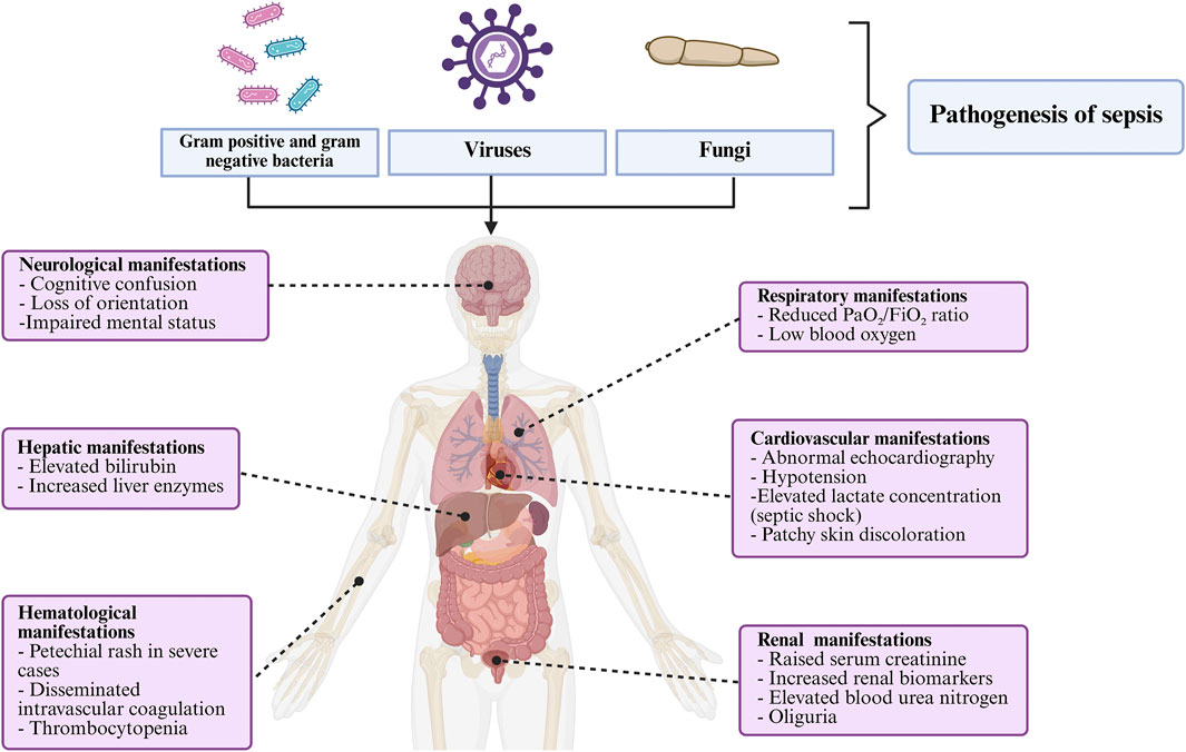
Figure 1. The pathological and clinical features of sepsis.
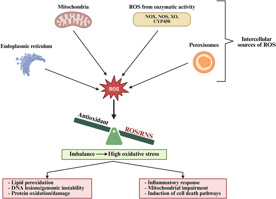
Figure 2. Intracellular sources and consequences of reactive oxygen species (ROS) generation.
Oxidative stress effect on sepsis pathology
Oxidative stress plays an essential role in the pathogenesis and progression of sepsis, primarily through the overproduction of ROS and impairment of antioxidant defence mechanisms. At the systemic level, oxidative stress contributes to vascular and endothelial dysfunction, promoting capillary leakage, microcirculatory derangements, and thrombosis. These effects impair oxygen and nutrient delivery to tissues, triggering hypoxia, metabolic acidosis, and organ failure (Higashi et al., 2014; Incalza et al., 2018). Studies have shown that oxidative stress induces endothelial cell apoptosis and disrupts the endothelial glycocalyx, facilitating leukocyte adhesion and extravasation, which worsens tissue damage (Joffre and Hellman, 2021). The combined impact on vascular tone, coagulation, and oxygen utilization is a major contributor to the development of shock and multi-organ dysfunction syndrome. During sepsis, immune cells such as neutrophils and macrophages are rapidly activated in response to microbial invasion, releasing large amounts of ROS, including superoxide anion (O2 −), hydrogen peroxide (H2O2), and hydroxyl radicals (OH−), as part of the host’s antimicrobial arsenal (Di Meo et al., 2016). While ROS are essential for pathogen clearance, excessive and unregulated production leads to widespread cellular and tissue injury (Figure 3) (Mantzarlis et al., 2017). However, research has also revealed that free radicals function as damaging agents and as critical mediators of intracellular signaling (Chandimali et al., 2025). Broadly, oxidants include a range of primary oxygen- and nitrogen-derived species that readily interact with biomolecules, inducing oxidative modifications to lipids, proteins, and nucleic acids (Juan et al., 2021). This pathological oxidative burden is closely linked to mitochondrial dysfunction. Mitochondria are both a source and target of ROS. Under physiological conditions, the, ETC tightly regulates ROS generation. However, during sepsis, ETC dysfunction leads to electron leakage and disproportionate ROS formation, resulting in the collapse of mitochondrial membrane potential, impaired ATP synthesis, and activation of mitochondrial-mediated apoptosis (Nagar et al., 2018). Cells with depleted ATP reserves are more vulnerable to metabolic or chemical stress and consequently progress to necrotic cell death. In apoptosis, the opening of the mitochondrial permeability transition pore (MPTP), which is mediated by oxidative stress, triggers a cascade of events, including Ca2+ efflux, collapse of mitochondrial membrane potential, enhanced ROS formation, and the release of cytochrome c along with other pro-apoptotic molecules into the cytoplasm (Bernardi et al., 2023; Giorgi et al., 2012).
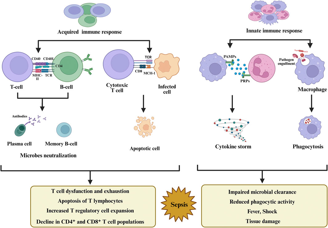
Figure 3. Sepsis triggers dysregulated innate and adaptive immunity, leading to inflammation, immune suppression, and organ damage.
Furthermore, nitric oxide (NO) serves multiple physiological functions, acting as a neurotransmitter in the nervous system, preserving vascular homeostasis, and regulating immune responses (Andrabi et al., 2023). In addition, it contributes to intracellular signaling, gene expression, and the regulation of cellular growth and differentiation (Tuteja et al., 2004; Lundberg and Weitzberg, 2022). Sepsis triggers the induction of inducible nitric oxide synthase (iNOS), resulting in excessive and sustained NO release. Excessive NO production interacts with ROS to generate peroxynitrite (ONOO−), a highly reactive nitrogen species capable of nitrating proteins and inactivating mitochondrial enzymes (Takatani et al., 2018; Cinelli et al., 2020). The resulting damage intensifies cellular dysfunction and suppresses mitochondrial respiration, thereby aggravating the bioenergetic failure that underlies severe sepsis (Víctor et al., 2009; Takatani et al., 2018). Pathogen-associated molecules such as lipopolysaccharide (LPS) play a critical upstream role in the oxidative cascade. In addition, the binding of LPS to the Toll-like receptor 4 (TLR4) complex activates NADPH oxidase and promotes mitochondrial ROS generation, while simultaneously inducing iNOS expression (Galley, 2011; Luo et al., 2025). This integrates microbial recognition with redox imbalance, ensuring both antimicrobial defense and collateral oxidative injury. At the same time, ROS and RNS act as signaling mediators that activate nuclear factor kappa B (NF-κB), a transcription factor central to the inflammatory response. Through ROS-mediated phosphorylation of IκB kinase, NF-κB is released from its inhibitor and translocates to the nucleus, where it induces transcription of pro-inflammatory cytokines, chemokines, adhesion molecules, and iNOS itself. This promotes further oxidant production and cytokine release, perpetuating systemic inflammation and tissue damage (Sun, 2017; Lawrence, 2009). Tsukamoto et al. found that LPS engagement with lipopolysaccharide-binding protein (LBP) and CD14 initiates TLR4 signaling that drives oxidative cascades through excessive ROS production (Tuteja et al., 2004). A study showed that LPS administration caused a strong, dose- and time-dependent activation of NF-κB across multiple organs, as demonstrated in transgenic NF-κB reporter mice. The activation was associated with significant IκB degradation and nuclear translocation of NF-κB, resulting in increased transcription of pro-inflammatory mediators (Blackwell et al., 2000). The outcome was a robust rise in cytokines such as TNF-α, IL-6, KC, MIP-2, and MCP-1, which promoted neutrophil infiltration and widespread inflammatory injury. Importantly, when NF-κB signaling was inhibited using a dominant-negative IκB mutant, both cytokine levels and tissue damage were markedly reduced, confirming that ROS-mediated NF-κB activation directly drives cytokine overproduction and multi-organ dysfunction in sepsis (Blackwell et al., 2000). Another study presented that NF-κB activity in peripheral blood mononuclear cells was significantly higher in patients with severe sepsis who did not survive compared to survivors, and strongly correlated with illness severity (APACHE II score) (Arnalich et al., 2000). Furthermore, elevated NF-κB activity was linked with increased plasma levels of IL-6, while survivors displayed higher concentrations of anti-inflammatory mediators such as IL-1 receptor antagonist (IL-1ra) and IL-10 (Arnalich et al., 2000). Moreover, Metnitz et al. showed that in sepsis-related ARDS, patients had depleted antioxidant defenses with persistently high malondialdehyde (MDA) levels, indicating ongoing lipid peroxidation. Elevated myeloperoxidase (MPO) activity in bronchoalveolar fluid reflected neutrophil-driven oxidative stress, contributing to sustained lung injury despite micronutrient supplementation (Metnitz et al., 1999). Nevertheless, sepsis induces the upregulation of numerous genes associated with inflammation, vascular dysfunction, and immune regulation. Classical pro-inflammatory cytokines (IL1B, IL6, TNF) and pattern recognition receptors (TLR2, TLR4) are markedly increased, along with adhesion molecules (VCAM1, ICAM1) and chemokines (CCL2, CXCL8) that promote leukocyte recruitment and vascular injury (Falcão-Holanda et al., 2021; Behairy et al., 2022; He et al., 2024; Warford et al., 2017). Furthermore, Zhang et al. reported that MMP-9 expression is upregulated in sepsis-induced acute lung injury, where it exerts a protective role by promoting the release of soluble RAGE (sRAGE). This mechanism suppresses RAGE/NF-κB signaling, thereby reducing inflammation, oxidative stress, and lung damage, ultimately improving survival (Zhang et al., 2021). Jakobsson et al. investigated the role of S100A8/A9 (calprotectin) in sepsis and its link to sepsis-induced myocardial dysfunction (SIMD). They found that plasma S100A8/A9 levels were significantly elevated in severe sepsis patients and correlated with left ventricular systolic dysfunction and higher SOFA scores. In a mouse model of endotoxemia, LPS rapidly induced S100A8/A9 release and acute cardiac depression, while genetic deficiency of S100A9 or pharmacological blockade of S100A8/A9 with ABR-238901 prevented or reversed myocardial dysfunction and reduced systemic inflammation (Jakobsson et al., 2023). Additional genes upregulated in sepsis include adrenomedullin (ADM), IRAK3, and arachidonate 5-lipoxygenase (ALOX5/5-LOX). Lundberg et al. showed that ADM levels are significantly elevated in sepsis and septic shock compared with non-septic patients. High circulating ADM was strongly associated with disease severity, organ failure, vasopressor need, and 30-day mortality, highlighting its value as both a biomarker and predictor of outcome in septic patients (Lundberg et al., 2020). Alongside, Deng et al. found that IRAK3 (IRAK-M) is markedly upregulated in sepsis, particularly in alveolar macrophages, where it suppresses TLR signaling and reduces cytokine production. This leads to impaired bacterial clearance and contributes to the immunosuppressive state of sepsis, while mice lacking IRAK3 exhibited improved host defense and survival (Deng et al., 2006). Xie et al. demonstrated that ALOX5 is markedly upregulated in sepsis, where it amplifies inflammatory and oxidative pathways contributing to septic cardiomyopathy. Increased ALOX5 expression elevated leukotriene B4 levels, promoted M1 macrophage polarization, and worsened cardiac injury, whereas ALOX5 knockout or pharmacological inhibition with zileuton reduced inflammation, preserved cardiac function, and improved survival (Xie et al., 2021). Recent evidence shows that long non-coding RNAs (lncRNAs) are critically involved in the pathogenesis and diagnosis of sepsis. Zheng et al. identified 14 lncRNA pairs (SepSigLnc) with reversed expression patterns between septic patients and healthy controls, achieving very high diagnostic accuracy (AUC >0.99 in some cohorts). These lncRNAs were enriched in pathways related to immune regulation, cell fate determination, and steroid hormone responses, linking them directly to mechanisms of sepsis progression (Zheng et al., 2021). In addition, Liao et al. reported that lncRNAs exhibit strong diagnostic and prognostic value in sepsis, with pooled AUCs of 0.88 for diagnosis and 0.84 for predicting 28-day mortality. Specific lncRNAs, such as MALAT1 and NEAT1, were consistently linked to sepsis severity and poor outcomes (Liao et al., 2022).
Therapeutic effect of antioxidants in sepsis
Enzyme-based antioxidants
Superoxide dismutase (SOD)
SOD is a key antioxidant enzyme that has a protective role in sepsis by mitigating oxidative stress through the detoxification of superoxide anions. Several studies have explored the therapeutic application of both native and mimetic forms of SOD to counteract this oxidative burden (Table 1). In septic models, SOD mimetics have demonstrated potential in limiting inflammation and improving survival outcomes by neutralizing superoxide and reducing peroxynitrite formation. This protective mechanism is particularly effective in preserving mitochondrial function and vascular responsiveness, two critical factors in the pathophysiology of sepsis-related organ failure (Salvemini and Cuzzocrea, 2003). Zainumi et al. investigated the therapeutic role of SOD in a rat model of sepsis induced by cecal ligation and puncture, a widely accepted method that mimics polymicrobial infection. The treatment markedly reduced superoxide accumulation, preserved mitochondrial respiratory function, and attenuated systemic oxidative damage. SOD administration also improved mean arterial pressure and reduced organ injury scores, particularly in the liver and kidneys (Zainumi et al., 2022). Bo et al. also investigated the protective effects of orally administered melon-derived SOD in a rat model of sepsis. Their results demonstrated that SOD supplementation significantly improved renal function by reducing serum creatinine and neutrophil gelatinase-associated lipocalin (NGAL) levels (Bo et al., 2022). Furthermore, Bao et al. foundthat in LPS-induced sepsis, neutrophils release extracellular vesicles containing mitochondrial SOD2, which play a protective role against oxidative endothelial injury and DIC. These vesicles significantly reduced ROS levels, maintained vascular barrier function, and decreased fibrin deposition. When transferred into septic or neutropenic mice, the SOD2-enriched vesicles improved tissue perfusion and survival, while inhibition of SOD2 activity abolished these benefits (Bao et al., 2022). In addition, Serena et al. study revealed that SOD1-overexpressing mice exhibited significantly higher survival rates, reduced histopathological damage in the liver and lungs, and decreased serum levels of pro-inflammatory cytokines. Additionally, SOD1 upregulation attenuated oxidative stress, as evidenced by lower MDA levels and preserved glutathione content. Experimental studies further support the use of low molecular weight SOD mimetics as promising therapeutic agents in septic shock. These compounds penetrate cells more efficiently and retain catalytic activity, offering targeted ROS neutralization in inflamed tissues (Albuszies and Brückner, 2003). For instance, melon-derived SOD formulations significantly reduced biomarkers of acute kidney injury (e.g., NGAL and serum creatinine) by restoring oxidative balance in septic mice, suggesting organ-specific therapeutic benefits (Ismy et al., 2022). Furthermore, adjunctive SOD therapy is linked to improved endothelial function and reduced expression of iNOS, thereby curbing the inflammatory cascade in sepsis (Prauchner, 2017). Coleman et al. investigated the therapeutic potential of a manganese porphyrin compound (MnTnBuOE-2-PyP5+), a SOD mimetic, in a mouse model of endotoxemia. Their study demonstrated that pre-treatment with this compound significantly improved survival, reduced serum cytokine levels, and mitigated liver and lung injury following LPS challenge (Coleman et al., 2017).
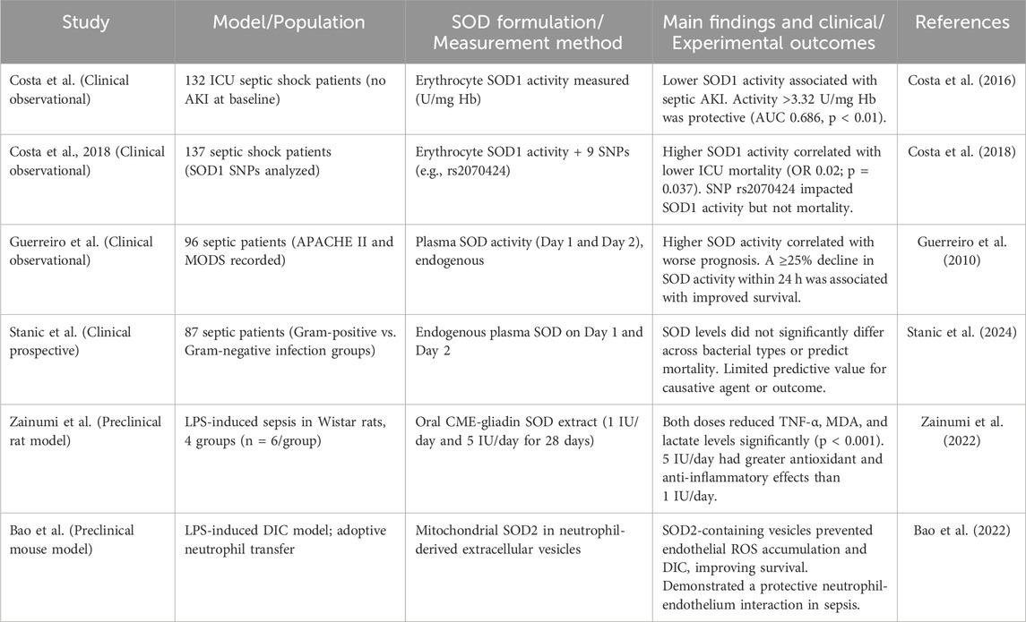
Table 1. Clinical and preclinical investigations of superoxide dismutase (SOD) as a biomarker and therapeutic modulator in sepsis pathophysiology.
Catalase
Catalase (CAT), a heme-containing enzyme, has a critical role in cellular redox homeostasis by catalyzing the decomposition of H2O2 into water and molecular oxygen, thereby mitigating oxidative stress. Several studies have explored its therapeutic application in experimental models of sepsis (Table 2). For instance, Li et al. developed a PEGylated form of catalase (CAT-PEG) to enhance its stability and bioavailability in vivo. In a murine model of sepsis, treatment with CAT-PEG significantly reduced systemic inflammation and improved survival outcomes, underscoring its potential as a viable antioxidant therapy in septic conditions (Li et al., 2021). Furthermore, comprehensive reviews of antioxidant therapies have emphasized the relevance of catalase and other ROS-scavenging enzymes in mitigating the molecular cascades triggered by oxidative and nitrosative stress during sepsis (Kumar et al., 2022; Victor et al., 2005). Che et al. developed a multifunctional polyphenol-copper nanozyme that mimics both catalase and superoxide dismutase activity. This nanozyme effectively suppressed endoplasmic reticulum stress, reduced lipid peroxidation, and improved hepatocellular architecture, demonstrating potent systemic protection (Che et al., 2025). Catalase upregulation appears to correlate with improved outcomes in various sepsis-associated organ injuries. Xia et al. demonstrated that the small molecule anemonin increased catalase expression in lung tissue, thereby reducing oxidative damage and inflammation in sepsis-induced acute lung injury via modulation of the NF-κB and Nrf2 pathways (Xia et al., 2025). Complementary data from Kumar et al. showed that catalase expression was significantly upregulated in rats treated with a dispiro-indanedione-parthenin hybrid compound, coinciding with suppression of MAPK/NF-κB signaling and improved redox status in septic tissues (Kumar et al., 2025). Interestingly, therapies that indirectly modulate catalase also confer benefit. Abdelmawgood et al. utilized chrysin-loaded PLGA nanoparticles to elevate catalase and GPx activity in the spleen of septic mice, reducing lipid peroxidation and modulating immune cell responses (Abdelmawgood et al., 2025). Moreover, Wang et al. found that mitochondrial dysfunction in sepsis-induced endothelial damage was associated with suppressed catalase activity. Pharmacological interventions that restored catalase levels helped reestablish mitochondrial integrity and barrier function in endothelial cells (Wang S. et al., 2025). Furthermore, Erel et al. reported increased hepatic catalase levels following ozone therapy in a rat model of cecal perforation-induced sepsis, suggesting catalase as both a therapeutic effector and a biomarker of response (Erel et al., 2024). Collectively, these studies underscore catalase as a multifaceted therapeutic agent in sepsis. Whether through direct enzyme delivery, gene regulation, or catalytic mimetics, catalase-targeted strategies effectively dampen oxidative injury, suppress inflammation, and enhance host resilience against septic insult.
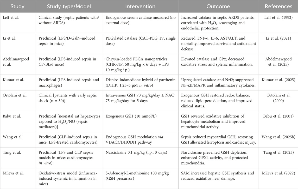
Table 2. Clinical and preclinical studies on catalase and glutathione activity and effects in sepsis.
Glutathione
Glutathione (GSH), a tripeptide composed of glutamate, cysteine, and glycine, serves as a critical intracellular antioxidant that regulates redox homeostasis, detoxifies ROS, and modulates immune responses (Narayanankutty et al., 2019). In the context of sepsis, GSH plays a multifaceted role in counteracting the oxidative stress and mitochondrial dysfunction that underlie organ failure and systemic inflammation. During sepsis, both the synthesis and bioavailability of GSH are significantly impaired, leading to uncontrolled ROS accumulation and cellular injury (Table 2). Biolo et al. study highlighted the dysregulation of glutathione metabolism in septic patients and emphasized that the impaired biosynthetic response may contribute to heightened oxidative damage and worsened clinical outcomes (Biolo et al., 2007). GSH has shown potential as a direct antioxidant and as an adjunctive therapy. A study by Liu et al. investigated the use of glutathione-assisted continuous renal replacement therapy (CRRT) in septic patients with acute kidney injury. This combination improved peripheral receptor expression and mitigated systemic inflammatory responses, suggesting synergistic effects between GSH and extracorporeal blood purification (Liu et al., 2024). Additionally, Ortolani et al. found that administration of GSH and its precursor NAC reduced lipid peroxidation and oxidative tissue damage in patients with early septic shock, affirming the therapeutic viability of antioxidant co-therapy in acute care settings (Ortolani et al., 2000). In addition, Mileva et al. showed that combining oseltamivir with SAM, a glutathione precursor, reduced oxidative stress during influenza by lowering MDA levels, restoring GSH, normalizing antioxidant enzymes, and improving survival (Mileva et al., 2022). Furthermore, Tang et al. demonstrated that restoring GSH levels protects against sepsis-induced cardiac dysfunction by inhibiting BNIP3-mediated mitophagy and ferroptosis, preserving mitochondrial function, and reducing lipid peroxidation. Enhancing GSH, via direct supplementation or activation of SLC7A11 and GPX4, also suppressed ferroptosis and myocardial injury in sepsis models. (Tang et al., 2025). Further evidence by Wang et al. highlighted glutathione’s role in mitochondrial regulation and inhibition of oxidative damage in septic myocardial tissue. The study identified a regulatory axis (VDAC3/DHODH) that enhances GSH-mediated ferroptosis resistance, demonstrating significant cardioprotective outcomes in vivo (Wang J. et al., 2025). Nevertheless, Jin et al. found that restoring GSH levels alleviated hepatic ferroptosis and oxidative stress by upregulating PI3K/Akt/Nrf2 signaling and enhancing the SLC7A11/GPX4 axis. This cascade effectively prevented mitochondrial membrane potential collapse and hepatocyte apoptosis (Yin Y. et al., 2025). Experimental data also support the hepatoprotective effects of GSH. Babu et al. found that glutamine and its dipeptides restored oxidative metabolism in neonatal hepatocytes impaired by septic mediators (H2O2, NO), likely through boosting glutathione synthesis rather than the Krebs cycle. Blocking glutathione synthesis abolished the effect, while exogenous glutathione replicated it, confirming the mechanism (Babu et al., 2001). Additionally, Zhao et al. utilized NAD+ -loaded neutrophils to activate antioxidant pathways, including GSH-related enzymes. The resulting downregulation of TLR4/NF-κB/NLRP3 signaling led to suppressed cytokine storms and enhanced organ protection in septic mice (Zhao et al., 2025). Li et al. discussed glutathione synthesis as a key determinant in mitigating cuproptosis and disulfidptosis, two novel, metal-dependent cell death pathways exacerbated in sepsis. By regulating mitochondrial ion homeostasis and redox-sensitive protein folding, glutathione supports cytoprotective responses under extreme oxidative burden (Li et al., 2025). Moreover, in a prospective study, reduced glutathione administered alongside blood purification therapy significantly decreased endotoxin levels, inflammatory mediators, and hepatic injury in septic shock patients (Liu and Meng, 2024).
Vitamin-based antioxidants
Vitamin A
Vitamin A is a fat-soluble micronutrient with immunomodulatory and antioxidant properties, and has a significant role in the regulation of innate and adaptive immune responses. In sepsis, vitamin A was investigated for its therapeutic potential due to its influence on epithelial barrier integrity, immune regulation, and oxidative stress mitigation (Table 3) (Willinger, 2019). One of the earliest mechanistic links between vitamin A and sepsis lies in its capacity to modulate inflammatory responses through its active metabolite, retinoic acid. In an experimental rodent model, Carlson et al. demonstrated that antioxidant vitamin therapy, including vitamin A at 417 U/kg, ameliorated myocardial apoptotic activity and reduced inflammatory signaling pathways associated with septic pathophysiology (Carlson et al., 2006). Vitamin A has been implicated in maintaining the integrity of epithelial and endothelial barriers, especially within the gastrointestinal tract (Pham et al., 2021). This is of particular importance, as gut barrier dysfunction is a recognized driver of systemic infection and endotoxemia in septic patients. Retinoic acid is found to enhance tight junction protein expression and reduce bacterial translocation in preclinical sepsis models, potentially mitigating multi-organ dysfunction (Martinez Ramos et al., 2025). However, the therapeutic efficacy of vitamin A in human sepsis remains contentious. A prospective, double-blind, placebo-controlled trial by Cherukuri et al. assessed high-dose vitamin A administration in septic adult patients, nearly half of whom were vitamin A deficient at baseline. The intervention failed to demonstrate significant improvements in outcomes compared to placebo, suggesting that supplementation alone may be insufficient without addressing broader metabolic and inflammatory dysfunctions inherent to sepsis (Cherukuri et al., 2019). In pediatric populations, emerging evidence is more promising. A recent randomized controlled trial investigated vitamin A supplementation in children with sepsis and reported improvements in clinical parameters, including shortened ICU stays and reduced inflammatory markers, indicating potential age-dependent therapeutic responsiveness (Fu et al., 2025). Moreover, epidemiological data support a high prevalence of vitamin A deficiency in septic patients. A study by Zhang et al. revealed a significant association between low serum retinol levels and illness severity in critically ill children with sepsis, highlighting the importance of baseline vitamin A status in determining treatment responsiveness (Zha et al., 2019). Garg et al. examined the impact of vitamin A supplementation on bronchopulmonary dysplasia (BPD) in extremely low birth weight neonates. Their findings showed a statistically significant reduction in BPD at 36 weeks postmenstrual age and a borderline significant decrease in the combined outcome of mortality or BPD. However, no significant benefit was observed at 28 days of life. While the results suggest potential benefit (Garg et al., 2019). Moreover, a randomized, double-blind, placebo-controlled trial evaluated the effect of weekly low-dose vitamin A supplementation (10,000 IU) on immune responses in pregnant and lactating Ghanaian women. Vitamin A significantly increased the ratio of proinflammatory (IFN-γ, TNF-α) to anti-inflammatory (IL-10) cytokines during pregnancy and postpartum, indicating a shift toward Th1-type immune responses. Clinically, this modulation suggests vitamin A may enhance resistance to intracellular infections such as malaria without adverse pregnancy outcomes when given in low doses (Binka et al., 1995).
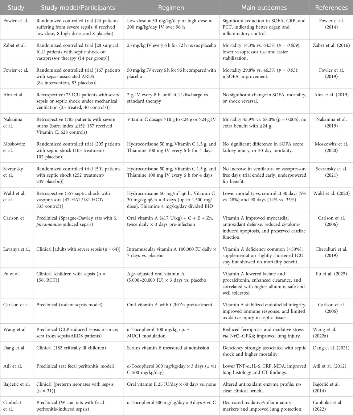
Table 3. Summary of major clinical studies evaluating vitamins therapy in sepsis.
Vitamin C
Vitamin C has emerged as a compelling adjunctive therapy in the management of sepsis, driven by its antioxidant, anti-inflammatory, and endothelial-stabilizing properties. Under intense oxidative stress and excessive ROS production, the requirement for vitamin C increases significantly as somatic and immune cells consume greater amounts of this antioxidant; moreover, the heightened proliferation and turnover of leukocytes amplify its depletion, since these cells can concentrate vitamin C up to a hundredfold higher than plasma levels, ultimately leading to a substantial reduction in systemic vitamin C availability during sepsis (Oudemans-van Straaten et al., 2014; Wilson, 2013). Moreover, vitamin C enhances neutrophil and lymphocyte activity, limits neutrophil necrosis and extracellular trap formation, and modulates nuclear responses to hypoxia and stress by regulating HIF-1α and inducing NF-κB-related epigenetic changes (Fisher et al., 2011; Carr and Maggini, 2017). Vitamin C deficiency is frequently observed in critically ill septic patients, and repletion strategies, particularly via high-dose intravenous vitamin C (HDIVC), have garnered significant scientific interest (Table 3) (Marik, 2018). Kashiouris et al. found that HDIVC was associated with improved organ function and a trend toward reduced mortality in septic patients (Kashiouris et al., 2020). A meta-analysis by Liang et al. further refined these findings, demonstrating that intravenous vitamin C significantly reduced vasopressor requirements and improved the Sequential Organ Failure Assessment (SOFA) score (Liang et al., 2023). Phase I randomized, double-blind, placebo-controlled clinical trial investigated intravenous vitamin C in 24 ICU patients with severe sepsis. Patients received placebo, low-dose (50 mg/kg/day), or high-dose (200 mg/kg/day) vitamin C every 6 hours for 4 days. Both doses were safe and well tolerated, but the high-dose regimen achieved markedly higher plasma concentrations and produced a faster, greater reduction in SOFA scores and inflammatory biomarkers (CRP and procalcitonin), while preventing thrombomodulin elevation (Fowler et al., 2014). A double-blind randomized clinical trial evaluated the effect of HDIVC on vasopressor requirements in 28 surgical ICU patients with septic shock. Patients received either vitamin C (25 mg/kg every 6 h for 72 h) or a placebo. Vitamin C administration significantly reduced the mean dose and duration of norepinephrine use and markedly lowered 28-day mortality (14.3% vs. 64.3%), with no adverse effects observed (Zabet et al., 2016). In addition, a sepsis with severe respiratory failure clinical trial by Fowler et al. identified that HDIVC did not significantly improve the modified SOFA score at 96 h but did reduce 28-day mortality (29.8% vs. 46.3%) and lower inflammatory and endothelial injury markers, including CRP and thrombomodulin (Fowler et al., 2019). Plasma syndecan-1, a marker of endothelial glycocalyx injury and a predictor of mortality in severe sepsis and acute respiratory distress syndrome, was significantly reduced following HDIVC treatment (Kashiouris et al., 2019). Furthermore, intravenous vitamin C was shown to reduce inflammatory and cardiac injury markers and lower SOFA scores, indicating enhanced inflammation control and cardioprotective effects (Jiang et al., 2024). In septic ICU patients with elevated lactate, treatment with vitamin C and thiamine lowered hospital mortality, showing the strongest benefit when both were given together, especially in vasopressor-dependent cases (Byerly et al., 2020). Marik et al. reported that combining hydrocortisone, vitamin C, and thiamine in patients with severe sepsis and septic shock significantly reduced hospital mortality (8.5% vs. 40.4%) and improved organ function (Marik et al., 2017).
Vitamin E
Vitamin E, a fat-soluble antioxidant best known for its α-tocopherol isoform, serves a vital function in shielding cell membranes from oxidative injury. In the context of sepsis, vitamin E was investigated for its ability to suppress inflammatory responses, preserve cellular integrity, and potentially improve clinical outcomes (Table 3). Pein et al. revealed that endogenous metabolites of vitamin E, particularly α-13′-COOH, exert anti-inflammatory effects by directly inhibiting 5-lipoxygenase (5-LO), a key enzyme in leukotriene biosynthesis. These metabolites reduced inflammatory lipid mediator production in human neutrophils and monocytes, with potency comparable to pharmaceutical 5-LO inhibitors (Pein et al., 2018). Minter et al. showed that MitoVitE, α-tocopherol, and Trolox all reduced oxidative stress, NFκB activation, and cytokine release in endothelial cells under sepsis-like conditions. MitoVitE provided the strongest effect by preserving mitochondrial membrane potential and broadly downregulating TLR2/4 pathway genes, highlighting the added benefit of mitochondrial targeting (Minter et al., 2020). Atli et al. investigated the protective effects of vitamin E and selenium against sepsis-induced lung injury in a rat model (Atli et al., 2012). Both antioxidants, administered individually or in combination, significantly improved blood gas parameters, reduced leukocyte and CRP levels, and elevated glutathione peroxidase activity compared to untreated septic rats. Histopathological analysis revealed decreased inflammation, congestion, edema, and emphysema in the lung tissues of treated groups. Mechanistically, vitamin E stabilizes cell membranes by neutralizing lipid peroxides (Atli et al., 2012). Similarly, Durant et al. observed that vitamin E reduced superoxide anion overproduction and improved microcirculatory flow, highlighting its role in redox regulation during septic states (Durant et al., 2004). Canbolat et al. showed that vitamin E significantly reduced sepsis-induced lung injury in a rat model by lowering inflammatory cytokines and MDA. It improved lung histopathology and gas exchange, likely through its antioxidant action and membrane-stabilizing effects, which help preserve alveolar-capillary integrity (Canbolat et al., 2022). Furthermore, Wang et al. demonstrated that Mucin one enhances the protective effect of vitamin E against sepsis-induced acute lung injury by inhibiting ferroptosis via the GSK3β/Keap1-Nrf2-GPX4 pathway. Vitamin E reduced oxidative stress and lipid peroxidation, with its efficacy significantly amplified by MUC1, suggesting a synergistic therapeutic potential (Wang Y. M. et al., 2022). Furthermore, vitamin E not only suppresses oxidative damage but also enhances endothelial NO bioavailability, thus improving vascular tone and preventing sepsis-related organ hypoperfusion. Novel delivery approaches, such as nano-emulsion formulations of Vitamin E, have been developed to optimize bioavailability, potentially broadening its therapeutic applications in sepsis and systemic inflammatory conditions (Yin W. et al., 2025). In pediatric populations, vitamin E deficiency appears common among children with sepsis and septic shock. Dang et al. reported a strong association between vitamin E deficiency and sepsis severity, proposing that correcting this deficit could support antioxidant defense mechanisms during critical illness (Dang et al., 2021). Adult-focused research has also gained traction. A retrospective cohort study by He et al. suggested that standalone vitamin E supplementation in ICU patients with sepsis was associated with a reduction in 28-day mortality (He et al., 2025). Furthermore, Bajčetić et al. studied preterm neonates with sepsis and found that while vitamin E supplementation improved glutathione peroxidase activity, it also suppressed glutathione reductase, potentially impairing redox balance. These changes may increase oxidative stress and infection risk, suggesting caution with vitamin E use in this vulnerable population (Bajčetić et al., 2014). Vitamin E has also been investigated as part of combination regimens. Wald et al. noted that vitamin E, alongside vitamins A and C, has been included in therapeutic protocols aiming to attenuate inflammatory damage and support organ function in septic patients (Wald et al., 2022). Kono et al. evaluated a novel water-soluble vitamin E derivative, E-Ant-S-GS, in a rat model of peritonitis-induced sepsis. The compound, combining vitamin E, glutathione, anthranilic acid, and succinic acid, significantly reduced IL-6 levels, neutrophil infiltration, and lung injury. Mechanistically, E-Ant-S-GS suppressed the expression of HMGB1 and PAR1, key mediators of inflammation and endothelial damage, thereby attenuating systemic inflammation and acute lung injury (Kono et al., 2012).
Novel and adjunctive antioxidant therapies
N-acetylcysteine (NAC)
NAC, a potent thiol-containing antioxidant, has garnered increasing attention for its therapeutic potential in mitigating the pathophysiological consequences of sepsis, particularly through modulation of oxidative stress and inflammation. Several studies support its efficacy in various organ systems affected during sepsis. NAC inhibits NF-κB activation and upregulates nuclear factor erythroid 2-related factor 2 (Nrf2), promoting cellular antioxidant defences (Table 4) (Chen et al., 2025). Additionally, Sainglers et al. found that NAC’s inhibition of neutrophil extracellular traps, which are implicated in sepsis-related tissue injury and organ failure (Sainglers et al., 2025). Le et al. demonstrated that NAC administration significantly attenuated acute lung injury in septic rats by reducing oxidative stress, inflammatory cytokine production, and apoptosis (Le et al., 2022). Similarly, Fan et al. reported that NAC pretreatment conferred protection against sepsis-induced acute kidney injury by alleviating inflammatory damage and oxidative insult in renal tissues (Fan et al., 2020). Furthermore, Oliva et al. showed that adjunctive NAC therapy in ICU patients with septic shock due to carbapenem-resistant infections reduced 30-day mortality, emphasizing its clinical value in high-risk patient populations (Oliva et al., 2021). An in vivo study included a rat model, found that NAC effectively prevented pulmonary edema and acute kidney injury during sepsis, particularly under mechanical ventilation stress, by preserving mitochondrial function and capillary integrity (Campos et al., 2012). Furthermore, Liu et al. investigated the impact of NAC on von Willebrand factor (vWF) release during septic conditions. Their findings suggested that NAC disrupts vWF multimers via disulfide bond reduction, reducing platelet aggregation and microthrombi formation, which are central to sepsis-induced organ failure (Liu et al., 2025). Clinical translation is evidenced by Fan et al., who explored NAC as a co-therapy with low molecular weight heparin, observing significant improvements in vascular function and inflammatory modulation in septic cardiovascular syndromes (Fan et al., 2024). NAC also demonstrates immunomodulatory activity. Yang et al. showed that NAC downregulated pro-inflammatory cytokines and restored macrophage function, aiding in pathogen clearance and limiting host tissue damage (Yang et al., 2025). Similarly, Sui et al. reported that NAC enhanced the efficacy of phage-antibiotic therapy against Pseudomonas aeruginosa and Klebsiella pneumoniae, highlighting its role in disrupting biofilms and restoring antibiotic susceptibility (Sui et al., 2025). A clinical study found that NAC, given at 600 mg every 12 h for 5 days as an adjuvant therapy in septic shock, significantly improved outcomes. NAC reduced procalcitonin levels, lowered the SOFA score by 42%–50%, and enhanced total antioxidant capacity by increasing GSH and glutathione peroxidase (GPx) activity (Aisa-Álvarez et al., 2023). Although results from randomized clinical trials remain limited, existing animal and translational studies strongly support the adjunctive use of NAC in early-phase sepsis to prevent multiorgan failure, improve microvascular perfusion, and reduce mortality.
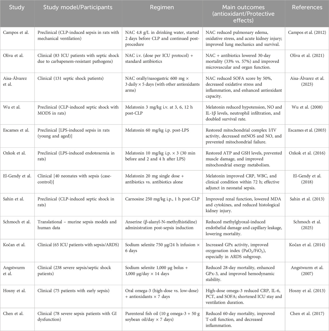
Table 4. Clinical and preclinical studies of novel and adjunctive antioxidant therapies.
Melatonin
Melatonin, an endogenously produced indoleamine best known for regulating circadian rhythms, has attracted increasing attention for its potent antioxidant, anti-inflammatory, and immunomodulatory properties in the context of sepsis. As a highly permeable molecule capable of crossing cellular and mitochondrial membranes, melatonin effectively scavenges reactive oxygen and nitrogen species and modulates the activity of pro-inflammatory cytokines (Srinivasan et al., 2010). Pharmacologically, melatonin’s appeal lies in its wide therapeutic index, high safety profile, and low toxicity even at high doses. Colunga et al. advocate for its inclusion in sepsis protocols due to these properties and its capacity to reduce mitochondrial dysfunction and immune dysregulation, hallmarks of septic pathophysiology (Colunga Biancatelli et al., 2020; Ibrahim et al., 2024). Escames et al. explored the role of melatonin in modulating the inflammatory response induced by LPS in mice, focusing on the expression and activity of inducible iNOS. They demonstrated that LPS administration markedly increased iNOS expression and NO production in liver and lung tissues, contributing to systemic inflammation. However, melatonin significantly counteracted these effects by reducing iNOS mRNA and protein levels, as well as NO production (Escames et al., 2003). A study showed that in LPS-induced septic skeletal muscle, melatonin preserves mitochondrial function by maintaining the activity of respiratory chain complexes I and IV, restoring ATP production, and stabilizing mitochondrial membrane potential. It reduces mitochondrial oxidative stress by scavenging ROS and upregulating antioxidant enzymes such as SOD2 and GPx1. Melatonin also prevents the opening of the mitochondrial permeability transition pore (mPTP) and activates AMPK, promoting cellular energy balance and mitochondrial biogenesis (Ozkok et al., 2016). Wu et al. investigated the therapeutic potential of melatonin in a rat model of peritonitis-induced septic shock and multiple organ dysfunction syndrome (Wu et al., 2008). They demonstrated that melatonin (3 mg/kg, administered intravenously at 3, 6, and 12 h post-cecal ligation and puncture) significantly improved survival, stabilized blood pressure, restored vascular responsiveness to norepinephrine, and reduced organ injury markers. Melatonin treatment suppressed inflammatory mediators such as interleukin-1β and NO, decreased superoxide production in the aorta, and mitigated polymorphonuclear neutrophil (PMN) infiltration in the lungs and liver (Wu et al., 2008). In addition, a clinical study by El-Gendy et al. reported significant reductions in inflammatory markers and oxidative stress among neonates with sepsis who received melatonin as adjunctive therapy, along with a decrease in mortality risk (El-Gendy et al., 2018). These findings were reinforced by a systematic review and meta-analysis by Henderson et al., which consolidated data from multiple neonatal studies and concluded that melatonin supplementation was associated with reduced oxidative damage and improved clinical outcomes (Henderson et al., 2018).
Omega-3 fatty acid supplementation
Omega-3 polyunsaturated fatty acids (PUFAs), particularly eicosapentaenoic acid (EPA) and docosahexaenoic acid (DHA) from fish oil, have demonstrated therapeutic relevance in modulating critical inflammatory and immune pathways implicated in septic progression (Shahidi and Ambigaipalan, 2018). By altering lipid mediator profiles, dampening cytokine cascades, and enhancing cellular resilience, omega-3 supplementation has shown potential to attenuate systemic inflammation and improve clinical outcomes in septic patients (Okut et al., 2025). Tseng et al. conducted a network meta-analysis demonstrating that high-dose omega-3 PUFAs were associated with improved clinical outcomes in septic patients, including reduced mortality and organ dysfunction, with favorable safety and acceptability profiles (Tseng et al., 2024). Similarly, Wang et al. reported in their meta-analysis that fish oil-enriched nutrition reduced ICU length of stay and improved overall prognosis when compared to standard nutrition in adult sepsis patients (Wang H. et al., 2022). Furthermore, a meta-analysis of 20 randomized controlled trials found that omega-3 fatty acid supplementation was associated with reduced mortality, shorter mechanical ventilation duration, and decreased ICU stay in patients with sepsis, with the greatest benefit observed in those with gastrointestinal dysfunction (Wang et al., 2020). Chen et al. examined the effect of omega-3 fatty acids on mortality in adults with sepsis and sepsis-induced ARDS. While some benefit was observed with enteral use, the overall impact on mortality was not statistically significant. Results suggest potential but remain inconclusive (Chen et al., 2018). Nevertheless, Omega-3 PUFAs exert their effects through modulation of the eicosanoid pathway, replacing arachidonic acid in cell membranes and subsequently reducing synthesis of pro-inflammatory mediators such as leukotriene B4 and prostaglandin E2 (Calviello et al., 2013; Lu et al., 2017). In terms of administration, both enteral and parenteral routes have demonstrated efficacy. A study by Chen et al. showed that enteral omega-3 fish oil reduced mortality in patients with severe sepsis and acute gastrointestinal injury, underscoring the gut’s role as a critical immunological interface in critical illness (Chen et al., 2017). Another trial using intravenous omega-3 administration reported beneficial modulation of plasma phospholipid profiles and inflammatory markers during early sepsis (Hosny et al., 2013). Recent experimental work also supports these findings. Velasque et al. demonstrated that omega-3-rich fish oil supplementation protected liver tissue from oxidative stress and injury in a rodent sepsis model, suggesting organ-specific cytoprotective effects that extend beyond systemic immunomodulation (Velasque et al., 2023).
Selenium
Selenium is an essential trace element critical for the function of various antioxidant enzymes, including glutathione peroxidases and thioredoxin reductases, and is investigated as a therapeutic adjunct in sepsis (Table 4) (Bai et al., 2024). In critically ill patients, plasma selenium levels are frequently depleted, correlating with heightened oxidative stress, impaired immune responses, and increased mortality risk. Restoring selenium homeostasis in this context has emerged as a targeted strategy to counteract sepsis-associated oxidative injury and immune dysregulation (Landucci et al., 2014). Prabhu et al. found that selenium deficiency in RAW 264.7 macrophages increases oxidative stress and enhances iNOS expression and NO production through heightened NF-κB activation, suggesting selenium’s key role in regulating inflammatory responses (Prabhu et al., 2002). A trial tested high-dose sodium selenite in 249 ICU patients with severe SIRS, sepsis, or septic shock. While mortality reduction in the full cohort was nonsignificant, the per-protocol group showed a significant 28-day survival benefit (42.4% vs. 56.7%). Benefits were strongest in patients with septic shock, high APACHE III scores, and lower multiple organ failure. Selenium supplementation increased blood selenium and glutathione peroxidase-3 activity without side effects, supporting it as a safe, low-cost adjuvant therapy in severe sepsis (Angstwurm et al., 2007). Another clinical trial enrolled 150 ICU patients with SIRS or sepsis to evaluate high-dose selenium (1,000 µg on day 1, then 500 µg/day for 14 days) versus standard supplementation. High-dose selenium significantly increased plasma selenium and glutathione peroxidase activity and was associated with reduced inflammatory markers and improved nutritional indices (prealbumin, cholesterol) (Valenta et al., 2011). More recent findings by Li et al. confirmed selenium’s potential benefits in reducing ICU and hospital stay durations, vasopressor dependency, and the incidence of nosocomial infections, reinforcing its role in improving clinical trajectories (Li et al., 2019). Kwon et al. demonstrated that selenium combined with niacin significantly reduced sepsis-induced lung injury via activation of the Nrf2 signaling pathway (Kwon et al., 2016). Kočan et al. investigated the effects of adjunctive selenium therapy in 65 septic patients stratified by oxygenation status (PaO2/FiO2 ≥200 or <200). Participants received continuous intravenous sodium selenite (750 µg/day for 6 days) or placebo. Selenium administration significantly enhanced glutathione peroxidase activity and improved oxygenation in patients with severe respiratory failure (PaO2/FiO2 <200), indicating potential pulmonary and antioxidant benefits, though without a significant impact on overall mortality (Kočan et al., 2014). Chelkeba et al. further supports selenium’s therapeutic value, showing improved outcomes in mechanically ventilated septic patients when selenium was added to standard care protocols (Chelkeba et al., 2015). Furthermore, Barchielli et al. showed that selenium, via key selenoproteins (GPx, TrxR, SelP), is essential for redox balance, thyroid function, immunity, fertility, and disease prevention. Adequate intake protects against cancer, cardiovascular, neurodegenerative, and inflammatory diseases, while deficiency worsens outcomes. However, excess selenium can be toxic, though its pro-oxidant properties may be exploited in cancer therapy (Barchielli et al., 2022).
Carnosine
Carnosine (β-alanyl-L-histidine), a naturally occurring dipeptide abundant in skeletal muscle and the brain, has gained interest as a multifunctional cytoprotective agent due to its antioxidant, anti-inflammatory, and metal-chelating properties (Caruso, 2022). In sepsis, carnosine has shown promise in several preclinical studies as a potential therapeutic intervention (Table 4). Experimental studies have demonstrated that carnosine alleviates sepsis-induced ALI by reducing oxidative stress and modulating inflammatory responses. In a rat model, carnosine significantly attenuated pulmonary damage, downregulated NF-κB activation, and improved overall survival rates, highlighting its protective role in septic lung pathology (Tanaka et al., 2017; Sun et al., 2017). Beyond pulmonary effects, carnosine demonstrates renoprotective actions in sepsis. In models of sepsis-induced acute kidney injury, carnosine administration mitigated tubular damage, suppressed pro-inflammatory cytokines, and improved renal function markers (Sahin and Burukoglu Donmez, 2018; Wang et al., 2021). Mechanistically, carnosine exerts its effects through antioxidant and carbonyl scavenging activity, thereby reducing lipid peroxidation and protein carbonylation. It also modulates inflammatory signaling pathways, helping restore immune balance during septic progression (Sahin et al., 2013; Chmielewska et al., 2020). Interestingly, carnosine metabolism may also generate anserine, a derivative that reduces capillary leakage and mortality in experimental sepsis, further supporting its translational potential (Table 5) (Schmoch et al., 2025).
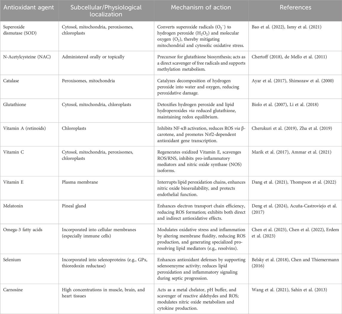
Table 5. Overview of key antioxidant systems and their mechanistic roles in the pathogenesis and modulation of sepsis.
Therapeutic limitations and risks associated with antioxidant treatment
The therapeutic use of antioxidants in sepsis has gained significant attention due to the well-established role of oxidative stress in the pathogenesis of this condition. Despite promising mechanistic insights and preclinical data, clinical translation has faced substantial limitations and risks that restrict widespread implementation. Previous antioxidant trials in critical illness, especially sepsis, have largely failed due to challenges in pharmacokinetics, patient selection, and trial design (Table 6). Antioxidants often exhibit poor bioavailability, rapid clearance, and inadequate tissue distribution in critically ill patients, making it difficult to achieve therapeutic concentrations at the right time. Additionally, most trials enrolled unstratified, heterogeneous patient populations without using oxidative stress biomarkers to identify individuals most likely to benefit. This lack of precision diluted any potential therapeutic signal. Comorbidities such as diabetes or liver dysfunction further distorted redox balance and response. While antioxidants such as vitamins C and E, melatonin, N-acetylcysteine, and mitochondrial-targeted compounds have shown beneficial effects in experimental models, their clinical efficacy remains inconsistent or inconclusive in humans. Many studies highlight the absence of statistically significant outcomes or improvement in mortality among septic patients receiving antioxidants, which undermines their routine clinical use (Prauchner, 2017; Galley, 2010). For example, HDIVC (50 mg/kg every 6 h for 96 h) failed to improve outcomes in vasopressor-treated septic ICU patients, instead increasing death or persistent organ dysfunction without secondary benefits (Lamontagne et al., 2022). Moreover, transcriptomic analysis from the LOVIT trial revealed that vitamin C lowered mortality in adaptive but worsened outcomes in inflammopathic sepsis, suggesting immune profiling may guide personalized therapy (Rynne et al., 2025). In addition, Manapurath et al. conducted a systematic review and meta-analysis of four trials involving 800 very low birth weight or very preterm infants to evaluate the effects of low-dose enteral vitamin A supplementation (≤10,000 IU/day). While supplementation significantly increased serum retinol levels, it had no significant effect on sepsis incidence. Despite the biological plausibility of vitamin A in supporting immune function, the findings do not currently support its use for sepsis prevention in this population (Manapurath et al., 2022). Another critical limitation lies in the complexity of dosing and timing. The optimal therapeutic window for antioxidant administration in sepsis remains poorly defined. Delayed treatment may fail to mitigate the oxidative cascade, while early intervention could interfere with beneficial immune responses that rely on ROS for pathogen clearance (Forman and Zhang, 2021). Additionally, inter-patient variability in oxidative stress levels complicates the establishment of standardized antioxidant protocols (Berger and Chioléro, 2007). From a risk perspective, antioxidants may exert unintended immunosuppressive effects. By scavenging ROS, antioxidants can potentially impair neutrophil and macrophage function, thereby weakening the host defence mechanisms critical in septic patients (Galley, 2010). Moreover, selenium and other trace element supplements, though biologically active, carry a narrow therapeutic window, and overdosing could exacerbate organ dysfunction or induce toxicity (Üstündağ, 2023). A phase II trial evaluated high-dose selenium (4,000 μg day 1, then 1,000 µg/day for 9 days) in 60 septic shock patients. Continuous infusion was well tolerated but showed no improvement in mortality or organ support duration, suggesting bolus dosing may be more effective (Forceville, 2007). A meta-analysis by Alhazzani et al. concluded that selenium supplementation, particularly in the form of intravenous sodium selenite, may reduce mortality in septic patients, although the evidence was heterogeneous and modest in effect size (Alhazzani et al., 2013). Finally, combination antioxidant therapies, while conceptually promising, present challenges in pharmacokinetics, potential interactions, and evaluation of synergistic effects. Trials employing multitherapy antioxidant regimens have encountered difficulties in isolating specific contributions of individual components and ensuring safe, effective dosage ratios (Abelli et al., 2022).
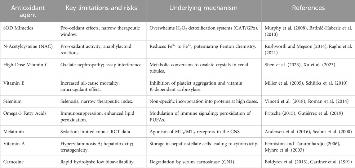
Table 6. Key limitations and risks of antioxidant therapies in sepsis.
Future directions
Although current antioxidant strategies in sepsis have yielded mixed clinical results, evolving insights into the disease’s redox biology are shaping a new generation of targeted and personalized interventions. Several future directions are emerging to overcome past limitations and optimize the therapeutic potential of antioxidants in sepsis. A critical area of focus is the targeted delivery of antioxidants to mitochondria, the primary source of ROS in sepsis-induced organ dysfunction. Mitochondria-targeted compounds, such as MitoQ and SkQ derivatives, are being investigated for their ability to localize antioxidant effects precisely where oxidative damage is most profound (Víctor et al., 2009). In parallel, natural antioxidants and phytochemicals (e.g., polyphenols, flavonoids) are gaining traction due to their multifaceted properties, including anti-inflammatory, antimicrobial, and antioxidant activities. These agents may provide a safer profile compared to synthetic antioxidants, especially when used in synergistic formulations (Üstündağ, 2023; Shrivastava et al., 2023). Biomarker-guided therapy is another promising avenue. Oxidative stress levels vary among patients, and identifying reliable biomarkers (e.g., isoprostanes, nitrotyrosine, mitochondrial DNA) could allow stratification of patients who are most likely to benefit from antioxidant treatment. This precision medicine approach may also guide dosage and timing, two factors previously identified as critical to therapeutic success (Santacroce et al., 2024). Furthermore, emerging technologies such as nanomedicine and nano-antioxidants offer new platforms for antioxidant delivery. Nanocarriers can encapsulate antioxidants, enhance bioavailability, and direct compounds to specific tissues or cells involved in sepsis pathology. Some nanomaterials even possess inherent redox-modulating properties, blurring the lines between vehicle and therapy (Lin et al., 2024). Guo et al. demonstrated that nano-parthenolide improved survival and intestinal barrier function in septic rats by reducing ROS and apoptosis through 5-HTR2A regulation, outperforming conventional formulations (Guo et al., 2023). Li et al. designed multifunctional nanoparticles capable of simultaneously scavenging LPS, ROS, and cell-free DNA, effectively suppressing cytokine storms and reducing mortality in animal sepsis models (Li et al., 2023b). Extending beyond systemic inflammation, Qu et al. developed a biomimetic nanomodulator targeting sepsis-associated encephalopathy, which crossed the blood-brain barrier, alleviated oxidative stress, polarized macrophages toward anti-inflammatory phenotypes, and improved cognition and survival (Qu et al., 2024). Complementing antioxidant-based strategies, Tang et al. introduced mRNA-lipid nanoparticles that reprogrammed macrophages in situ into CAR-MΦs, restoring immune clearance against multidrug-resistant bacteria in septic mice (Tang et al., 2024). Finally, the integration of artificial intelligence and real-time monitoring presents a transformative opportunity; developing machine learning algorithms that dynamically adjust antioxidant infusion based on continuous clinical data could enable adaptive, personalized dosing, while federated learning models could predict patients at high risk for oxidative organ failure, facilitating pre-emptive intervention. Alam et al. proposed FedSepsis, a federated deep-learning framework using electronic health records and IoMT devices for early sepsis detection, underscoring the potential of integrating predictive analytics with antioxidant-based therapeutics (Alam and Rahmani, 2023). Collectively, these directions point toward multifunctional nanotherapies, precision redox modulation, and AI-driven early detection systems as transformative approaches for harnessing antioxidants in sepsis management.
Conclusion
The intricate interplay among oxidative stress, mitochondrial dysfunction, and immune dysregulation constitutes a central axis in the pathogenesis of sepsis. Although antioxidant-based therapies are biologically compelling, their clinical application remains challenged by variability in efficacy, timing of administration, and patient-specific factors. Among emerging strategies, mitochondria-targeted antioxidants such as melatonin and MitoQ, along with adjunctive agents like N-acetylcysteine and vitamin C, offer promising therapeutic avenues. Moreover, biomarker-guided treatment and nanomedicine-enabled delivery systems provide a foundation for precision medicine approaches that could enhance both safety and effectiveness. Moving forward, integrated efforts to establish reliable biomarkers, refine dosing protocols, and align interventions with distinct immunometabolic endotypes are essential. Such a mechanistically informed and patient-tailored strategy may ultimately shift sepsis management from generalized supportive care toward targeted, disease-modifying therapy.
Author contributions
YL: Supervision, Validation, Writing – original draft. AA: Data curation, Writing – original draft, Writing – review and editing. LC: Data curation, Writing – review and editing. SM: Supervision, Validation, Writing – review and editing.
Funding
The author(s) declare that no financial support was received for the research and/or publication of this article.
Conflict of interest
The authors declare that the research was conducted in the absence of any commercial or financial relationships that could be construed as a potential conflict of interest.
Generative AI statement
The author(s) declare that no Generative AI was used in the creation of this manuscript.
Any alternative text (alt text) provided alongside figures in this article has been generated by Frontiers with the support of artificial intelligence and reasonable efforts have been made to ensure accuracy, including review by the authors wherever possible. If you identify any issues, please contact us.
Publisher’s note
All claims expressed in this article are solely those of the authors and do not necessarily represent those of their affiliated organizations, or those of the publisher, the editors and the reviewers. Any product that may be evaluated in this article, or claim that may be made by its manufacturer, is not guaranteed or endorsed by the publisher.
References
Abdelmawgood, I. A., Badr, A. M., Abdelkader, A. E., Mahana, N. A., Mohamed, A. S., and Abdelfattah, H. H. (2025). Chrysin-loaded poly (lactic-co-glycolic acid) nanoparticles alleviate sepsis-induced splenic injury by regulating myeloid-derived suppressor cells. Immunol. Res. 73 (1), 80. doi:10.1007/s12026-025-09634-5
Abelli, J., Méndez-Valdés, G., Gómez-Hevia, F., Bragato, M. C., Chichiarelli, S., Saso, L., et al. (2022). Potential antioxidant multitherapy against complications occurring in sepsis. Biomedicines 10 (12), 3088. doi:10.3390/biomedicines10123088
Acuña-Castroviejo, D., Rahim, I., Acuña-Fernández, C., Fernández-Ortiz, M., Solera-Marín, J., Sayed, R. K. A., et al. (2017). Melatonin, clock genes and mitochondria in sepsis. Cell Mol. Life Sci. 74 (21), 3965–3987. doi:10.1007/s00018-017-2610-1
Ahn, J. H., Oh, D. K., Huh, J. W., Lim, C. M., Koh, Y., and Hong, S. B. (2019). Vitamin C alone does not improve treatment outcomes in mechanically ventilated patients with severe sepsis or septic shock: a retrospective cohort study. J. Thorac. Dis. 11 (4), 1562–1570. doi:10.21037/jtd.2019.03.03
Aisa-Álvarez, A., Pérez-Torres, I., Guarner-Lans, V., Manzano-Pech, L., Cruz-Soto, R., Márquez-Velasco, R., et al. (2023). Randomized clinical trial of antioxidant therapy patients with septic shock and organ dysfunction in the ICU: SOFA score reduction by improvement of the enzymatic and non-enzymatic antioxidant system. Cells 12 (9), 1330. doi:10.3390/cells12091330
Alam, M. U., and Rahmani, R. (2023). FedSepsis: a federated multi-modal deep learning-based internet of medical things application for early detection of sepsis from electronic health records using raspberry Pi and Jetson Nano devices. Sensors (Basel) 23 (2), 970. doi:10.3390/s23020970
Albuszies, G., and Brückner, U. B. (2003). Antioxidant therapy in sepsis. Intensive Care Med. 29 (10), 1632–1636. doi:10.1007/s00134-003-1861-5
Alhazzani, W., Jacobi, J., Sindi, A., Hartog, C., Reinhart, K., Kokkoris, S., et al. (2013). The effect of selenium therapy on mortality in patients with sepsis syndrome: a systematic review and meta-analysis of randomized controlled trials. Crit. Care Med. 41 (6), 1555–1564. doi:10.1097/CCM.0b013e31828a24c6
Ammar, M. A., Ammar, A. A., Condeni, M. S., and Bell, C. M. (2021). Vitamin C for sepsis and septic shock. Am. J. Ther. 28 (6), e649–e679. doi:10.1097/MJT.0000000000001423
Andersen, L. P., Gögenur, I., Rosenberg, J., and Reiter, R. J. (2016). The safety of melatonin in humans. Clin. Drug Investig. 36 (3), 169–175. doi:10.1007/s40261-015-0368-5
Andrabi, S. M., Sharma, N. S., Karan, A., Shahriar, S. M. S., Cordon, B., Ma, B., et al. (2023). Nitric oxide: physiological functions, delivery, and biomedical applications. Adv. Sci. (Weinh) 10 (30), e2303259. doi:10.1002/advs.202303259
Angstwurm, M. W., Engelmann, L., Zimmermann, T., Lehmann, C., Spes, C. H., Abel, P., et al. (2007). Selenium in intensive care (SIC): results of a prospective randomized, placebo-controlled, multiple-center study in patients with severe systemic inflammatory response syndrome, sepsis, and septic shock. Crit. Care Med. 35 (1), 118–126. doi:10.1097/01.CCM.0000251124.83436.0E
Arnalich, F., Garcia-Palomero, E., López, J., Jiménez, M., Madero, R., Renart, J., et al. (2000). Predictive value of nuclear factor kappaB activity and plasma cytokine levels in patients with sepsis. Infect. Immun. 68 (4), 1942–1945. doi:10.1128/IAI.68.4.1942-1945.2000
Atli, M., Erikoglu, M., Kaynak, A., Esen, H. H., and Kurban, S. (2012). The effects of selenium and vitamin E on lung tissue in rats with sepsis. Clin. Invest Med. 35 (2), E48–E54. doi:10.25011/cim.v35i2.16288
Ayar, G., Atmaca, Y. M., Alışık, M., and Erel, Ö. (2017). Effects of paraoxonase, arylesterase, ceruloplasmin, catalase, and myeloperoxidase activities on prognosis in pediatric patients with sepsis. Clin. Biochem. 50 (7-8), 414–417. doi:10.1016/j.clinbiochem.2016.12.013
Babu, R., Eaton, S., Drake, D. P., Spitz, L., and Pierro, A. (2001). Glutamine and glutathione counteract the inhibitory effects of mediators of sepsis in neonatal hepatocytes. J. Pediatr. Surg. 36 (2), 282–286. doi:10.1053/jpsu.2001.20690
Bai, S., Zhang, M., Tang, S., Li, M., Wu, R., Wan, S., et al. (2024). Effects and impact of selenium on human health, A review. Molecules 30 (1), 50. doi:10.3390/molecules30010050
Bajčetić, M., Otašević, B., Prekajski, N. B., Spasić, S., and Spasojević, I. (2014). Antioxidative system in the erythrocytes of preterm neonates with sepsis: the effects of vitamin E supplementation. Ann. Clin. Biochem. 51 (Pt 5), 550–556. doi:10.1177/0004563213503317
Bao, W., Xing, H., Cao, S., Long, X., Liu, H., Ma, J., et al. (2022). Neutrophils restrain sepsis associated coagulopathy via extracellular vesicles carrying superoxide dismutase 2 in a murine model of lipopolysaccharide induced sepsis. Nat. Commun. 13 (1), 4583. doi:10.1038/s41467-022-32325-w
Barchielli, G., Capperucci, A., and Tanini, D. (2022). The role of selenium in pathologies: an updated review. Antioxidants (Basel) 11 (2), 251. doi:10.3390/antiox11020251
Batinić-Haberle, I., Rebouças, J. S., and Spasojević, I. (2010). Superoxide dismutase mimics: chemistry, pharmacology, and therapeutic potential. Antioxid. Redox Signal 13 (6), 877–918. doi:10.1089/ars.2009.2876
Behairy, M. Y., Abdelrahman, A. A., Toraih, E. A., Ibrahim, E. E. D. A., Azab, M. M., Sayed, A. A., et al. (2022). Investigation of TLR2 and TLR4 polymorphisms and sepsis susceptibility: computational and experimental approaches. Int. J. Mol. Sci. 23 (18), 10982. doi:10.3390/ijms231810982
Belsky, J. B., Wira, C. R., Jacob, V., Sather, J. E., and Lee, P. J. (2018). A review of micronutrients in sepsis: the role of thiamine, l-carnitine, vitamin C, selenium and vitamin D. Nutr. Res. Rev. 31 (2), 281–290. doi:10.1017/S0954422418000124
Berger, M. M., and Chioléro, R. L. (2007). Antioxidant supplementation in sepsis and systemic inflammatory response syndrome. Crit. Care Med. 35 (9 Suppl. l), S584–S590. doi:10.1097/01.CCM.0000279189.81529.C4
Bernardi, P., Gerle, C., Halestrap, A. P., Jonas, E. A., Karch, J., Mnatsakanyan, N., et al. (2023). Identity, structure, and function of the mitochondrial permeability transition pore: controversies, consensus, recent advances, and future directions. Cell Death Differ. 30 (8), 1869–1885. doi:10.1038/s41418-023-01187-0
Binka, F. N., Ross, D. A., Morris, S. S., Kirkwood, B. R., Arthur, P., Dollimore, N., et al. (1995). Vitamin A supplementation and childhood malaria in northern Ghana. Am. J. Clin. Nutr. 61 (4), 853–859. doi:10.1093/ajcn/61.4.853
Biolo, G., Antonione, R., and De Cicco, M. (2007). Glutathione metabolism in sepsis. Crit. Care Med. 35 (9 Suppl. l), S591–S595. doi:10.1097/01.CCM.0000278913.19123.13
Blackwell, T. S., Yull, F. E., Chen, C. L., Venkatakrishnan, A., Blackwell, T. R., Hicks, D. J., et al. (2000). Multiorgan nuclear factor kappa B activation in a transgenic mouse model of systemic inflammation. Am. J. Respir. Crit. Care Med. 162 (3 Pt 1), 1095–1101. doi:10.1164/ajrccm.162.3.9906129
Bo, J. H., Wang, J. X., Wang, X. L., Jiao, Y., Jiang, M., Chen, J. L., et al. (2022). Dexmedetomidine attenuates lipopolysaccharide-induced sympathetic activation and sepsis via suppressing superoxide signaling in paraventricular nucleus. Antioxidants (Basel) 11 (12), 2395. doi:10.3390/antiox11122395
Boldyrev, A. A., Aldini, G., and Derave, W. (2013). Physiology and pathophysiology of carnosine. Physiol. Rev. 93 (4), 1803–1845. doi:10.1152/physrev.00039.2012
Brealey, D., Brand, M., Hargreaves, I., Heales, S., Land, J., Smolenski, R., et al. (2002). Association between mitochondrial dysfunction and severity and outcome of septic shock. Lancet 360 (9328), 219–223. doi:10.1016/S0140-6736(02)09459-X
Byerly, S., Parreco, J. P., Soe-Lin, H., Parks, J. J., Lee, E. E., Shnaydman, I., et al. (2020). Vitamin C and thiamine are associated with lower mortality in sepsis. J. Trauma Acute Care Surg. 89 (1), 111–117. doi:10.1097/TA.0000000000002613
Calviello, G., Su, H. M., Weylandt, K. H., Fasano, E., Serini, S., and Cittadini, A. (2013). Experimental evidence of ω-3 polyunsaturated fatty acid modulation of inflammatory cytokines and bioactive lipid mediators: their potential role in inflammatory, neurodegenerative, and neoplastic diseases. Biomed. Res. Int. 2013, 743171. doi:10.1155/2013/743171
Campos, R., Shimizu, M. H. M., Volpini, R. A., de Bragança, A. C., Andrade, L., Lopes, F. D. T. Q. D. S., et al. (2012). N-acetylcysteine prevents pulmonary edema and acute kidney injury in rats with sepsis submitted to mechanical ventilation. Am. J. Physiol. Lung Cell Mol. Physiol. 302 (7), L640–L650. doi:10.1152/ajplung.00097.2011
Canbolat, N., Ozkul, B., Sever, I. H., Sogut, I., Eroglu, E., Uyanikgil, Y., et al. (2022). Vitamins C and E protect from sepsis-induced lung damage in rat and CT correlation. Bratisl. Lek. Listy 123 (11), 828–832. doi:10.4149/BLL_2022_132
Carlson, D., Maass, D. L., White, D. J., Tan, J., and Horton, J. W. (2006). Antioxidant vitamin therapy alters sepsis-related apoptotic myocardial activity and inflammatory responses. Am. J. Physiol. Heart Circ. Physiol. 291 (6), H2779–H2789. doi:10.1152/ajpheart.01258.2005
Carr, A. C., and Maggini, S. (2017). Vitamin C and immune function. Nutrients 9 (11), 1211. doi:10.3390/nu9111211
Caruso, G. (2022). Unveiling the hidden therapeutic potential of carnosine, a molecule with a multimodal mechanism of action: a position paper. Molecules 27 (10), 3303. doi:10.3390/molecules27103303
Chandimali, N., Bak, S. G., Park, E. H., Lim, H. J., Won, Y. S., Kim, E. K., et al. (2025). Free radicals and their impact on health and antioxidant defenses: a review. Cell Death Discov. 11 (1), 19. doi:10.1038/s41420-024-02278-8
Che, D., Xiao, Y., Zhang, X., Zhu, K., Chen, W., You, G., et al. (2025). Multifunctional integrated polyphenol-copper nanozymes for sepsis-induced acute liver injury via ameliorating endoplasmic reticulum stress and reprogramming inflammatory microenvironment. Chem. Eng. J. 507, 160293. doi:10.1016/j.cej.2025.160293
Chelkeba, L., Ahmadi, A., Abdollahi, M., Najafi, A., Ghadimi, M. H., Mosaed, R., et al. (2015). The effect of parenteral selenium on outcomes of mechanically ventilated patients following sepsis: a prospective randomized clinical trial. Ann. Intensive Care 5 (1), 29. doi:10.1186/s13613-015-0071-y
Chen, J., and Thiemermann, C. (2016). Selenium and Niacin for sepsis therapy: the sum is greater than its parts. Crit. Care Med. 44 (6), 1256–1257. doi:10.1097/CCM.0000000000001493
Chen, H., Wang, W., Hong, C., Zhang, M., Hong, Y., Wang, S., et al. (2017). Omega-3 fish oil reduces mortality due to severe sepsis with acute gastrointestinal injury grade III. Pharmacogn. Mag. 13 (51), 407–412. doi:10.4103/pm.pm_418_16
Chen, H., Wang, S., Zhao, Y., Luo, Y., Tong, H., and Su, L. (2018). Correlation analysis of omega-3 fatty acids and mortality of sepsis and sepsis-induced ARDS in adults: data from previous randomized controlled trials. Nutr. J. 17 (1), 57. doi:10.1186/s12937-018-0356-8
Chen, Y. L., Xie, Y. J., Liu, Z. M., Chen, W. B., Zhang, R., Ye, H. X., et al. (2022). Omega-3 fatty acids impair miR-1-3p-dependent Notch3 down-regulation and alleviate sepsis-induced intestinal injury. Mol. Med. 28 (1), 9. doi:10.1186/s10020-021-00425-w
Chen, Z. S., Yu, M. M., Wang, K., Meng, X. L., Liu, Y. C., Shou, S. T., et al. (2023). Omega-3 polyunsaturated fatty acids inhibit cardiomyocyte apoptosis and attenuate sepsis-induced cardiomyopathy. Nutrition 106, 111886. doi:10.1016/j.nut.2022.111886
Chen, S., Hou, Z., Xiao, M., Wu, P., Yang, Y., Han, S., et al. (2025). Quaternized chitosan-based photothermal antibacterial hydrogel with pro-vascularization and on-demand degradation properties for enhanced infected wound healing. Carbohydr. Polym. 355, 123350. doi:10.1016/j.carbpol.2025.123350
Chertoff, J. (2018). N-Acetylcysteine's role in sepsis and potential benefit in patients with microcirculatory derangements. J. Intensive Care Med. 33 (2), 87–96. doi:10.1177/0885066617696850
Cherukuri, L., Gewirtz, G., Osea, K., and Tayek, J. A. (2019). Vitamin A treatment for severe sepsis in humans; a prospective randomized double blind placebo-controlled clinical trial. Clin. Nutr. ESPEN 29, 49–51. doi:10.1016/j.clnesp.2018.10.011
Chmielewska, K., Dzierzbicka, K., Inkielewicz-Stępniak, I., and Przybyłowska, M. (2020). Therapeutic potential of carnosine and its derivatives in the treatment of human diseases. Chem. Res. Toxicol. 33 (7), 1561–1578. doi:10.1021/acs.chemrestox.0c00010
Cinelli, M. A., Do, H. T., Miley, G. P., and Silverman, R. B. (2020). Inducible nitric oxide synthase: regulation, structure, and inhibition. Med. Res. Rev. 40 (1), 158–189. doi:10.1002/med.21599
Coleman, M. C., Brouillette, M. J., Andresen, N. S., Oberley-Deegan, R. E., and Martin, J. M. (2017). Differential effects of superoxide dismutase mimetics after mechanical overload of articular cartilage. Antioxidants (Basel) 6 (4), 98. doi:10.3390/antiox6040098
Colunga Biancatelli, R. M. L., Berrill, M., Mohammed, Y. H., and Marik, P. E. (2020). Melatonin for the treatment of sepsis: the scientific rationale. J. Thorac. Dis. 12 (Suppl. 1), S54–s65. doi:10.21037/jtd.2019.12.85
Costa, N. A., Gut, A. L., Azevedo, P. S., Tanni, S. E., Cunha, N. B., Magalhães, E. S., et al. (2016). Erythrocyte superoxide dismutase as a biomarker of septic acute kidney injury. Ann. Intensive Care 6 (1), 95. doi:10.1186/s13613-016-0198-5
Costa, N. A., Cunha, N. B., Gut, A. L., Azevedo, P. S., Polegato, B. F., Zornoff, L. A. M., et al. (2018). Erythrocyte SOD1 activity, but not SOD1 polymorphisms, is associated with ICU mortality in patients with septic shock. Free Radic. Biol. Med. 124, 199–204. doi:10.1016/j.freeradbiomed.2018.06.013
Dang, H., Li, J., Liu, C., and Xu, F. (2021). The association between vitamin E deficiency and critically ill children with sepsis and septic shock. Front. Nutr. 8, 648442. doi:10.3389/fnut.2021.648442
de Mello, R. O., Lunardelli, A., Caberlon, E., de Moraes, C. M. B., Christ Vianna Santos, R., da Costa, V. L., et al. (2011). Effect of N-acetylcysteine and fructose-1,6-bisphosphate in the treatment of experimental sepsis. Inflammation 34 (6), 539–550. doi:10.1007/s10753-010-9261-9
Deng, J. C., Cheng, G., Newstead, M. W., Zeng, X., Kobayashi, K., Flavell, R. A., et al. (2006). Sepsis-induced suppression of lung innate immunity is mediated by IRAK-M. J. Clin. Invest 116 (9), 2532–2542. doi:10.1172/JCI28054
Deng, Z., He, M., Hu, H., Zhang, W., Zhang, Y., Ge, Y., et al. (2024). Melatonin attenuates sepsis-induced acute kidney injury by promoting mitophagy through SIRT3-mediated TFAM deacetylation. Autophagy 20 (1), 151–165. doi:10.1080/15548627.2023.2252265
Di Meo, S., Reed, T. T., Venditti, P., and Victor, V. M. (2016). Role of ROS and RNS sources in physiological and pathological conditions. Oxid. Med. Cell Longev. 2016, 1245049. doi:10.1155/2016/1245049
Durant, R., Klouche, K., Delbosc, S., Morena, M., Amigues, L., Beraud, J. J., et al. (2004). Superoxide anion overproduction in sepsis: effects of vitamin e and simvastatin. Shock 22 (1), 34–39. doi:10.1097/01.shk.0000129197.46212.7e
El-Gendy, F. M., El-Hawy, M. A., and Hassan, M. G. (2018). Beneficial effect of melatonin in the treatment of neonatal sepsis. J. Matern. Fetal Neonatal Med. 31 (17), 2299–2303. doi:10.1080/14767058.2017.1342794
Erdem, D., Segmen, F., Uysal, E., and Kilicarslan, G. (2023). Effect of omega-3 fatty acid use on sepsis and mortality in patients with Covıd-19. Niger. J. Clin. Pract. 26 (1), 102–108. doi:10.4103/njcp.njcp_415_22
Erel, S., Küçükk, A., Dikmen, K., Tekin, E., Yığman, Z., Ceyhan, M. Ş., et al. (2024). Evaluation of the efficacy of ozone therapy on liver tissue in the treatment of sepsis in rats with cecal perforation. Med. Kaunas. 60 (9), 1552. doi:10.3390/medicina60091552
Escames, G., León, J., Macías, M., Khaldy, H., and Acuña-Castroviejo, D. (2003). Melatonin counteracts lipopolysaccharide-induced expression and activity of mitochondrial nitric oxide synthase in rats. Faseb J. 17 (8), 932–934. doi:10.1096/fj.02-0692fje
Falcão-Holanda, R. B., Brunialti, M. K. C., Jasiulionis, M. G., and Salomão, R. (2021). Epigenetic regulation in sepsis, role in pathophysiology and therapeutic perspective. Front. Med. (Lausanne) 8, 685333. doi:10.3389/fmed.2021.685333
Fan, H., Le, J. W., and Zhu, J. H. (2020). Protective effect of N-Acetylcysteine pretreatment on acute kidney injury in septic rats. J. Surg. Res. 254, 125–134. doi:10.1016/j.jss.2020.04.017
Fan, D., Wang, L., Chakraborty, A., Zhang, C. L., and Yang, F. H. (2024). Editorial: novel treatments for cardiovascular diseases by targeting inflammation, oxidative stress, and cell death. Front. Cardiovasc Med. 11, 1538222. doi:10.3389/fcvm.2024.1538222
Fisher, B. J., Seropian, I. M., Kraskauskas, D., Thakkar, J. N., Voelkel, N. F., Fowler, A. A., et al. (2011). Ascorbic acid attenuates lipopolysaccharide-induced acute lung injury. Crit. Care Med. 39 (6), 1454–1460. doi:10.1097/CCM.0b013e3182120cb8
Forceville, X. (2007). Effects of high doses of selenium, as sodium selenite, in septic shock patients a placebo-controlled, randomized, double-blind, multi-center phase II study--selenium and sepsis. J. Trace Elem. Med. Biol. 21 (Suppl. 1), 62–65. doi:10.1016/j.jtemb.2007.09.021
Forman, H. J., and Zhang, H. (2021). Targeting oxidative stress in disease: promise and limitations of antioxidant therapy. Nat. Rev. Drug Discov. 20 (9), 689–709. doi:10.1038/s41573-021-00233-1
Fowler, A. A., Syed, A. A., Knowlson, S., Sculthorpe, R., Farthing, D., DeWilde, C., et al. (2014). Phase I safety trial of intravenous ascorbic acid in patients with severe sepsis. J. Transl. Med. 12 (1), 32. doi:10.1186/1479-5876-12-32
Fowler, A. A., Truwit, J. D., Hite, R. D., Morris, P. E., DeWilde, C., Priday, A., et al. (2019). Effect of vitamin C infusion on organ failure and biomarkers of inflammation and vascular injury in patients with sepsis and severe acute respiratory failure: the CITRIS-ALI randomized clinical trial. Jama 322 (13), 1261–1270. doi:10.1001/jama.2019.11825
Fritsche, K. L. (2015). The science of fatty acids and inflammation. Adv. Nutr. 6 (3), 293s–301s. doi:10.3945/an.114.006940
Fu, Q., Liu, M., Zhang, X., Fu, J., Zhang, G., Ji, Y., et al. (2025). A randomized controlled trial on the efficacy and safety of vitamin A supplementation in children with sepsis. Front. Pediatr. 13, 1579006. doi:10.3389/fped.2025.1579006
Galley, H. F. (2010). Bench-to-bedside review: targeting antioxidants to mitochondria in sepsis. Crit. Care 14 (4), 230. doi:10.1186/cc9098
Galley, H. F. (2011). Oxidative stress and mitochondrial dysfunction in sepsis. Br. J. Anaesth. 107 (1), 57–64. doi:10.1093/bja/aer093
Gardner, M. L., Illingworth, K. M., Kelleher, J., and Wood, D. (1991). Intestinal absorption of the intact peptide carnosine in man, and comparison with intestinal permeability to lactulose. J. Physiol. 439, 411–422. doi:10.1113/jphysiol.1991.sp018673
Garg, B. D., Bansal, A., and Kabra, N. S. (2019). Role of vitamin A supplementation in prevention of bronchopulmonary dysplasia in extremely low birth weight neonates: a systematic review of randomized trials. J. Matern. Fetal Neonatal Med. 32 (15), 2608–2615. doi:10.1080/14767058.2018.1441282
Giamarellos-Bourboulis, E. J., Aschenbrenner, A. C., Bauer, M., Bock, C., Calandra, T., Gat-Viks, I., et al. (2024). The pathophysiology of sepsis and precision-medicine-based immunotherapy. Nat. Immunol. 25 (1), 19–28. doi:10.1038/s41590-023-01660-5
Giorgi, C., Baldassari, F., Bononi, A., Bonora, M., De Marchi, E., Marchi, S., et al. (2012). Mitochondrial Ca2+ and apoptosis. Cell Calcium 52 (1), 36–43. doi:10.1016/j.ceca.2012.02.008
Guerreiro, M. O., Petronilho, F., Andrades, M., Constantino, L., Mina, F. G., Moreira, J. C. F., et al. (2010). Plasma superoxide dismutase activity and mortality in septic patients [corrected]. J. Trauma 69 (6), E102–E106. doi:10.1097/TA.0b013e3181dbb289
Guo, N. K., She, H., Tan, L., Zhou, Y. Q., Tang, C. Q., Peng, X. Y., et al. (2023). Nano parthenolide improves intestinal barrier function of sepsis by inhibiting apoptosis and ROS via 5-HTR2A. Int. J. Nanomedicine 18, 693–709. doi:10.2147/IJN.S394544
Gutiérrez, S., Svahn, S. L., and Johansson, M. E. (2019). Effects of Omega-3 fatty acids on immune cells. Int. J. Mol. Sci. 20 (20), 5028. doi:10.3390/ijms20205028
Habimana, R., Choi, I., Cho, H. J., Kim, D., Lee, K., and Jeong, I. (2020). Sepsis-induced cardiac dysfunction: a review of pathophysiology. Acute Crit. Care 35 (2), 57–66. doi:10.4266/acc.2020.00248
He, J., Duan, M., and Zhuang, H. (2024). ICAM1 and VCAM1 are associated with outcome in patients with sepsis: a systematic review and meta-analysis. Heliyon 10 (21), e40003. doi:10.1016/j.heliyon.2024.e40003
He, Y., Li, Y., Lao, Q., Qin, T., Xie, X., and Jiang, W. (2025). Association between vitamin E and 28-Day mortality in adult sepsis patients: a retrospective cohort analysis. Am. J. Med. Sci. S0002-9629 (25), 01148–01156. doi:10.1016/j.amjms.2025.04.020
Henderson, R., Kim, S., and Lee, E. (2018). Use of melatonin as adjunctive therapy in neonatal sepsis: a systematic review and meta-analysis. Complement. Ther. Med. 39, 131–136. doi:10.1016/j.ctim.2018.06.002
Higashi, Y., Maruhashi, T., Noma, K., and Kihara, Y. (2014). Oxidative stress and endothelial dysfunction: clinical evidence and therapeutic implications. Trends Cardiovasc Med. 24 (4), 165–169. doi:10.1016/j.tcm.2013.12.001
Hosny, M., Nahas, R., Ali, S., Elshafei, S. A., and Khaled, H. (2013). Impact of oral omega-3 fatty acids supplementation in early sepsis on clinical outcome and immunomodulation. Egypt. J. Crit. Care Med. 1 (3), 119–126. doi:10.1016/j.ejccm.2013.11.002
Ibrahim, A. E. H., El Sallab, S. M., Hamed, A. M., and El Ghareeb, A. H. (2024). Anti-inflammatory effect of melatonin therapy in sepsis. Alexandria J. Pediatr. 37 (2), 95–99. doi:10.4103/ajop.ajop_14_24
Incalza, M. A., D'Oria, R., Natalicchio, A., Perrini, S., Laviola, L., and Giorgino, F. (2018). Oxidative stress and reactive oxygen species in endothelial dysfunction associated with cardiovascular and metabolic diseases. Vasc. Pharmacol. 100, 1–19. doi:10.1016/j.vph.2017.05.005
Ismy, J., Syukri, M., Emril, D. R., Sekarwana, N., Ismy, J., and Suhanda, R. (2021). The effect of superoxide dismutase on inhibition of acute kidney injury induced by sepsis based on kidney tissue histology and murine sepsis score. ScientificWorldJournal 2021, 1827296. doi:10.1155/2021/1827296
Ismy, J., Syukri, M., Emril, D. R., Sekarwana, N., and Ismy, J. (2022). Superoxide dismutase reduces creatinine and NGAL by restoring oxidative balance during sepsis. Emerg. Sci. J. 6, 286–294. doi:10.28991/esj-2022-06-02-06
Jakobsson, G., Papareddy, P., Andersson, H., Mulholland, M., Bhongir, R., Ljungcrantz, I., et al. (2023). Therapeutic S100A8/A9 blockade inhibits myocardial and systemic inflammation and mitigates sepsis-induced myocardial dysfunction. Crit. Care 27 (1), 374. doi:10.1186/s13054-023-04652-x
Jiang, N., Li, N., Huang, J., Ning, K., Zhang, H., Gou, H., et al. (2024). Impact of vitamin C on inflammatory response and myocardial injury in sepsis patients. Altern. Ther. Health Med. 30 (10), 427–431.
Joffre, J., and Hellman, J. (2021). Oxidative stress and endothelial dysfunction in sepsis and acute inflammation. Antioxid. Redox Signal 35 (15), 1291–1307. doi:10.1089/ars.2021.0027
Juan, C. A., Pérez de la Lastra, J. M., Plou, F. J., and Pérez-Lebeña, E. (2021). The chemistry of reactive oxygen Species (ROS) revisited: outlining their role in biological macromolecules (DNA, lipids and proteins) and induced pathologies. Int. J. Mol. Sci. 22 (9), 4642. doi:10.3390/ijms22094642
Kashiouris, M. G., Qiao, X., Fisher, B., Truwit, J., Hite, R., Morris, P., et al. (2019). “A multicenter parsimonious biomarker mortality prediction model for sepsis-induced ARDS”, in A95. critical care: alice in wonderland - biomarkers in critical illness: clues to be unravelled, A2381. doi:10.1164/ajrccm-conference.2019.199.1_meetingabstracts.a2381
Kashiouris, M. G., L'Heureux, M., Cable, C. A., Fisher, B. J., Leichtle, S. W., and Fowler, A. A. (2020). The emerging role of vitamin C as a treatment for sepsis. Nutrients 12 (2), 292. doi:10.3390/nu12020292
Kočan, L., Vašková, J., Vaško, L., Simonová, J., Simon, R., and Firment, J. (2014). Selenium adjuvant therapy in septic patients selected according to carrico index. Clin. Biochem. 47 (15), 44–50. doi:10.1016/j.clinbiochem.2014.07.004
Kono, Y., Inomata, M., Hagiwara, S., Shiraishi, N., Noguchi, T., and Kitano, S. (2012). A newly synthetic vitamin E derivative, E-Ant-S-GS, attenuates lung injury caused by cecal ligation and puncture-induced sepsis in rats. Surgery 151 (3), 420–426. doi:10.1016/j.surg.2011.08.003
Kumar, S., Saxena, J., Srivastava, V. K., Kaushik, S., Singh, H., Abo-El-Sooud, K., et al. (2022). The interplay of oxidative stress and ROS scavenging: antioxidants as a therapeutic potential in sepsis. Vaccines (Basel) 10 (10), 1575. doi:10.3390/vaccines10101575
Kumar, D., Ahmed, M., Andrabi, N. I., Singh, C. P., Saroch, D., Bharitkar, Y. P., et al. (2025). Anti-inflammatory and anti-oxidant potential of dispiro-indanedione hybrid of parthenin via regulating Nrf2 and NF-κB/MAPK pathways. Eur. J. Pharmacol. 996, 177547. doi:10.1016/j.ejphar.2025.177547
Kwon, W. Y., Suh, G. J., Kim, K. S., Jung, Y. S., Kim, S. H., Kim, J. S., et al. (2016). Niacin and selenium attenuate sepsis-induced lung injury by up-regulating nuclear factor erythroid 2-related factor 2 signaling. Crit. Care Med. 44 (6), e370–e382. doi:10.1097/CCM.0000000000001422
Lamontagne, F., Masse, M. H., Menard, J., Sprague, S., Pinto, R., Heyland, D. K., et al. (2022). Intravenous vitamin C in adults with sepsis in the intensive care unit. N. Engl. J. Med. 386 (25), 2387–2398. doi:10.1056/NEJMoa2200644
Landucci, F., Mancinelli, P., De Gaudio, A. R., and Virgili, G. (2014). Selenium supplementation in critically ill patients: a systematic review and meta-analysis. J. Crit. Care 29 (1), 150–156. doi:10.1016/j.jcrc.2013.08.017
Lawrence, T. (2009). The nuclear factor NF-kappaB pathway in inflammation. Cold Spring Harb. Perspect. Biol. 1 (6), a001651. doi:10.1101/cshperspect.a001651
Le, J. W., Sun, M., Zhu, J. H., and Fan, H. (2022). Protective effect of N-acetylcysteine on acute lung injury in septic rats by inhibiting inflammation, oxidation, and apoptosis. Iran. J. Basic Med. Sci. 25 (7), 859–864. doi:10.22038/IJBMS.2022.65350.14384
Leff, J. A., Parsons, P. E., Day, C. E., Moore, E. E., Moore, F. A., Oppegard, M. A., et al. (1992). Increased serum catalase activity in septic patients with the adult respiratory distress syndrome. Am. Rev. Respir. Dis. 146 (4), 985–989. doi:10.1164/ajrccm/146.4.985
Lelubre, C., and Vincent, J. L. (2018). Mechanisms and treatment of organ failure in sepsis. Nat. Rev. Nephrol. 14 (7), 417–427. doi:10.1038/s41581-018-0005-7
Li, J., Kwon, Y., Chung, K. S., Lim, C. S., Lee, D., Yue, Y., et al. (2018). Naphthalene-based fluorescent probes for glutathione and their applications in living cells and patients with sepsis. Theranostics 8 (5), 1411–1420. doi:10.7150/thno.22252
Li, S., Tang, T., Guo, P., Zou, Q., Ao, X., Hu, L., et al. (2019). A meta-analysis of randomized controlled trials: efficacy of selenium treatment for sepsis. Med. Baltim. 98 (9), e14733. doi:10.1097/MD.0000000000014733
Li, F., Yan, R., Wu, J., Han, Z., Qin, M., Liu, C., et al. (2021). An antioxidant enzyme therapeutic for sepsis. Front. Bioeng. Biotechnol. 9, 800684. doi:10.3389/fbioe.2021.800684
Li, Z., Qiao, O., Wang, Y., Li, N., and Gong, Y. (2023a). Potential therapeutic targets for trauma management. Trends Pharmacol. Sci. 44, 891–901. doi:10.1016/j.tips.2023.10.001
Li, Z., Feng, Y., Zhang, S., Li, T., Li, H., Wang, D., et al. (2023b). A multifunctional nanoparticle mitigating cytokine storm by scavenging multiple inflammatory mediators of sepsis. ACS Nano 17 (9), 8551–8563. doi:10.1021/acsnano.3c00906
Li, J., Liu, H., Shan, Z., Zhong, K., and Liang, Q. (2025). Molecular mechanisms and potential implications of ferroptosis, cuproptosis, and disulfidptosis in septic lung injury. Front. Med. (Lausanne). 12, 1615264. doi:10.3389/fmed.2025.1615264
Liang, B., Su, J., Shao, H., Chen, H., and Xie, B. (2023). The outcome of IV vitamin C therapy in patients with sepsis or septic shock: a meta-analysis of randomized controlled trials. Crit. Care 27 (1), 109. doi:10.1186/s13054-023-04392-y
Liao, Y., Wang, R., and Wen, F. (2022). Diagnostic and prognostic value of long noncoding RNAs in sepsis: a systematic review and meta-analysis. Expert Rev. Mol. Diagn 22 (8), 821–831. doi:10.1080/14737159.2022.2125801
Lin, L., Liu, H., Zhang, D., Du, L., and Zhang, H. (2024). Nanolevel immunomodulators in sepsis: novel roles, current perspectives, and future directions. Int. J. Nanomedicine 19, 12529–12556. doi:10.2147/IJN.S496456
Liu, R., and Meng, Y. (2024). Effect of continuous blood purification combined with reduced glutathione on endotoxin, inflammatory mediators and severity of liver injury in patients with septic shock. Trop. J. Pharm. Res. 23, 595–601. doi:10.4314/tjpr.v23i3.14
Liu, H., Tian, Y., Liu, C., Wang, D., and Wang, H. (2024). Effect of glutathione-assisted continuous renal replacement therapy in sepsis patients with acute renal injury. Clin. Nephrol. 101 (4), 191–198. doi:10.5414/CN111220
Liu, Y., Zhao, W., Huang, Q., Wan, L., Ren, Z., Zhang, B., et al. (2025). Advances in research on the release of von Willebrand factor from endothelial cells through the membrane attack complex C5b-9 in sepsis. J. Inflamm. Res. 18, 6719–6733. doi:10.2147/JIR.S520726
Lu, C., Sharma, S., McIntyre, L., Rhodes, A., Evans, L., Almenawer, S., et al. (2017). Omega-3 supplementation in patients with sepsis: a systematic review and meta-analysis of randomized trials. Ann. Intensive Care 7 (1), 58. doi:10.1186/s13613-017-0282-5
Lundberg, J. O., and Weitzberg, E. (2022). Nitric oxide signaling in health and disease. Cell 185 (16), 2853–2878. doi:10.1016/j.cell.2022.06.010
Lundberg, O. H. M., Lengquist, M., Spångfors, M., Annborn, M., Bergmann, D., Schulte, J., et al. (2020). Circulating bioactive adrenomedullin as a marker of sepsis, septic shock and critical illness. Crit. Care 24 (1), 636. doi:10.1186/s13054-020-03351-1
Luo, R., Yao, Y., Chen, Z., and Sun, X. (2025). An examination of the LPS-TLR4 immune response through the analysis of molecular structures and protein-protein interactions. Cell Commun. Signal 23 (1), 142. doi:10.1186/s12964-025-02149-4
Manapurath, R. M., Kumar, M., Pathak, B. G., Chowdhury, R., Sinha, B., Choudhary, T., et al. (2022). Enteral low-dose vitamin A supplementation in preterm or low birth weight infants to prevent morbidity and mortality: a systematic review and meta-analysis. Pediatrics 150 (Suppl. 1), e2022057092L. doi:10.1542/peds.2022-057092L
Mantzarlis, K., Tsolaki, V., and Zakynthinos, E. (2017). Role of oxidative stress and mitochondrial dysfunction in sepsis and potential therapies. Oxid. Med. Cell Longev. 2017, 5985209. doi:10.1155/2017/5985209
Marik, P. E. (2018). Vitamin C for the treatment of sepsis: the scientific rationale. Pharmacol. Ther. 189, 63–70. doi:10.1016/j.pharmthera.2018.04.007
Marik, P. E., Khangoora, V., Rivera, R., Hooper, M. H., and Catravas, J. (2017). Hydrocortisone, vitamin C, and thiamine for the treatment of severe sepsis and septic shock: a retrospective before-after study. Chest 151 (6), 1229–1238. doi:10.1016/j.chest.2016.11.036
Martinez Ramos, A., Galindo Salom, H. M., Prieto Lozano, H. A., Lopez Posada, P. A., Martinez, J. M., Guerrero, M., et al. (2025). Effects of the intravenous application of sodium ascorbate in patients with the diagnosis of sepsis in the intensive care unit - observational, analytical, retrospective, comparative study. J. Complement. Integr. Med. doi:10.1515/jcim-2025-0066
Metnitz, P. G., Bartens, C., Fischer, M., Fridrich, P., Steltzer, H., and Druml, W. (1999). Antioxidant status in patients with acute respiratory distress syndrome. Intensive Care Med. 25 (2), 180–185. doi:10.1007/s001340050813
Mileva, M., Dimitrova-Koleva, A., Tsvetanova, E., Krastev, D., Georgieva, A., Alexandrova, A., et al. (2022). Mitigation of the oxidative damage in liver caused by influenza virus infection in mice by an effective combination of oseltamivir and S-adenosyl-L-methionine (SAM). J. Pharm. Nutr. Sci. 12, 128–138. doi:10.29169/1927-5951.2022.12.11
Miller, E. R., Pastor-Barriuso, R., Dalal, D., Riemersma, R. A., Appel, L. J., and Guallar, E. (2005). Meta-analysis: high-dosage vitamin E supplementation may increase all-cause mortality. Ann. Intern Med. 142 (1), 37–46. doi:10.7326/0003-4819-142-1-200501040-00110
Minter, B. E., Lowes, D. A., Webster, N. R., and Galley, H. F. (2020). Differential effects of MitoVitE, α-Tocopherol and trolox on oxidative stress, mitochondrial function and inflammatory signalling pathways in endothelial cells cultured under conditions mimicking sepsis. Antioxidants (Basel) 9 (3), 195. doi:10.3390/antiox9030195
Moskowitz, A., Huang, D. T., Hou, P. C., Gong, J., Doshi, P. B., Grossestreuer, A. V., et al. (2020). Effect of ascorbic acid, corticosteroids, and thiamine on organ injury in septic shock: the ACTS randomized clinical trial. Jama 324 (7), 642–650. doi:10.1001/jama.2020.11946
Murphy, C. K., Fey, E. G., Watkins, B. A., Wong, V., Rothstein, D., and Sonis, S. T. (2008). Efficacy of superoxide dismutase mimetic M40403 in attenuating radiation-induced oral mucositis in hamsters. Clin. Cancer Res. 14 (13), 4292–4297. doi:10.1158/1078-0432.CCR-07-4669
Myhre, A. M., Carlsen, M. H., Bøhn, S. K., Wold, H. L., Laake, P., and Blomhoff, R. (2003). Water-miscible, emulsified, and solid forms of retinol supplements are more toxic than oil-based preparations. Am. J. Clin. Nutr. 78 (6), 1152–1159. doi:10.1093/ajcn/78.6.1152
Nagar, H., Piao, S., and Kim, C. S. (2018). Role of mitochondrial oxidative stress in sepsis. Acute Crit. Care 33 (2), 65–72. doi:10.4266/acc.2018.00157
Nakajima, M., Kojiro, M., Aso, S., Matsui, H., Fushimi, K., Kaita, Y., et al. (2019). Effect of high-dose vitamin C therapy on severe burn patients: a nationwide cohort study. Crit. Care 23 (1), 407. doi:10.1186/s13054-019-2693-1
Narayanankutty, A., Job, J. T., and Narayanankutty, V. (2019). Glutathione, an antioxidant tripeptide: dual roles in carcinogenesis and chemoprevention. Curr. Protein Pept. Sci. 20 (9), 907–917. doi:10.2174/1389203720666190206130003
Neri, A., Pezzotti, P., Fazio, C., Vacca, P., D'Ancona, F. P., Caporali, M. G., et al. (2015). Epidemiological and molecular characterization of invasive meningococcal disease in Italy, 2008/09-2012/13. PLoS One 10 (10), e0139376. doi:10.1371/journal.pone.0139376
Okut, S., Ozan, M., Buzdağli, Y., Uçar, H., İnaç, M. R., Han, M. T., et al. (2025). The effects of Omega-3 supplementation combined with strength training on neuro-biomarkers, inflammatory and antioxidant responses, and the lipid profile in physically healthy adults. Nutrients 17 (13), 2088. doi:10.3390/nu17132088
Oliva, A., Bianchi, A., Russo, A., Ceccarelli, G., Cancelli, F., Aloj, F., et al. (2021). Effect of N-Acetylcysteine administration on 30-Day mortality in critically ill patients with septic shock caused by carbapenem-resistant Klebsiella pneumoniae and acinetobacter baumannii: a retrospective case-control study. Antibiot. (Basel) 10 (3), 271. doi:10.3390/antibiotics10030271
Ortolani, O., Conti, A., De Gaudio, A. R., Moraldi, E., Cantini, Q., and Novelli, G. (2000). The effect of glutathione and N-acetylcysteine on lipoperoxidative damage in patients with early septic shock. Am. J. Respir. Crit. Care Med. 161 (6), 1907–1911. doi:10.1164/ajrccm.161.6.9903043
Oudemans-van Straaten, H. M., Man, A. M. E. S.-d., and de Waard, M. C. (2014). Vitamin C revisited. Crit. Care 18 (4), 460. doi:10.1186/s13054-014-0460-x
Ozkok, E., Yorulmaz, H., Ates, G., Aksu, A., Balkis, N., Şahin, Ö., et al. (2016). Amelioration of energy metabolism by melatonin in skeletal muscle of rats with LPS induced endotoxemia. Physiol. Res. 65 (5), 833–842. doi:10.33549/physiolres.933282
Park, H., Ko, R. E., Oh, H. S., Moon, J. Y., Chang, Y., and Suh, G. Y. (2025). Evaluating the diagnostic performance of adult sepsis event criteria in the emergency department: impact of including isolated serum lactate elevations. J. Intensive Care 13 (1), 44. doi:10.1186/s40560-025-00815-w
Peerapornratana, S., Manrique-Caballero, C. L., Gómez, H., and Kellum, J. A. (2019). Acute kidney injury from sepsis: current concepts, epidemiology, pathophysiology, prevention and treatment. Kidney Int. 96 (5), 1083–1099. doi:10.1016/j.kint.2019.05.026
Pein, H., Ville, A., Pace, S., Temml, V., Garscha, U., Raasch, M., et al. (2018). Endogenous metabolites of vitamin E limit inflammation by targeting 5-lipoxygenase. Nat. Commun. 9 (1), 3834. doi:10.1038/s41467-018-06158-5
Penniston, K. L., and Tanumihardjo, S. A. (2006). The acute and chronic toxic effects of vitamin A. Am. J. Clin. Nutr. 83 (2), 191–201. doi:10.1093/ajcn/83.2.191
Pham, V. T., Dold, S., Rehman, A., Bird, J. K., and Steinert, R. E. (2021). Vitamins, the gut microbiome and gastrointestinal health in humans. Nutr. Res. 95, 35–53. doi:10.1016/j.nutres.2021.09.001
Prabhu, K. S., Zamamiri-Davis, F., Stewart, J. B., Thompson, J. T., Sordillo, L. M., and Reddy, C. C. (2002). Selenium deficiency increases the expression of inducible nitric oxide synthase in RAW 264.7 macrophages: role of nuclear factor-kappaB in up-regulation. Biochem. J. 366 (Pt 1), 203–209. doi:10.1042/BJ20020256
Prauchner, C. A. (2017). Oxidative stress in sepsis: pathophysiological implications justifying antioxidant co-therapy. Burns 43 (3), 471–485. doi:10.1016/j.burns.2016.09.023
Qu, H., Wu, J., Pan, Y., Abdulla, A., Duan, Z., Cheng, W., et al. (2024). Biomimetic nanomodulator regulates oxidative and inflammatory stresses to treat sepsis-associated encephalopathy. ACS Nano 18 (41), 28228–28245. doi:10.1021/acsnano.4c08157
Raghu, G., Berk, M., Campochiaro, P. A., Jaeschke, H., Marenzi, G., Richeldi, L., et al. (2021). The multifaceted therapeutic role of N-Acetylcysteine (NAC) in disorders characterized by oxidative stress. Curr. Neuropharmacol. 19 (8), 1202–1224. doi:10.2174/1570159X19666201230144109
Rocha, M., Herance, R., Rovira, S., Hernández-Mijares, A., and Victor, V. M. (2012). Mitochondrial dysfunction and antioxidant therapy in sepsis. Infect. Disord. Drug Targets 12 (2), 161–178. doi:10.2174/187152612800100189
Roman, M., Jitaru, P., and Barbante, C. (2014). Selenium biochemistry and its role for human health. Metallomics 6 (1), 25–54. doi:10.1039/c3mt00185g
Rudd, K. E., Johnson, S. C., Agesa, K. M., Shackelford, K. A., Tsoi, D., Kievlan, D. R., et al. (2020). Global, regional, and national sepsis incidence and mortality, 1990–2017: analysis for the global burden of disease study. Lancet 395 (10219), 200–211. doi:10.1016/S0140-6736(19)32989-7
Rushworth, G. F., and Megson, I. L. (2014). Existing and potential therapeutic uses for N-acetylcysteine: the need for conversion to intracellular glutathione for antioxidant benefits. Pharmacol. Ther. 141 (2), 150–159. doi:10.1016/j.pharmthera.2013.09.006
Rynne, J., Mosavie, M., Masse, M. H., Ménard, J., Battista, M. C., Maslove, D. M., et al. (2025). Sepsis subtypes and differential treatment response to vitamin C: biological sub-study of the LOVIT trial. Intensive Care Med. 51 (1), 82–93. doi:10.1007/s00134-024-07733-9
Sahin, S., and Burukoglu Donmez, D. (2018). Effects of carnosine (Beta-Alanyl-L-Histidine) in an experimental rat model of acute kidney injury due to septic shock. Med. Sci. Monit. 24, 305–316. doi:10.12659/msm.905181
Sahin, S., Oter, S., Burukoğlu, D., and Sutken, E. (2013). The effects of carnosine in an experimental rat model of septic shock. Med. Sci. Monit. Basic Res. 19, 54–61. doi:10.12659/msmbr.883758
Sainglers, W., Khamwong, M., and Chareonsudjai, S. (2025). N-acetylcysteine inhibits NETs, exhibits antibacterial and antibiofilm properties and enhances neutrophil function against Burkholderia pseudomallei. Sci. Rep. 15 (1), 29943. doi:10.1038/s41598-025-13506-1
Salvemini, D., and Cuzzocrea, S. (2003). Therapeutic potential of superoxide dismutase mimetics as therapeutic agents in critical care medicine. Crit. Care Med. 31 (1 Suppl. l), S29–S38. doi:10.1097/00003246-200301001-00005
Santacroce, E., D'Angerio, M., Ciobanu, A. L., Masini, L., Lo Tartaro, D., Coloretti, I., et al. (2024). Advances and challenges in sepsis management: modern tools and future directions. Cells 13 (5), 439. doi:10.3390/cells13050439
Schmoch, T., Gallenstein, N., Peters, V., Bartosova, M., Uhle, F., Kummer, L., et al. (2025). Anserine reduces mortality in experimental sepsis by preventing methylglyoxal-induced capillary leakage. EBioMedicine 114, 105644. doi:10.1016/j.ebiom.2025.105644
Schürks, M., Glynn, R. J., Rist, P. M., Tzourio, C., and Kurth, T. (2010). Effects of vitamin E on stroke subtypes: meta-analysis of randomised controlled trials. Bmj 341, c5702. doi:10.1136/bmj.c5702
Seabra, M. L., Bignotto, M., Pinto, L. R., and Tufik, S. (2000). Randomized, double-blind clinical trial, controlled with placebo, of the toxicology of chronic melatonin treatment. J. Pineal Res. 29 (4), 193–200. doi:10.1034/j.1600-0633.2002.290401.x
Sevransky, J. E., Rothman, R. E., Hager, D. N., Bernard, G. R., Brown, S. M., Buchman, T. G., et al. (2021). Effect of vitamin C, thiamine, and hydrocortisone on Ventilator- and vasopressor-free days in patients with sepsis: the VICTAS randomized clinical trial. Jama 325 (8), 742–750. doi:10.1001/jama.2020.24505
Shahidi, F., and Ambigaipalan, P. (2018). Omega-3 polyunsaturated Fatty acids and their health benefits. Annu. Rev. Food Sci. Technol. 9, 345–381. doi:10.1146/annurev-food-111317-095850
Shen, Z. Y., Chen, Y. R., Wang, M. C., and Chang, S. S. (2023). High-dose vitamin C-induced acute oxalate nephropathy in a renal transplant recipient: a case report and literature review. Asian J. Surg. 46 (5), 2223–2224. doi:10.1016/j.asjsur.2022.11.112
Shimozawa, N., Zhang, Z., Imamura, A., Suzuki, Y., Fujiki, Y., Tsukamoto, T., et al. (2000). Molecular mechanism of detectable catalase-containing particles, peroxisomes, in fibroblasts from a PEX2-defective patient. Biochem. Biophys. Res. Commun. 268 (1), 31–35. doi:10.1006/bbrc.1999.2082
Shrivastava, A. K., Sahu, P. K., Cecchi, T., Shrestha, L., Shah, S. K., Gupta, A., et al. (2023). An emerging natural antioxidant therapy for COVID-19 infection patients: current and future directions. Food Front. 4 (3), 1179–1205. doi:10.1002/fft2.207
Sinha, P., Meyer, N. J., and Calfee, C. S. (2023). Biological phenotyping in sepsis and acute respiratory distress syndrome. Annu. Rev. Med. 74, 457–471. doi:10.1146/annurev-med-043021-014005
Srinivasan, V., Pandi-Perumal, S. R., Spence, D. W., Kato, H., and Cardinali, D. P. (2010). Melatonin in septic shock: some recent concepts. J. Crit. Care 25 (4), 656.e1–656.e6566. doi:10.1016/j.jcrc.2010.03.006
Stanic, M., Dragic, S., Travar, M., Uletilovic, S., Mandic-Kovacevic, N., and Kovacevic, P. (2024). The potential predictive role of oxidative stress and antioxidant parameters regarding mortality and the type of causative agent in sepsis. Cureus 16 (11), e73456. doi:10.7759/cureus.73456
Sui, B., Li, X., Li, N., Tao, Y., Wang, L., Xu, Y., et al. (2025). Synergistic action of mucoactive drugs and phages against Pseudomonas aeruginosa and Klebsiella pneumoniae. Microbiol. Spectr. 13 (3), e0160124. doi:10.1128/spectrum.01601-24
Sun, S. C. (2017). The non-canonical NF-κB pathway in immunity and inflammation. Nat. Rev. Immunol. 17 (9), 545–558. doi:10.1038/nri.2017.52
Sun, C., Wu, Q., Zhang, X., He, Q., and Zhao, H. (2017). Mechanistic evaluation of the protective effect of carnosine on acute lung injury in sepsis rats. Pharmacology 100 (5-6), 292–300. doi:10.1159/000479879
Takatani, Y., Ono, K., Suzuki, H., Inaba, M., Sawada, M., and Matsuda, N. (2018). Inducible nitric oxide synthase during the late phase of sepsis is associated with hypothermia and immune cell migration. Lab. Invest 98 (5), 629–639. doi:10.1038/s41374-018-0021-z
Tanaka, K. I., Sugizaki, T., Kanda, Y., Tamura, F., Niino, T., and Kawahara, M. (2017). Preventive effects of carnosine on lipopolysaccharide-induced lung injury. Sci. Rep. 7, 42813. doi:10.1038/srep42813
Tang, C., Jing, W., Han, K., Yang, Z., Zhang, S., Liu, M., et al. (2024). mRNA-Laden lipid-nanoparticle-enabled in situ CAR-macrophage engineering for the eradication of multidrug-resistant bacteria in a sepsis mouse model. ACS Nano 18 (3), 2261–2278. doi:10.1021/acsnano.3c10109
Tang, R., Jiang, M., Tang, X., Chen, S., Xu, H., Pan, Y., et al. (2025). Narciclasine mitigates sepsis-induced cardiac dysfunction by enhancing BNIP3-mediated mitophagy and suppressing ferroptosis. Free Radic. Biol. Med. 238, 220–234. doi:10.1016/j.freeradbiomed.2025.06.051
Thompson, M. A., Zuniga, K., Sousse, L., Christy, R., and Gurney, C. J. (2022). The role of vitamin E in thermal burn injuries, infection, and sepsis: a review. J. Burn Care Res. 43 (6), 1260–1270. doi:10.1093/jbcr/irac100
Tseng, P. T., Zeng, B. Y., Zeng, B. S., Yeh, P. Y., Stubbs, B., Kuo, J. S., et al. (2024). The efficacy and acceptability of anti-inflammatory omega-3 polyunsaturated fatty acid supplements in sepsis management: a network meta-analysis of randomized controlled trials. Prostagl. Leukot. Essent. Fat. Acids 202, 102633. doi:10.1016/j.plefa.2024.102633
Tuteja, N., Chandra, M., Tuteja, R., and Misra, M. K. (2004). Nitric oxide as a unique bioactive signaling messenger in physiology and pathophysiology. J. Biomed. Biotechnol. 2004 (4), 227–237. doi:10.1155/S1110724304402034
Üstündağ, H. (2023). The role of antioxidants in sepsis management: a review of therapeutic applications. Eur. J. Mol. Biochem. Sci. 2, 38–48.
Valenta, J., Brodska, H., Drabek, T., Hendl, J., and Kazda, A. (2011). High-dose selenium substitution in sepsis: a prospective randomized clinical trial. Intensive Care Med. 37 (5), 808–815. doi:10.1007/s00134-011-2153-0
Velasque, M., Branchini, G., Catarina, A. V., Bettoni, L., Fernandes, R. S., Da Silva, A. F., et al. (2023). Fish oil - omega-3 exerts protective effect in oxidative stress and liver dysfunctions resulting from experimental sepsis. J. Clin. Exp. Hepatol. 13 (1), 64–74. doi:10.1016/j.jceh.2022.07.001
Victor, V. M., Rocha, M., Esplugues, J. V., and De la Fuente, M. (2005). Role of free radicals in sepsis: antioxidant therapy. Curr. Pharm. Des. 11 (24), 3141–3158. doi:10.2174/1381612054864894
Víctor, V. M., Espulgues, J. V., Hernández-Mijares, A., and Rocha, M. (2009). Oxidative stress and mitochondrial dysfunction in sepsis: a potential therapy with mitochondria-targeted antioxidants. Infect. Disord. Drug Targets 9 (4), 376–389. doi:10.2174/187152609788922519
Vinceti, M., Filippini, T., Del Giovane, C., Dennert, G., Zwahlen, M., Brinkman, M., et al. (2018). Selenium for preventing cancer. Cochrane Database Syst. Rev. 1 (1), Cd005195. doi:10.1002/14651858.CD005195.pub4
Wald, E. L., Sanchez-Pinto, L. N., Smith, C. M., Moran, T., Badke, C. M., Barhight, M. F., et al. (2020). Hydrocortisone-ascorbic acid-thiamine use associated with lower mortality in pediatric septic shock. Am. J. Respir. Crit. Care Med. 201 (7), 863–867. doi:10.1164/rccm.201908-1543LE
Wald, E. L., Badke, C. M., Hintz, L. K., Spewak, M., and Sanchez-Pinto, L. N. (2022). Vitamin therapy in sepsis. Pediatr. Res. 91 (2), 328–336. doi:10.1038/s41390-021-01673-6
Wang, C., Han, D., Feng, X., and Wu, J. (2020). Omega-3 fatty acid supplementation is associated with favorable outcomes in patients with sepsis: an updated meta-analysis. J. Int. Med. Res. 48 (12), 300060520953684. doi:10.1177/0300060520953684
Wang, H., Fang, Z., Qiu, G., Zhang, C., Tang, M., and Zhou, B. (2021). Bioprotective and functional effect of carnosine on sepsis induced renal damage in Male albino rat model through targeting IL-1β and TNF-α production. Dokl. Biochem. Biophys. 500 (1), 408–414. doi:10.1134/S1607672921050033
Wang, Y. M., Gong, F. C., Qi, X., Zheng, Y. J., Zheng, X. T., Chen, Y., et al. (2022a). Mucin 1 inhibits ferroptosis and sensitizes vitamin E to alleviate sepsis-induced acute lung injury through GSK3β/Keap1-Nrf2-GPX4 pathway. Oxid. Med. Cell Longev. 2022, 2405943. doi:10.1155/2022/2405943
Wang, H., Su, S., Wang, C., Hu, J., Dan, W., and Peng, X. (2022b). Effects of fish oil-containing nutrition supplementation in adult sepsis patients: a systematic review and meta-analysis. Burns Trauma 10, tkac012. doi:10.1093/burnst/tkac012
Wang, S., Liu, Z., Li, R., Wang, L., Wu, Y., Zhang, W., et al. (2025a). Acetaldehyde dehydrogenase 2 attenuates lipopolysaccharide -induced endothelial barrier damage by inhibiting mitochondrial fission in sepsis-associated encephalopathy. Eur. J. Pharmacol. 997, 177468. doi:10.1016/j.ejphar.2025.177468
Wang, J., Zhang, F., Tan, C., Wang, N., Xia, X., Ye, Y., et al. (2025b). The VDAC3/DHODH axis ameliorates sepsis-induced myocardial injury by regulating ferroptosis. Front. Biosci. (Landmark Ed.) 30 (6), 39559. doi:10.31083/FBL39559
Warford, J., Lamport, A. C., Kennedy, B., and Easton, A. S. (2017). Human brain chemokine and cytokine expression in sepsis: a report of three cases. Can. J. Neurol. Sci. 44 (1), 96–104. doi:10.1017/cjn.2016.310
Willinger, T. (2019). Metabolic control of innate lymphoid cell migration. Front. Immunol. 10, 2010. doi:10.3389/fimmu.2019.02010
Wilson, J. X. (2013). Evaluation of vitamin C for adjuvant sepsis therapy. Antioxid. Redox Signal 19 (17), 2129–2140. doi:10.1089/ars.2013.5401
Wu, J. Y., Tsou, M. Y., Chen, T. H., Chen, S. J., Tsao, C. M., and Wu, C. C. (2008). Therapeutic effects of melatonin on peritonitis-induced septic shock with multiple organ dysfunction syndrome in rats. J. Pineal Res. 45 (1), 106–116. doi:10.1111/j.1600-079X.2008.00567.x
Xia, Q., Liu, G., Zhang, L., Xie, B., and Deng, L. (2025). Anemonin suppresses sepsis-induced acute lung injury by inactivation of nuclear factor-kappa B and activation of nuclear factor erythroid 2-related factor-2/heme oxygenase-1 pathway. Faseb J. 39 (2), e70328. doi:10.1096/fj.202401987RR
Xie, S., Qi, X., Wu, Q., Wei, L., Zhang, M., Xing, Y., et al. (2021). Inhibition of 5-lipoxygenase is associated with downregulation of the leukotriene B4 receptor 1/Interleukin-12p35 pathway and ameliorates sepsis-induced myocardial injury. Free Radic. Biol. Med. 166, 348–357. doi:10.1016/j.freeradbiomed.2021.02.034
Xu, C., Yi, T., Tan, S., Xu, H., Hu, Y., Ma, J., et al. (2023). Association of oral or intravenous vitamin C supplementation with mortality: a systematic review and meta-analysis. Nutrients 15 (8), 1848. doi:10.3390/nu15081848
Yang, S., Zhang, Y., Xu, J., Chen, Z., Ren, Y., Long, Y., et al. (2025). N-Acetylcysteine as a host-directed therapy against clarithromycin-resistant Mycobacterium abscessus. Pathogens 14 (4), 302. doi:10.3390/pathogens14040302
Yin, Y., Mu, F., Zhang, L., Zhao, J., Gong, R., Yin, Y., et al. (2025a). Wedelolactone activates the PI3K/AKT/NRF2 and SLC7A11/GPX4 signalling pathways to alleviate oxidative stress and ferroptosis and improve sepsis-induced liver injury. J. Ethnopharmacol. 344, 119557. doi:10.1016/j.jep.2025.119557
Yin, W., Chang, C., Zhu, Y., Ma, F., Wang, H., Lu, Y., et al. (2025b). Construction of vitamin E-based oil-in-water nano-emulsion adjuvant enhances immunogenicity of inactivated pseudorabies virus vaccine. Virology 610, 110616. doi:10.1016/j.virol.2025.110616
Zabet, M. H., Mohammadi, M., Ramezani, M., and Khalili, H. (2016). Effect of high-dose ascorbic acid on vasopressor's requirement in septic shock. J. Res. Pharm. Pract. 5 (2), 94–100. doi:10.4103/2279-042X.179569
Zainumi, C. M., Siregar, G. A., Wijaya, D. W., and Ichwan, M. (2022). Comparison enteral superoxide dismutase 1 IU and 5 IU from Cucumis melo L.C extract combined with gliadin as an antioxidant and anti-inflammatory in LPS-induced sepsis model rats. Heliyon 8 (8), e10236. doi:10.1016/j.heliyon.2022.e10236
Zhang, X., Yang, K., Chen, L., Liao, X., Deng, L., Chen, S., et al. (2019). Vitamin A deficiency in critically ill children with sepsis. Crit. Care 23 (1), 267. doi:10.1186/s13054-019-2548-9
Zhang, Q., Raoof, M., Chen, Y., Sumi, Y., Sursal, T., Junger, W., et al. (2010). Circulating mitochondrial DAMPs cause inflammatory responses to injury. Nature 464 (7285), 104–107. doi:10.1038/nature08780
Zhang, H., Mao, Y. F., Zhao, Y., Xu, D. F., Wang, Y., Xu, C. F., et al. (2021). Upregulation of matrix metalloproteinase-9 protects against sepsis-induced acute lung injury via promoting the release of soluble receptor for advanced glycation end products. Oxid. Med. Cell Longev. 2021, 8889313. doi:10.1155/2021/8889313
Zhao, Y., Qu, Y., Huang, C., Li, C., Zhang, W., Wang, X., et al. (2025). Neutrophils loaded NAD+ impede TLR4/NF-κB/NLRP3 pathway for sepsis treatment. Mater Today Bio 34, 102168. doi:10.1016/j.mtbio.2025.102168
Zheng, X., Leung, K. S., Wong, M. H., and Cheng, L. (2021). Long non-coding RNA pairs to assist in diagnosing sepsis. BMC Genomics 22 (1), 275. doi:10.1186/s12864-021-07576-4
Keywords: sepsis, oxidative stress, superoxide dismutase, vitamins, glutathione
Citation: Lin Y, Alhaskawi A, Chen L and Moqbel SAA (2025) Recent advances in understanding oxidative stress in sepsis: pathogenic roles and antioxidant therapeutic prospects - a narrative review. Front. Pharmacol. 16:1695992. doi: 10.3389/fphar.2025.1695992
Received: 31 August 2025; Accepted: 27 October 2025;
Published: 10 November 2025.
Edited by:
Galina Sud’ina, Lomonosov Moscow State University, RussiaReviewed by:
Sarah Farid, Ain Shams University, EgyptMustafa Can Guler, Atatürk University, Türkiye
Copyright © 2025 Lin, Alhaskawi, Chen and Moqbel. This is an open-access article distributed under the terms of the Creative Commons Attribution License (CC BY). The use, distribution or reproduction in other forums is permitted, provided the original author(s) and the copyright owner(s) are credited and that the original publication in this journal is cited, in accordance with accepted academic practice. No use, distribution or reproduction is permitted which does not comply with these terms.
*Correspondence: Yijuan Lin, NzA3MzMyOTQyQHFxLmNvbQ==
†These authors have contributed equally to this work to the work.