 Cristian Sandoval-Vásquez1,2
 Isabella Cárcamo3Paula Lagos3Anaís Muñoz3Francisco Zavala3Valentina Colil3
 Francisca Villagrán-Silva4
 Edgar Vásquez-Carrasco5,6,7*
 Jordan Hernandez-Martinez8,9
 Pablo Valdés-Badilla10,11
 Francisco Torrens12Paola Fincheira13
 Paulina Sepúlveda14*
- 1Escuela de Tecnología Médica, Facultad de Salud, Universidad Santo Tomás, Los Carreras, Osorno, Chile
- 2Departamento de Medicina Interna, Facultad de Medicina, Universidad de La Frontera, Temuco, Chile
- 3Carrera de Tecnología Médica, Facultad de Medicina, Universidad de La Frontera, Temuco, Chile
- 4Programa de Doctorado en Ciencias Morfológicas, Facultad de Medicina, Universidad de La Frontera, Temuco, Chile
- 5School of Occupational Therapy, Faculty of Psychology, Universidad de Talca, Talca, Chile
- 6Centro de Investigación en Ciencias Cognitivas, Faculty of Psychology, Universidad de Talca, Talca, Chile
- 7VITALIS Longevity Center, Universidad de Talca, Talca, Chile
- 8Department of Physical Activity Sciences, Universidad de Los Lagos, Osorno, Chile
- 9Department of Education, Faculty of Humanities, Universidad de la Serena, La Serena, Chile
- 10Department of Physical Activity Sciences, Faculty of Education Sciences, Universidad Católica del Maule, Talca, Chile
- 11Sports Coach Career, Faculty of Life Sciences, Universidad Viña del Mar, Viña del Mar, Chile
- 12Institut Universitari de Ciència Molecular, Universitat de València, València, Spain
- 13Departamento de Ciencias Preclínicas, Facultad de Medicina, Universidad de La Frontera, Temuco, Chile
- 14Departamento de Ciencias Básicas, Facultad de Medicina, Universidad de La Frontera, Temuco, Chile
Background: Breast cancer treatment remains a major challenge to modern medicine and has driven the need for nanotechnology-based strategies to improve drug delivery and overcome chemoresistance. Poly(ethylene glycol) and poly (lactic-co-glycolic acid) (PEG–PLGA) nanoparticles (NPs) are a type of FDA-approved biodegradable copolymer (lactic + glycolic acids) that degrades into non-toxic metabolites (lactic acid and glycolic acid); it has emerged as a promising drug carrier owing to its biocompatibility, sustained release properties, and ability to enhance the cellular uptake of chemotherapeutic agents. This systematic review examines the efficacies of PEG–PLGA nanoparticles loaded with antineoplastic drugs on in vitro models of breast cancer cell lines.
Methods: Following PRISMA guidelines, we conducted a comprehensive search of the Web of Science, Embase, MEDLINE, and Scopus databases to identify experimental studies published between 2014 and August 2025 that evaluated PEG–PLGA formulations applied to breast cancer cell lines. The methodological quality of each study was appraised using the National Institute for Health and Care Excellence (NICE) criteria.
Results: Thirteen studies were chosen based on our inclusion criteria. Here, the PEG–PLGA nanoparticles were predominantly spherical (30–210 nm) and exhibited controlled release kinetics. Compared with free drugs, the nanoformulations significantly reduced cell viability, increased apoptosis, and induced cell-cycle arrest. Functionalization with ligands such as folic acid enhanced drug targeting and cytotoxicity, while the molecular analyses revealed upregulation of p53, Bax, and caspases as well as downregulation of Bcl-2 and hTERT genes.
Conclusion: PEG–PLGA nanoparticles can substantially improve the selectivity, bioavailability, and cytotoxic efficacies of anticancer drugs in breast cancer in vitro. These findings underscore their translational potential as next-generation drug-delivery systems, warranting in vivo validation as well as development of theranostic- and stimulus-responsive designs for personalized oncology.
Systematic Review Registration: https://www.crd.york.ac.uk/PROSPERO/view/CRD420251076570.

GRAPHICAL ABSTRACT |
1 Introduction
1.1 Breast cancer burden and challenges
Breast cancer is the second most-prevalent malignancy globally, surpassed only by lung cancer, and remains as the leading cause of cancer-related mortality among women (Sung et al., 2021). Its etiology is multifactorial and involves demographic, genetic, hormonal, reproductive, and lifestyle-related risk factors, including age, female sex, obesity, family history, and unhealthy behaviors (Colditz and Bohlke, 2014; Key et al., 2001). Clinically, this condition often manifests as a palpable breast mass that is either self-detected or identified during routine examinations. Suspected cases of cancerous breast masses are often confirmed diagnostically through imaging techniques such as mammography and ultrasonography, followed by histopathological evaluation through biopsy (D’Orsi et al., 2013). Most breast carcinomas originate from the ductal or lobular epithelium and are categorized as carcinoma in situ or invasive carcinomas. Carcinoma in situ is confined within the ducts or lobules without breaching the basement membrane, whereas invasive carcinomas infiltrate the stroma and surrounding tissues (Schnitt, 2010). The most frequent histological subtypes are invasive ductal carcinoma and invasive lobular carcinoma (Lakhani et al., 2012). Therapeutic strategies against these carcinomas depend on the stage of the disease and may include surgery, radiotherapy, chemotherapy, endocrine therapy, or targeted agents. In advanced or metastatic settings, the treatment is mainly palliative, with the aim of extending survival and preserving the quality of life (Cardoso et al., 2020). Despite numerous therapeutic advances in recent times, challenges persist with respect to systemic toxicity, multidrug resistance, and suboptimal tumor bioavailability, underscoring the need for more effective drug-delivery strategies.
1.2 Nanoparticles in drug delivery
Nanotechnology has emerged as a transformative platform to enhance the pharmacokinetic and pharmacodynamic profiles of anticancer agents. Among the various polymers investigated in related literature, poly(lactic-co-glycolic acid) (PLGA) nanoparticles (NPs) have been extensively employed in drug delivery owing to their favorable biocompatibility and biodegradability profiles (Danhier et al., 2012). However, their rapid clearance via plasma protein opsonization and recognition by the reticuloendothelial system have limited clinical translation (Owens and Peppas, 2006). Among the various fabrication techniques available, nanoprecipitation is widely used because of its simplicity; this process involves dissolving the polymer in an organic solvent (e.g., acetone), introducing the solution into an aqueous medium, allowing spontaneous self-assembly, and removing the solvent subsequently (Fessi et al., 1989). In oncology, poly(ethylene glycol) with PLGA (PEG–PLGA) NPs offer a promising alternative to conventional chemotherapy by enabling controlled and targeted drug release, thereby improving the therapeutic efficacy while minimizing systemic toxicity and drug resistance (Shi et al., 2010; Parveen et al., 2012).
1.3 PEG–PLGA NPs: advantages and limitations
Surface modification of the PLGA NPs with PEG was developed to address issues related to immunogenicity and rapid clearance. PEGylation reduces immunogenicity by shielding the antigenic sites, enhances NP hydrophilicity and stability, and prolongs systemic circulation (Jokerst et al., 2011; Makadia and Siegel, 2011). Structurally, PEG–PLGA NPs exhibit a core-shell configuration, in which the PLGA core encapsulates therapeutic molecules, while the PEG shell provides protection against immune detection (Mu and Feng, 2003). Despite these advantages, the PEG–PLGA NPs have some critical limitations: i) the effectiveness of PEGylation depends on the chain density and length, which influence protein corona formation and biodistribution; ii) repeated administration may induce accelerated blood clearance or anti-PEG immune responses; iii) physicochemical characterizations of the particles are often incomplete, along with limited knowledge regarding the zeta potential, polydispersity, and long-term stability under physiologically relevant conditions; iv) ligand functionalization may improve active targeting but can compromise the stealth effects of PEG, increase formulation complexity, and limit scalability. Therefore, we aimed to assess the effectiveness of drug-loaded PEG–PLGA NPs in the treatment of breast cancer based on in vitro studies reported over the last decade.
2 Methods
2.1 Protocol and registration
We conducted a systematic evaluation of quantitative studies to assess the effectiveness of drug-loaded PEG-PLGA NPs in breast cancer therapy by focusing exclusively on in vitro experimental models. Our review was conducted in accordance with the preferred reporting items for systematic reviews and meta-analyses (PRISMA) guidelines (Page et al., 2021). The study protocol was prospectively registered with the PROSPERO database (CRD420251076570) (Vásquez and Sandoval, 2025).
2.2 Search strategy and selection criteria
2.2.1 Search strategy
A comprehensive literature search was conducted across four electronic databases, namely, MEDLINE, Embase, Scopus, and Web of Science, covering all available records up to August 2025. The search was designed to identify peer-reviewed in vitro studies without restrictions on language or publication date. The exclusions included conference abstracts, books, book chapters, editorials, letters to the editor, protocol records, reviews, case reports, and reviews or in vivo studies given the purpose of this review, which is to synthesize the mechanistic and cytotoxic effects of PEG–PLGA drug-loaded NPs under controlled in vitro conditions independent of the confounding physiological variables (e.g., metabolism, immune clearance, and vascular barriers) present in in vivo models. The inclusion of studies was guided by the population, intervention, comparator, outcome, and study design (PICOS) framework, as outlined in Table 1.

Table 1. PICOS criteria used in the systematic review.
The search strategy involved a combination of medical subject headings (MeSH) and free-text terms related to drug-loaded systems, PEG–PLGA NPs, breast cancer, and treatment. The Boolean search strings used were as follows: (“PEG–PLGA” OR “PLGA-PEG” OR “Poly (lactic-co-glycolic acid)” OR “PLGA nanoparticles” OR “PEGylated PLGA”) AND (“drug-loaded” OR “drug delivery system” OR “nanocarrier” OR “controlled release” OR “targeted delivery”) AND (“nanoparticles” OR “nanomedicine” OR “nanosystem”) AND (“cancer” OR “neoplasm” OR “tumor” OR “carcinoma” OR “oncology” OR “breast cancer” OR “mammary carcinoma”) AND (“treatment” OR “therapy” OR “therapeutic efficacy”). The lists of references of eligible studies and relevant reviews were manually screened to ensure comprehensive coverage, and only original studies that reported primary quantitative data were considered. The search and selection process implemented is summarized in the PRISMA flow diagram shown in Figure 1.

Figure 1. Flowchart of the systematic review.
2.2.2 Identification of relevant studies
An independent expert was consulted on the included articles and the inclusion and exclusion criteria to help find more relevant studies. The chosen expert had to meet two established requirements, namely, hold a PhD in health sciences and have peer-reviewed works published in journals with an impact factor, in accordance with the Journal Citation Reports® on drug-loaded treatments, NPs, and breast cancer. Our search approach was not revealed to the expert so as to avoid bias in their searches. After completing these procedures, on 30 August 2025, we searched the database to find relevant retractions or errata related to the included articles.
2.2.3 Types of study and design
The studies included herein were used to investigate the effectiveness of drug-loaded PEG–PLGA NPs in breast cancer therapy and the impacts of PEG–PLGA NPs on various biological pathways. Furthermore, we deemed it essential that these investigations were conducted in the English language. The inclusion criteria for the studies considered are as follows: (1) the research included studies focusing on the collection of numerical data or a combination of both numerical and qualitative data; (2) the statistical analysis techniques used in the research included both descriptive and inferential methods involving parametric and non-parametric approaches; (3) the types of studies included in the research were randomized controlled trials, cross-sectional studies, experimental research, and clinical trials. The exclusion criteria for the studies considered are as follows: (1) there was lack of quantitative data or specific numerical values in the studies; (2) the investigations were not reported in peer-reviewed scholarly publications; (3) the sources included conference abstracts, systematic reviews, or letters to the editor; (4) the studies did not primarily focus on investigating the use of drug-loaded PEG–PLGA NPs in breast cancer treatment or provide comprehensive descriptions of the anti-inflammatory activities and/or excluded anti-oncogenic qualities.
2.2.4 Cell lines
For the in vitro studies, breast cancer cell lines including MCF-7, MDA-MB-231, T47D, MCF-10A, MDA-MB-436, and SKBR3 were analyzed. These models were employed to evaluate the anticancer efficacies of the PEG–PLGA NPs, particularly their effects on the anti-inflammatory activities and/or anti-oncogenic qualities.
2.2.5 Quality assessment and risk of bias
The methodological quality of each quantitative study was initially assessed by a single reviewer based on the National Institute for Health and Care Excellence (NICE) rating methodology (National Institute for Health and Care Excellence, 2012). Then, a second reviewer autonomously confirmed the precision of the evaluations. All differences were addressed through dialogue and agreement. None of the studies were rejected on the grounds of problems related to methodological quality.
2.2.6 Data extraction and synthesis
Two independent reviewers assessed the titles, abstracts, and full texts of all the retrieved records to confirm eligibility. Each reviewer prepared the summary tables and flow diagrams aligned with their search outcomes. The extracted data were then compared, and discrepancies were resolved with the involvement of a third reviewer, who carefully re-examined the textual content, tables, and figures of the included studies. If any disagreements remained unresolved, the original study authors were contacted for clarification. Additionally, two researchers independently reviewed the results and discussion sections of eligible studies to determine the therapeutic roles of drug-loaded PEG–PLGA NPs in cancer treatment and their influences on different biological pathways. The synthesis focused on parameters related to the efficacy of PEG–PLGA NPs in cancer progression based exclusively on in vitro experimental models. The search procedures were in accordance with the PRISMA guidelines, as presented in Figure 1.
3 Results
3.1 Study selection
A total of 635 original studies were identified through the database search, and six studies were excluded from this list owing to duplication. Of the remaining 629 records, 583 were excluded on the basis of publication year, article type, titles unrelated to the topic, and abstracts that did not include the terms PEG–PLGA or breast cancer. Following a full-text review of the remaining 46 works, 33 studies were excluded for not matching the required study type. Ultimately, 13 studies were included in the systematic review described herein (Esim et al., 2024; Jafari-Gharabaghlou et al., 2023; Farajzadeh et al., 2018; Yildiz et al., 2018; Haggag et al., 2020; El-Hammadi et al., 2017; Kordi et al., 2016; Singh et al., 2015; Houdaihed et al., 2020; Sharma et al., 2018; Javan et al., 2019; Tabatabaei Mirakabad et al., 2016; Gorjikhah et al., 2017) (Figure 1).
3.2 Summary of the included studies
The 13 experimental studies shortlisted herein all examined PEG–PLGA-based polymeric NPs encapsulating antineoplastic agents for in vitro testing on human breast cancer cell lines. These studies were conducted across multiple countries, namely, Turkey (Esim et al., 2024), Iran (Jafari-Gharabaghlou et al., 2023; Farajzadeh et al., 2018; Kordi et al., 2016; Javan et al., 2019; Tabatabaei Mirakabad et al., 2016; Gorjikhah et al., 2017), United States (Yildiz et al., 2018), United Kingdom (Haggag et al., 2020; Sharma et al., 2018), Canada (Houdaihed et al., 2020), Spain (El-Hammadi et al., 2017), and India (Singh et al., 2015). The main cell models used in these studies were MCF-7 (Esim et al., 2024; Haggag et al., 2020; El-Hammadi et al., 2017; Singh et al., 2015; Houdaihed et al., 2020; Tabatabaei Mirakabad et al., 2016), MDA-MB-231 (Esim et al., 2024; Jafari-Gharabaghlou et al., 2023; Yildiz et al., 2018; Houdaihed et al., 2020; Sharma et al., 2018; Javan et al., 2019), and T47D (Farajzadeh et al., 2018; Kordi et al., 2016; Gorjikhah et al., 2017); additional cell lines such as SKBR3 and MDA-MB-436 were also tested in one study (Houdaihed et al., 2020). A comprehensive summary of each study and its outcome measures is presented in Table 2.

Table 2. Characteristics of the included studies.
3.3 Quality evaluation
The quality evaluation results of the studies are summarized in Table 3; most of the studies demonstrated moderate-to-high methodological quality, particularly in terms of randomization, blinding of the in vitro replicates, and reproducibility of assays. None of the studies were excluded on quality grounds.

Table 3. National Institute for Health and Care Excellence methodology checklist: quantitative studies.
3.4 NP design, synthesis, and characterization
A series of polymeric NPs was successfully synthesized using PLGA and its PEGylated derivatives (PEG–PLGA, mPEG-b-PLGA, and folate-functionalized PEG–PLGA) as carrier matrices (Esim et al., 2024; Jafari-Gharabaghlou et al., 2023; Farajzadeh et al., 2018; Yildiz et al., 2018; Haggag et al., 2020; El-Hammadi et al., 2017; Kordi et al., 2016; Singh et al., 2015; Houdaihed et al., 2020; Sharma et al., 2018; Javan et al., 2019; Tabatabaei Mirakabad et al., 2016; Gorjikhah et al., 2017). The most common fabrication techniques included nanoprecipitation and double-emulsion solvent evaporation (W1/O/W2), which enabled high encapsulation efficiencies (EEs) and narrow particle size distributions (Jafari-Gharabaghlou et al., 2023; Farajzadeh et al., 2018; Haggag et al., 2020; Kordi et al., 2016; Sharma et al., 2018; Javan et al., 2019). Table 4 shows the comparative physicochemical data for these PEG–PLGA NPs. The formulations were optimized to enhance the solubilities, bioavailabilities, and tumor-targeting potentials of hydrophobic anticancer agents such as regorafenib (Esim et al., 2024), metformin (Jafari-Gharabaghlou et al., 2023; Farajzadeh et al., 2018), curcumin (Javan et al., 2019; Tabatabaei Mirakabad et al., 2016), paclitaxel (Houdaihed et al., 2020), everolimus (Houdaihed et al., 2020), saquinavir (Singh et al., 2015), and honokiol (Haggag et al., 2020), in addition to nucleic acid cargos such as shRNA (Sharma et al., 2018). PEGylation markedly improved the colloidal stability and prolonged circulation, while folate or antibody conjugation enhanced active targeting to folate-receptor-positive or HER2/EGFR-expressing breast cancer cells (Houdaihed et al., 2020). The co-loading strategy (regorafenib + fluorouracil; metformin + curcumin; paclitaxel + everolimus) was noted to achieve synergistic therapeutic ratios while reducing the drug concentrations required for cytotoxicity, confirming the advantages of combination-loaded PEG–PLGA nanosystems (Esim et al., 2024; Farajzadeh et al., 2018; Houdaihed et al., 2020). The zeta-potential measurements showed negative surface charges (in millivolts), which were attributed to the deprotonated carboxyl groups of PLGA (Esim et al., 2024; Jafari-Gharabaghlou et al., 2023; Farajzadeh et al., 2018; Yildiz et al., 2018; Haggag et al., 2020; El-Hammadi et al., 2017; Kordi et al., 2016; Singh et al., 2015; Houdaihed et al., 2020; Sharma et al., 2018; Javan et al., 2019; Tabatabaei Mirakabad et al., 2016; Gorjikhah et al., 2017). This negative potential reduced NP aggregation and prolonged suspension stability under physiological conditions.

Table 4. Comparative physicochemical data for PEG–PLGA NPs.
3.5 Cytotoxicity, cellular uptake, apoptosis, and molecular mechanisms
Controlled drug release was most commonly evaluated using dialysis-based assays (Esim et al., 2024; Jafari-Gharabaghlou et al., 2023; Yildiz et al., 2018; Haggag et al., 2020; Singh et al., 2015; Javan et al., 2019) or high-performance liquid chromatography (HPLC) (Houdaihed et al., 2020), with all studies reporting sustained-release patterns. A summary of the outcomes is shown in Table 5. The cellular uptake was visualized via fluorescence microscopy, HPLC, and flow cytometry (Esim et al., 2024; Jafari-Gharabaghlou et al., 2023; Yildiz et al., 2018; Haggag et al., 2020; Houdaihed et al., 2020; Javan et al., 2019). PEGylation and folate targeting significantly enhanced the internalization and cytoplasmic localization of NPs without nuclear entry (Jafari-Gharabaghlou et al., 2023; Singh et al., 2015; Houdaihed et al., 2020; Sharma et al., 2018). Esim et al. (2024) demonstrated increased intracellular accumulation of regorafenib when encapsulated in NPs, particularly in the MDA-MB-231 cells; however, fluorouracil uptake remained unchanged, likely owing to differences in P-glycoprotein (P-gp) expression. Similarly, Jafari-Gharabaghlou et al. (2023) reported that metformin-loaded folate-PEG–PLGA NPs had higher cellular uptake than non-targeted PEG–PLGA formulations. In the MDA-MB-231 cells, Haggag et al. (2020) found that optimal uptake occurred for NPs composed of 10% PEGylated PLGA. In contrast, Sharma et al. (2018) observed that coumarin-6-labeled NPs were localized exclusively in the cytoplasm of the MDA-MB-231 cells without nuclear penetration. Javan et al. (2019) further indicated that NP internalization occurred predominantly through endocytic pathways. In the MCF-7 cells, Singh et al. (2015) showed that folate-targeted PEG–PLGA NPs encapsulating saquinavir (SQV-FA-PEG–PLGA) exhibited greater fluorescence intensity than either free saquinavir or non-targeted SQV-PLGA NPs.
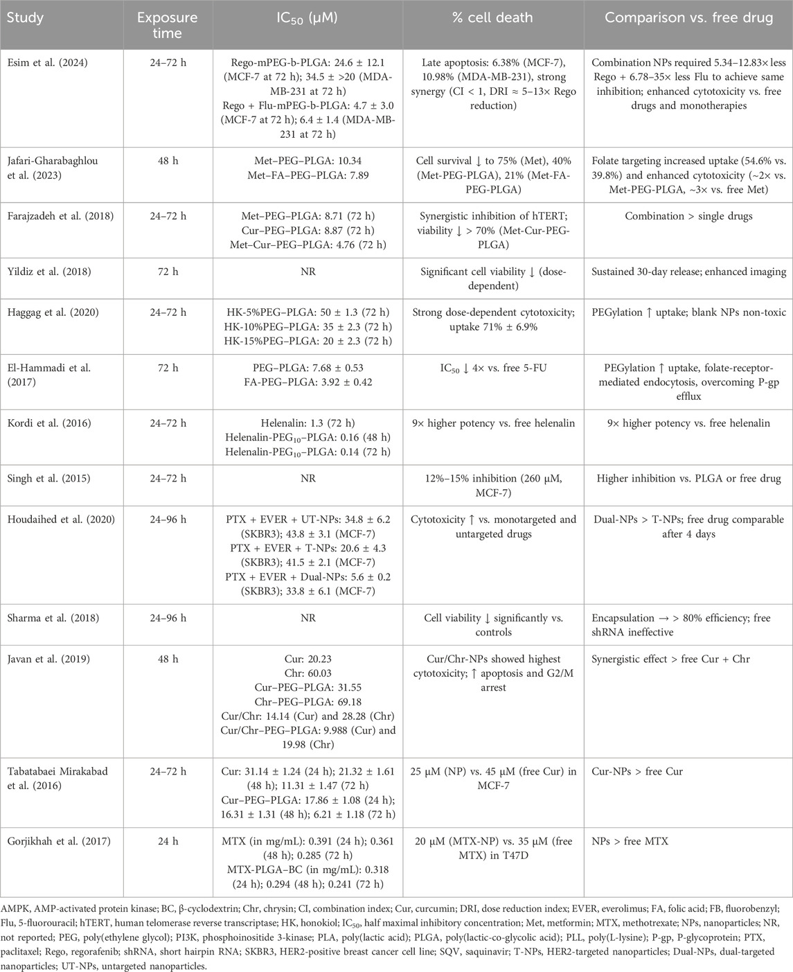
Table 5. Summary of cytotoxicity outcomes.
The apoptotic effects were mostly verified by flow cytometry and reverse transcription polymerase chain reaction (RT-PCR) (Esim et al., 2024; Jafari-Gharabaghlou et al., 2023; Farajzadeh et al., 2018; Haggag et al., 2020; Kordi et al., 2016; Javan et al., 2019). Esim et al. (2024) showed that the regorafenib +fluorouracil-mPEG-b-PLGA formulation produced greater cytotoxicity than either regorafenib alone or rego@mPEG-b-PLGA in both the MCF-7 and MDA-MB-231 cell lines. In the MDA-MB-231 cells, Jafari-Gharabaghlou et al. (2023) demonstrated that metformin-loaded FA-PEG–PLGA NPs significantly upregulated proapoptotic genes such as p53, Bax, and caspases while downregulating antiapoptotic markers such as Bcl-2 and hTERT. Similarly, Farajzadeh et al. (2018) found that metformin–curcumin-loaded NPs induced cell-cycle arrest in the T47D cells and promoted Bax and caspase expressions, whereas both free metformin–curcumin and metformin–curcumin-PLGA/PEG NPs decreased hTERT mRNA expression in a dose-dependent manner. In the MDA-MB-231 cells, Haggag et al. (2020) reported that peptide-functionalized NPs caused cell-cycle arrest at the G0/G1 and G2/M phases, while Kordi et al. (2016) observed that NC-helenalin NPs markedly reduced hTERT expression compared to free helenalin. Sharma et al. (2018) noted a pronounced reduction in MDA-MB-231 cell viability following treatment with 10% PEG–PLGA NPs. Among the included studies, the highest apoptosis rate was recorded in MDA-MB-231 cells treated with curcumin–chrysin co-loaded NPs (Javan et al., 2019).
3.6 Additional assessments
Several studies incorporated complementary analyses to further characterize the biological effects of the PEG–PLGA NP formulations. These included metabolomics (Esim et al., 2024), Ran-GTP expression (Haggag et al., 2020), and blood compatibility testing (El-Hammadi et al., 2017). Haggag et al. (2020) demonstrated that peptide-functionalized NPs not only reduced breast cancer cell viability but also suppressed the expression of Ran-GTP, which is a protein associated with tumor growth and progression. In addition, El-Hammadi et al. (2017) confirmed the biocompatibility of their system and reported no cytotoxic effects in normal human cell lines, including CCD-18 colon fibroblasts and MCF-10A non-tumorigenic breast epithelial cells.
4 Discussion
4.1 Summary of key findings and interpretation
This systematic review of 13 experimental studies highlights the promising roles of PEG–PLGA NPs as drug-delivery systems for antitumor agents in the treatment of breast cancer. Across all the cell models examined in these studies (MCF-7, MDA-MB-231, and T47D), the nanocarriers consistently improved drug encapsulation, sustained release, and cytotoxicity compared to the free formulations. Formulations with optimal physicochemical properties (150–250 nm size, polydispersity index (PDI) <0.2, zeta potential (ζ) = −20 to −35 mV) were reported to achieve the strongest antiproliferative effects, confirming the relevance of nanoscale stability in biological performance. For example, regorafenib/fluorouracil-mPEG-b-PLGA NPs (225.5 ± 7.3 nm, ζ = −24.3 mV, EE = 32.6%) showed late apoptosis rates of 6.38% (MCF-7) and 10.98% (MDA-MB-231), reducing IC50 from 24.6 to 4.7 µM and allowing 5–13 fewer regorafenib and 6–35 fewer fluorouracil doses (Esim et al., 2024). Furthermore, metformin-PEG–PLGA NPs (242 ± 6 nm, ζ = −32.1 mV, EE = 64%) and folate-modified variants (269 ± 4 nm, ζ = −33.6 mV, EE = 83.5%) decreased cell survival to 40% and 21%, respectively, confirming enhanced folate receptor uptake and cytotoxicity by nearly thrice those of free metformin (Jafari-Gharabaghlou et al., 2023). Similarly, metformin/curcumin–PEG–PLGA (257 ± 11 nm, ζ = −3.2 mV) was reported to induce >70% viability loss with synergistic hTERT inhibition, outperforming single-drug systems (Farajzadeh et al., 2018). Natural-compound-based NPs comprising honokiol, curcumin, chrysin, and helenalin showed EEs of 80%–90% and up to 9-fold greater cytotoxicity than free agents (Haggag et al., 2020; El-Hammadi et al., 2017; Kordi et al., 2016; Javan et al., 2019; Tabatabaei Mirakabad et al., 2016). Even large systems such as shRNA-PLGA (237–426 nm, EE ∼80%) were noted to achieve gene silencing and reduce invasiveness in the MDA-MB-231 cells (Sharma et al., 2018). These consistent outcomes underline that PEG–PLGA nanocarriers can potentiate drug performance through improved solubility, controlled release, and receptor-mediated uptake.
4.2 Mechanistic determinants of enhanced efficacy
Certain formulations such as co-loaded or folate-functionalized PEG–PLGA NPs can achieve superior performance through both physicochemical and biological mechanisms. For example, regorafenib/fluorouracil dual-loaded NPs (rego/flu-mPEG-b-PLGA) achieved a dose reduction of up to 12 times with increased late apoptosis (6.38% in MCF-7 and 10.98% in MDA-MB-231), which are attributable to combined inhibition of the glycolytic and tricarboxylic acid cycle pathways (Esim et al., 2024). Similarly, folate-functionalized metformin-loaded PEG–PLGA NPs induced strong downregulation of hTERT and Bcl-2 while upregulating Bax, Caspase-3/7, and p53 (Jafari-Gharabaghlou et al., 2023), consistent with the activation of mitochondrial apoptosis through modulation of the AMPK/mTOR axis. These observations reveal that improved cytotoxicity is not merely a consequence of the higher intracellular drug concentration but a sign of coordinated metabolic and transcriptional reprogramming triggered by nanodelivery. Moreover, PEG–PLGA NPs have been repeatedly shown to prolong circulation time, reduce immunogenicity, and enhance targeting when functionalized with ligands such as folic acid and peptides (Gao et al., 2023; Liu et al., 2021a; Liu et al., 2021b; Liu et al., 2019; Liu et al., 2022).
Targeted uptake, PEGylation effects, and intracellular signaling modulation are all considered to increase the efficacy. Dual-targeted paclitaxel + everolimus NPs (104 ± 5 nm) showed the best selectivity in HER2^high and EGFR^mod cells, lowering the IC50 value to 5.6 µM from 34.8 µM for constructs that were not targeted (Houdaihed et al., 2020). In addition, folate functionalization significantly increased internalization (54.6% vs. 39.8% for non-targeted NPs) and nearly doubled the cytotoxicity (Jafari-Gharabaghlou et al., 2023), as observed in formulations containing 5-fluorouracil (El-Hammadi et al., 2017), saquinavir (Singh et al., 2015), and metformin (Jafari-Gharabaghlou et al., 2023). These improvements are owed to the folate-receptor-mediated endocytosis, which selectively boosts the uptake of NPs in breast tumor cells that have too many folate receptors. Furthermore, PEGylation further prolongs NP circulation and minimizes premature opsonization, producing smaller particle sizes (typically 100–220 nm) and narrower PDIs (p < 0.05) that are characteristics known to favor the enhanced permeability and retention (EPR) effect.
At the molecular level, several formulations activated intrinsic apoptosis via the p53/Bax/Caspase-3/7 pathway and inhibited the Bcl-2, PI3K/Akt/mTOR, and Ras-MAPK pathways (Jafari-Gharabaghlou et al., 2023; Farajzadeh et al., 2018; Kordi et al., 2016). Metabolomic analyses demonstrated the inhibition of glycolysis and tricarboxylic acid intermediates subsequent to co-delivery of regorafenib and fluorouracil (Esim et al., 2024), suggesting metabolic reprogramming as a cytotoxic mechanism. Curcumin–chrysin NPs were reported to inhibit NF-κB/p65 and HN1 while enhancing miR-132 and miR-502c, establishing a connection between nanoencapsulation and epigenetic remodeling (Javan et al., 2019). Similarly, shRNA-PLGA systems achieved RAN gene knockdown, thereby limiting metastasis-related signaling (Sharma et al., 2018). Collectively, these findings demonstrate that PEG–PLGA nanocarriers act through combined physical targeting and molecular pathway modulation.
4.3 Patterns of cytotoxicity and apoptosis
Across the studies considered herein, PEG–PLGA NPs produced consistent dose-dependent cytotoxic effects with significant reductions in the IC50 values compared to free drugs. For example, metformin/curcumin–PLGA-PEG NPs achieved synergistic growth inhibition and greater suppression of hTERT expression than either agent alone (Farajzadeh et al., 2018); honokiol-loaded PEG–PLGA nanocapsules produced up to 80% growth inhibition in MCF-7 cells and significantly reduced the tumor volume in vivo (Haggag et al., 2020). The integration of apoptosis-related assays (MTT, Annexin-V/PI, and qPCR) corroborated these cytotoxic findings, highlighting the roles of nanodelivery systems in restoring apoptotic sensitivity in resistant phenotypes like the triple-negative MDA-MB-231. The data in Tables 2, 4, 5 indicate well-defined structure–activity relationships among the PEG–PLGA NP formulations. A decrease in particle size was consistently associated with enhanced cellular uptake and cytotoxicity; for instance, honokiol NPs of size 125 nm exhibited an IC50 value of 20 μM, whereas larger particles of size 216 nm reached 50 µM (Haggag et al., 2020). Moderately negative surface charges (ζ = −25 to −35 mV) enhanced the colloidal stability and facilitated electrostatic interactions with negatively charged cellular membranes (Esim et al., 2024; Jafari-Gharabaghlou et al., 2023). High EEs (>75%), such as those reported for curcumin (Tabatabaei Mirakabad et al., 2016), methotrexate (Gorjikhah et al., 2017), and phytochemical formulations (Javan et al., 2019), were linked to prolonged intracellular drug retention and sustained therapeutic levels. Most of these systems displayed a biphasic release pattern characterized by an initial ≈20% burst, followed by 48–96 h of controlled release, thereby maintaining effective cytotoxic concentrations over time (Farajzadeh et al., 2018; Haggag et al., 2020). These findings indicate that the most effective formulations possess ideal physicochemical profiles characterized by particle sizes ranging from 150 to 250 nm, PDIs less than 0.2, zeta potentials between −25 and −35 mV, and EEs exceeding 75% (Esim et al., 2024; Jafari-Gharabaghlou et al., 2023; Farajzadeh et al., 2018; Yildiz et al., 2018; Haggag et al., 2020; El-Hammadi et al., 2017; Kordi et al., 2016; Singh et al., 2015; Houdaihed et al., 2020; Sharma et al., 2018; Javan et al., 2019; Tabatabaei Mirakabad et al., 2016; Gorjikhah et al., 2017).
The cytotoxic performances of the PEG–PLGA formulations demonstrated marked improvements over the corresponding free drugs, with enhancements ranging from 2-fold to 12-fold depending on the active compound and targeting strategy. For example, rego/flu-mPEG-b-PLGA NPs achieved an IC50 value of 4.7 µM versus 24.6 µM for the free combination, accompanied by 6%–11% increase in the apoptotic activity (Esim et al., 2024). Metformin functionalized with folic acid (met-FA-PEG–PLGA) showed an IC50 value of 7.9 µM compared to >20 µM for the unencapsulated drug, corresponding to a three-fold increase in cytotoxicity (Jafari-Gharabaghlou et al., 2023). Similarly, metformin–curcumin–PEG–PLGA NPs reached an IC50 value of 4.8 µM versus >8 μM for the free formulations, reducing cell viability by approximately 70% (Farajzadeh et al., 2018). Helenalin–PEG–PLGA showed the largest improvement, with an IC50 value of 0.14 µM compared to 1.3 µM for the free drug, amounting to nine-fold better efficacy (Kordi et al., 2016). Dual-loaded curcumin/chrysin–PEG–PLGA NPs (IC50 ≈ 10 µM) exhibited synergistic cytotoxic effects that surpassed those of the free drug mixture (Javan et al., 2019). Finally, MTX-PLGA-β-CD NPs ensured better solubility of methotrexate that was more toxic to the cells, which lowered the IC50 value from 35 μM to 20 µM (Gorjikhah et al., 2017). Collectively, these results confirm that PEG–PLGA encapsulation enhances drug solubility, stability, and cellular penetration while effectively lowering the required systemic dose for comparable or superior therapeutic outcomes.
4.4 Critical synthesis and future directions
The collective findings of this survey highlight a coherent mechanistic framework underlying the therapeutic performances of PEG–PLGA nanocarriers. Encapsulation within the PEG–PLGA matrix significantly enhances drug solubility and promotes tumoral accumulation via the EPR effect (Esim et al., 2024; Jafari-Gharabaghlou et al., 2023; Farajzadeh et al., 2018; Yildiz et al., 2018; Haggag et al., 2020; El-Hammadi et al., 2017; Kordi et al., 2016; Singh et al., 2015; Houdaihed et al., 2020; Sharma et al., 2018; Javan et al., 2019; Tabatabaei Mirakabad et al., 2016; Gorjikhah et al., 2017). Surface modification through PEGylation and ligand conjugation further optimize the cellular uptake and receptor-mediated selectivity, thereby improving the targeting efficiency (Jafari-Gharabaghlou et al., 2023; Farajzadeh et al., 2018; El-Hammadi et al., 2017). Co-encapsulation strategies like regorafenib + fluorouracil, metformin + curcumin, and curcumin + chrysin produce synergistic activation of the metabolic and apoptotic pathways, thereby amplifying the anticancer efficacies beyond monotherapy outcomes (Esim et al., 2024; Farajzadeh et al., 2018; Javan et al., 2019). Additionally, modulation of the gene and miRNA expressions can broaden the therapeutic spectra of these nanoplatforms (Sharma et al., 2018; Javan et al., 2019), while theranostic formulations like doxorubicin-loaded NPs integrate imaging capabilities with chemotherapeutic action to support real-time treatment monitoring (Yildiz et al., 2018). Previous studies have described the properties of doxorubicin-loaded folate-biotin NPs (Dox-FB) as an initial burst release on the first day followed by a prolonged and controlled release phase (Yusuf et al., 2023; Lee et al., 2008), while peptide-loaded NPs reportedly maintained their activity against MDA-MB-231 cells for up to 4 days (Samec et al., 2022). To advance clinical translation of these findings, future studies should consider incorporating 3D tumor spheroid models, biodistribution and pharmacokinetic mapping, as well as development of good-manufacturing-practice-compliant PEG–PLGA prototypes to ensure scalability, reproducibility, and regulatory readiness for human trials.
4.5 Scope and limitations
This systematic review demonstrates several strengths that reinforce its scientific value. The inclusion of studies from diverse geographic regions, encompassing multiple breast cancer cell lines and drug types, enhances the robustness and generalizability of the findings. The methodologies employed in these studies were comprehensive, integrating physicochemical characterizations, cytotoxicity assays, mechanistic analyses, and safety assessments, thereby providing a holistic understanding of the performance of NP systems. Comparative approaches, such as ligand functionalization and dual-drug-delivery strategies, were also reported in some cases, offering additional insights into therapeutic optimization. Furthermore, mechanistic evaluations using advanced techniques such as flow cytometry, RT-PCR, and metabolomic profiling enriched the analyses and strengthened the evidence base. Irrespective of these strengths, several limitations must be acknowledged. The reliance on in vitro models without complementary in vivo or clinical investigations can be considered a restriction to the translational applicability of these findings. Considerable heterogeneities in the drug dosages, NP compositions, and experimental protocols also complicate direct comparisons while limiting the feasibility of meta-analytic integration. Some critical pharmacological parameters such as biodistribution, clearance, and inflammatory responses were rarely assessed in the studies, leaving a gap in the understanding of the systemic behaviors of these formulations. Moreover, the absence of real-time imaging approaches precluded evaluation of any theranostic applications or stimulus-responsive release mechanisms. Future studies should therefore emphasize in vivo preclinical investigations that address the pharmacokinetics, biodistribution, and tumor-specific accumulation under physiologically relevant conditions. Expanding the research efforts into theranostics and remotely triggered PLGA-based platforms could further accelerate the clinical translation of these findings to breast cancer therapy.
5 Conclusion
This systematic review indicates that polymeric NPs, especially those composed of PEG–PLGA as the loading matrix, could serve as highly efficient vehicles for the targeted delivery of antineoplastic drugs in breast cancer models. These nanoformulations, when optimized to a size of 150–250 nm, zeta potential of approximately −25 to −35 mV, and EE > 75%, can consistently demonstrate superior performances relative to their free drug formulations, including enhanced cellular uptake, prolonged drug release, increased cytotoxic effects, and modulation of the apoptosis-related pathways. The incorporation of targeted ligands such as folic acid can enhance therapeutic selectivity, highlighting the potential to mitigate the drawbacks of traditional chemotherapy, such as chemoresistance and off-target damage. Through its convergent mechanisms, PEG–PLGA constitutes a clinically promising nanoplatform for multidrug and multitarget breast cancer therapy that aligns bioengineering control with translational oncology goals.
Data availability statement
The original contributions presented in the study are included in the article/Supplementary Material; further inquiries can be directed to the corresponding authors.
Author contributions
CS-V: Conceptualization, Data curation, Formal analysis, Funding acquisition, Methodology, Supervision, Writing – original draft, Writing – review and editing. IC: Formal analysis, Methodology, Software, Writing – original draft, Writing – review and editing. PL: Formal analysis, Methodology, Software, Writing – original draft, Writing – review and editing. AM: Formal analysis, Investigation, Methodology, Software, Writing – original draft, Writing – review and editing. FZ: Formal analysis, Investigation, Methodology, Software, Writing – original draft, Writing – review and editing. VC: Formal analysis, Investigation, Software, Writing – original draft, Writing – review and editing. FV-S: Investigation, Writing – original draft, Writing – review and editing. EV-C: Conceptualization, Data curation, Investigation, Supervision, Validation, Writing – original draft, Writing – review and editing. JH-M: Data curation, Investigation, Validation, Writing – original draft, Writing – review and editing. PV-B: Data curation, Investigation, Validation, Writing – original draft, Writing – review and editing. FT: Validation, Writing – original draft, Writing – review and editing. PF: Investigation, Writing – original draft, Writing – review and editing. PS: Conceptualization, Funding acquisition, Investigation, Supervision, Writing – original draft, Writing – review and editing.
Funding
The authors declare that financial support was received for the research and/or publication of this article. This study was partially funded by the Dirección de Investigación, Universidad de La Frontera, Apoyo (PP25-0007), and ANID, FONDECYT (11240623) Project. The APC was funded by Universidad de La Frontera.
Conflict of interest
The authors declare that the research was conducted in the absence of any commercial or financial relationships that could be construed as a potential conflict of interest.
Generative AI statement
The authors declare that no Generative AI was used in the creation of this manuscript.
Any alternative text (alt text) provided alongside figures in this article has been generated by Frontiers with the support of artificial intelligence and reasonable efforts have been made to ensure accuracy, including review by the authors wherever possible. If you identify any issues, please contact us.
Publisher’s note
All claims expressed in this article are solely those of the authors and do not necessarily represent those of their affiliated organizations, or those of the publisher, the editors and the reviewers. Any product that may be evaluated in this article, or claim that may be made by its manufacturer, is not guaranteed or endorsed by the publisher.
References
Cardoso, F., Senkus, E., Costa, A., Papadopoulos, E., Aapro, M., André, F., et al. (2020). 4th ESO-ESMO international consensus guidelines for advanced breast cancer (ABC 4). Ann. Oncol. 31, 1623–1649. doi:10.1016/j.annonc.2020.09.010
Colditz, G. A., and Bohlke, K. (2014). Priorities for the primary prevention of breast cancer. CA Cancer J. Clin. 64, 186–194. doi:10.3322/caac.21225
Danhier, F., Ansorena, E., Silva, J. M., Coco, R., Le Breton, A., and Préat, V. (2012). PLGA-based nanoparticles: an overview of biomedical applications. J. Control. Release 161, 505–522. doi:10.1016/j.jconrel.2012.01.043
D’Orsi, C. J., Bassett, L. W., Berg, W. A., Feig, S. A., Jackson, V. P., Kopans, D. B., et al. (2013). Breast imaging reporting and data system: ACR BI-RADS—Breast imaging Atlas. 4th ed. Reston: American College of Radiology.
El-Hammadi, M. M., Delgado, Á. V., Melguizo, C., Prados, J. C., and Arias, J. L. (2017). Folic acid-decorated and PEGylated PLGA nanoparticles for improving the antitumour activity of 5-fluorouracil. Int. J. Pharm. 516 (1-2), 61–70. doi:10.1016/j.ijpharm.2016.11.012
Esim, O., Adatepe, S., Sarper, M., Bakirhan, N. K., Erdoğan Kablan, S., Kocak, E., et al. (2024). The potential role of hot flashes treatment strategies and regorafenib combinations in breast cancer therapy via co-drug loaded polymeric nanoparticles. J. Drug Deliv. Sci. Technol. 98, 105898. doi:10.1016/j.jddst.2024.105898
Farajzadeh, R., Pilehvar-Soltanahmadi, Y., Dadashpour, M., Javidfar, S., Lotfi-Attari, J., Sadeghzadeh, H., et al. (2018). Nano-encapsulated metformin-curcumin in PLGA/PEG inhibits synergistically growth and hTERT gene expression in human breast cancer cells. Artif. Cells Nanomed. Biotechnol. 46 (5), 917–925. doi:10.1080/21691401.2017.1347879
Fessi, H., Puisieux, F., Devissaguet, J. P., Ammoury, N., and Benita, S. (1989). Nanocapsule formation by interfacial polymer deposition following solvent displacement. Int. J. Pharm. 55, R1–R4. doi:10.1016/0378-5173(89)90281-0
Gao, Y., Wang, K., Zhang, J., Duan, X., Sun, Q., and Men, K. (2023). Multifunctional nanoparticle for cancer therapy. MedComm 4 (1), e187. doi:10.1002/mco2.187
Gorjikhah, F., Azizi Jalalian, F., Salehi, R., Panahi, Y., Hasanzadeh, A., Alizadeh, E., et al. (2017). Preparation and characterization of PLGA-β-CD polymeric nanoparticles containing methotrexate and evaluation of their effects on T47D cell line. Artif. Cells Nanomed. Biotechnol. 45 (3), 432–440. doi:10.3109/21691401.2016.1160915
Haggag, Y. A., Ibrahim, R. R., and Hafiz, A. A. (2020). Design, formulation and in vivo evaluation of novel honokiol-loaded PEGylated PLGA nanocapsules for treatment of breast cancer. Int. J. Nanomed. 15, 1625–1642. doi:10.2147/IJN.S241428
Houdaihed, L., Evans, J. C., and Allen, C. (2020). Dual-targeted delivery of nanoparticles encapsulating paclitaxel and everolimus: a novel strategy to overcome breast cancer receptor heterogeneity. Pharm. Res. 37 (3), 39. doi:10.1007/s11095-019-2684-6
Jafari-Gharabaghlou, D., Dadashpour, M., Joodi Khanghah, O., Salmani-Javan, E., and Zarghami, N. (2023). Potentiation of folate-functionalized PLGA-PEG nanoparticles loaded with metformin for the treatment of breast cancer: possible clinical application. Mol. Biol. Rep. 50 (4), 3023–3033. doi:10.1007/s11033-022-08171-w
Javan, N., Khadem Ansari, M. H., Dadashpour, M., Khojastehfard, M., Bastami, M., Rahmati-Yamchi, M., et al. (2019). Synergistic antiproliferative effects of co-nanoencapsulated curcumin and chrysin on MDA-MB-231 breast cancer cells through upregulating miR-132 and miR-502c. Nutr. Cancer 71 (7), 1201–1213. doi:10.1080/01635581.2019.1599968
Jokerst, J. V., Lobovkina, T., Zare, R. N., and Gambhir, S. S. (2011). Nanoparticle PEGylation for imaging and therapy. Nanomedicine (London) 6, 715–728. doi:10.2217/nnm.11.19
Key, T. J., Verkasalo, P. K., and Banks, E. (2001). Epidemiology of breast cancer. Lancet Oncol. 2, 133–140. doi:10.1016/S1470-2045(00)00254-0
Kordi, S., Zarghami, N., Akbarzadeh, A., Rahmati, Y. M., Ghasemali, S., Barkhordari, A., et al. (2016). A comparison of the inhibitory effect of nano-encapsulated helenalin and free helenalin on telomerase gene expression in the breast cancer cell line, by real-time PCR. Artif. Cells Nanomed. Biotechnol. 44 (2), 695–703. doi:10.3109/21691401.2014.981270
S. R. Lakhani, I. O. Ellis, S. J. Schnitt, P. H. Tan, and M. J. van de Vijver (2012). WHO classification of tumours of the breast. 4th ed. (Lyon: IARC).
Lee, E. S., Gao, Z., Kim, D., Park, K., Kwon, I. C., and Bae, Y. H. (2008). Super pH-sensitive multifunctional polymeric micelle for tumor pH(e) specific TAT exposure and multidrug resistance. J. Control. Release 129, 228–236. doi:10.1016/j.jconrel.2008.04.024
Liu, M., Wang, B., Guo, C., Hou, X., Cheng, Z., and Chen, D. (2019). Novel multifunctional triple folic acid, biotin and CD44 targeting pH-sensitive nano-actiniaes for breast cancer combinational therapy. Drug Deliv. 26 (1), 1002–1016. doi:10.1080/10717544.2019.1669734
Liu, J., Ai, X., Cabral, H., Liu, J., Huang, Y., and Mi, P. (2021a). Tumor hypoxia-activated combinatorial nanomedicine triggers systemic antitumor immunity to effectively eradicate advanced breast cancer. Biomaterials 273, 120847. doi:10.1016/j.biomaterials.2021.120847
Liu, J., Tian, B., Liu, Y., Wan, J. B., Suhovskih, A. V., Kazanskaya, G. M., et al. (2021b). Cyclodextrin-containing hydrogels: a review of preparation method, drug delivery, and degradation behavior. Int. J. Mol. Sci. 22 (24), 13350. doi:10.3390/ijms222413350
Liu, Y., Wang, L., Song, Q., Ali, M., Crowe, W. N., Kucera, G. L., et al. (2022). Intrapleural nano-immunotherapy promotes innate and adaptive immune responses to enhance anti-PD-L1 therapy for malignant pleural effusion. Nat. Nanotechnol. 17, 206–216. doi:10.1038/s41565-021-01032-w
Makadia, H. K., and Siegel, S. J. (2011). Poly lactic-co-glycolic acid (PLGA) as biodegradable controlled drug delivery carrier. Polymers (Basel) 3, 1377–1397. doi:10.3390/polym3031377
Mu, L., and Feng, S. S. (2003). A novel controlled release formulation for the anticancer drug paclitaxel (Taxol®): PLGA nanoparticles containing vitamin E TPGS. J. Control. Release 86, 33–48. doi:10.1016/S0168-3659(02)00320-6
National Institute for Health and Care Excellence (2012). “Appendix F: quality appraisal checklist—Quantitative intervention studies,” in Methods for the development of NICE public health guidance (London, UK: National Institute for Health and Care Excellence). Available online at: https://www.nice.org.uk/process/pmg4/chapter/about-this-document (Accessed August 1, 2025).
Owens, D. E., and Peppas, N. A. (2006). Opsonization, biodistribution, and pharmacokinetics of polymeric nanoparticles. Int. J. Pharm. 307, 93–102. doi:10.1016/j.ijpharm.2005.10.010
Page, M. J., McKenzie, J. E., Bossuyt, P. M., Boutron, I., Hoffmann, T. C., Mulrow, C. D., et al. (2021). PRISMA 2020 statement: an updated guideline for the publication of systematic reviews. BMJ 74, 790–799. doi:10.1136/bmj.n71
Parveen, S., Misra, R., and Sahoo, S. K. (2012). Nanoparticles: a boon to drug delivery, therapeutics, diagnostics and imaging. Nanomedicine 8, 147–166. doi:10.1016/j.nano.2011.05.016
Samec, T., Boulos, J., Gilmore, S., Hazelton, A., and Alexander-Bryant, A. (2022). Peptide-based delivery of therapeutics in cancer treatment. Mater. Today Bio. 14, 100248. doi:10.1016/j.mtbio.2022.100248
Schnitt, S. J. (2010). Classification and prognosis of invasive breast cancer: from morphology to molecular taxonomy. Mod. Pathol. 23, S60–S64. doi:10.1038/modpathol.2010.33
Sharma, A., McCarron, P., Matchett, K., Hawthorne, S., and El-Tanani, M. (2018). Anti-invasive and anti-proliferative effects of shRNA-loaded poly(lactide-co-glycolide) nanoparticles following RAN silencing in MDA-MB231 breast cancer cells. Pharm. Res. 36 (2), 26. doi:10.1007/s11095-018-2555-6
Shi, J., Votruba, A. R., Farokhzad, O. C., and Langer, R. (2010). Nanotechnology in drug delivery and tissue engineering: from discovery to applications. Nano Lett. 10, 3223–3230. doi:10.1021/nl102184c
Singh, R., Kesharwani, P., Mehra, N. K., Singh, S., Banerjee, S., and Jain, N. K. (2015). Development and characterization of folate-anchored saquinavir entrapped PLGA nanoparticles for anti-tumor activity. Drug Dev. Ind. Pharm. 41 (11), 1888–1901. doi:10.3109/03639045.2015.1019355
Sung, H., Ferlay, J., Siegel, R. L., Laversanne, M., Soerjomataram, I., Jemal, A., et al. (2021). Global cancer statistics 2020: GLOBOCAN estimates of incidence and mortality worldwide for 36 cancers in 185 countries. CA Cancer J. Clin. 71, 209–249. doi:10.3322/caac.21660
Tabatabaei Mirakabad, F. S., Akbarzadeh, A., Milani, M., Zarghami, N., Taheri-Anganeh, M., Zeighamian, V., et al. (2016). A comparison between the cytotoxic effects of pure curcumin and curcumin-loaded PLGA-PEG nanoparticles on the MCF-7 human breast cancer cell line. Artif. Cells Nanomed. Biotechnol. 44 (1), 423–430. doi:10.3109/21691401.2014.955108
Vásquez, E., and Sandoval, C. (2025). Effectiveness of drug-loaded PEG-PLGA nanoparticles in cancer treatment: a systematic review. PROSPERO CRD420251076570. Available online at: https://www.crd.york.ac.uk/PROSPERO/view/CRD420251076570.
Yildiz, T., Gu, R., Zauscher, S., and Betancourt, T. (2018). Doxorubicin-loaded protease-activated near-infrared fluorescent polymeric nanoparticles for imaging and therapy of cancer. Int. J. Nanomed. 13, 6961–6986. doi:10.2147/IJN.S174068
Keywords: breast cancer, PEG–PLGA nanoparticles, drug-delivery systems, antineoplastic agents, theranostics
Citation: Sandoval-Vásquez C, Cárcamo I, Lagos P, Muñoz A, Zavala F, Colil V, Villagrán-Silva F, Vásquez-Carrasco E, Hernandez-Martinez J, Valdés-Badilla P, Torrens F, Fincheira P and Sepúlveda P (2026) Effectiveness of drug-loaded poly(ethylene glycol) and poly(lactic-co-glycolic-acid) nanoparticles in the in vitro treatment of breast cancer: a systematic review. Front. Pharmacol. 16:1710176. doi: 10.3389/fphar.2025.1710176
Received: 21 September 2025; Accepted: 12 November 2025;
Published: 05 January 2026.
Edited by:
Donato Cosco, University of Catanzaro “Magna Graecia”, ItalyReviewed by:
Agnese Gagliardi, University Magna Graecia of Catanzaro, ItalyJonaid Ahmad Malik, Indian Institute of Technology Ropar, India
Copyright © 2026 Sandoval-Vásquez, Cárcamo, Lagos, Muñoz, Zavala, Colil, Villagrán-Silva, Vásquez-Carrasco, Hernandez-Martinez, Valdés-Badilla, Torrens, Fincheira and Sepúlveda. This is an open-access article distributed under the terms of the Creative Commons Attribution License (CC BY). The use, distribution or reproduction in other forums is permitted, provided the original author(s) and the copyright owner(s) are credited and that the original publication in this journal is cited, in accordance with accepted academic practice. No use, distribution or reproduction is permitted which does not comply with these terms.
*Correspondence: Edgar Vásquez-Carrasco, ZWRnYXIudmFzcXVlekB1dGFsY2EuY2w=; Paulina Sepúlveda, cGF1bGluYS5zZXB1bHZlZGFAdWZyb250ZXJhLmNs