 Natasha Irrera1
 Gabriele Pizzino1Margherita Calò2
 Giovanni Pallio1
 Federica Mannino1
 Fausto Famà3
 Vincenzo Arcoraci1
 Vincenzo Fodale3Antonio David3Francesca Cosentino3
 Letteria Minutoli1Emanuela Mazzon4Placido Bramanti4
 Francesco Squadrito1*
 Domenica Altavilla5
 Alessandra Bitto1
- 1Department of Clinical and Experimental Medicine, AOU Policlinico G. Martino, University of Messina, Messina, Italy
- 2Department of Veterinary Sciences, University of Messina, Messina, Italy
- 3Department of Human Pathology, AOU Policlinico G. Martino, University of Messina, Messina, Italy
- 4IRCCS Centro Neurolesi “Bonino-Pulejo”, Messina, Italy
- 5Department of Biomedical and Dental Sciences and Morphological and Functional Sciences, AOU Policlinico G. Martino, University of Messina, Messina, Italy
A Correction on
Lack of the Nlrp3 inflammasome improves mice recovery following traumatic brain injury
by Irrera N, Pizzino G, Calò M, Pallio G, Mannino F, Famà F, Arcoraci V, Fodale V, David A, Cosentino F, Minutoli L, Mazzon E, Bramanti P, Squadrito F, Altavilla D and Bitto A (2017). Front. Pharmacol. 8:459. doi: 10.3389/fphar.2017.00459
In the published article, there was an error in Figures 3B,D, 5B,D,G,I as published. The previously published images were erroneously labeled in the source file. The corrected Figures 3B,D, 5B,D,G,I and their captions appear below.
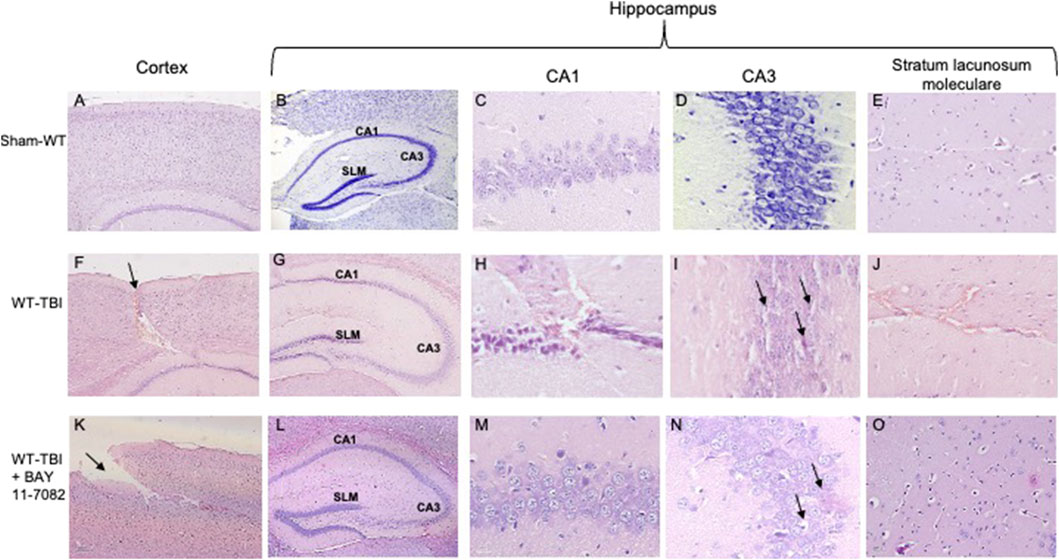
Figure 3. Representative H&E staining of brain tissue from WT animals 24 h following TBI. (A) Cortex of Sham-WT animal. Original magnification X5. (B) Hippocampus of Sham-WT animal. The rectangle indicates the Ca1, Ca3, and stratus lacunosum moleculare areas used for enlargement. Original magnification X5. (C) Ca1 area of Sham-WT animal. Original magnification X40. (D) Ca3 area of Sham-WT animal. Original magnification X40. (E) Stratus lacunosum moleculare of Sham-WT animal. Original magnification X20. (F) Cortex of WT-TBI animal, the arrow indicates the impact point, hemorrhage and edema are markedly visible. Original magnification X5. (G) Hippocampus of WT-TBI animal. The rectangle indicates the Ca1 and Ca3 areas used for enlargement. Original magnification X5. (H) Ca1 area of WT-TBI animal, showing loss of normal architecture, presence of hemorrhage and edema, with shrank neurons. Original magnification X40. (I) Ca3 area of WT-TBI animal, showing diffused neuronal loss and presence of eosinophil neurons (arrows). Original magnification X40. (J) Stratus lacunosum moleculare of WT-TBI animal, showing hemorrhage and edema. Original magnification X20. (K) Cortex of WT-TBI + BAY 11-7082 animal, the arrow indicates the impact point, hemorrhage and edema are markedly visible. Original magnification X5. (L) Hippocampus of WT-TBI + BAY 11-7082 animal. The rectangle indicates the Ca1, Ca3, and stratus lacunosum moleculare areas used for enlargement. Original magnification X5. (M) Ca1 area of WT-TBI + BAY 11-7082 animal, showing a more preserved architecture, without hemorrhage or edema. Original magnification X40. (N) Ca3 area of WT-TBI + BAY 11-7082 animal, showing slight neuronal loss and presence of shrank and eosinophil neurons (arrows). Original magnification X40. (O) Stratus lacunosum moleculare of WT-TBI + BAY 11-7082 animal, showing a more preserved architecture. Original magnification X20.
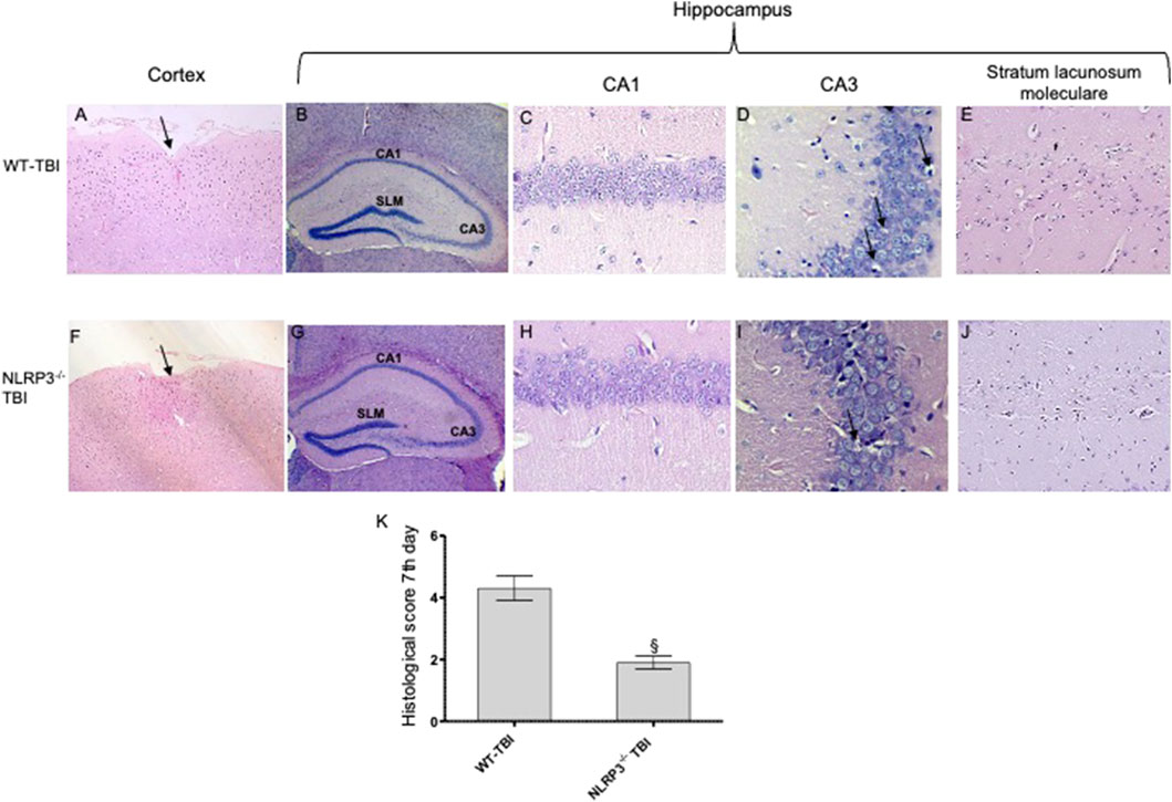
Figure 5. Representative H&E staining of brain tissue from WT and NLRP3−/− mice 7 days following TBI. (A) Cortex of WT-TBI animal, the arrow indicates the impact point, hemorrhage and edema are still markedly visible. Original magnification X5. (B) Hippocampus of WT-TBI animal. The rectangle indicates the Ca1, Ca3, and stratus lacunosum moleculare areas used for enlargement. Original magnification X5. (C) Ca1 area of WT-TBI animal, showing partial restoration of normal architecture with shrank neurons. Original magnification X40. (D) Ca3 area of WT-TBI animal, showing neuronal loss and presence of eosinophil neurons (arrows). Original magnification X40. (E) Stratus lacunosum moleculare of WT-TBI animal, showing partially restored architecture. Original magnification X20. (F) Cortex of NLRP3−/−-TBI animal, the arrow indicates the impact point, hemorrhage and edema are markedly visible. Original magnification X5. (G) Hippocampus of NLRP3−/−-TBI animal. The rectangle indicates the Ca1, Ca3, and stratus lacunosum moleculare areas used for enlargement. Original magnification X5. (H) Ca1 area of NLRP3−/−-TBI animal, showing a preserved architecture, absence of hemorrhage and edema, with few shrank neurons. Original magnification X40. (I) Ca3 area of NLRP3−/−-TBI animal, showing neuronal loss and presence of eosinophil neurons (arrows). Original magnification X40. (J) Stratus lacunosum moleculare of NLRP3−/−-TBI animal, showing an almost normal architecture. Original magnification X20. (K) The graph represents the cumulative histological score evaluated at 7 days from each group of animals. §p < 0.05 vs. WT-TBI. Each bar represents the mean and SD of seven animals.
Author Francesca Cosentino was incorrectly written as Cosentino Francesca.
The original article has been updated.
Generative AI statement
Any alternative text (alt text) provided alongside figures in this article has been generated by Frontiers with the support of artificial intelligence and reasonable efforts have been made to ensure accuracy, including review by the authors wherever possible. If you identify any issues, please contact us.
Publisher’s note
All claims expressed in this article are solely those of the authors and do not necessarily represent those of their affiliated organizations, or those of the publisher, the editors and the reviewers. Any product that may be evaluated in this article, or claim that may be made by its manufacturer, is not guaranteed or endorsed by the publisher.
Keywords: NLRP3 inflammasome, traumatic brain injury, inflammation, cytokines, apoptosis
Citation: Irrera N, Pizzino G, Calò M, Pallio G, Mannino F, Famà F, Arcoraci V, Fodale V, David A, Cosentino F, Minutoli L, Mazzon E, Bramanti P, Squadrito F, Altavilla D and Bitto A (2025) Correction: Lack of the Nlrp3 inflammasome improves mice recovery following traumatic brain injury. Front. Pharmacol. 16:1733683. doi: 10.3389/fphar.2025.1733683
Received: 27 October 2025; Accepted: 03 November 2025;
Published: 26 November 2025.
Edited and reviewed by:
Jacob Raber, Oregon Health and Science University, United StatesCopyright © 2025 Irrera, Pizzino, Calò, Pallio, Mannino, Famà, Arcoraci, Fodale, David, Cosentino, Minutoli, Mazzon, Bramanti, Squadrito, Altavilla and Bitto. This is an open-access article distributed under the terms of the Creative Commons Attribution License (CC BY). The use, distribution or reproduction in other forums is permitted, provided the original author(s) and the copyright owner(s) are credited and that the original publication in this journal is cited, in accordance with accepted academic practice. No use, distribution or reproduction is permitted which does not comply with these terms.
*Correspondence: Francesco Squadrito, ZnNxdWFkcml0b0B1bmltZS5pdA==