 Romelia Pop
 Alexandru-Flaviu Tăbăran
 Iosif Vasiu
 Joshua Milgram
 Ciprian Andrei Ober
- 1Department of Pathology, Faculty of Veterinary Medicine, University of Agricultural Sciences and Veterinary Medicine of Cluj-Napoca, Cluj-Napoca, Romania
- 2Department of Anesthesiology and Surgery, University of Agricultural Sciences and Veterinary Medicine, Cluj-Napoca, Romania
- 3Department of Small Animal Surgery, The Koret School of Veterinary Medicine, The Hebrew University of Jerusalem, Rehovot, Israel
Ossifying fibroma (OF) is a rare, benign fibro-osseous neoplasm that primarily originates from membranous bones. While most frequently documented in equines, OF has also been reported in other species, including dogs, though it remains uncommon. The condition poses significant diagnostic challenges due to its ambiguous presentation, often requiring differentiation from other benign and malignant intraosseous lesions. This case report describes an ossifying fibroma localized to the zygomatic bone in a 9-year-old Hungarian Vizsla. A zygomatic arch ostectomy was successfully performed, and long-term follow-up was excellent. This is only the second documented case of zygomatic localization of OF in a dog, highlighting the rarity of this presentation. The discussion emphasizes the importance of distinguishing OF from other proliferative fibro-osseous lesions, such as fibrous dysplasia (FD) and cemento-osseous dysplasia (COD), and considering the potential for malignancies, such as low-grade osteosarcoma (LG-OSA), to mimic these benign growths. This case contributes valuable insights to the limited veterinary literature on ossifying fibroma, particularly regarding its atypical presentations in canine patients.
1 Introduction
Ossifying fibroma (OF) represents a rare and benign fibro-osseous neoplasm (1) that primarily arises from membranous bones (2). Its infrequent occurrence and ambiguous clinical presentation pose significant diagnostic and therapeutic challenges in veterinary medicine. OF is most commonly found in equines, particularly horses (3–7), but it can also be observed in other species such as dogs (1, 8, 9) and cats (10). Rare instances have been reported in rabbits (11), llamas (12), and canaries (13) (see Table 1). In humans, according to the World Health Organization (WHO), the most frequent localization of ossifying fibroma is in the posterior mandible (14). Similarly, literature on canine cases indicates that the most common sites of ossifying fibroma are the mandible and maxilla (see Figure 1). This case report details the occurrence of ossifying fibroma in a 9-year-old Hungarian Vizsla with zygomatic localization. Through this detailed case study, we aim to contribute to the limited veterinary literature on ossifying fibroma, providing valuable insights for clinicians encountering similar cases in their practice. To the authors' knowledge, this is the second report of an ossifying fibroma with zygomatic bone localization in this species, the first one being described by Best, E. in a Corgi (15).
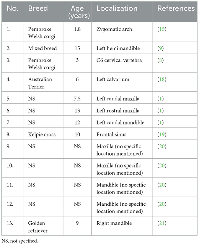
Table 1. Documented cases of ossifying fibroma in dogs: breed, age, and localization.
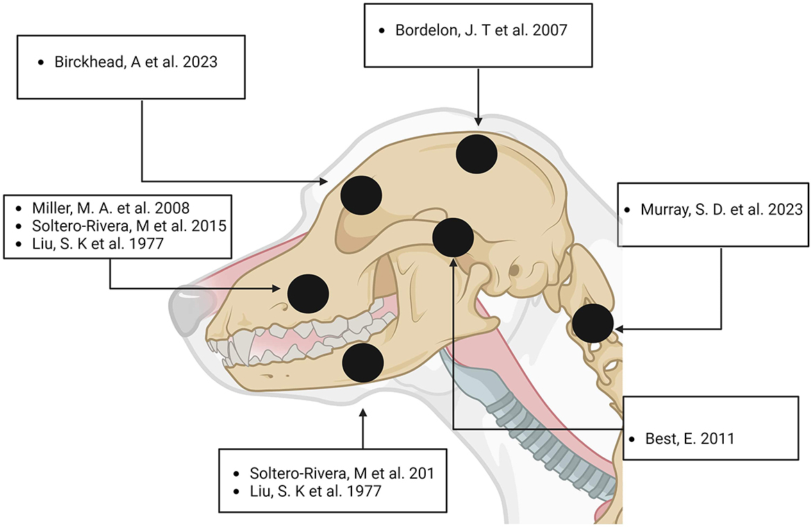
Figure 1. Distribution of Ossifying Fibroma in the Cranio-Cervical Region of Dogs. Created with Biorender.
2 Case description
2.1 Clinical examination
A 9.4-year-old male Hungarian Vizsla was referred to the Department of Surgery and Intensive Care for evaluation of a slow-growing zygomatic arch mass near the right eye, present for 2 years (see Figure 2A). Upon examination, the mass measured ~2.5 cm in diameter, with a multilobulated, firm, non-mobile, and non-painful consistency upon palpation. Occasional signs included blepharospasm, photosensitivity, hyperemic conjunctiva, and purulent epiphora. Non-contrast computed tomography (CT) revealed a focal, round, expansile bone lesion on the right zygomatic arch, measuring 2.0 × 2.3 × 1.5 cm (L, H, W) (see Figure 3). The inner bone structure, nasolacrimal duct, and teeth were unaffected, and no lymphadenopathy was noted. Thoracic CT showed no evidence of metastasis. Given the lesion's slow growth, the owner opted for an excisional biopsy. Cefazolin (22 mg/kg IV) was administered preoperatively. The surgical approach was well documented (16), and the cosmetic outcome was excellent following the zygomatic arch ostectomy. The procedure involved an incision of the skin and temporalis muscle aponeurosis along the dorsal margin of the zygomatic arch. Both cranial and caudal osteotomies were performed with an oscillating saw, preserving the orbital ligament. Histopathological analysis of the excised tissue confirmed the diagnosis of ossifying fibroma. The dog's recovery from surgery and anesthesia was uneventful. Postoperative pain management was maintained using a constant rate infusion (CRI) of lidocaine (20 μg/kg/h) combined with ketamine (10 μg/kg/h) and metamizole (25 mg/kg IV every 8 h). The dog was discharged with robenacoxib (2 mg/kg SC every 24 h). The owner was advised to provide a soft kibble diet, avoid toys or mouth play, and restrict the dog's exercise to short-lead walks for two weeks.
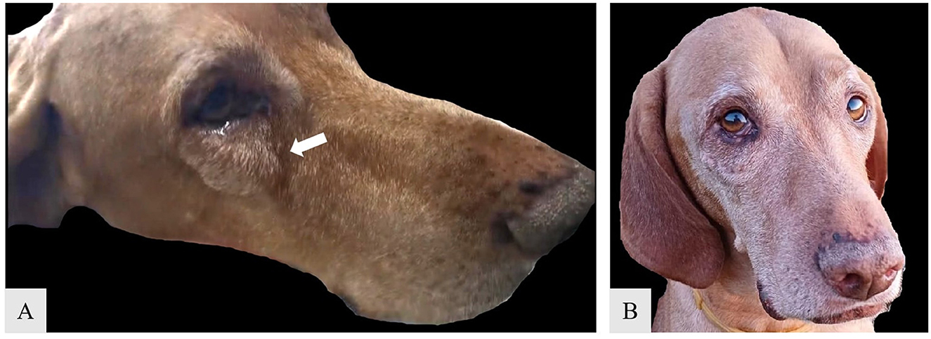
Figure 2. Preoperative appearance of neoplastic growth on the zygomatic arch in male Hungarian Viszla [(A) white arrow]; Ten-months postoperative aspect of the zygomatic area (B).
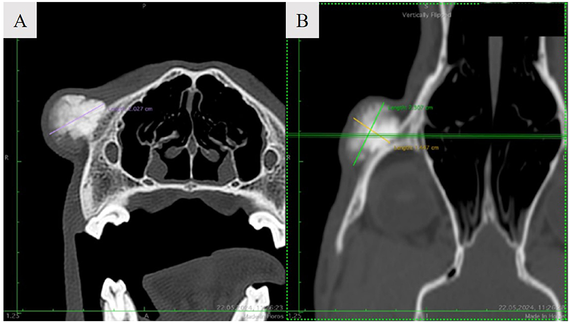
Figure 3. Noncontrast axial (A) and dorsal (B) CT image demonstrating a focal, round, expansile bone cortical lesion of the right zygomatic arch.
2.2 Histopathological examination
For histological analysis, the mass was fixed in 10% neutral buffered formalin (NBF) for 24 h, followed by decalcification in a rapid decalcifier for 5 days. The tissue was then routinely processed for paraffin embedding. Sections, 2 micrometers thick, were cut and stained with hematoxylin and eosin (H&E). Histopathological examination revealed a well-demarcated mass composed of a fibrous component consisting of spindle-shaped fibroblasts arranged in a whorled or storiform pattern, embedded within a collagenous stroma. Interspersed throughout the fibrous stroma were varying amounts of mineralized material, including woven bone, lamellar bone, and cementum-like calcifications. The mineralized component often appeared as trabeculae of osteoid and mature bone, occasionally rimmed by osteoblasts. These trabeculae were typically surrounded by osteoclast-like giant cells involved in bone remodeling. The transition between the fibrous tissue and the mineralized material was gradual, with no signs of anaplasia or atypia. The presence of well-formed bone trabeculae within a cellular fibroblastic stroma is characteristic of ossifying fibroma. Additionally, areas of hemorrhage and cystic degeneration were observed in some cases (see Figure 4).
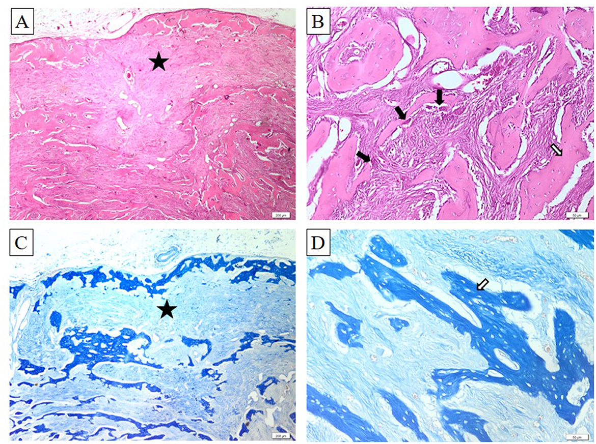
Figure 4. Ossifying fibroma. Bony trabeculae (white arrows) bordered by a single layer of osteoblasts (black arrows) and embedded within abundant fibrous connective tissue (black asterisk). Within this connective tissue, numerous well-differentiated spindle-shaped fibroblasts are dispersed. H&E stain (A, B), Masson's trichrome stain (C, D). Ob. ×4 (A, C) and ob. ×20 (B, D). Barr 50 μm (B, D) and 200 μm (A, C).
2.3 Follow-up
Two weeks after discharge, the incision site showed proper healing, with a good cosmetic outcome. The owner reported normal prehension and behavior. The dog was bright, alert, tolerated food and water well, and was walked daily on a leash. Monthly follow-ups were conducted via telephone, and the owner was instructed to return for in-person evaluations at three-month intervals during the first year post-surgery. At 10 months after the procedure, a clinical examination revealed no signs of tumor recurrence (Figure 2B), and the owner reported no changes in the dog's eating, drinking, or behavior. Twelve months after surgery, the owner was very satisfied with the cosmetic appearance and the comfort of the right eye.
3 Discussion
Ossifying fibroma (OF) is a rare, benign fibro-osseous neoplasm that requires careful differentiation from other benign intraosseous proliferative fibro-osseous lesions (PFOLs). PFOLs are characterized by the replacement of normal bone with a fibrous matrix containing varying degrees of mineralization and ossification (1). In humans, this category includes conditions such as ossifying fibroma (OF), fibrous dysplasia (FD), and cemento-osseous dysplasia (COD) (17). It is important to recognize that some malignant lesions, like low-grade osteosarcoma (LG-OSA), can mimic these benign growths, especially in the skull.
A review of the literature highlights the rarity of ossifying fibroma across species, with most cases documented in equines, particularly horses (3–7). In small animals, reports are limited, although ossifying fibroma has been observed in dogs (1, 8, 9, 18–21) and cats (10). This limited occurrence poses challenges in diagnosis and treatment, particularly when the lesion arises in atypical locations, such as the zygomatic bone, as in the case presented here. The uniqueness of this case—only the second report of such localization in a dog—emphasizes the need for continued documentation and study of these rare cases to enhance understanding and management of OF in veterinary practice.
In veterinary medicine, other potential differential diagnoses for intraosseous lesions, besides true PFOLs like OF and FD, include osteoma, osteitis/osteomyelitis, fibrous osteodystrophy, conventional osteosarcoma (OSA), and multilobular tumor of bone (MLTB). FD is a rare benign condition in which fibrous tissue replaces normal bone, leading to deformities and swelling. It commonly affects young animals, with expansile growth potentially causing decreased bone strength and pathologic fractures, often in the skull and jawbones. Radiographically, fibrous dysplasia typically presents a more homogeneous “ground glass” appearance, lacking the well-defined margins characteristic of ossifying fibroma (17). Cemento-osseous dysplasia (COD) primarily affects the jawbones (maxilla and mandible) and is characterized by a disorganized mixture of fibrous tissue, irregular bone, and cementum-like material (22). However, in our case, the lesion was uniquely located in the zygomatic bone, underscoring the importance of careful diagnostic evaluation to distinguish ossifying fibroma from other intraosseous lesions. Given the rarity of OF in dogs and its variable presentation, this case report contributes valuable insights to the limited veterinary literature. The zygomatic localization presents a unique diagnostic challenge, requiring a comprehensive understanding of potential differential diagnoses. Since the literature primarily documents ossifying fibroma in the mandible and maxilla of dogs (1), this case highlights the importance of considering less common sites when diagnosing PFOLs in small animals. Continued reporting and review of such cases are critical to refining diagnostic and therapeutic approaches for ossifying fibroma and similar lesions in veterinary practice.
Data availability statement
The original contributions presented in the study are included in the article/supplementary material, further inquiries can be directed to the corresponding author.
Ethics statement
The animal studies were approved by University Ethics Committee, The Bioethics Subcommittee for Experiments on Animals, Plants, or Human Subjects. University of Agriculture Science and Veterinary Medicine Cluj-Napoca, Romania. The studies were conducted in accordance with the local legislation and institutional requirements. Written informed consent was obtained from the owners for the participation of their animals in this study.
Author contributions
RP: Conceptualization, Methodology, Writing – original draft. A-FT: Investigation, Supervision, Validation, Visualization, Writing – review & editing. IV: Conceptualization, Methodology, Writing – original draft. JM: Methodology, Supervision, Validation, Writing – review & editing. CO: Investigation, Supervision, Validation, Writing – review & editing.
Funding
The author(s) declare financial support was received for the research, authorship, and/or publication of this article. The University of Agriculture Science and Veterinary Medicine Cluj-Napoca, Romania.
Conflict of interest
The authors declare that the research was conducted in the absence of any commercial or financial relationships that could be construed as a potential conflict of interest.
Publisher's note
All claims expressed in this article are solely those of the authors and do not necessarily represent those of their affiliated organizations, or those of the publisher, the editors and the reviewers. Any product that may be evaluated in this article, or claim that may be made by its manufacturer, is not guaranteed or endorsed by the publisher.
References
1. Soltero-Rivera M, Engiles JB, Reiter AM, Reetz J, Lewis JR, Sánchez MD. Benign and malignant proliferative fibro-osseous and osseous lesions of the oral cavity of dogs. Vet Pathol. (2015) 52:894–902. doi: 10.1177/0300985815583096
2. Thompson KG, Dittmer KE. Tumors of Bone. In:Meuten DJ, , editors. Tumors in Domestic Animals. Hoboken, NJ: John Wiley & Sons, Inc (2016). p. 356–424. doi: 10.1002/9781119181200.ch10
3. Crijns CP, Vlaminck L, Verschooten F, van Bergen T, De Cock HE, Huylebroek F, et al. Multiple mandibular ossifying fibromas in a yearling B elgian D raught horse filly. Equine Vet Educ. (2015) 27:11–5. doi: 10.1111/eve.12246
4. Puff C, Ohnesorge B, Wagels R, Baumgärtner W. An unusual mucinous osteoma with features of an ossifying fibroma in the nasal cavity of a horse. J Comp Pathol. (2006) 135:52–5. doi: 10.1016/j.jcpa.2006.02.008
5. Kodaira K, Muranaka M, Naito Y, Ode H, Oku K, Nukada T, et al. Histopathological characteristics of an ossifying fibroma formed in the maxilla of a racehorse. J Equine Sci. (2010) 21:7–10. doi: 10.1294/jes.21.7
6. Turek B, Górski K, Drewnowska O, Buczkowska R, Kozłowska N, Sapierzyński R. Ossifying fibroma in the nasal cavity of a 2-year-old horse. Animals. (2021) 11:317. doi: 10.3390/ani11020317
7. Morse CC, Saik JE, Richardson DW, Fetter AW. Equine juvenile mandibular ossifying fibroma. Vet Pathol. (1988) 25:415–21. doi: 10.1177/030098588802500603
8. Murray SD, Cameron S, Tolliver SE, Cole CY, Aschenbroich SA. Ossifying fibroma in the cervical vertebra of a dog. Can Vet J. (2023) 64:367–71.
9. Miller MA, Towle HAM, Heng HG, Greenberg CB, Pool RR. Mandibular ossifying fibroma in a dog. Vet Pathol. (2008) 45:203–6. doi: 10.1354/vp.45-2-203
10. Turrel JM, Pool RR. Primary bone tumors in the cat: a retrospective study of 15 cats and a literature review. Vet Radiol. (1982) 23:152–66. doi: 10.1111/j.1740-8261.1982.tb01099.x
11. Whitten KA, Popielarczyk MM, Belote DA, McLeod GC, Mense MG. Ossifying fibroma in a miniature rex rabbit (Oryctolagus cuniculus). Vet Pathol. (2006) 43:62–4. doi: 10.1354/vp.43-1-62
12. McCauley CT, Campbell GA, Cummings CA, Drost WT. Ossifying fibroma in a llama. J Vet Diagn Invest. (2000) 12:473–6. doi: 10.1177/104063870001200517
13. Razmyar J, Dezfoulian O, Peighambari SM. Ossifying fibroma in a canary (Serinus canaria). J Avian Med Surg. (2008) 22:320–2. doi: 10.1647/2007-027.1
14. Thompson LDR. World Health Organization classification of tumours: pathology and genetics of head and neck tumours. Ear Nose Throat J. (2006) 85:74. doi: 10.1177/014556130608500201
15. Best E. Ossifying fibroma of the zygomatic arch in a 20-month-old male corgi. (2011). Available at: https://www.vettimes.co.uk/app/uploads/wp-post-to-pdf-enhanced-cache/1/ossifying fibroma of the zygomatic arch in a 20 month old male corgi.pdf (accessed December 1, 2024).
16. Dörner J, Oberbacher S, Dupré G. Comparison of three surgical approaches for zygomatic sialoadenectomy in canine cadavers. Vet Surg. (2021) 50:564–70. doi: 10.1111/vsu.13589
17. Alawi F. Benign fibro-osseous diseases of the maxillofacial bones: a review and differential diagnosis. Pathol Patterns Rev. (2002) 118:S50–70. doi: 10.1309/NUXA-JUT9-HA09-WKMV
18. Bordelon JT, Rochat MC. Use of a titanium mesh for cranioplasty following radical rostrotentorial craniectomy to remove an ossifying fibroma in a dog. J Am Vet Med Assoc. (2007) 231:1692–5. doi: 10.2460/javma.231.11.1692
19. Birckhead A, Carstens A, Geiss E, Yap F, Huizing X. Computed tomographic characteristics of frontal sinus ossifying fibroma in a dog. Vet Radiol. Ultrasound. (2023) 64:E60–3. doi: 10.1111/vru.13285
20. Liu S, Dorfman HD, Hurvitz AI, Patnaik AK. Primary and secondary bone tumours in the dog. J Small Anim Pract. (1977) 18:313–26. doi: 10.1111/j.1748-5827.1977.tb05890.x
21. Speltz MC, Pool RR, Hayden DW. Pathology in practice. J Am Vet Med Assoc. (2009) 235:1283–5. doi: 10.2460/javma.235.11.1283
Keywords: fibroma, ossifying, zygomatic, radiology, histopathology, dog
Citation: Pop R, Tăbăran A-F, Vasiu I, Milgram J and Ober CA (2025) Surgical management of ossifying fibroma in a 9-year-old Hungarian Vizsla: a case report and review of the literature. Front. Vet. Sci. 11:1497077. doi: 10.3389/fvets.2024.1497077
Received: 16 September 2024; Accepted: 04 December 2024;
Published: 10 January 2025.
Edited by:
Leonardo Leonardi, University of Perugia, ItalyReviewed by:
Robert Michael Baratt, Salem Valley Veterinary Clinic, United StatesAurelian-Sorin Pasca, Iasi University of Life Sciences (IULS), Romania
Copyright © 2025 Pop, Tăbăran, Vasiu, Milgram and Ober. This is an open-access article distributed under the terms of the Creative Commons Attribution License (CC BY). The use, distribution or reproduction in other forums is permitted, provided the original author(s) and the copyright owner(s) are credited and that the original publication in this journal is cited, in accordance with accepted academic practice. No use, distribution or reproduction is permitted which does not comply with these terms.
*Correspondence: Alexandru-Flaviu Tăbăran, YWxleGFuZHJ1LnRhYmFyYW5AdXNhbXZjbHVqLnJv