You're viewing our updated article page. If you need more time to adjust, you can return to the old layout.

CORRECTION article

Front. Vet. Sci., 10 January 2025

Sec. Veterinary Clinical, Anatomical, and Comparative Pathology

Volume 11 - 2024 | https://doi.org/10.3389/fvets.2024.1533729

Corrigendum: Dogs with acute myeloid leukemia have clonal rearrangements in T and B cell receptors

  • 1. Department of Population Medicine and Diagnostic Sciences, College of Veterinary Medicine, Cornell University, Ithaca, NY, United States

  • 2. Animal Health Diagnostic Center, College of Veterinary Medicine, Cornell University, Ithaca, NY, United States

Article metrics

View details

797

Views

280

Downloads

In the published article, there was an error in Figure 1, Table 1, Table 2, Table 3, and Table 6 as published. CD1c in the Figures and Tables should be CD1a. The authors discovered this error after publication of the article when the source of the antibody sent a notification stating that the antigen was not CD1c as originally indicated. The corrected Figure and Tables and their captions appear below.
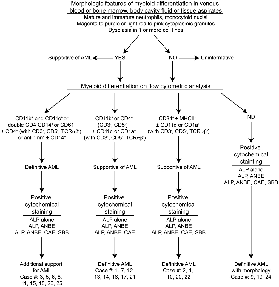
Figure 1

Figure 1

Algorithm used to diagnose acute myeloid leukemia (AML) in the 25 dogs of this study. This diagnostic algorithm was based on the order in which tests were generally performed in our laboratory, i.e., morphologic assessment of blood, bone marrow, or body cavity fluid or tissue aspirates, followed by flow cytometric analysis (performed routinely twice a week), followed by cytochemical staining (performed as needed). After completion of all the tests, the results were reevaluated, and a diagnosis of AML was based on the combined data. The path used to diagnose each case (#) is also shown. More details on the criteria are provided in Table 1.
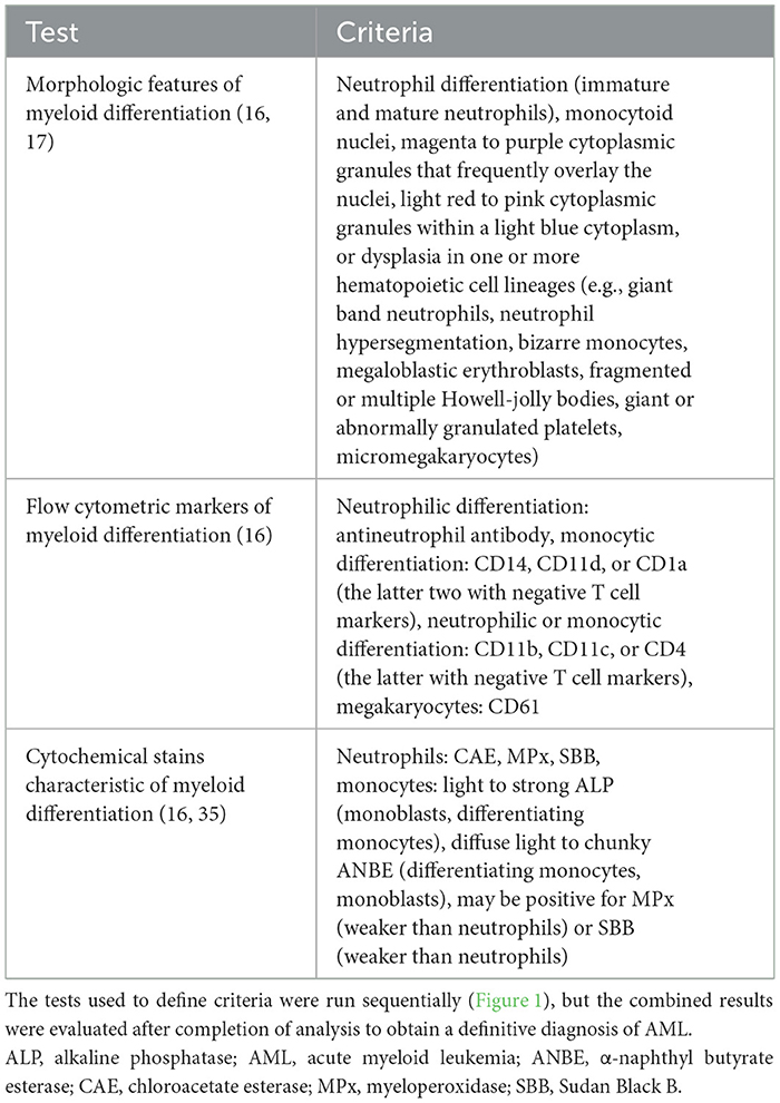
Table 1

Test Criteria
Morphologic features of myeloid differentiation (16, 17) Neutrophil differentiation (immature and mature neutrophils), monocytoid nuclei, magenta to purple cytoplasmic granules that frequently overlay the nuclei, light red to pink cytoplasmic granules within a light blue cytoplasm, or dysplasia in one or more hematopoietic cell lineages (e.g., giant band neutrophils, neutrophil hypersegmentation, bizarre monocytes, megaloblastic erythroblasts, fragmented or multiple Howell-jolly bodies, giant or abnormally granulated platelets, micromegakaryocytes)
Flow cytometric markers of myeloid differentiation (16) Neutrophilic differentiation: antineutrophil antibody, monocytic differentiation: CD14, CD11d, or CD1a (the latter two with negative T cell markers), neutrophilic or monocytic differentiation: CD11b, CD11c, or CD4 (the latter with negative T cell markers), megakaryocytes: CD61
Cytochemical stains characteristic of myeloid differentiation (16, 35) Neutrophils: CAE, MPx, SBB, monocytes: light to strong ALP (monoblasts, differentiating monocytes), diffuse light to chunky ANBE (differentiating monocytes, monoblasts), may be positive for MPx (weaker than neutrophils) or SBB (weaker than neutrophils)

Criteria used to support myeloid lineage of leukemia in 25 dogs.
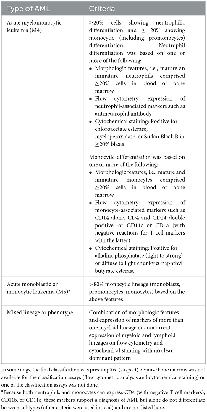
The tests used to define criteria were run sequentially (Figure 1), but the combined results were evaluated after completion of analysis to obtain a definitive diagnosis of AML.

ALP, alkaline phosphatase; AML, acute myeloid leukemia; ANBE, α-naphthyl butyrate esterase; CAE, chloroacetate esterase; MPx, myeloperoxidase; SBB, Sudan Black B.

Table 2

Type of AML Criteria
Acute myelomonocytic leukemia (M4) ≥20% cells showing neutrophilic differentiation and ≥ 20% showing monocytic (including promonocytes) differentiation. Neutrophil differentiation was based on one or more of the following:
• Morphologic features, i.e., mature an immature neutrophils comprised ≥20% cells in blood or bone marrow
• Flow cytometry: expression of neutrophil-associated markers such as antineutrophil antibody
• Cytochemical staining: Positive for chloroacetate esterase, myeloperoxidase, or Sudan Black B in ≥20% blasts
Monocytic differentiation was based on one or more of the following:
• Morphologic features, i.e., mature and immature monocytes comprised ≥20% cells in blood or bone marrow
• Flow cytometry: expression of monocyte-associated markers such as CD14 alone, CD4 and CD14 double positive, or CD11c or CD1a (with negative reactions for T cell markers with the latter)
• Cytochemical staining: Positive for alkaline phosphatase (light to strong) or diffuse to light chunky α-naphthyl butyrate esterase
Acute monoblastic or monocytic leukemia (M5)a >80% monocytic lineage (monoblasts, promonocytes, monocytes) based on the above features
Mixed lineage or phenotype Combination of morphologic features and expression of markers of more than one myeloid lineage or concurrent expression of myeloid and lymphoid lineages on flow cytometry and cytochemical staining with no clear dominant pattern

Criteria for classification of the subtype of acute myeloid leukemia (AML) based on the World Health Organization scheme – not otherwise specified (17, 18).
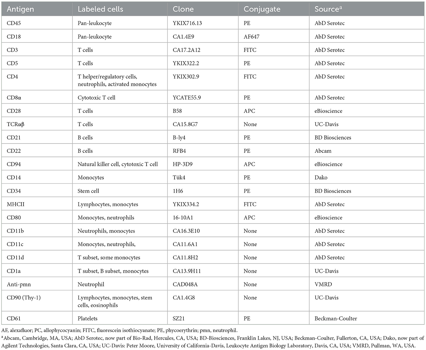
In some dogs, the final classification was presumptive (suspect) because bone marrow was not available for the classification assays (flow cytometric analysis and cytochemical staining) or one of the classification assays was not done.

aBecause both neutrophils and monocytes can express CD4 (with negative T cell markers), CD11b, or CD11c, these markers support a diagnosis of AML but alone do not differentiate between subtypes (other criteria were used instead) and are not listed here.

Table 3

Antigen Labeled cells Clone Conjugate Sourcea
CD45 Pan-leukocyte YKIX716.13 PE AbD Serotec
CD18 Pan-leukocyte CA1.4E9 AF647 AbD Serotec
CD3 T cells CA17.2A12 FITC AbD Serotec
CD5 T cells YKIX322.2 PE AbD Serotec
CD4 T helper/regulatory cells, neutrophils, activated monocytes YKIX302.9 FITC AbD Serotec
CD8α Cytotoxic T cell YCATE55.9 PE AbD Serotec
CD28 T cells B58 APC eBioscience
TCRαβ T cells CA15.8G7 None UC-Davis
CD21 B cells B-ly4 PE BD Biosciences
CD22 B cells RFB4 PE Abcam
CD94 Natural killer cell, cytotoxic T cell HP-3D9 APC eBioscience
CD14 Monocytes Tük4 PE Dako
CD34 Stem cell 1H6 PE BD Biosciences
MHCII Lymphocytes, monocytes YKIX334.2 FITC AbD Serotec
CD80 Monocytes, neutrophils 16-10A1 APC eBioscience
CD11b Neutrophils, monocytes CA16.3E10 None AbD Serotec
CD11c Monocytes, neutrophils, CA11.6A1 None AbD Serotec
CD11d T subset, some monocytes CA11.8H2 None AbD Serotec
CD1a T subset, B subset, monocytes CA13.9H11 None UC-Davis
Anti-pmn Neutrophil CAD048A None VMRD
CD90 (Thy-1) Lymphocytes, monocytes, stem cells, eosinophils CA1.4G8 None UC-Davis
CD61 Platelets SZ21 PE Beckman-Coulter

Antibodies used in flow cytometry at Cornell University to label antigens on tumor cells in liquid samples (blood or bone marrow, body cavity fluid, or tissue aspirates) from dogs with acute myeloid leukemia.
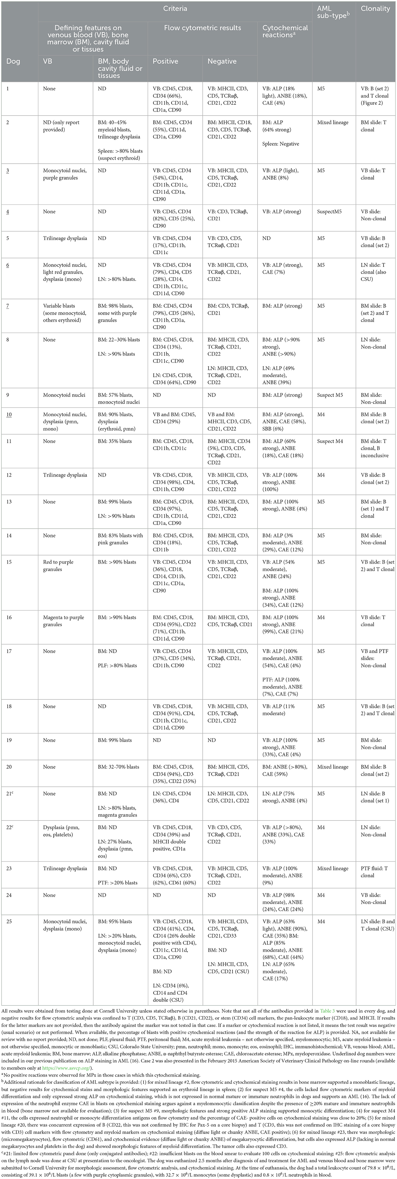
AF, alexafluor; PC, allophycocyanin; FITC, fluorescein isothiocyanate; PE, phycoerythrin; pmn, neutrophil.

aAbcam, Cambridge, MA, USA; AbD Serotec, now part of Bio-Rad, Hercules, CA, USA; BD-Biosciences, Franklin Lakes, NJ, USA; Beckman-Coulter, Fullerton, CA, USA; Dako, now part of Agilent Technologies, Santa Clara, CA, USA; UC-Davis: Peter Moore, University of California-Davis, Leukocyte Antigen Biology Laboratory, Davis, CA, USA; VMRD, Pullman, WA, USA.

Table 6

Criteria AML sub-typeb Clonality
Defining features on venous blood (VB), bone marrow (BM), cavity fluid or tissues Flow cytometric results Cytochemical reactions a
Dog VB BM, body cavity fluid or tissues Positive Negative
1 None ND VB: CD45, CD18, CD34 (66%), CD11b, CD11d, CD1a, CD90 VB: MHCII, CD3, CD5, TCRαβ, CD21, CD22 VB: ALP (18% light), ANBE (18%), CAE (4%) M5 VB: B (set 2) and T clonal (Figure 2)
2 ND (only report provided) BM: 40–45% myeloid blasts, trilineage dysplasia Spleen: >80% blasts (suspect erythroid) BM: CD45, CD34 (55%), CD11d, CD1a, CD90 BM: MHCII, CD18, CD3, CD5, TCRαβ, CD21, CD22 BM: ALP (64% strong) Spleen: Negative Mixed lineage BM slide: T clonal
3 Monocytoid nuclei, purple granules ND VB: CD45, CD34 (54%), CD14, CD11b, CD11c, CD11d, CD1a, CD90 VB: MHCII, CD3, CD5, TCRαβ, CD21, CD22 VB: ALP (light), ANBE (8%) M5 VB slide: T clonal
4 None ND VB: CD45, CD34 (82%), CD5 (25%), CD90 VB: CD3, TCRαβ, CD21 VB: ALP (strong) SuspectM5 VB slide: Non-clonal
5 Trilineage dysplasia ND VB: CD45, CD34 (17%), CD11b, CD11c VB: CD3, CD5, TCRαβ, CD21 ND M5 VB slide: B clonal (set 2)
6 Monocytoid nuclei, light red granules, dysplasia (mono) ND LN: >80% blasts. VB: CD45, CD34 (79%), CD4, CD5 (28%), CD14, CD11b, CD11c, CD11d, CD90 VB: MHCII, CD3, TCRαβ, CD21, CD22 VB: ALP (strong), CAE (7%) M5 LN slide: T clonal (also CSU)
7 Variable blasts (some monocytoid, others erythroid) BM: 98% blasts, some with purple granules BM: CD45, CD34 (79%), CD5 (26%), CD11b, CD1a, CD90 BM: CD3, TCRαβ, CD21 BM: ALP (strong) M5 BM slide: B (set 2) and T clonal
8 None BM: 22–30% blasts LN: >90% blasts BM: CD45, CD18, CD34 (13%), CD11b, CD11c, CD90 LN: CD45, CD18, CD34 (64%), CD90 BM: MHCII, CD3, TCRαβ, CD21, CD22 LN: MHCII, CD3, TCRαβ, CD21, CD22 BM: ALP (>90% strong), ANBE (>90%) LN: ALP (49% moderate), ANBE (39%) M5 LN slide: Non-clonal
9 Monocytoid nuclei BM: 57% blasts, monocytoid nuclei ND ND BM: ALP (strong) Suspect M5 BM slide: Non-clonal
10 Monocytoid nuclei, dysplasia (pmn, mono) BM: 90% blasts, dysplasia (erythroid, pmn) VB and BM: CD45, CD34 (29%) VB and BM: MHCII, CD3, CD5, CD21, CD22 BM: ALP (strong), ANBE, CAE (58%), SBB (6%) M4 BM slide: B clonal (set 2)
11 None BM: 35% blasts BM: CD45, CD18, CD11b, CD11c BM: MHCII, CD34 (5%), CD3, CD5, TCRαβ, CD21, CD22 BM: ALP (60% strong), ANBE (18%), CAE (18%) Suspect M4 BM slide: T clonal, B inconclusive
12 Trilineage dysplasia ND VB: CD45, CD18, CD34 (98%), CD4, CD11b, CD90 VB: MHCII, CD3, CD5, TCRαβ, CD21, CD22 VB: ALP (100% strong), ANBE (100%) M4 VB slide: B clonal (set 2)
13 None BM: 99% blasts LN: >90% blasts BM: CD45, CD18, CD34 (97%), CD11b, CD11d, CD1a, CD90 BM: MHCII, CD3, CD5, TCRαβ, CD21, CD22 BM: ALP (100% strong), ANBE (4%) M5 BM slide: B (set 1) and T clonal
14 None BM: 83% blasts with pink granules BM: CD45, CD18, CD34 (18%), CD11b BM: MHCII, CD3, CD5, TCRαβ, CD21, CD22 BM: ALP (3% moderate), ANBE (29%), CAE (12%) M5 BM slide: Non-clonal
15 Red to purple granules BM: >90% blasts VB: CD45, CD34 (36%), CD18, CD14, CD11b, CD11c, CD1a, CD90 VB: MHCII, CD3, CD5, TCRαβ, CD21, CD22 VB: ALP (54% moderate), ANBE (24%) BM: ALP (100% strong), ANBE (34%), CAE (12%) M5 VB slide: B (set 2) and T clonal
16 Magenta to purple granules BM: >90% blasts BM: CD45, CD18, CD34 (95%), CD22 (71%), CD11b, CD11d, CD90 BM: MHCII, CD3, CD5, TCRαβ, CD21 BM: ALP (100% strong), ANBE (99%), CAE (21%) M4 VB slide: T clonal
17 None BM: ND PLF: >80% blasts VB: CD45, CD34 (37%), CD5 (34%), CD11b, CD90 VB: MHCII, CD3, TCRαβ, CD21, CD22 VB: ALP (100% moderate), ANBE (54%), CAE (4%) PTF: ALP (100% moderate), ANBE (7%), CAE (7%) M5 VB and PTF slides: Non-clonal
18 None ND VB: CD45, CD18, CD34 (91%), CD4, CD11b, CD11c, CD11d, CD90 VB: MCHII, CD3, CD5, TCRαβ, CD21, CD22 VB: ALP (11% moderate) M5 VB slide: B (set 2) and T clonal
19 None BM: 99% blasts ND ND VB: ALP (100% strong), ANBE (33%), CAE (4%) M5 BM slide: Non-clonal
20 None BM: 32-70% blasts BM: CD45, CD18, CD34 (94%), CD3 (35%), CD22 (35%) BM: MHCII, CD5, TCRαβ, CD21 BM: ANBE (>80%), CAE (59%) Mixed lineage BM slide: B clonal (set 2)
21c None BM: ND LN: >80% blasts, magenta granules LN: CD45, CD34 (36%), CD4 LN: MHCII, CD3, CD5, CD21, CD22 LN: ALP (75% strong), ANBE (4%) M5 LN slide: B clonal (set 1)
22c Dysplasia (pmn, eos, platelets) BM: ND LN: 27% blasts, dysplasia (pmn, eos) VB: CD45, CD18, CD34 (39%) and MHCII double positive, CD1a VB: CD3, CD5, TCRαβ, CD21, CD22 VB: ALP (>80%), ANBE (33%), CAE (33%) M4 LN slide: Non-clonal
23 Trilineage dysplasia BM: ND PTF: >20% blasts VB: CD45, CD18, CD34 (6%), CD3 (62%), CD61 (60%) VB: MHCII, CD5, TCRαβ, CD21, CD22 VB: ALP (100% moderate), ANBE (9%) Mixed lineage PTF fluid: T clonal
24 None ND ND ND VB: ALP (98% moderate), ANBE (24%), CAE (24%) M4 VB slide: Non-clonal
25 Monocytoid nuclei, dysplasia (mono) BM: 95% blasts LN: >20% blasts, monocytoid nuclei, dysplasia (mono) VB: CD45, CD18, CD34 (41%), CD4, CD14 (26% double positive with CD4), CD11c, CD11d, CD1a, CD90 BM: ND LN: CD34 (6%), CD14 and CD4 double (CSU) VB: MHCII, CD3, CD5, TCRαβ, CD21, CD33 BM: ND LN: MHCII, CD3, CD5, CD21 (CSU) VB: ALP (63% light), ANBE (90%), CAE (35%) BM: ALP (85% moderate), ANBE (68%), CAE (44%) LN: ALP (65% moderate), CAE (17%) M4 LN slide: B and T clonal (CSU)

Morphologic findings from blood or cytologic smears and results from flow cytometric labeling, cytochemical staining, and clonality testing in 25 dogs with AML.

All results were obtained from testing done at Cornell University unless stated otherwise in parentheses. Note that not all of the antibodies provided in Table 3 were used in every dog, and negative results for flow cytometric analysis was confined to T (CD3, CD5, TCRαβ), B (CD21, CD22), or stem (CD34) cell markers, the pan-leukocyte marker (CD18), and MHCII. If results for the latter markers are not provided, then the antibody against the marker was not tested in that case. If a marker or cytochemical reaction is not listed, it means the test result was negative (usual scenario) or not performed. When available, the percentage of blasts with positive cytochemical reactions (and the strength of the reaction for ALP) is provided. NA, not available for review with no report provided; ND, not done; PLF, pleural fluid; PTF, peritoneal fluid; M4, acute myeloid leukemia – not otherwise specified, myelomonocytic; M5, acute myeloid leukemia – not otherwise specified, monocytic or monoblastic; CSU, Colorado State University; pmn, neutrophil; mono, monocyte; eos, eosinophil; IHC, immunohistochemical; VB, venous blood; AML, acute myeloid leukemia; BM, bone marrow; ALP, alkaline phosphatase; ANBE, α-naphthyl butyrate esterase; CAE, chloroacetate esterase; MPx, myeloperoxidase. Underlined dog numbers were included in our previous publication on ALP staining in AML (16). Case 2 was also presented in the February 2015 American Society of Veterinary Clinical Pathology on-line rounds (available to members only at https://www.asvcp.org/).

aNo positive reactions were observed for MPx in those cases in which this cytochemical staining.

bAdditional rationale for classification of AML subtype is provided: (1) for mixed lineage #2, flow cytometric and cytochemical staining results in bone marrow supported a monoblastic lineage, but negative results for cytochemical stains and morphologic features supported an erythroid lineage in spleen; (2) for suspect M5 #4, the cells lacked flow cytometric markers of myeloid differentiation and only expressed strong ALP on cytochemical staining, which is not expressed in normal mature or immature neutrophils in dogs and supports an AML (16). The lack of expression of the neutrophil enzyme CAE in blasts on cytochemical staining argues against a myelomonocytic classification despite the presence of ≥20% mature and immature neutrophils in blood (bone marrow not available for evaluation); (3) for suspect M5 #9, morphologic features and strong positive ALP staining supported monocytic differentiation; (4) for suspect M4 #11, the cells expressed neutrophil or monocyte differentiation antigens on flow cytometry and the percentage of CAE- positive cells on cytochemical staining was close to 20%; (5) for mixed lineage #20, there was concurrent expression of B (CD22, this was not confirmed by IHC for Pax-5 on a core biopsy) and T (CD3, this was not confirmed on IHC staining of a core biopsy with CD3) cell markers with flow cytometry and myeloid markers on cytochemical staining (diffuse light or chunky ANBE, CAE positive); (6) for mixed lineage #23, there was morphologic (micromegakaryocytes), flow cytometric (CD61), and cytochemical evidence (diffuse light or chunky ANBE) of megakaryocytic differentiation, but cells also expressed ALP (lacking in normal megakaryocytes and platelets in the dog) and showed morphologic features of myeloid differentiation. The tumor cells also expressed CD3.

c#21: limited flow cytometric panel done (only conjugated antibodies); #22: insufficient blasts on the blood smear to evaluate 100 cells on cytochemical staining; #25: flow cytometric analysis on the lymph node was done at CSU at presentation to the oncologist. The dog was euthanized 2.5 months after diagnosis of and treatment for AML and venous blood and bone marrow were submitted to Cornell University for morphologic assessment, flow cytometric analysis, and cytochemical staining. At the time of euthanasia, the dog had a total leukocyte count of 79.8 × 10⁶/L, consisting of 39.1 × 10⁶/L blasts (a few with purple cytoplasmic granules), with 32.7 × 10⁶/L monocytes (some dysplastic) and 0.8 × 10⁶/L neutrophils in blood.

The authors apologize for this error and state that this does not change the scientific conclusions of the article in any way. The original article has been updated.

Statements

Publisher’s note

All claims expressed in this article are solely those of the authors and do not necessarily represent those of their affiliated organizations, or those of the publisher, the editors and the reviewers. Any product that may be evaluated in this article, or claim that may be made by its manufacturer, is not guaranteed or endorsed by the publisher.

Summary

Keywords

acute myelogenous leukemia, canine, polymerase testing for antigen receptor rearrangements, clonality testing, phenotyping, leukemia, flow cytometry, cytochemistry

Citation

Stokol T, Nickerson GA, Shuman M and Belcher N (2025) Corrigendum: Dogs with acute myeloid leukemia have clonal rearrangements in T and B cell receptors. Front. Vet. Sci. 11:1533729. doi: 10.3389/fvets.2024.1533729

Received

24 November 2024

Accepted

03 December 2024

Published

10 January 2025

Volume

11 - 2024

Edited and reviewed by

Stefano Comazzi, University of Milan, Italy

Updates

Copyright

*Correspondence: Tracy Stokol

Disclaimer

All claims expressed in this article are solely those of the authors and do not necessarily represent those of their affiliated organizations, or those of the publisher, the editors and the reviewers. Any product that may be evaluated in this article or claim that may be made by its manufacturer is not guaranteed or endorsed by the publisher.

Outline

Figures

Cite article

Copy to clipboard


Export citation file


Share article

Article metrics