 Helder Camilo da Silva Pereira1Wesley Marinho Brandão Barbosa2
 Mariana Ferreira Venceslau1Ricardo Romão Guerra3
 Lucas Rannier Ribeiro Antonino Carvalho4*
- 1Department of Veterinary Science, IESP Centro Universitário, João Pessoa, Brazil
- 2Department of Veterinary Medicine, UNIPÊ Centro Universitário de João Pessoa, João Pessoa, Brazil
- 3Postgraduate Program in Animal Science (PPGCAn), Federal University of Paraíba, Areia, Brazil
- 4Department of Physiology and Pharmacology, Karolinska Institutet, Stockholm, Sweden
Introduction: Cannabis intoxication is increasingly reported in small animal practice, often resulting from accidental ingestion of human products or unsupervised access to the plant. Although clinical signs are well documented, laboratory alterations remain poorly described. This study aimed to confirm cannabis intoxication in dogs using a rapid urinary test for THC metabolites and to investigate associated hematological, biochemical, and urinary alterations.
Methods: In this study, nine dogs that were admitted to the emergency veterinary clinic in João Pessoa, Brazil, between 2021 and 2022, with suspected cannabis ingestion and owner confirmation, were evaluated. The animals were included regardless of breed, sex or age, and the results compared with healthy animals (control). Clinical signs were treated supportively. Urine and blood samples were collected 6 h after admission. Dogs were classified as “positive” or “negative/control” based on chromatographic detection of THC metabolites (PoCT COC/THC Assure Tech, Westlake Eco Zone/China; ANVISA 80885650015). Hematological analysis evaluated anemia, leukocyte, and platelet counts; serum biochemistry focused on hepatic and renal markers; urinalysis assessed color, clarity, specific gravity, and pH.
Results: No statistically significant differences were observed between intoxicated and non-intoxicated dogs in any of the laboratory parameters. Additionally, most animals showed only discrete or absent clinical signs.
Discussion: These findings suggest that accidental cannabis ingestion in dogs tends to result in mild physiological effects and minimal laboratory alterations, indicating a low risk of severe toxicity. Nonetheless, accurate identification of exposure using diagnostic tools is essential for appropriate case management. A deeper understanding of the endocannabinoid system in companion animals is key to improving clinical decision-making and assessing potential risks associated with cannabis exposure.
Introduction
Intoxications are commonly observed in small animal clinical practice (1). These events often result from the accidental ingestion of toxic plants, the consumption of foods inappropriate for the species, or the incorrect administration of medications without veterinary guidance (2). Cannabis is a shrub native to Asia, belonging to the family Cannabaceae. It contains variable amounts of flavonoids, terpenes, and cannabinoids, with the latter being the main intoxicating active compounds when ingested beyond the animal's metabolic capacity (3).
In recent years, Cannabis sp. has emerged as a relevant toxic agent in domestic environments, particularly in regions where its recreational or medicinal use has been legalized. A study conducted in the United States evaluated trends in marijuana toxicosis in dogs between 2005 and 2010 in a state with legalized medical cannabis, reporting a fourfold increase in cases during the study period, totaling more than 120 reports (4). In Colombia (2023–2024), 113 cases of marijuana intoxication in dogs were documented, representing approximately 12% of all veterinary toxicology cases. In Brazil, a previous study reported that 3.3% of recorded intoxications cases in dogs were associated with cannabis ingestion (5). These findings highlight the growing relevance of cannabis intoxication in veterinary practice and emphasize the need for prompt diagnosis and treatment.
Because of their exploratory behavior, dogs are the species most frequently affected by accidental cannabis ingestion (6, 19, 20). In such cases, clinical management is improved when the causative agent is confirmed (7). Dogs intoxicated with cannabis may exhibit a variety of clinical signs, most commonly lethargy, ataxia, urinary incontinence, hypersalivation, and mydriasis (4), as well as vomiting (8). Other reported symptoms include disorientation, vocalization and bradycardia. However, these changes are generally mild and inconsistent among cases. This variability in both clinical presentation and laboratory results underscores the importance of combining clinical evaluation with confirmatory diagnostic tests—such as the rapid detection of urinary THC metabolites—to ensure accurate diagnosis and appropriate therapeutic decision-making (4).
While confirming the identity of the ingested agent in suspected cases on cannabis intoxication in dogs can provide certainty for clients, laboratory confirmation is not typically required for an adequate diagnosis to be made, nor will it alter clinical decision-making or treatment strategies. Laboratory confirmation of marijuana intoxication is often costly and time-consuming, requiring specialized expertise and equipment (9). However, the use of point-of-care (POC) tests has become increasingly feasible due to their low cost, ease of use, and rapid turnaround time. These tests enable timely clinical decisions and allow veterinarians to implement targeted therapeutic approaches (4).
The present study aimed to detect the presence of Cannabis metabolites in the urine of dogs using a rapid diagnostic test designed for the detection of human THC metabolites and to compare hematological, clinical biochemical, and urinalysis findings between cannabis-positive and control (cannabis-negative) dogs, as well as with established laboratory reference values. This approach facilitates a better understanding of the physiological effects of accidental cannabis ingestion in dogs and evaluates the diagnostic utility of human rapid urinary THC metabolite tests in veterinary emergency situations.
Methods
This was a prospective observational study conducted in João Pessoa, in the Northeast region of Brazil, which analyzed cases of accidental ingestion of cannabis-derived products by dogs. Due to the exploratory nature of the investigation, no exclusion criteria were established regarding breed, age, or sex. The analysis included dogs admitted to the emergency service with a history of marijuana ingestion and clinical signs suggestive of cannabis intoxication, as well as cases in which owners reported possible ingestion of Cannabis or its derivatives.
Initially, an anamnesis was performed with the animal owners. During the interview, the main complaint was recorded, along with the estimated time since suspected cannabis ingestion, the product/compound and the clinical signs observed by the owner. Clinical management was tailored to each patient's general condition. Regardless of the source of intoxication, biological samples were collected for hematological and urinary analyses at two time points. First, upon admission, blood samples were obtained for hematological and biochemical analyses to assess the systemic impact of intoxication. Second, after clinical stabilization, urine samples were collected via ultrasound-guided cystocentesis approximately 6 h after hospitalization for THC metabolite testing.
Urinalysis included assessment of volume, color, appearance, specific gravity, and pH. Complete blood counts and serum biochemical analyses were also performed to evaluate renal, hepatic, and metabolic biomarkers; electrolytes were not measured.
Vital parameters, including temperature and blood glucose, were monitored, along with clinical signs such as vomiting, diarrhea, and dehydration, which were corrected as needed using conventional protocols. The dose of marijuana ingestion could not be estimated due to imprecision in the reported amount consumed by the animal, and at the time of the study, no laboratory in the state was equipped to quantify cannabis-derived compounds, which is a limitation of this work.
All patients were hospitalized, and a rapid urine test for detection of THC metabolites was performed approximately 6 h after hospital admission. Facing of the emergency nature of the cases, admission times were not standardized, and the interval prior to testing was estimated at an average of 6 h to align with the operational routine of the veterinary clinic.
Urine samples were collected by ultrasound-guided cystocentesis and immediately tested using the PoCT COC/THC Assure Tech (Westlake Eco Zone, Hangzhou, China; ANVISA Registration MS 80885650015, Minas Gerais, Brazil). This urine drug test is validated for human urine, and according to the manufacturer, the main urinary metabolite of marijuana is 11-nor-Δ9-tetrahydrocannabinol-9-carboxylic acid (THC-COOH) and its glucuronide conjugate in humans. However, other metabolites may also produce a positive reaction when present at the concentrations listed in Table 1, indicating possible cross-reactivity. The evaluation of other urinary parameters was performed using Labtest UriAction 10 colorimetric strips, ref. 122/100 (Labtest Diagnóstica S.A., Brazil).

Table 1. THC-related compounds that react to the Assure Tech PoCT COC/THC test.
All data were tabulated and analyzed using GraphPad Prism 9 software. Based on the PoCT results, dogs were classified as “cannabis-positive” or “control (cannabis-negative).” Data distribution was assessed using the Shapiro–Wilk normality test. For variables showing a normal distribution, comparisons between groups were performed using the paired Student's t-test, whereas non-normally distributed data were analyzed using the Mann–Whitney U test. Differences were considered statistically significant at p ≤ 0.05. Results are expressed as mean ± standard deviation and were compared with established laboratory reference values.
Results
During the study period, nine dogs were admitted to the veterinary emergency department presenting with clinical signs consistent with cannabis intoxication and a history of accidental ingestion reported by their owners. All nine tested positive for THC metabolites using the referenced point-of-care (PoC) test. As a control group, urine samples from nine healthy dogs—whose owners denied any exposure to toxic agents—were analyzed, and all tested negative.
Among the nine dogs testing positive for urinary THC metabolites, owners were asked during anamnesis about potential sources of intoxication. In most confirmed cases (seven), the primary route of exposure was the ingestion of marijuana cigarette remnants. Two cases differed in etiology: one involved the ingestion of butter infused with THC extract, and the other involved direct ingestion of a cannabis-containing cake.
All animals included in the study were admitted to the clinic during nighttime hours and were hospitalized for supportive medical care. Hospitalization not only allowed monitoring of the progression of intoxication but was also necessary to accommodate the clinic's daytime operational schedule and the start of laboratory and imaging services the following day.
For the test procedure, a small volume of urine was applied to the reaction area of the test cassette. A positive result was indicated by the complete absence of coloration in the specific test zone for THC metabolites, as illustrated in Figure 1.

Figure 1. Chromatographic test for the detection of THC metabolites showing a positive result (arrow). PocT COC/THC Assure Tech, Westlake Eco Zone/China (Registered with ANVISA/MS 80885650015 – Minas Gerais, Brazil).
The clinical parameters of the patients evaluated in this study are presented in Table 2. The clinical signs observed were generally mild and subtle, characterized mainly by slight apathy and mild reluctance to move, with or without vomiting or diarrhea, and variable changes in body temperature and glycemia. No consistent pattern of abnormalities could be identified among the affected animals. Heart and respiratory rates remained within normal limits or showed only minor variations. All transient clinical alterations were promptly corrected with supportive care.
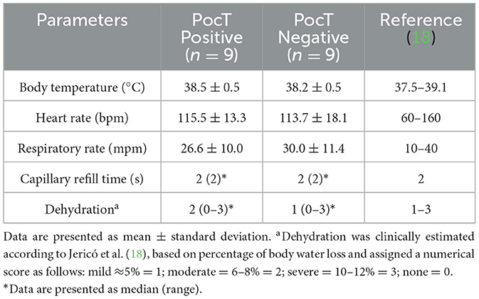
Table 2. Clinical parameters of dogs classified as cannabis-positive, or control (cannabis-negative) based on the PoCT COC/THC Assure Tech (Westlake Eco Zone, China; ANVISA/MS registration 80885650015 – Minas Gerais, Brazil).
Hematological analyses were performed within a maximum interval of 12 h after blood collection. The results are presented in Table 3. No significant differences were observed when comparing the “positive” and “negative” groups. Furthermore, no alterations indicative of anemia, infection, inflammation, anaphylactic reaction, or platelet abnormalities were detected when compared to reference values.

Table 3. Hematological parameters of dogs classified as cannabis-positive, or control (cannabis-negative) based on the PoCT COC/THC Assure Tech (Westlake Eco Zone, China; ANVISA/MS registration 80885650015 – Minas Gerais, Brazil).
Urinalysis and the diagnostic test for intoxication were performed immediately after urine sample collection. The urinary parameters are presented in Table 4. Similar to the hematological evaluation, no notable urinary alterations were observed.
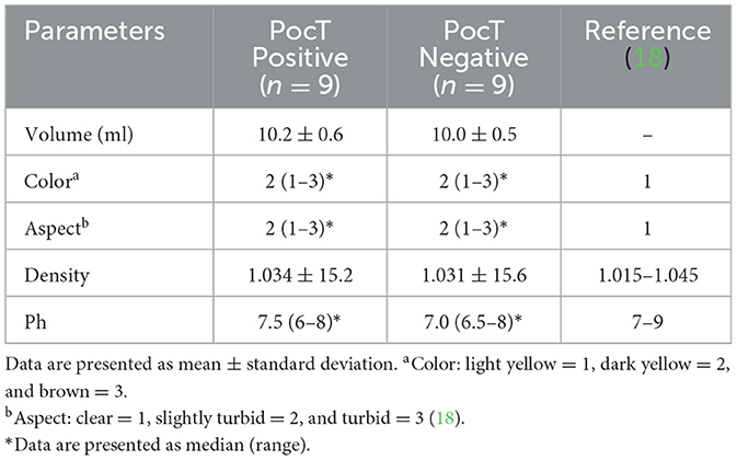
Table 4. Urinary parameters of dogs classified as cannabis-positive, or control (cannabis-negative) based on the PoCT COC/THC Assure Tech (Westlake Eco Zone, China; ANVISA/MS registration 80885650015 – Minas Gerais, Brazil).
Regarding clinical biochemical analyses, serum measurements of creatinine, urea, alanine aminotransferase (ALT), alkaline phosphatase (ALP), and glucose were performed. The data are presented in Table 5. No significant alterations were observed in the evaluated parameters that would indicate renal or hepatic impairment, nor were there changes in glycemia suggestive of acute disturbances in glucose metabolism.
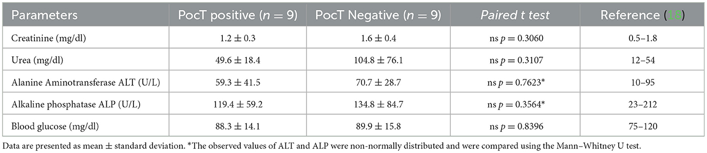
Table 5. Biochemical parameters of dogs classified as cannabis-positive, or control (cannabis-negative) based on the PoCT COC/THC Assure Tech (Westlake Eco Zone, China; ANVISA/MS registration 80885650015 – Minas Gerais, Brazil).
During treatment, non-specific clinical signs such as vomiting, diarrhea, and dehydration were managed supportively and resolved completely. Other clinical manifestations, including ataxia and behavioral alterations, were also monitored until full recovery. The duration of hospitalization ranged from 24 to 72 h, depending on the severity of clinical presentation. No animals died or were euthanized due to complications related to cannabis intoxication, as confirmed by clinical evaluation and/or point-of-care testing during the study period.
Discussion
This study highlights the application of point-of-care (POC) testing for the rapid diagnosis of cannabis intoxication in dogs. The POC test proved to be a valuable tool for timely and accurate identification of the toxic agent, facilitating targeted clinical management (10). Therefore, in contrast to previous reports in the literature (4, 11), the human-based rapid test performed reliably in this veterinary setting.
Importantly, the comparison of laboratory results between cannabis-positive and negative animals revealed minimal physiological disturbances, suggesting that, although clinical signs may be present, cannabis intoxication in dogs does not typically result in significant hematological or biochemical alterations. This finding reinforces the notion that supportive care remains the cornerstone of treatment, while POC testing enhances diagnostic confidence and clinical decision-making.
The clinical alterations observed in this study were mild and consistent with those previously described in dogs exposed to cannabis. In the large retrospective cohort of 223 dogs with confirmed or suspected THC ingestion reported by Binagia et al. (8), the most frequent signs included neurological manifestations such as ataxia, lethargy, urinary incontinence, and hypersensitivity to stimuli, as well as cardiovascular and gastrointestinal alterations including tachycardia, bradycardia, vomiting, and hypersalivation. These clinical signs generally vary according to the amount and concentration of cannabinoids absorbed by the animal (8, 12, 13).
Regarding the routes of intoxication, oral ingestion was the exclusive exposure pathway identified in this study. The dogs ingested cannabis through various sources, including marijuana cigarette remnants, homemade foods containing cannabis, and butter infused with THC. These findings align closely with previous reports by Meola et al. (4), Miranda et al. (12), Amissah et al. (13), and Binagia et al. (8), which also identified oral consumption as the primary route of exposure in canine cannabis toxicosis. Oral ingestion is particularly concerning due to the high variable bioavailability and prolonged effects of cannabinoids when absorbed via the gastrointestinal tract, compared to inhalation. Furthermore, edible cannabis products pose an increased risk because they often contain concentrated amounts of cannabinoids and may be more appealing to pets due to their palatability. Understanding these common exposure routes is crucial for veterinary practitioners to accurately assess risk, provide appropriate client education, and tailor clinical interventions.
Similar to the hematological evaluation, the biochemical findings observed in this study appeared to reflect preexisting or non-specific conditions rather than direct effects of cannabis intoxication. No evidence of renal impairment was detected, which indicates that mild accidental exposures did not result in measurable kidney dysfunction. Although cannabinoids have been investigated for potential renal effects, our results do not support any protective or deleterious influence in this context. Likewise, hepatic enzyme values remained within reference limits, suggesting the absence of acute hepatic injury; however, previous studies have reported that cannabis or its derivatives may cause hepatocellular alterations under certain exposure conditions (14).
Regarding the clinical findings of temperature, heart rate, respiratory rate, etc., as well as any hematological changes observed in this study, they are likely related to pre-existing comorbidities, and not to cannabis intoxication itself. The literature does not report significant changes in red or white blood cell counts, nor platelet abnormalities, associated with cannabis intoxication in dogs. However, hematological analysis remains a valuable tool for assessing the patient's hydration status and overall physiological condition, allowing for more accurate correction of dehydration and supportive care tailored to individual needs.
In this study, the clinical findings related to hepatic evaluation are consistent with previous reports indicating that administration of relatively high doses of cannabinoids did not alter hepatic function in the animals studied (15). Regarding the unchanged levels of hepatic enzymes and glycemic index observed, other research has shown no significant changes in hepatic metabolites and even highlighted a positive effect of cannabis on insulin metabolism, demonstrating decreased insulin and triglyceride levels 30 min after ingestion of food or CBD, without impairing liver or kidney function (16).
Regarding urinalysis, no significant alterations related to cannabis intoxication were identified in the parameters of the urine samples. For this study, the most critical parameter was the presence or absence of cannabinoid compounds in the urine, for which complementary evaluation was necessary.
The chromatographic urine test used in this study is a lateral flow immunochromatographic test designed for the qualitative detection of tetrahydrocannabinol (THC) and its metabolites in human urine, with a cutoff concentration of 50 ng/ml for THC. The method is based on competitive antigen–antibody binding, in which the presence of THC or its metabolites above the cutoff inhibits the appearance of the test line. The primary metabolite detected by this test is THC-COOH. In veterinary context, unlike in humans, the production of THC-COOH has not been clearly demonstrated in dogs, and experimental and pharmacokinetic evidence indicates that canine metabolism favors the formation of 8-hydroxy-Δ9-THC (8-OH-THC) and 11-hydroxy-Δ9-THC (11-OH-THC) as the predominant metabolites (4, 11, 17). Therefore, the positive results obtained in this study likely reflect cross-reactivity of the assay with one or more of these canine metabolites rather than direct detection of THC-COOH.
Another relevant point for discussion is the interval between admission and testing. In this study, urine samples were analyzed approximately 6 h after clinical presentation, rather than immediately. This interval may have influenced the results, as most urinary immunoassays for cannabinoids detect the metabolite THC-COOH about 4 h after ingestion (8). Therefore, delayed testing likely increased the urinary concentration of detectable metabolites, thereby enhancing the apparent sensitivity of the assay without necessarily reflecting superior analytical performance. The manufacturer's instructions do not specify the ideal interval between exposure and testing; the recommendation is that the assay must be performed promptly after urine sample collection.
With respect to the limitations of this study, the relatively small sample size, inherent to its observational nature, reduces the statistical power to detect mild physiological variations previously associated with THC intoxication, such as subtle fluctuations in heart rate, respiratory rate, or body temperature. The absence of additional physiological parameters, including blood pressure, blood gas analysis, and electrolyte monitoring, also limited the evaluation of systemic responses that could have provided further insight into the cardiovascular and metabolic effects of THC in dogs. Moreover, confirmatory analytical testing using liquid chromatography–mass spectrometry (LC–MS) was not available, precluding definitive identification of the metabolites responsible for positive immunoassay results. Future studies with larger populations, comprehensive physiological monitoring, and validated confirmatory methods are warranted to better elucidate the metabolic profile of THC in dogs and to establish the diagnostic accuracy of point-of-care urine testing in veterinary settings.
Conclusions
In this study, the point-of-care chromatographic urine test (PoCT COC/THC Assure Tech, Westlake Eco Zone/China) correctly identified all nine dogs with accidental cannabis ingestion and yielded negative results in control animals. Hematological, biochemical, and urinalysis parameters showed no significant differences between groups, suggesting that mild accidental exposures may not cause detectable systemic alterations. Although the sample size was limited, the findings indicate that this urine drug screen may offer greater sensitivity than those previously evaluated in veterinary medicine. Further studies with larger populations are warranted to confirm its diagnostic performance and clinical applicability in veterinary practice.
Data availability statement
The original contributions presented in the study are included in the article/supplementary material, further inquiries can be directed to the corresponding author.
Ethics statement
Ethical approval was not required for this study, as it involved the evaluation of client-owned animals presented to a veterinary emergency service. All diagnostic and therapeutic procedures were performed as part of routine clinical care, and no experimental interventions were conducted. Written informed consent was obtained from all owners for their animals' participation and for the publication of this study.
Author contributions
HP: Conceptualization, Formal analysis, Investigation, Methodology, Visualization, Writing – original draft, Writing – review & editing. WB: Investigation, Methodology, Visualization, Writing – review & editing. MV: Formal analysis, Investigation, Methodology, Visualization, Writing – review & editing. RG: Investigation, Methodology, Supervision, Visualization, Writing – review & editing. LC: Conceptualization, Formal analysis, Funding acquisition, Investigation, Methodology, Project administration, Resources, Supervision, Validation, Writing – original draft, Writing – review & editing.
Funding
The author(s) declare that financial support was received for the research and/or publication of this article. The publication was supported by Karolinska Institutet through an institutional publication agreement. The funding covered the open access publication fees.
Acknowledgments
The authors thank the Paraíba State Research Support Foundation (FAPESQ-PB) and the Postgraduate Program in Animal Science (PPGCAn) of the Federal University of Paraíba, for their support and assistance during the conduct of the research.
Conflict of interest
The authors declare that the research was conducted in the absence of any commercial or financial relationships that could be construed as a potential conflict of interest.
Generative AI statement
The author(s) declare that no Gen AI was used in the creation of this manuscript.
Any alternative text (alt text) provided alongside figures in this article has been generated by Frontiers with the support of artificial intelligence and reasonable efforts have been made to ensure accuracy, including review by the authors wherever possible. If you identify any issues, please contact us.
Publisher's note
All claims expressed in this article are solely those of the authors and do not necessarily represent those of their affiliated organizations, or those of the publisher, the editors and the reviewers. Any product that may be evaluated in this article, or claim that may be made by its manufacturer, is not guaranteed or endorsed by the publisher.
References
1. Silva DR, Silva CS. Frequency of intoxication in small animals in a veterinary clinic in the city of Patos de Minas – MG: analysis of the number of cases in 2021. GETEC. (2022) 11:29–49.
2. Nascimento AH, Domingues AI, Feio JV, De Vasconcelos MVN, Da Silva Alves WF. Intoxicação por fipronil em felino – relato de caso. Rev Multidiscip Saúde. (2021) 2:18. doi: 10.51161/rems/1830
4. Meola SD, Tearney, CC, Haas SA, Hackett TB, Mazzaferro EM. Evaluation of trends in marijuana toxicosis in dogs living in a state with legalized medical marijuana: 125 dogs (2005–2010). J Vet Emerg Crit Care. (2012) 22:690–6. doi: 10.1111/j.1476-4431.2012.00818.x
5. Hansen DTK. Prevalence of Intoxications in Dogs and Cats in Curitiba [master's thesis]. Curitiba, Brazil: Universidade Federal do Paraná (2006).
6. Ribeiro UR, Pinto PN. Intoxicação de cães domésticos por Cannabis sativa: review. PubVet. (2022) 16, a1199.1–5. doi: 10.31533/pubvet.v16n08a1199.1-5
7. Bulcão RP, Tonello R, Piva SJ, Schmitt GC. Intoxicação em cães e gatos: diagnóstico toxicológico empregando cromatografia em camada delgada e cromatografia líquida de alta pressão com detecção ultravioleta em amostras estomacais. Ciência Rural. (2010) 40:1109–13. doi: 10.1590/S0103-84782010000500017
8. Binagia EM, Gregory, EA, Yankin I. Clinical examination findings and electrolyte abnormalities in dogs with marijuana/tetrahydrocannabinol toxicity: 223 cases (January 2017 to July 2021). J Am Vet Med Assoc. (2024) 262:1047–54. doi: 10.2460/javma.24.02.0092
9. Fitzgerald KT, Bronstein AC, Newquist KL. Marijuana poisoning. Top Companion Anim Med. (2013) 28:8–12. doi: 10.1053/j.tcam.2013.03.004
10. Claus BB, Specka M, McAnally, H, Scherbaum N, Schifano F, Bonnet U. Is the urine cannabinoid level measured via a commercial point-of-care semiquantitative immunoassay a cannabis withdrawal syndrome severity predictor? Front Psychiatry. (2020) 11:598150. doi: 10.3389/fpsyt.2020.598150
11. Teitler JB. Evaluation of a human on-site urine multidrug test for emergency use with dogs. J Am Anim Hosp Assoc. (2009) 45:59–66. doi: 10.5326/0450059
12. Miranda ALS, Soto-Blanco B, Marília M. Intoxicações de cães por drogas recreativas: maconha e cocaína. Rev Cient Med Vet. (2017) 28:1–8.
13. Amissah RQ, Vogt NA, Chen C, Urban K, Khokhar J. Prevalence and characteristics of induced toxicoses for cannabis in pets: results of a survey of veterinarians in North America. PLoS ONE. (2022) 17:e0261909. doi: 10.1371/journal.pone.0261909
14. González-Mariscal I, Carmona-Hidalgo B, Winkler M, Unciti-Broceta JD, Escamilla A, Gómez-Cañas M, et al. (+)-trans-Cannabidiol-2-hydroxy pentyl is a dual CB1R antagonist/CB2R agonist that prevents diabetic nephropathy in mice. Pharmacol Res. (2021) 169:105492. doi: 10.1016/j.phrs.2021.105492
15. Nasrin S, Watson CJW, Perez-Paramo YX, Lazarus P. Cannabinoid metabolites as inhibitors of major hepatic CYP450 enzymes, with implications for Cannabis-drug interactions. Drug Metab Dispos. (2021) 49:1070–80. doi: 10.1124/dmd.121.000442
16. Abbotts KSS, Ewell TR, Butterklee HM, Bomar MC, Akagi N, Dooley GP, et al. Cannabidiol and cannabidiol metabolites: pharmacokinetics, interaction with food and influence on liver function. Nutrients. (2022) 14:2152. doi: 10.3390/nu14102152
17. Kitts-Morgan SE, Sams RA, Muir WW. Pharmacokinetics of cannabidiol, (-)-trans-Δ9-tetrahydrocannabinol, and their oxidative metabolites after intravenous and oral administration of a cannabidiol-dominant full-spectrum hemp product to beagle dogs. Front Vet Sci. (2025) 12:1556975. doi: 10.3389/fvets.2025.1556975
18. Jericó MM, Andrade Neto JP, Kogika MM. Tratado de medicina interna de cães e gatos, 2 vols. Rio de Janeiro, Brazil: Guanabara Koogan (2023).
19. Janczyk P, Donaldson CW, Gwaltney, S. Two hundred thirteen cases of marijuana toxicosis in dogs. Vet Hum Toxicol. (2004) 46:19–21.
Keywords: cannabis intoxication, dogs, THC metabolites, veterinary toxicology, endocannabinoid system
Citation: Pereira HCdS, Barbosa WMB, Venceslau MF, Guerra RR and Carvalho LRRA (2025) Comparison of clinicopathologic findings and urine drug screen results in cannabis-positive and control dogs. Front. Vet. Sci. 12:1679400. doi: 10.3389/fvets.2025.1679400
Received: 04 August 2025; Revised: 13 November 2025;
Accepted: 17 November 2025; Published: 08 December 2025.
Edited by:
Heather Knych, University of California, Davis, United StatesReviewed by:
Alan Chicoine, University of Saskatchewan, CanadaErin Binagia, Texas A and M University, United States
Copyright © 2025 Pereira, Barbosa, Venceslau, Guerra and Carvalho. This is an open-access article distributed under the terms of the Creative Commons Attribution License (CC BY). The use, distribution or reproduction in other forums is permitted, provided the original author(s) and the copyright owner(s) are credited and that the original publication in this journal is cited, in accordance with accepted academic practice. No use, distribution or reproduction is permitted which does not comply with these terms.
*Correspondence: Lucas Rannier Ribeiro Antonino Carvalho, bHVjYXMuY2FydmFsaG9Aa2kuc2U=