
Biology of Ly-6 Supergene Family in Health and Disease

56kviews
Editorial23 June 2022
Editorial: Biology of Ly-6 Supergene Family in Health and Disease

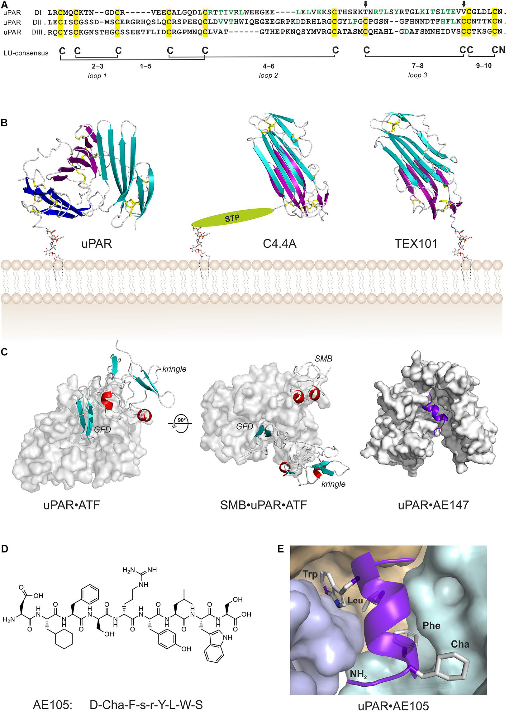
PI3K/AKT/mTOR. This report shows, in addition to interaction with α-7nAChR, SLURP-1 binds to platelet-derived growth factor receptor type α (PDGFRβ) and epidermal growth factor receptor (EGFR). Furthermore, their data highlights the role of SLURP-1's "loop I" in binding to α7-nAChR and altering cell proliferative response with druggable synthetic SLURP-1 loop-1 mimics. Doss et.al., report on LYNX1, specifically examining the interaction between LYNX1 and nicotinic acetylcholine receptor (nAChR) subtypes in skeletal neuromuscular junctions (NMJs). The authors' data support of the role of LYNX1 in maintaining the structure and function of adult and aging NMJs. Kristensen et.al., in their review article summarize recent developments in the biology of GPIHBP1, a GPIanchored LU protein. This member of LU family regulates plasma triglyceride metabolism by interacting and functionally charging the Lipoprotein lipase (LPL) enzyme. These authors discuss the role of GPIHBP1's intrinsically disordered, highly acidic N-terminal extension and the folded disulfide bond-rich three-fingered LU domain in ushering LPL for intravascular lipolysis. In another review article, Leth and Ploug., discuss recent development in the role of urokinase plasminogen activation receptor, known to bind uPA and regulating uPA's extravesicular serine protease activity for fibrinolysis, cell adhesion and migration. Authors highlight the upregulated uPAR protein expression at the boundary between the solid tumor and the normal tissue allowing for recognition and complete recitation (surgical/non-surgical) of the tumor tissue. Authors discuss targeting tumors with killer T cells or uPAR-targeted reportermolecules containing positron emitting radionuclides or near-infrared (NIR) florescence probes. These probes allow tumor detection with tomography (PET) and/or its removal after illuminating it.Perspectives: Over the last 50 years the use of LU proteins as markers of hematopoietic stem cells and their progeny has been vital for identifying and understanding the functional responses generated by a variety of immune cell subsets. The reports in this volume and summarized above highlight the broader tissue expression of LU proteins and their role in myriad biological process. LU proteins are known to function as a signaling rheostat by fine tuning and/or regulating signals triggered by other membrane receptor as previously reviewed (Bamezai 2004). For example, Ly-6A expression regulates signaling through the antigen receptor in T lymphocytes (Stanford et.al., 1997, Henderson et.al., 2002, while LYNX1 binds and regulates signaling through acetylcholine receptor (Miwa et.al., 1999), the Sleepless protein regulates a potassium ion channel (Shaker) in neurons (Koh et.al., 2008). These interactions between the membrane anchored LU protein and other receptors appear to occur either "in-cis", meaning within the plane of plasma membrane through a cell-autonomous mechanism, or "in-trans" where the secreted LU protein engages its target membrane receptor to drive the autocrine/paracrine cellular signaling. Additionally, the reported ligand(s) engaging GPI-anchored LU protein may contribute indirectly by regulating signaling in non-cell autonomous manner "in-trans". Putative soluble or cell-bound ligands directly interacting with LU proteins have been reported (English et.al., 2000, Plugh et.al., 2002; however, much needs to be done to fully characterize ligands for some Ly-6 family members and to fully understand the signal transduction through these tail-less, GPIanchored proteins in the absence of their transmembrane and cytoplasmic domains.Contribution of cholesterol-rich lipid rafts on the membrane in signaling through LU proteins has been observed (Lang et.al., 2017) and is currently under investigation. LUproteins are housed in these membrane nanodomains because of their lipid anchor to allow for their interaction and regulation of transmembrane receptors/ion channels residing in lipid rafts (Koh et.al., 2008, Ozhan et.al., 2013. Another emerging area of investigation is the use of some LU-domain proteins as markers of tumors (Treister et.al., 1998, Upadhay, 2019, Gravekamp, 2021) and as possible targets of either antibody directed and/or cell-based immune therapies (Lo et.al., 2021).

AKB wrote the manuscript. JM edited and approved the manuscript. Both authors provided intellectual input in co-editing this volume.

2,800 views

2 citations

Human Three-Finger Protein Lypd6 Is a Negative Modulator of the Cholinergic System in the Brain
ORIGINAL RESEARCH
21 September 2021
Human Three-Finger Protein Lypd6 Is a Negative Modulator of the Cholinergic System in the Brain
Dmitrii Kulbatskii, 15 more and Ekaterina Lyukmanova
Lypd6 is a GPI-tethered protein from the Ly-6/uPAR family expressed in the brain. Lypd6 enhances the Wnt/β-catenin signaling, although its action on nicotinic acetylcholine receptors (nAChRs) have been also proposed. To investigate a cholinergic activity of Lypd6, we studied a recombinant water-soluble variant of the human protein (ws-Lypd6) containing isolated ‘three-finger’ LU-domain. Experiments at different nAChR subtypes expressed in Xenopus oocytes revealed the negative allosteric modulatory activity of ws-Lypd6. Ws-Lypd6 inhibited ACh-evoked currents at α3β4- and α7-nAChRs with IC50 of ~ 35 and 10 μM, respectively, and the maximal amplitude of inhibition of 30 – 50 %. EC50 of ACh at α3β4-nAChRs (~ 30 μM) was not changed in the presence of 35 μM ws-Lypd6, while the maximal amplitude of ACh-evoked current was reduced by ~ 20 %. Ws-Lypd6 did not elicit currents through nAChRs in the absence of ACh. Application of 1 μM ws-Lypd6 significantly inhibited (up to ~ 28 %) choline-evoked current at α7-nAChRs in rat hippocampal slices. Similar to snake neurotoxin α-bungarotoxin, ws-Lypd6 suppressed the long-term potentiation in mouse hippocampal slices. Colocalization of endogenous GPI-tethered Lypd6 with α3β4- and α7-nAChRs was detected in primary cortical and hippocampal neurons. Ws-Lypd6 interaction with the extracellular domain of α7-nAChR was modelled using ensemble protein-protein docking protocol. The interaction of all three Lypd6 loops (‘fingers’) with the entrance to the orthosteric ligand-binding site and the loop C of the primary receptor subunit was predicted. The results obtained allow us to consider Lypd6 as the endogenous negative modulator involved in the regulation of the cholinergic system in the brain.

6,393 views

20 citations

Targeting the Urokinase-Type Plasminogen Activator Receptor (uPAR) in Human Diseases With a View to Non-invasive Imaging and Therapeutic Intervention
The interaction between the serine protease urokinase-type plasminogen activator (uPA) and its glycolipid-anchored receptor (uPAR) focalizes plasminogen activation to cell surfaces, thereby regulating extravascular fibrinolysis, cell adhesion and migration. uPAR belongs to the Ly6/uPAR (LU) gene superfamily and the high-affinity binding site for uPA is assembled by a dynamic association of its three consecutive LU domains. In most human solid cancers, uPAR is expressed at the invasive areas of the tumor-stromal microenvironment. High levels of uPAR in resected tumors or shed to the plasma of cancer patients are robustly associated with poor prognosis and increased risk of relapse and metastasis. Over the years, a plethora of different strategies to inhibit uPA and uPAR function have been designed and investigated in vitro and in vivo in mouse models, but so far none have been implemented in the clinics. In recent years, uPAR-targeting with the intent of cytotoxic eradication of uPAR-expressing cells have nonetheless gained increasing momentum. Another avenue that is currently being explored is non-invasive imaging with specific uPAR-targeted reporter-molecules containing positron emitting radionuclides or near-infrared (NIR) florescence probes with the overarching aim of being able to (i) localize disease dissemination using positron emission tomography (PET) and (ii) assist fluorescence guided surgery using optical imaging. In this review, we will discuss these advancements with special emphasis on applications using a small 9-mer peptide antagonist that targets uPAR with high affinity.

11,869 views

36 citations

ORIGINAL RESEARCH
14 September 2021
SLURP-1 Controls Growth and Migration of Lung Adenocarcinoma Cells, Forming a Complex With α7-nAChR and PDGFR/EGFR Heterodimer
Maxim L. Bychkov, 10 more and Ekaterina N. Lyukmanova

7,537 views

30 citations

Abstract Intravascular processing of triglyceride-rich lipoproteins (TRLs) is crucial for delivery of dietary lipids fueling energy metabolism in heart and skeletal muscle and for storage in white adipose tissue. During the last decade, mechanisms underlying focal lipolytic processing of TRLs along the luminal surface of capillaries have been clarified by fresh insights into the functions of lipoprotein lipase (LPL); LPL’s dedicated transporter protein, glycosylphosphatidylinositol-anchored high density lipoprotein–binding protein 1 (GPIHBP1); and its endogenous inhibitors, angiopoietin-like (ANGPTL) proteins 3, 4 and 8. Key discoveries in LPL biology include solving the crystal structure of LPL, showing LPL is catalytically active as a monomer rather than as a homodimer, and that the borderline stability of LPL’s hydrolase domain is crucial for the regulation of LPL activity. Another key discovery was understanding how ANGPTL4 regulates LPL activity. The binding of ANGPTL4 to LPL sequences adjacent to the catalytic cavity triggers cooperative and sequential unfolding of LPL’s hydrolase domain resulting in irreversible collapse of the catalytic cavity and loss of LPL activity. Recent studies have highlighted the importance of the ANGPTL3–ANGPTL8 complex for endocrine regulation of LPL activity in oxidative organs (e.g., heart, skeletal muscle, brown adipose tissue), but the molecular mechanisms have not been fully defined. New insights have also been gained into LPL–GPIHBP1 interactions and how GPIHBP1 moves LPL to its site of action in the capillary lumen. GPIHBP1 is an atypical member of the LU (Ly6/uPAR) domain protein superfamily, containing an intrinsically disordered and highly acidic N-terminal extension and a disulfide bond–rich three-fingered LU domain. Both the disordered acidic domain and the folded LU domain are crucial for the stability and transport of LPL, and for modulating its susceptibility to ANGPTL4-mediated unfolding. This review focuses on recent advances in the biology and biochemistry of crucial proteins for intravascular lipolysis.

11,239 views

34 citations