 Chenchen Song1
 Xuan Sun2,3Yao Wang1
 Leif Bülow2Michael Mecklenburg4
 Changxin Wu1
 Qinglai Meng1*
 Bin Xie2*
- 1Institute of Biomedical Sciences, The Key Laboratory of Chemical Biology and Molecular Engineering of National Ministry of Education, Shanxi Provincial Key Laboratory of Medical Molecular Cell Biology, Shanxi University, Taiyuan, China
- 2Division of Pure and Applied Biochemistry, Department of Chemistry, Lund University, Lund, Sweden
- 3School of Basic Medical Sciences, Hubei University of Chinese Medicine, Wuhan, China
- 4Omik Bioscience AB, Lund, Sweden
Antibiotic resistance has become a serious threat to global public health and economic development. Rapid and accurate identification of a patient status for antimicrobial resistance (AMR) are urgently needed in clinical diagnosis. Here we describe the development of an assay method for activity fingerprinting of AMR β-lactamases using panels of 7 β-lactam antibiotics in 35 min. New Deli Metallo β-lactamase-1 (NDM-1) and penicillinase were demonstrated as two different classes of β-lactamases. The panel consisted of three classes of antibiotics, including: penicillins (penicillin G, piperacillin), cephalosporins (cefepime, ceftriaxone, cefazolin) and carbapenems (meropenem and imipenem). The assay employed a scheme combines the catalytic reaction of AMR β-lactamases on antibiotic substrates with a flow-injected thermometric biosensor that allows the direct detection of the heat generated from the enzymatic catalysis, and eliminates the need for custom substrates and multiple detection schemes. In order to differentiate classes of β-lactamases, characterization of the enzyme activity under different catalytic condition, such as, buffer composition, ion strength and pH were investigated. This assay could provide a tool for fast diagnosis of patient AMR status which makes possible for the future accurate treatment with selected antibiotics.
Introduction
Since the discovery of penicillin, a broad range of antibiotics have been discovered which have enhanced our ability to treat bacterial infections. Antibiotics have played a critical role in medicine, as well as in other fields (Bennett and Chung, 2001). However, in recent years, the dramatic increase in use of antibiotics has resulted in the spread of antibiotic resistance. Initially, resistance was limited to a single antibiotic and was easily addressed by simply choosing another antibiotic. As resistance has spread, a class of pathogens emerged that are resistant to multiple antibiotics, so-called multi-resistant pathogens. This trend has continued and spawned a new class of pathogens which are resistant to all known antibiotics, i.e. super-bugs. Patients infected with these pathogens have dramatically higher hospitalization and mortality rates (Hansen et al., 2015). This trend in AMR threatens to undermine global public health and economic development (English and Gaur, 2010; Qiao et al., 2018).
Physicians are on the front lines in the effort to treat patients infected with multi-resistant pathogens. However, in order to determine the most effective course of treatment, physicians require AMR diagnostics capable of rapidly and specifically determining the patient’s AMR profile. Although there are multiple mechanisms by which pathogens achieve resistance, the most common mechanism is the production of enzymes that catalytically inactivate the antibiotics. Numerous physical and activity based methods have been developed to detect these AMR enzymes ranging from traditional methods, such as the disk method to new techniques, such as multiplex PCR, DNA microarrays, colorimetric assays, immune-chromatographic technique and thermometric analysis. Each technique has advantages and disadvantages. However, none of these techniques is capable of providing results/diagnostics of multi-resistance in a clinically relevant timeframe. Efforts must be renewed to address this issue (Rodriguez-Rojas et al., 2013; Blair et al., 2014). Rapid, simple, and reliable AMR analytical solution are urgently needed that provide doctors with the ability to profile AMR in order to allow tailored, personalized therapeutics for patients infected with AMR pathogens.
There are several methods that have been conventionally used to detect antibiotic resistance in recent decades, such as the double dilution method and susceptibility testing (Ge et al., 2013). However, these traditional detection methods have low sensitivity, are complicated process and time-consuming. Thus, researchers have exploited some other methods, including multiplex PCR, immune-chromatographic techniques, colorimetric methods, DNA microarray, and thermometric biosensor (March-Rosselló, 2017). Of all the above methods, the thermometric method has attracted our attention because of its rapid analysis, simple operation, and high throughput (Chen et al., 2015; Wang et al., 2018).
Here we describe the development of a novel AMR strategy that profiles AMR enzymes by testing a panel of antibiotics. The diagnostic strategy specifically and efficiently identifies the AMR enzymatic activity profile using a flow injected thermal biosensor device. For these studies, we have chosen to use a panel of antibiotics that are widely used in clinical settings. The panel consisted of three different classes of antibiotics: Penicillins (penicillin G, piperacillin), Cephalosprins (cefazolin, ceftriaxone, cefepime) and Carbapenems (imipenem, meropenem). In order to verify the profile based characterization, two classes of AMR β-lactamases: New Deli Metallo β-lactamase-1 (NDM-1) and penicillinase were chosen as examples in the study. The effects of the reaction conditions, such as: ion strength, buffer composition, and pH on the activity profiling of the AMR β-lactamases catalyzing this panel of antibiotics were investigated.
Materials and methods
Chemicals and materials
Penicillin G, Piperacillin, Ceftriaxone, Cefepime, Cefazolin, Meropenem were purchased from Solarbio Co., Ltd. (Beijing, China). Imipenem was bought from Fresenius Kabi (Bad Homburg, Germany). HEPES was obtained from Sangon Biotech Co., Ltd. (Shanghai, China). All other reagents were purchased from Sigma-Aldrich Co (St. Louis, USA). The controlled pore glass (CPG) beads (125–140 μm particle diameter and 50 nm pore size) were originally purchased from VEB Trisola, Steinach, Germany. The construction of plasmid encoding NDM-1, expression and purification of recombinant NDM-1(rNDM-1) were performed as previously described (Meng et al., 2021a; Meng et al., 2021b). The purity of the purified rNDM-1 is > 95%, The unit activity of rNDM-1 to hydrolyze meropenem is 108 IU/mg.
Buffer and sample preparation
Phosphate buffer (PB) and HEPES buffer in different ionic strengths (0.01, 0.05 0.1 M) and pHs (pH 6.8, 7.2, 7.6, 8.0) were prepared as running buffers and for sample preparation. All water used for preparing the buffers and samples was from a Milli-Q system (Millipore, Bedford, MA, USA).
Stock solutions of each of the antibiotics: Penicillin G, Piperacillin, Ceftriaxone, Cefepime, Cefazolin, Meropenem, and Imipenem, were prepared in PB and HEPES at the various ionic strengths and pHs, which were then serially diluted: 10 mM, 5 mM, 2.5 mM, 1.25 mM, and 0.625 mM.
The analytical strategy
The Enzyme Thermal biosensor (ET) is a flow-injected analytical device (Omik Bioscience AB, Sweden) has been employed in this study. The enzyme, e.g. β-lactamase, was immobilized on spherical porous glass beads (CPG), loaded in an enzyme column and substrate samples were injected (e.g. antibiotics) with running buffers at various concentrations. Meanwhile the simultaneously generated heat can be precisely and continuously detected with high resolution thermistor sensors (Ramanathan and Danielsson, 2001; Zhou et al., 2013). The thermistor sensor converts the heat generated by the reaction into a temperature signal (Xie et al., 2000; Qie et al., 2013). In an adiabatic system, if all the heat released is used to increase the temperature of the reaction system, the following formula can be used:
Where n is the number of moles of product; ΔH is the enthalpy change of the biochemical reaction; ΔT is the temperature change of the reaction system; CS is the heat capacity of the reaction system. This formula shows that there is a linear relationship between the temperature change and the substrate concentration. Thus, the peak height or area can be used for determination of an analyte concentration (Xie et al., 2000; Yakovleva et al., 2013). Thermometric biosensors have numerous advantages including: the universality of the sensing mechanism, label-free analysis and high specificity. In addition, thermal sensors are less sensitive to sample interference than optical and electrical biosensors. The technology has been widely used to detect sample concentration and enzyme activity in clinical analysis, fermentation process control, food safety and environmental monitoring (Xie et al., 1994; Yakovleva et al., 2012; Chen et al., 2013; Yao et al., 2015; Andersson et al., 2017; Xu et al., 2017; Adlerberth et al., 2020).
Here, we establish a novel method for fast identification and classification of AMR β-lactamases by thermometric biosensor profiling of the enzyme catalytic activity for a panel of antibiotics (Figure 1). The device is simple to operate and automatically monitors the degradation reactions of various antibiotics. Considering the clinical interesting, 7 mostly used antibiotics including penicillin G, piperacillin, cefepime, ceftriaxone, cefazolin, meropenem, and imipenem were selected for the NDM-1 and penicillinase profiling studies.
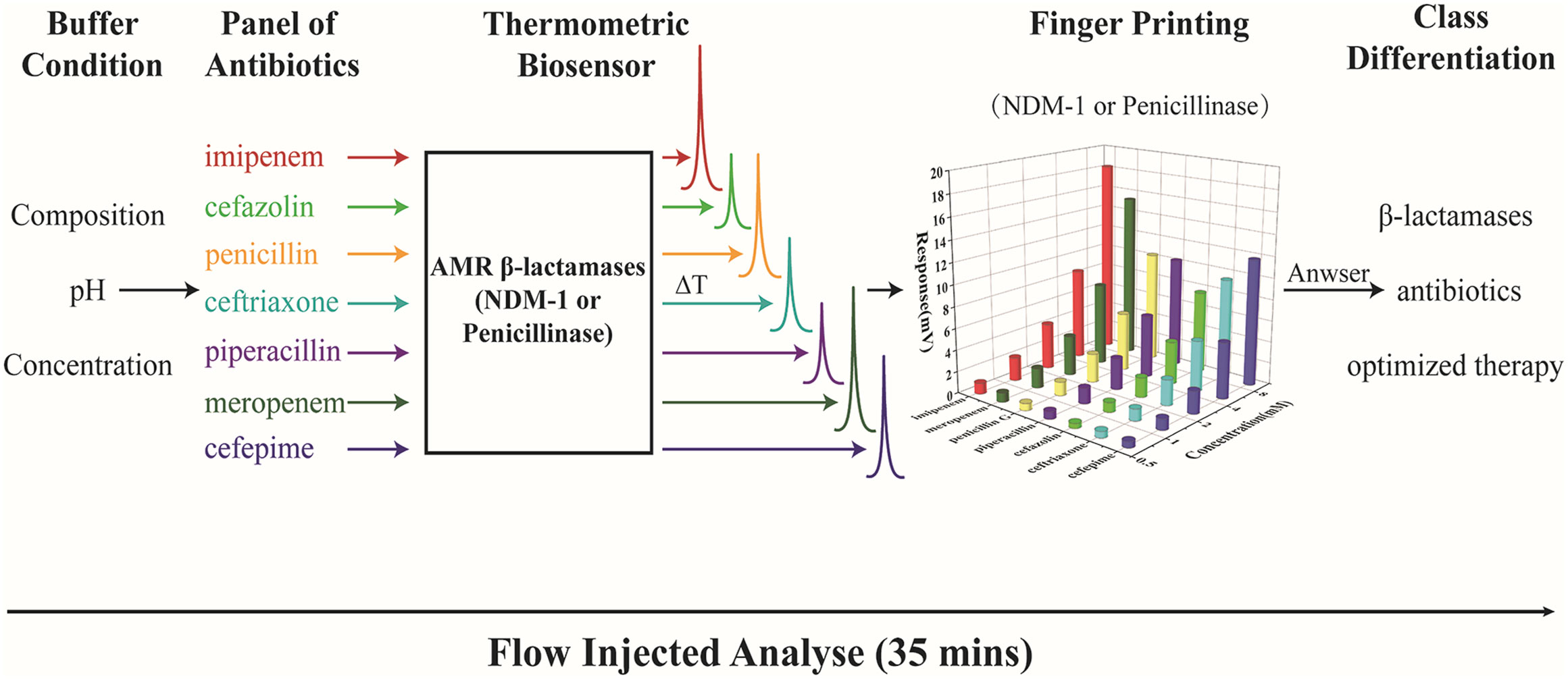
Figure 1 Diagram for fast classification of AMR β-lactamases using a thermometric biosensor profiling of the catalytic activity.
Instrumental setup
The Enzyme Thermal biosensor used in this study was composed of a pump, a sample valve, split two enzyme columns (one as measurement and the other as reference) and high resolution thermistors in coupled with signal amplification.
The enzyme column was loaded with ca. 50 IU immobilized β-lactamase (e.g. penicillinase or NDM-1) onto CPG. The working temperature was set at 25°C. A flow rate of 0.6 mL min-1 and sample volumes of 30 and 75 μL were used. However, for the comparative study, the response signals were adjusted to 10 μL/analyte. Each sample measurement was performed in triplicate.
Results
Effects of buffer composition and concentration on profiling of the AMR β-lactamases
We first evaluated effects of different buffers on degradation of 7 β-lactam antibiotics: penicillin G, piperacillin, ceftriaxone, cefepime, cefazolin, meropenem and imipenem. Among these 7 antibiotics, penicillin G and piperacillin belong to penicillin class; ceftriaxone, cefepime, cefazolin belong to cephalosporin class; and meropenem and imipenem belong to carbapenem class. Cefazolin, ceftriaxone and cefepime are representative of the 1st, 3rd and 4th generation cephalosporins, respectively.
Studies were performed to determine the effect that buffer had on assay sensitivity (Figures 2, 3). In this study, thermometric biosensor was employed for instant detection of the catalytic enthalpy (e.g. NDM-1 or penicillinase catalysis of antibiotics) that was converted to a corresponding temperature changes. The intensity of the thermal signal was proportional to the concentration of the substrates, e.g. antibiotics. In this study, a panel of 7 different antibiotics at concentrations of 0.625, 1.25, 2.5, 5.0 and 10 mM were determined sequentially. Therefore, a complete profile for one AMR enzyme could be generated in 35 min based on a cycle time of 5 min.
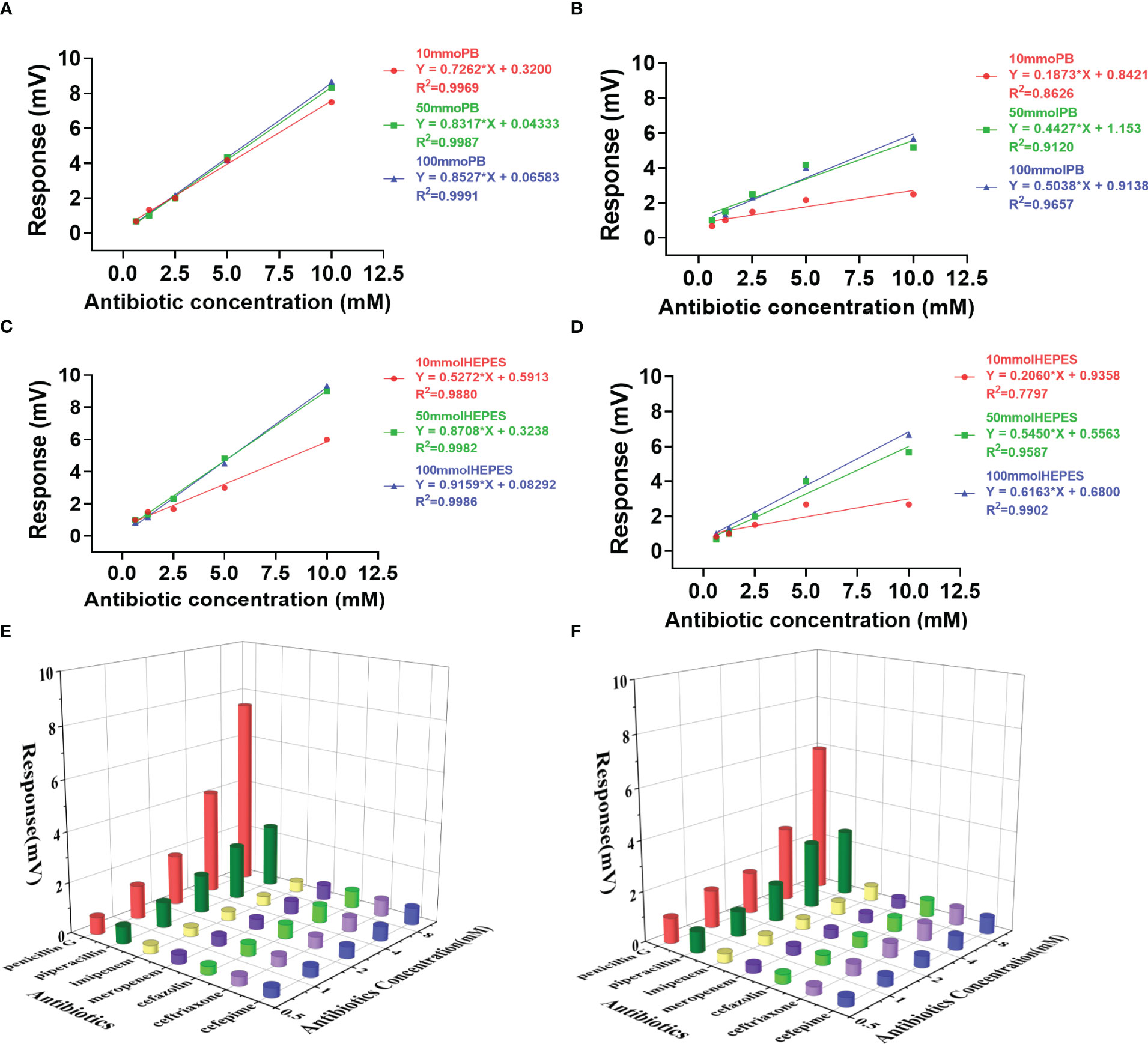
Figure 2 Effects of buffer on the activity and profiling of penicillinase catalysis. Response slopes in different concentrations of Penicillin G and Piperacillin in PB buffer (A, B, respectively) and HEPES buffer (C, D, respectively). The profile generated with a panel of 7 antibiotics in PB buffer (E) and HEPES buffer (F) using 10 mM concentration.
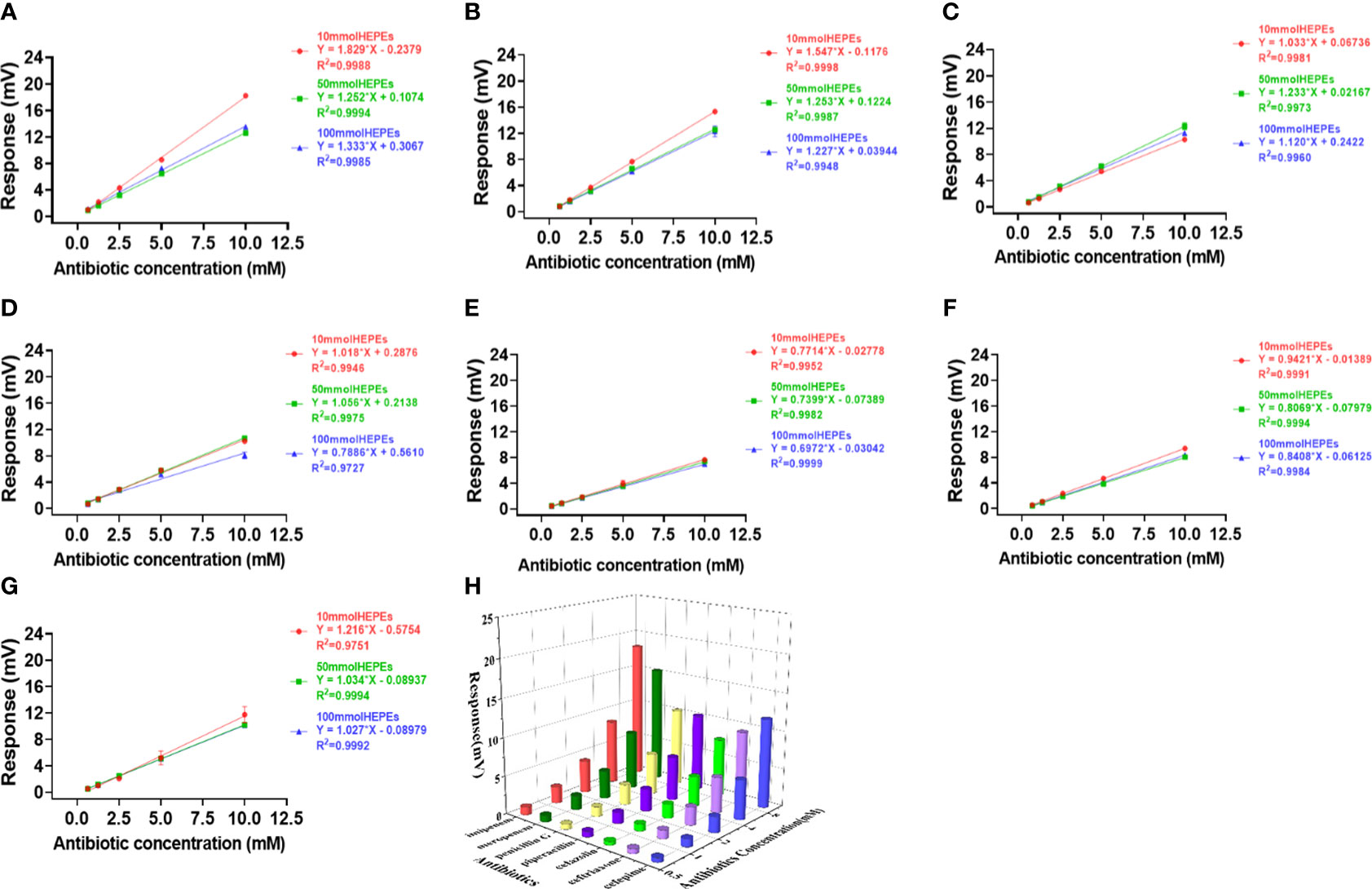
Figure 3 Effects of HEPES buffer on the activity and profiling of NDM-1 catalysis. The response slopes of 7 antibiotics in buffer concentrations of 10 mM (red), 50 mM (green) and 100 mM (blue) were presented by (A) Imipenem, (B) Meropenem, (C) Penicillin G, (D) Piperacillin, (E) Cefazolin, (F) Ceftriaxone, (G) Cefepime. The catalytic profile was generated using a 10 mM PB buffer (H).
Penicillinase profiling
As penicillinase can only efficiently catalyze penicillin class, only penicillin G and piperacillin response curves were presented in buffers of PB (Figures 2A, B) and HEPES (Figures 2C, D) at a concentration of 10, 50 and 100 mM. However, the 3D profiles of the catalytic activities for the 7 antibiotics were generated using a concentration of 10 mM using PB and HEPES buffers (Figures 2E, F).
The two buffer systems presented similar sensitivities for the individual antibiotics. However, when comparing the three buffer concentrations, the sensitivity was the highest at 100 mM and lowest at 10 mM. Meanwhile the highest variation in sensitivity was seen at 10 mM buffer concentration for both PB and HEPES (Figure 2). The lowest sensitivity variation was observed for Penicillin G in PB, which means penicillinase catalyzing penicillin G was relatively stable in PB buffer.
NDM-1 profiling
In order to verify the catalytic efficiency of NDM-1 enzyme, three classes of antibiotics including: Penicillins (penicillin G, piperacillin), cephalosporins (cefepime, ceftriaxone, cefazolin) and Carbapenems (meropenem, imipenem), were investigated using PB (Figure 4) and HEPES (Figure 3) buffers at concentrations of 10, 50 and 100 mM.
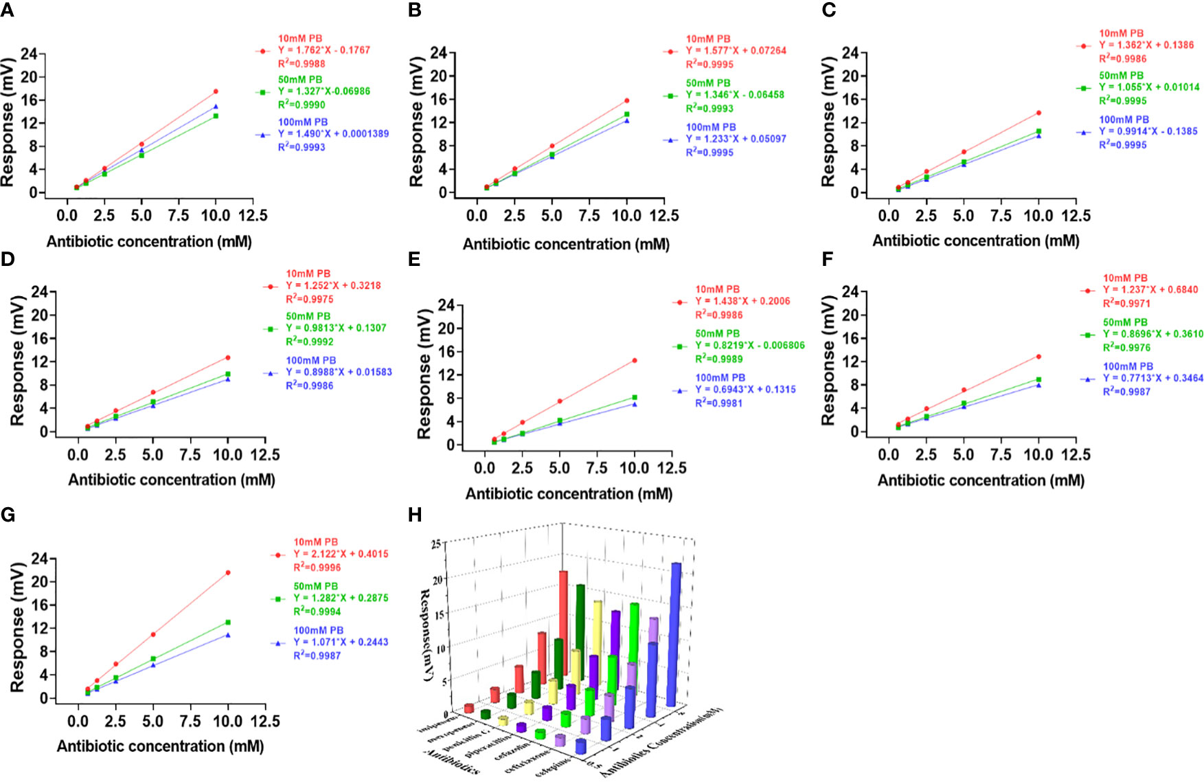
Figure 4 Effects of PB buffer on the activity and profiling of NDM-1 catalysis. The response slopes of 7 antibiotics in buffer concentrations of 10 mM (red), 50 mM (green) and 100 mM (blue) were presented by (A) Imipenem, (B) Meropenem, (C) Penicillin G, (D) Piperacillin, (E) Cefazolin, (F) Ceftriaxone, (G) Cefepime. The catalytic profile was generated using 10 mM PB buffer (H).
In contrast to Penicillinase in Figure 2, the highest sensitivity was obtained with 10 mM buffer and the lowest with 100 mM buffer for both PB (Figure 4) and HEPES (Figure 3) for all 7 antibiotics with the exception of penicillin G in HEPES buffer (Figure 3C). In addition, effect of the buffer concentration on profile variation was the highest in PB (Figures 4E-G), but the lowest in HEPES buffer (Figures 3E-G) for the Cephalosprins antibiotic class (cefepime, ceftriaxone, cefazolin). This finding could be possibly used for differentiating cephalosprins from other classes of antibiotics.
Figure 5 showing the signal response comparison between PB and HEPES buffers was similar to those seen in Figure 3 and 4, i.e. that the highest sensitivity was obtained at 10 mM buffer for almost all antibiotic reactions. Cefazolin showed the lowest response indicating that it generated the least catalytic enthalpy as compared with the other antibiotics. In general, higher sensitivity was observed in PB than HEPES buffer.
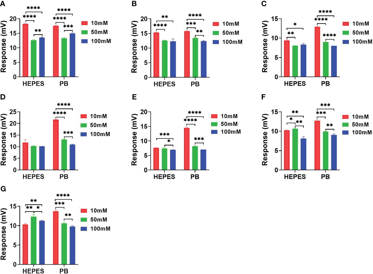
Figure 5 The response comparison between PB and HEPES buffers in 10, 50 and 100 mM concentrations under NDM-1 catalyzation of 7 antibiotics. (A) Imipenem, (B) Meropenem, (C) Penicillin G, (D) Piperacillin, (E) Cefazolin, (F) Ceftriaxone, (G) Cefepime. P value: * < 0.05, ** < 0.01, *** < 0.001, **** < 0.0001.
pH effects on the profiling
The pH effects on the catalytic reactions were evaluated in PB and HEPES buffers at pHs 6.8, 7.2, 7.6 and 8.0 (Figure 6). Using the HEPES buffer, an apparent pH dependent effect on catalytic activity of NDM-1 was observed in both carbapenem antibiotics (Figures 6A, B). In addition, an apparent pH dependent effect on catalytic activity of NDM-1 was observed in both carbapenem and penicillin antibiotics using PB buffer (Figures 6A-D). In addition, the results show that higher sensitivity was observed using PB than HEPES, and the sensitivity variation was also higher for PB than HEPES. Meanwhile, the cefazolin showed the smallest response in HEPES buffer and was least effected by pH. Overall, the differential effect of pH on the catalytic activity of NDM-1 and penicillinase might facilitate differentiation of these two β-lactamases.
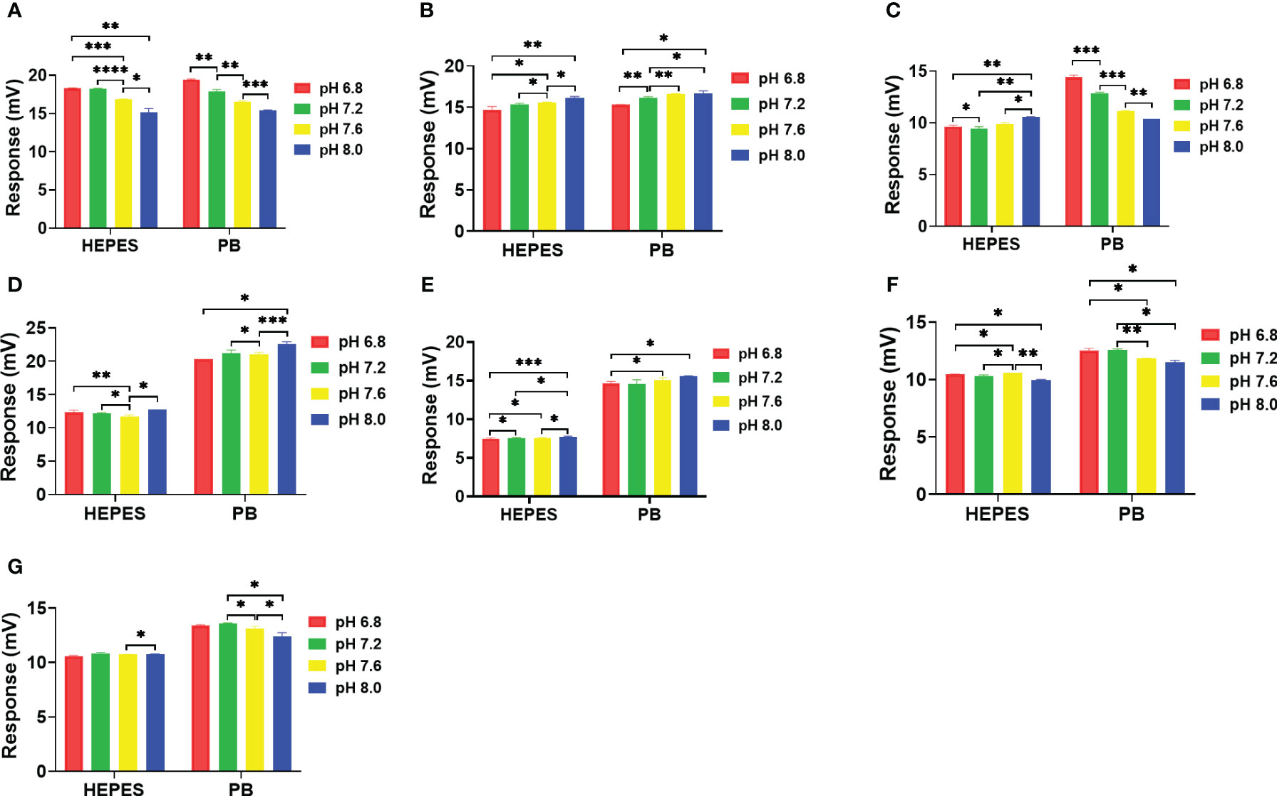
Figure 6 Effect of pH on NDM-1catalysis of 7 antibiotics in 10 mM. (A) Imipenem, (B) Meropenem, (C) Penicillin G, (D) Piperacillin, (E) Cefazolin, (F) Ceftriaxone, (G) Cefepime. P value: * < 0.05, ** < 0.01, *** < 0.001, **** < 0.0001.
Discussion
As enzymatic degradation of antibiotics is the most common mechanism of AMR, we have developed a thermometric biosensor strategy for the rapid identification of AMR enzymes in 35 min using a panel of antibiotics to profile their substrate specificity. In this study, a substrate specificity profile for NDM-1 and penicillinase has been created using a panel of 7 antibiotics including penicillin G, piperacillin, cefepime, ceftriaxone, cefazolin, meropenem, and imipenem. The results show that the strategy is both feasible and reliable.
The concentration dependent calibration curves were prepared using PB and HEPES. The results show that buffer conditions can be employed to further increase the informational content of the assay scheme should the antibiotic profile alone be inadequate to identify the AMR enzyme. The results of the buffer and pH effects on the profiles provided the possibility for differentiation of classes of antibiotics, e.g. carbapenem and peniciilin or cephalosporin in which the carbapenem antibiotics showed highest sensitivity vs cepalosprin which showed the smallest changes in buffer concentration of 100 mM. This conclusion is also supported by the pH studies. Furthermore, the results also indicate that it is feasible to differentiate β-lactamases, e.g. penicillinase and NDM-1 by varying the buffer concentrations. The lowest buffer concentration (10 mM) gave the highest sensitivity for penicillinase reactions vs the lowest sensitivity for NDM-1 reactions but the highest sensitivity in 100 mM buffer.
As expected, penicillinase was most active against the penicillin class of antibiotics, penicillin and Piperacillin. In contrast, NDM-1 could hydrolize all three classes of antibiotics in particular carbapenems. Compared to other methods (see Supplement Table 1), our strategy has a number of distinct advantages. First, the flow injected assays are cost effective and simple to automate. Second, the immobilized β-lactamases were very stable and could be repeatedly used for hundreds of samples which reduces recalibration requirements. Third, substrate specificity profiles for other AMR enzymes can easily be obtained by simply exchanging the enzyme column and repeating the analysis of the panel of antibiotics. Identification of very related AMR enzymes may require the antibiotic panel to be modified. This is a simple to implement since no specialized substrates or assay modifications are needed. By expanding the β-lactamase activity profiling database, the assay will not only be able to identify known AMR enzymes, but could, in the future also be used to identify novel AMR enzymes.
Data availability statement
The raw data supporting the conclusions of this article will be made available by the authors, without undue reservation.
Author contributions
CS: Investigation, comparative study, data curation, formal analysis, visualization, writing - review & editing, XS: Data curation, formal analysis, investigation, methodology, validation, visualization, writing - original draft, writing - review & editing. YW, Investigation, data curation, formal analysis, LB: Funding acquisition, review & editing, supervision, resources. MM: Review &editing, English proofreading. CW: funding acquisition, supervision QM: project administration, formal analysis, writing - review & editing, funding acquisition, resources. BX: Conceptualization, project administration, data curation, formal analysis, methodology, writing - review & editing, funding acquisition, resources, supervision. All authors contributed to the article and approved the submitted version.
Funding
This work was supported by Swedish Research council [VR 5607,2014], Swedish Foundation for Strategic Research [RBP14-0055], the four “batches” Innovation Project of Invigorating Medical Through Science and Technology of Shanxi Province (No. 2022XM13), the Programme of Introducing Talents of Discipline to Universities (No. D21004), National Key Research and Development Project of China (No. 2019YFF0216504), the Innovation Program of graduate in Shanxi (No. SXU2022Y220).
Conflict of interest
Author MM was employed by the company Omik Bioscience AB.
The remaining authors declare that the research was conducted in the absence of any commercial or financial relationships that could be construed as a potential conflict of interest.
Publisher’s note
All claims expressed in this article are solely those of the authors and do not necessarily represent those of their affiliated organizations, or those of the publisher, the editors and the reviewers. Any product that may be evaluated in this article, or claim that may be made by its manufacturer, is not guaranteed or endorsed by the publisher.
Supplementary material
The Supplementary Material for this article can be found online at: https://www.frontiersin.org/articles/10.3389/fcimb.2023.1222156/full#supplementary-material
Supplementary Figure 1 | Reproducibility and life-time of the assay.
Supplementary Table 1 | For comparison of different technologies.
References
Adlerberth, J., Meng, Q., Mecklenburg, M., Tian, Z., Zhou, Y., Bülow, L., et al. (2020). Thermometric analysis of blood metabolites in ICU patients. J. Therm Anal. Calorim 140, 763–771. doi: 10.1007/s10973-019-08873-7
Andersson, A., Chen, Q., Groop, L., Bülow, L., Xie, B. (2017). Continuous and simultaneous determination of venous blood metabolites. Talanta 171, 270–274. doi: 10.1016/j.talanta.2017.05.010
Bennett, J. W., Chung, K. T. (2001). Alexander fleming and the discovery of penicillin. Adv. Appl. Microbiol. 49, 163–184. doi: 10.1016/S0065-2164(01)49013-7
Blair, J. M. A., Webber, M. A., Baylay, A. J., Ogbolu, D. O., Piddock, L. J. V. (2014). Molecular mechanisms of antibiotic resistance. Nat. Rev. Microbiol. 13, 42–51. doi: 10.1038/nrmicro3380
Chen, Q., Andersson, A., Mecklenburg, M., Xie, B. (2013). Fast determination of antibiotics in whole blood. Clin. Microbiol. Infect. 19, 869–874. doi: 10.1111/1469-0691.12074
Chen, Q., Andersson, A., Mecklenburg, M., Xie, B. (2015). A biosensing strategy for the rapid detection and classification of antibiotic resistance. Biosens Bioelectron 73, 251–255. doi: 10.1016/j.bios.2015.06.011
English, B. K., Gaur, A. H. (2010). The use and abuse of antibiotics and the development of antibiotic resistance. Adv. Exp. Med. Biol. 659, 73–82. doi: 10.1007/978-1-4419-0981-7_6
Ge, B., Wang, F., Sjolund-Karlsson, M., McDermott, P. F. (2013). Antimicrobial resistance in campylobacter: susceptibility testing methods and resistance trends. J. Microbiol. Methods 95, 57–67. doi: 10.1016/j.mimet.2013.06.021
Hansen, M. P., Hoffmann, T. C., McCullough, A. R., van Driel, M. L., Del Mar, C. B. (2015). Antibiotic resistance: what are the opportunities for primary care in alleviating the crisis? Front. Public Health 3 (35),1–7. doi: 10.3389/fpubh.2015.00035
March-Rosselló, G. A. (2017). Rapid methods for detection of bacterial resistance to antibiotics. Enferm Infecc Microbiol. Clin. 35 (3), 182–188. doi: 10.1016/j.eimce.2017.02.007
Meng, Q., Liu, S., Meng, J., Feng, J., Mecklenburg, M., Zhu, L., et al. (2021a). Rapid personalized AMR diagnostics using two-dimensional antibiotic resistance profiling strategy employing a thermometric NDM-1 biosensor. Biosens Bioelectron 193, 113526. doi: 10.1016/j.bios.2021.113526
Meng, Q., Wang, Y., Long, Y., Yue, A., Mecklenburg, M., Tian, S., et al. (2021b). Rapid detection of multiple classes of β-lactam antibiotics in blood using an NDM-1 biosensing assay. Antibiotics 10 (9), 1110. doi: 10.3390/antibiotics10091110
Qiao, M., Ying, G. G., Singer, A. C., Zhu, Y. G. (2018). Review of antibiotic resistance in China and its environment. Environ. Int. 110, 160–172. doi: 10.1016/j.envint.2017.10.016
Qie, Z., Ning, B., Liu, M., Bai, J., Peng, Y., Song, N., et al. (2013). Fast detection of atrazine in corn using thermometric biosensors. Analyst 138, 5151–5156. doi: 10.1039/C3AN00490B
Ramanathan, K., Danielsson, B. (2001). Principles and applications of thermal biosensors. Biosens Bioelectron 16, 417–423. doi: 10.1016/S0956-5663(01)00124-5
Rodriguez-Rojas, A., Rodriguez-Beltran, J., Couce, A., Blazquez, J. (2013). Antibiotics and antibiotic resistance: a bitter fight against evolution. Int. J. Med. Microbiol. 303, 293–297. doi: 10.1016/j.ijmm.2013.02.004
Wang, Q., He, Y., Lu, R., Wang, W. M., Yang, K. W., Fan, H. M., et al. (2018). Thermokinetic profile of NDM-1 and its inhibition by small carboxylic acids. Biosci. Rep. 38 (2). doi: 10.1042/BSR20180244
Xie, B., Harborn, U., Mecklenburg, M., Danielsson, B. (1994). Urea and lactate determined in 1-microL whole-blood samples with a miniaturized thermal biosensor. Clin. Chem. 40, 2282–2287. doi: 10.1093/clinchem/40.12.2282
Xie, B., Ramanathan, K., Danielsson, B. (2000). Mini / micro thermal biosensors and other related devices for biochemical / clinical analysis and monitoring. TrAC Trends Anal. Chem. 19, 340–349. doi: 10.1016/S0165-9936(99)00211-3
Xu, N., Bai, J., Peng, Y., Qie, Z., Liu, Z., Tang, H., et al. (2017). Pretreatment-free detection of diazepam in beverages based on a thermometric biosensor. Sensors Actuators B 241, 504–512. doi: 10.1016/j.snb.2016.10.081
Yakovleva, M., Bhand, S., Danielsson, B. (2013). The enzyme thermistor–a realistic biosensor concept. A critical review. Anal. Chim. Acta 766, 1–12. doi: 10.1016/j.aca.2012.12.004
Yakovleva, M., Buzas, O., Matsumura, H., Samejima, M., Igarashi, K., Larsson, P.-O., et al. (2012). A novel combined thermometric and amperometric biosensor for lactose determination based on immobilised cellobiose dehydrogenase. Biosens Bioelectron 31, 251–256. doi: 10.1016/j.bios.2011.10.027
Yao, N., Liu, Z., Chen, Y., Zhou, Y., Xie, B. (2015). A novel thermal sensor for the sensitive measurement of chemical oxygen demand. Sensors (Basel) 15, 20501–20510. doi: 10.3390/s150820501
Keywords: antibiotics, antimicrobial resistance, NDM-1, thermometric biosensor, β-lactamase
Citation: Song C, Sun X, Wang Y, Bülow L, Mecklenburg M, Wu C, Meng Q and Xie B (2023) Activity fingerprinting of AMR β-lactamase towards a fast and accurate diagnosis. Front. Cell. Infect. Microbiol. 13:1222156. doi: 10.3389/fcimb.2023.1222156
Received: 16 May 2023; Accepted: 16 August 2023;
Published: 05 September 2023.
Edited by:
Zichen Yang, Xinqiao Hospital, ChinaReviewed by:
Saeed Ahmed, National University of Medical Sciences (NUMS), PakistanQun Chen, Xi’an Jiaotong University, China
Copyright © 2023 Song, Sun, Wang, Bülow, Mecklenburg, Wu, Meng and Xie. This is an open-access article distributed under the terms of the Creative Commons Attribution License (CC BY). The use, distribution or reproduction in other forums is permitted, provided the original author(s) and the copyright owner(s) are credited and that the original publication in this journal is cited, in accordance with accepted academic practice. No use, distribution or reproduction is permitted which does not comply with these terms.
*Correspondence: Bin Xie, YmluLnhpZUB0Ymlva2VtLmx0aC5zZQ==; Qinglai Meng, cW1lbmdAc3h1LmVkdS5jbg==