- Department of Pulmonary and Critical Care Medicine, Tianjin Medical University Baodi Hospital, Tianjin, China
Objective: To investigate the diagnostic value of metagenomic next-generation sequencing (mNGS) and targeted next-generation sequencing (tNGS) in identifying pathogens in patients with pulmonary infections.
Methods: A retrospective analysis was conducted on 155 patients with suspected lung infections who underwent alveolar lavage and were admitted to the Department of Respiratory and Critical Care Medicine at Baodi Hospital, Tianjin Medical University, from July 2023 to December 2023. The bronchoalveolar lavage fluid (BALF) samples obtained were subjected to mNGS, tNGS and culture methods to compare their diagnostic efficacy in identifying lung infection pathogens.
Results: The results indicated that both tNGS and mNGS methods exhibit comparable detection efficiencies in identifying pathogens in patients with pulmonary infections, significantly outperforming BALF culture approach. In terms of diagnostic accuracy, tNGS exhibited a higher sensitivity than mNGS, with rates of 96.1% and 75.7% respectively (P>0.05). However, the specificity of tNGS was slightly lower than that of mNGS, with rates of 59.1% and 68.2% respectively (P>0.05). It is noteworthy that this difference in specificity was not statistically significant.
Conclusion: tNGS exhibits a diagnostic efficacy comparable to mNGS, particularly in its sensitivity for identifying lung infections, as evidenced by expert insights and clinical applications. Furthermore, tNGS offers advantages in convenience, time efficiency, and cost-effectiveness, hinting at its potential to serve as an alternative to mNGS in clinical settings.
1 Introduction
Lung infections constitute a prevalent and substantial global health issue, imposing a considerable socioeconomic burden on societies worldwide. Early identification of the underlying pathogen is paramount for the formulation of an appropriate and effective clinical treatment plan. Traditional methods of pathogen detection, such as pathogen culture, antigen and antibody detection, and smear microscopy, each possess their unique advantages and disadvantages. However, the overall detection rates achieved through these methods remain unsatisfactory, with approximately 50% of respiratory infections remaining unidentified (Zhu et al., 2018).
In recent years, high-throughput sequencing technologies have garnered increasing attention in clinical settings due to their enhanced sensitivity and reduced detection time. Among these, metagenomic next-generation sequencing (mNGS) and targeted next-generation sequencing (tNGS) have emerged as prominent tools. mNGS possesses the capability to detect a wide array of pathogenic microorganisms, including emerging and rare species (Hilt and Ferrieri, 2022). However, mNGS also has limitations, such as lower sensitivity compared to PCR at low viral loads, inability to distinguish infecting pathogens from colonizing bacteria, and a lack of standardized reporting and interpretation criteria (Chiu and Miller, 2019; Diao et al., 2021; Liu, 2024). Currently, mNGS is typically considered when patients remain undiagnosed by traditional microbiological methods and empirical anti-infective therapy has been ineffective. On the other hand, tNGS utilizes polymerase chain reaction (PCR) technology to specifically amplify target genes, thereby minimizing interference from host nucleic acids. This method demonstrates enhanced specificity and sensitivity in detecting pathogens (Leo et al., 2017). Despite the growing utilization of mNGS and tNGS, there is a scarcity of comprehensive studies comparing their efficacy in diagnosing pulmonary infectious diseases.
The present study compared the results of mNGS and tNGS in bronchoalveolar lavage fluid (BALF) samples obtained from 155 patients with suspected pulmonary infections, primarily to evaluate their effectiveness and differences in pathogen detection in these patients. Furthermore, this study aimed to explore the respective advantages and disadvantages in the clinical application of mNGS and tNGS, thereby contributing to the ongoing advancement in the diagnosis and management of pulmonary infectious diseases.
2 Methods
2.1 Study subjects
A retrospective analysis was conducted on the clinical data of patients admitted to the Department of Respiratory and Critical Care Medicine, Baodi Hospital of Tianjin Medical University, between July 2023 and December 2023. Patients suspected of lung infections underwent BALF mNGS and tNGS, which are advanced diagnostic techniques that enable the detection of a wide range of pathogens, including bacteria, viruses, fungi, and parasites, in respiratory samples. Inclusion criteria encompassed: 1) patients admitted to the aforementioned department with suspected lung infections during the specified timeframe; and 2) patients who consented to bronchoscopy and BALF collection for mNGS and tNGS analysis. Exclusion criteria comprised: 1) individuals who did not complete bronchoscopy or failed to successfully obtain lavage fluid samples; and 2) patients with incomplete clinical data. Clinicians determine the clinical relevance of NGS-detected microorganisms (categorized as pathogenic, opportunistic, or colonizing) by integrating findings from the patient’s clinical presentation, immune status, blood biochemical parameters, imaging features, and conventional microbiological tests. Additionally, the interpretation of negative NGS results requires correlation with clinical manifestations and supplementary diagnostic evidence. The interpretation of these results was conducted by three trained physicians. This study was approved by The Ethics Committee of Baodi Hospital affiliated to Tianjin Medical University (No. BDYYLL202506) and was conducted according to the principles of the Helsinki Declaration.
2.2 Bronchoscope irrigation procedure
Following the insertion of the bronchoscope’s tip into the selected bronchial branch, 20–40 mL of saline was administered. The resulting BALF was then collected and transferred into a sterilized container for subsequent culture, mNGS detection, and tNGS detection. Samples were stored on dry ice for 2 hours before being dispatched to the Tianjin Kingmed Diagnostics Laboratory Co.,Ltd. for further mNGS and tNGS analysis.
2.3 Microbiological culture
BALF specimens were inoculated onto blood agar plates, MacConkey agar plates, Sabouraud dextrose agar plates, and chocolate agar plates using a sterile inoculation loop. Blood agar and chocolate agar plates were incubated at 35°Cunder 5% CO2 atmosphere, while MacConkey agar and Sabouraud dextrose agar plates were cultured in a conventional incubator at 35°C. Following 18–24 hours of continuous culture, bacterial growth was examined across all plates. Preliminary identification was performed based on colonial morphology and Gram-staining characteristics. Subsequent species-level identification was conducted according to the biological characteristics of each bacterial type, accompanied by antimicrobial susceptibility testing.
2.4 mNGS assay and report
Upon receipt, the samples underwent centrifugation and processing. Nucleic acids were extracted using the Universal DNA/RNA Extraction Kit for Environmental Samples (TR202JY-50, Jianshi Biotechnology, Beijing, China) through centrifugal column-based protocols. After qubit quantification, the nucleic acids were subjected to standardized DNA library preparation procedures. PCR amplification and sequencing on an MGISEQ-200 sequencer were then executed, and the raw sequencing data were filtered using Fastp (v0.20.0) (Chen et al., 2018) with culling the reads containing more than 40% of low-quality bases (mass value less than 15), the reads containing more than 5 n-bases, the reads matched to the sequencing primer, the PCR repeat sequence, and the human nucleic acid sequences. The non-human high-quality sequences were then compared with the microbial genome database by BWA (v0.7.17-r1198-dirty) (Li, 2013), and the results of microbial species identification were obtained. The microbial database was collected from the NCBI RefSeq and GenBank databases.
2.5 tNGS assay and report
After liquefying the treated samples, nucleic acid extraction and quantification were performed. Total nucleic acid extraction was performed either manually or through automation using the Nucleic Acid Extraction/Purification Kit (KS118-BYTQ-96, Guangzhou KingCreate Biotechnology Co. Ltd., Guangzhou, China) on the KingFisher™ Flex Purification Platform (Thermo Fisher Scientific, Waltham, USA). The RP100 Respiratory Pathogen Microorganisms Multiplex Testing Kit (Guangzhou KingCreate Biotechnology Co. Ltd., Guangzhou, China) on BALF were used to conduct library preparation. PCR amplification was used to enrich the target pathogens, and steps were taken to eliminate the interference from human background nucleic acid within the sample. Qualified libraries were used as input for sequencing, which was performed on the KM MiniSeqDx-CN Platform (Guangzhou KingCreate Biotechnology Co. Ltd., Guangzhou, China). The raw sequencing data were filtered with criteria consistent with mNGS to obtain the non-human high-quality sequences. The resulting data were then aligned against a reference database using Bowtie2 v2.4.1 (Langmead and Salzberg, 2012) in “very-sensitive” mode. The reference database was compiled from multiple sources, including the GenBank database, RefSeq database, and NT database from the National Center for Biotechnology Information (NCBI)
2.6 Statistical analysis
All statistical analyses were conducted using R software (version 4.3.1). The Wilcoxon test was used to evaluate differences between the two groups, considering P<0.05 as statistically significant. ROC curves were generated to assess the diagnostic efficacy of the assays, providing a visual representation of their sensitivity and specificity, and enabling the comparison of their diagnostic performance through the area under the curve (AUC). Count data were presented as cases or percentages (%), and the chi-squared test was used to compare the sensitivity and specificity between mNGS and tNGS.
2.7 Data availability
The raw sequence data reported in this paper have been deposited in the Genome Sequence Archive (Genomics, Proteomics & Bioinformatics 2021) in National Genomics Data Center (Nucleic Acids Res 2022), China National Center for Bioinformation/Beijing Institute of Genomics, Chinese Academy of Sciences (GSA: CRA021644) that are publicly accessible at https://ngdc.cncb.ac.cn/gsa (Chen et al., 2021; CNCB-NGDC Members and Partners, 2022).
3 Result
3.1 Patient characteristics
In our study, a total of 155 patients were enrolled in this analysis. Among them, 125 patients were confirmed to have pulmonary infection, while the remaining 30 patients were diagnosed with non-pulmonary infections. The demographic and clinical information of all cases between two groups were collected, including gender, age, length of stay, white blood cell count, neutrophil ratio and primary disease, as summarized in Table 1. We observed significant differences in length of stay, white blood cell count, and neutrophil ratio between patients with pulmonary infections and those with non-pulmonary infections, highlighting clinical distinctions between the two groups.
3.2 Pathogen detection using mNGS, tNGS and culture methods
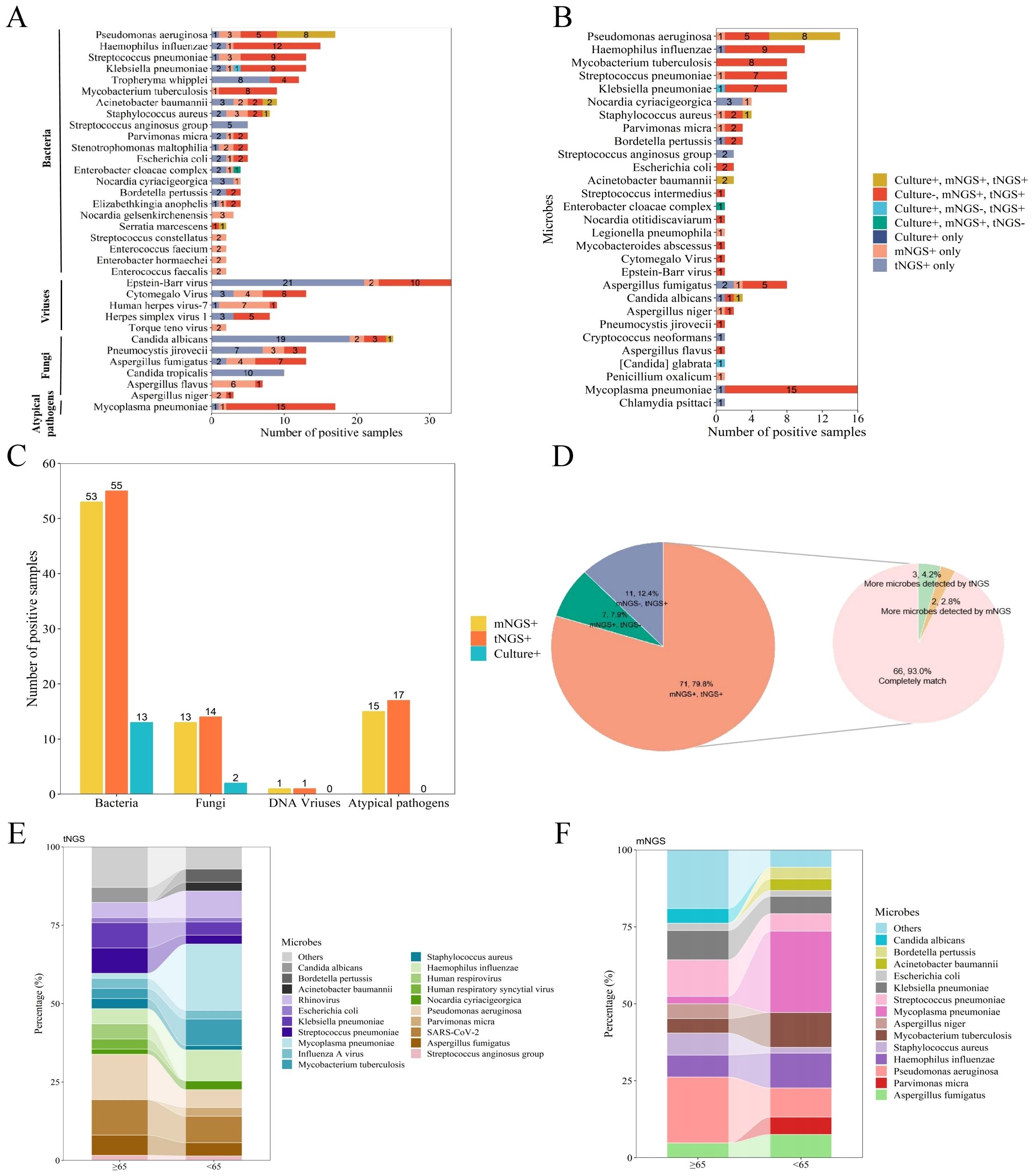
A total of 155 specimens were subjected to test through mNGS, tNGS, and BALF culture protocols. The mNGS technique identified 210 strains belonging to 49 different microorganisms, while tNGS detected 248 strains from 44 microorganisms. BALF culture, on the other hand, revealed 19 strains from 11 microorganisms. Among the bacteria, the three most frequently detected were Pseudomonas aeruginosa (5.2%), Haemophilus influenzae (4.6%), and Streptococcus pneumoniae (4.0%). For viral detection, the top three pathogens identified were Epstein-Barr virus (10.0%), cytomegalo virus (4.0%), and human herpes virus 7 (2.7%). For fungal detection Candida albicans (7.6%), Pneumocystis jirovecii (4.0%), and Aspergillus fumigatus (4.0%) were the predominant fugal pathogens (Figure 1A).

Figure 1. An evaluation of the efficacy in identifying pathogens utilizing mNGS, tNGS, and culture methods. (A) A categorization of the distribution of microorganisms detected in more than two samples through the application of mNGS, tNGS, and culture assays. (B) The distribution of potential pathogens and the respective contributions of tNGS, mNGS, and culture in the detection of these pathogens. (C) A comparison of the number of positive samples for bacteria, fungi, DNA viruses, and atypical pathogens among tNGS, mNGS, and culture methods. (D) A pie chart illustrating the consistency of results between mNGS and tNGS, with double positive results further categorized into completely matches, more microbes detected by mNGS, and more microbes detected by tNGS. (E) The variability in the distribution of pathogens across different age groups when utilizing tNGS. (F) The variability in the distribution of pathogens across different age groups when utilizing mNGS.
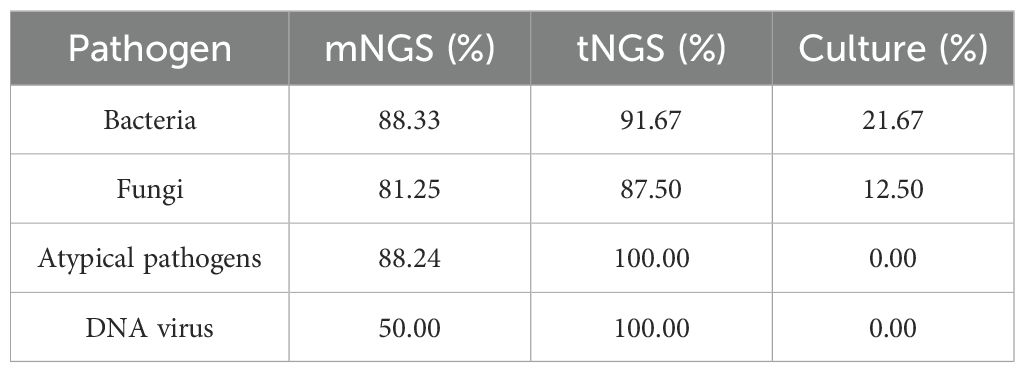
Among the 125 patients diagnosed with pulmonary infections, pathogenic microorganisms were identified in 103 cases (82.4%), with 30 of these cases diagnosed as mixed infections. A total of 110 pathogens were detected among 103 patients. As demonstrated in Figure 1B, the majority of pathogens were successfully detected by both mNGS and tNGS, showcasing their complementary strengths in pathogen identification. In contrast, BALF culture identified fewer pathogens, indicating its limitations in sensitivity compared to the sequencing technologies. Additionally, tNGS and mNGS demonstrated similar efficacy in detecting specific microbial types and co-infection, and significantly outperformed traditional BALF culture, as shown in Figure 1C, Table 2 and Supplementary Table 1.

Table 2. Positive sample detection rates for mNGS, tNGS, and culture methods across various microbial types.
Furthermore, we conducted a comparative analysis to evaluate the detection rates of positive samples and the discrepancies in identifying pathogenic microorganisms using two distinct approaches. As showed in Figure 1D, 71 cases (79.8%) were positive with both tNGS and mNGS, while 11 cases (12.4%) were positive only with tNGS and 7 cases (7.9%) were exclusively positive with mNGS. Among patients with positive results in both tNGS and mNGS, the pathogens detected by both methods matched exactly in 66 cases (93.0%). More pathogens were detected by tNGS in three cases (4.2%), while more pathogens were detected by mNGS in two cases (2.8%). The results indicate that both tNGS and mNGS methods have comparable detection efficiencies in identifying pathogens in patients with pulmonary infections, significantly surpassing the BALF culture approach, thereby highlighting the superior performance of NGS detection methods in pathogen detection.
Concurrently, we performed a comparative analysis to examine the variations in the distribution of BALF pathogens across different age cohorts. We found that, for both mNGS and tNGS, the predominant pathogens in patients aged 65 and above were Pseudomonas aeruginosa, Streptococcus pneumoniae, Klebsiella pneumoniae, and Candida albicans. Among patients younger than 65 years, microorganisms such as Parvimonas micra, Haemophilus influenzae, Mycoplasma pneumoniae and Bordetella pertussis were more prevalent and ranked higher in the pathogen spectrum (Figures 1E, F). Previous studies have shown that Klebsiella pneumoniae and Pseudomonas aeruginosa in acute respiratory infections follow an age-related pattern, with the positive detection rates increasing with patient age, consistent with the findings of this study. The consistency between mNGS and tNGS in the analyzing pathogen distribution across different age groups further supports the reliability and effectiveness of both in pathogen detection.
3.3 Clinical evaluation of mNGS, tNGS and culture methods
To assess the clinical diagnostic performance of the three methodologies, we calculated the sensitivity, specificity, and accuracy for the different detection methods based on the clinical diagnosis of each sample, as shown in Table 3. Among the three detection techniques, tNGS exhibited the highest sensitivity of 96.1%, followed by mNGS with 75.7%, while culture demonstrated the lowest sensitivity at 14.6%. Conversely, the specificity trend was inverse to that of sensitivity, with culture achieving 100.0% specificity, mNGS at 68.2%, and tNGS at 59.1%, respectively. In summary, tNGS demonstrated a marginally superior clinical diagnostic performance compared to mNGS, while culture fared the worst, with F1 score of 0.830, 0.938, and 0.254, respectively. Our comparative analysis of tNGS and mNGS demonstrated a diagnostic concordance rate of 70.4% between the two methods (Table 4). Besides, a chi-squared test was subsequently performed to assess the sensitivity and specificity of tNGS and mNGS. The results demonstrated that there was no statistically significant statistical difference in the sensitivity and specificity of the two methods, with P values of 0.248 and 0.290, respectively (Tables 5, 6).
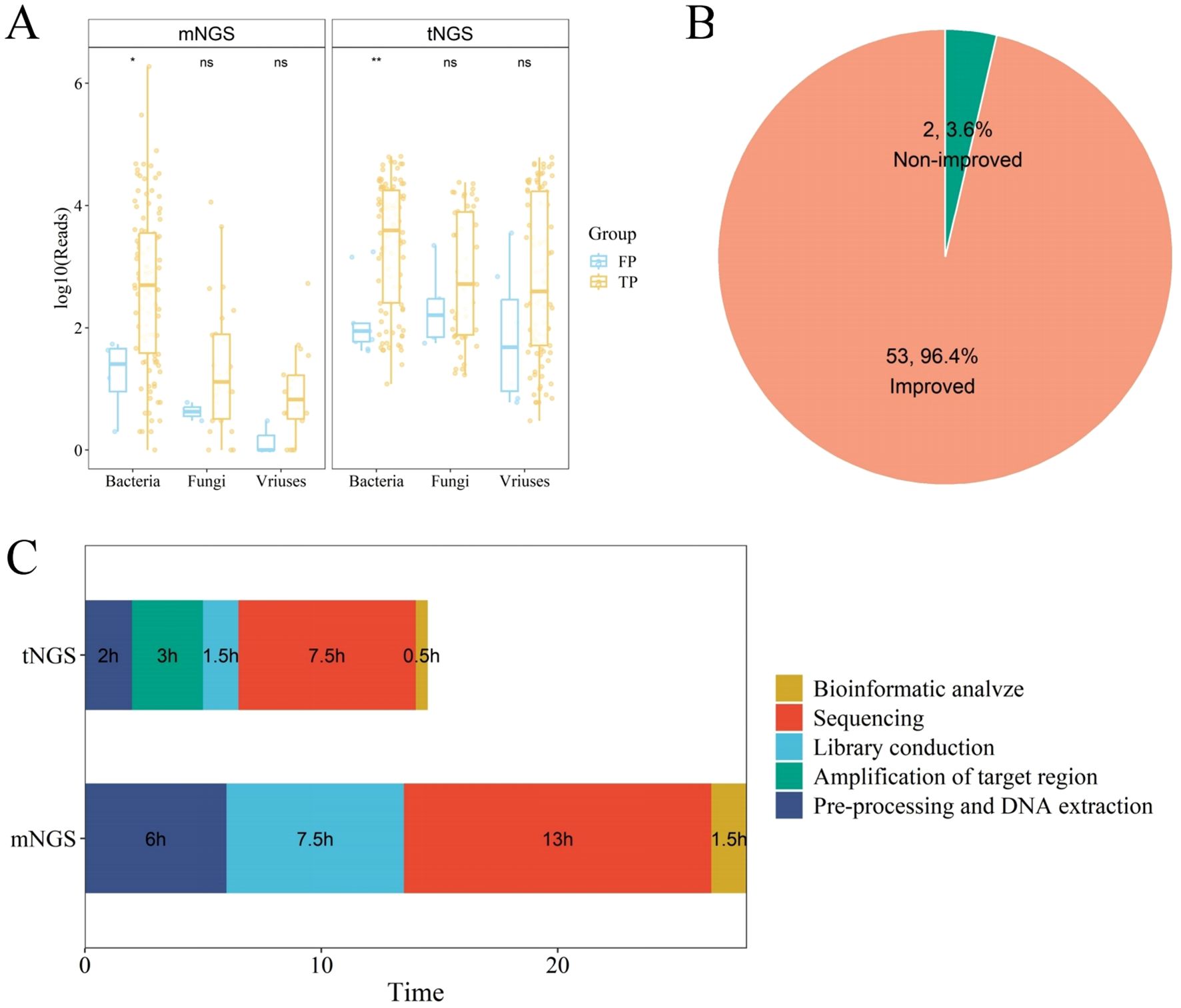
Samples categorized into true positive (TP) and false positive (FP) groups were further compared based on either mNGS or tNGS. For mNGS method, 78 samples were regard as TP, while 7 samples were classified as FP. For tNGS method, 99 and 9 samples were grouped into TP and FP, respectively. A comparative analysis of the sequence counts for specific microbial types in TP and FP groups revealed that the count of bacterial sequences in TP samples was significantly higher than that in FP samples. Similarly, the counts of fungal and viral sequences also exhibited this pattern (Figure 2A). This suggests that the quantity of microbial sequences could be considered as a criterion for determining the authenticity of pathogenic infection.

Figure 2. Evaluation of Clinical Diagnostic Efficacy of mNGS and tNGS Methods. (A) Box plot showing the log10(reads) in the true positive (TP) group compared with that in the false positive (FP) group. (B) Pipe chart revealing the improvement of 55 patients adjusted the anti-infective treatment. (C) Comparison of turnaround time between mNGS and tNGS.
A review of the treatment records of 125 patients with lung infections revealed that 55 patients had their anti-infective treatment adjusted based on the results of the NGS test. Among these 55 patients, 53 (96.4%) experienced a significant clinical improvement, whereas 2 (3.6%) did not respond to the treatment (Figure 2B; Supplementary Table 2). Additionally, the NGS test results of 47 patients were concordant with the initial clinical assessments, allowing the continuation of the initial anti-infective treatment regimen. The treatment plans of two patients were altered for reasons not associated with NGS test results, and no changes were made for 21 patients whose pathogens were not detected by NGS. Moreover, the average testing duration for tNGS was 14.5 hours, compared to 28 hours for mNGS (Figure 2C). Compared to mNGS and traditional culture methods, tNGS offers a swift and reliable turnaround time (TAT), which can aid physicians in delivering more accurate anti-infective treatments to patients.
4 Discussion
With the emergence of new pathogens and the increasing drug resistance of pathogens, it is difficult to meet the needs of disease diagnosis and treatment with the traditional clinical pathogen detection methods, and there is an urgent need for new faster and more sensitive pathogen detection methods to guide clinical diagnosis and treatment. Recently, the utilization of mNGS in clinical settings has progressively gained traction. Numerous reports have highlighted its superiority over conventional detection methods. However, it has its limitations, such as the requirement for DNA and RNA separation, susceptibility to interference from host nucleic acids, and a comparatively high cost (Diao et al., 2021; Moragues-Solanas et al., 2021). On the contrary, tNGS combines the advantages of ultra-multiplexed PCR amplification with high-throughput sequencing, allowing for targeted sequencing of specific pathogenic microorganisms. This approach boasts advantages such as cost-effectiveness, customizability, and a shortened detection timeframe. The objective of this study was to comprehensively assess the diagnostic efficacy of both mNGS and tNGS in BALF samples, ultimately providing a valuable reference for clinical practice.
In this study, we conducted a retrospective analysis of the outcomes from mNGS and tNGS tests performed on BALF samples from 155 patients suspected of having lung infections, with a focus on understanding the technical differences and clinical applications of these next-generation sequencing technologies. Among the microorganisms ultimately identified as causative agents, the three most commonly detected bacteria were Pseudomonas aeruginosa, Haemophilus influenzae, and Streptococcus pneumoniae. This prevalence may be attributed to the high incidence of comorbid underlying diseases and the advanced average age of the patients in our study. Previous research has indicated that in elderly patients or those with underlying diseases, the pathogens responsible for lung infections are predominantly gram-negative bacteria, such as Pseudomonas aeruginosa and Klebsiella pneumoniae (Han et al., 2018; Zhang et al., 2023). As for fungal pathogens, the most commonly identified species include Candida albicans, Pneumocystis jirovecii, and Aspergillus fumigatus, findings that are consistent with relevant epidemiological studies (Peng et al., 2018; Li et al., 2021). The top three DNA viruses detected were EBV, cytomegalo virus, and human herpes virus 7, with their distribution patterns mirroring those reported by Liu et al (Liu et al., 2022). Notably, the number of EBV strains detected by tNGS was significantly higher than that by mNGS (31 vs. 12 strains), but most were considered to be part of the respiratory tract’s normal flora.
Except Epstein-Barr virus, Gram-negative bacilli such as Haemophilus influenzae, Klebsiella pneumoniae, and Pseudomonas aeruginosa may exist as colonizing organisms. Their colonization patterns are typically associated with host immune status, environmental exposures, and microbiome interactions, particularly in immunocompromised individuals or those with prolonged hospitalization (Man et al., 2017). In immunocompetent populations, Pneumocystis jirovecii colonization is frequently observed in the absence of overt clinical symptoms or imaging abnormalities (Aguilar et al., 2021), and clinicians should comprehensively evaluate clinical context to determine their pathogenic role. The disparity in detection rates between mNGS and tNGS might be explained by the fact that mNGS sequencing necessitates the elimination of host background genes, which could impact its detection efficiency for gram-negative bacteria, viruses, and intracellular bacteria (Thorburn et al., 2015; Zhou et al., 2019). Furthermore, this result underscores the exceptional sensitivity of tNGS in detecting viruses. Comparative analyses revealed that mNGS and tNGS exhibited similar detection capabilities for various types of respiratory pathogens, both surpassing the traditional culture method in effectiveness, in line with the findings of Yin et al. (Yin et al., 2024). However, it was also concluded that mNGS demonstrated significantly superior performance than tNGS in identifying pathogenic respiratory pathogens within BALF samples (Simner et al., 2018).
An in-depth analysis of the distribution patterns of pathogens across diverse age groups revealed that, in patients aged 65 and above, the predominant pathogens were Pseudomonas aeruginosa, Streptococcus pneumoniae, Klebsiella pneumoniae, and Candida albicans. Conversely, Parvimonas micra, Haemophilus influenzae, Mycoplasma pneumoniae, and Bordetella pertussis were more prevalent among patients younger than 65 years. Prior research has established that, in the context of acute respiratory infections, Klebsiella pneumoniae and Pseudomonas aeruginosa exhibit an age-dependent distribution, with positive detection rates escalating with advancing age. These findings are in concordance with the outcomes of the present study (Prina et al., 2015; Cassini et al., 2019; Hu et al., 2020).
Regarding diagnostic efficacy, the sensitivity of tNGS was found to be superior to both mNGS and culture methods, with a sensitivity of 96.1% compared to 75.7% for mNGS and 14.6% for culture. However, in terms of specificity, the culture method demonstrated the highest specificity at 100.0%, followed by mNGS at 68.2% and tNGS at 59.1%. It is noteworthy that the statistical difference in sensitivity and specificity between mNGS and tNGS was insignificant. The results indicated that mNGS and tNGS exhibited notable advantages compared to BALF culture in detecting a wide range of pathogens, albeit with a lower specificity than the culture method. Numerous studies have been conducted to validate the superiority of mNGS and tNGS over traditional culture techniques in BALF samples (Wu et al., 2020; Liu et al., 2022; Yin et al., 2024), particularly for pathogens that are recalcitrant to cultivation.
A comparative analysis of the sequence counts of specific microbial types within the TP and FP groups, utilizing mNGS and tNGS assays, has shown that TP samples exhibit significantly higher sequence counts of bacteria, fungi, and viruses compared to FP samples. It is evident that a heightened sequence count is indicative of a greater likelihood of infection and can serve as a marker for clinicians in determining the underlying pathogens. However, it is important to note that the level of pathogen sequence count is influenced by a multitude of factors. Even if two samples exhibit an equivalent number of detected pathogen sequence counts, the actual concentration of the pathogens may vary. Consequently, sequence counts cannot be solely utilized as a definitive criterion for judgment. Rather, they must be interpreted in conjunction with the types of microorganisms detected, the clinical manifestations presented by the patients, and the outcomes of other laboratory tests.
Among all patients diagnosed with pulmonary infections, a substantial proportion of 44.0% (55/125) adjusted their anti-infective treatment regimens based on the outcomes derived from NGS. Notably, an overwhelming 96.4% (53/55) of these patients exhibited improvements in their clinical symptoms following the adjustment. The incorporation of NGS technology has markedly enhanced the precision of pathogen detection, a factor that is indispensable for the effective guidance of clinical treatment strategies. Moreover, tNGS boasts a significantly shorter average testing duration than mNGS. This advantage allows physicians to swiftly identify potential pathogens in patients, enabling more precise and efficient tailoring of treatment plans.
In summary, the clinical diagnostic efficacy of tNGS is comparable to that of mNGS in the context of clinical pulmonary infections. While tNGS does not fully supplant mNGS in identifying emerging and rare pathogens, it is significant to note that the predominant pathogens involved in clinical pulmonary infections are common ones, effectively detectable by tNGS. tNGS possesses the capability to detect both DNA and RNA simultaneously, exhibiting high sensitivity for pathogens present in low abundance within samples. Consequently, tNGS emerges as a novel solution for detecting the pathogens of clinical pulmonary infections.
However, it is imperative to acknowledge several limitations of this study: Firstly, as a retrospective single-center study, the inclusion of a relatively small sample size may introduce potential biases in the results. Secondly, RNA virus sequencing was not performed using mNGS, thereby limiting the ability to assess the diagnostic efficacy of mNGS specifically for RNA viruses. Lastly, the interpretation of the results obtained from both mNGS and tNGS relies heavily on the clinician’s combined judgment of the patient’s clinical presentation and the test results, which introduces a degree of subjectivity in the interpretation process.
Data availability statement
The datasets presented in this study can be found in online repositories. The names of the repository/repositories and accession number(s) can be found below: https://ngdc.cncb.ac.cn/gsa, CRA021644.
Ethics statement
The studies involving humans were approved by The Ethics Committee of Baodi Hospital affiliated to Tianjin Medical University, Tianjin, China. The studies were conducted in accordance with the local legislation and institutional requirements. The human samples used in this study were acquired from a by- product of routine care or industry. Written informed consent for participation was not required from the participants or the participants’ legal guardians/next of kin in accordance with the national legislation and institutional requirements.
Author contributions
YT: Writing – original draft. ST: Writing – review & editing. YZ: Writing – review & editing. ZL: Writing – review & editing. SS: Writing – review & editing, Methodology.
Funding
The author(s) declare that no financial support was received for the research and/or publication of this article.
Acknowledgments
We thank the team of Clinical Genome Center in Tianjin Kingmed Diagnostics Laboratory Co. Ltd for their contributions. We also thank the Guangzhou KingCreate Biotechnology Co. Ltd. for their technical assistance.
Conflict of interest
The authors declare that the research was conducted in the absence of any commercial or financial relationships that could be construed as a potential conflict of interest.
Generative AI statement
The author(s) declare that no Generative AI was used in the creation of this manuscript.
Publisher’s note
All claims expressed in this article are solely those of the authors and do not necessarily represent those of their affiliated organizations, or those of the publisher, the editors and the reviewers. Any product that may be evaluated in this article, or claim that may be made by its manufacturer, is not guaranteed or endorsed by the publisher.
Supplementary material
The Supplementary Material for this article can be found online at: https://www.frontiersin.org/articles/10.3389/fcimb.2025.1552236/full#supplementary-material
References
Aguilar, Y. A., Rueda, Z. V., Maya, M. A., Vera, C., Rodiño, J., Muskus, C., et al. (2021). Is it possible to differentiate pneumocystis jirovecii pneumonia and colonization in the immunocompromised patients with pneumonia? J. Fungi (Basel). 7, 1036. doi: 10.3390/jof7121036
Cassini, A., Högberg, L. D., Plachouras, D., Quattrocchi, A., Hoxha, A., Simonsen, G. S., et al. (2019). Attributab le deaths and disability-adjusted life-years caused by infections with antibiotic-resistant bacteria in the EU and the European Economic Area in 2015: a population-level modelling analysis. Lancet Infect. Dis. 19, 56–66. doi: 10.1016/S1473-3099(18)30605-4
Chen, T., Chen, X., Zhang, S., Zhu, J., Tang, B., Wang, A., et al. (2021). The genome sequence archive family: toward explosive data growth and diverse data types. Genomics Proteomics Bioinf. 19, 578–583. doi: 10.1016/j.gpb.2021.08.001
Chen, S., Zhou, Y., Chen, Y., and Gu, J. (2018). fastp: an ultra-fast all-in-one FASTQ preprocessor. Bioinformatics 34, i884–i890. doi: 10.1093/bioinformatics/bty560
Chiu, C. Y. and Miller, S. A. (2019). Clinical metagenomics. Nat. Rev. Genet. 20, 341–355. doi: 10.1038/s41576-019-0113-7
CNCB-NGDC Members and Partners (2022). Database resources of the national genomics data center, China national center for bioinformation in 2022. Nucleic Acids Res. 50, D27–D38. doi: 10.1093/nar/gkab951
Diao, Z., Han, D., Zhang, R., and Li, J. (2021). Metagenomics next-generation sequencing tests take the stage in the diagnosis of lower respiratory tract infections. J. Adv. Res. 38, 201–212. doi: 10.1016/j.jare.2021.09.012
Han, X., Zhou, F., Li, H., Xing, X., Chen, L., Wang, Y., et al. (2018). Effects of age, comorbidity and adherence to current antimicrobial guidelines on mortality in hospitalized elderly patients with community-acquired pneumonia. BMC Infect. Dis. 18, 192. doi: 10.1186/s12879-018-3098-5
Hilt, E. E. and Ferrieri, P. (2022). Next generation and other sequencing technologies in diagnostic microbiology and infectious diseases. Genes (Basel). 13, 1566. doi: 10.3390/genes13091566
Hu, Y., Liu, C., Shen, Z., Zhou, H., Cao, J., Chen, S., et al. (2020). Prevalence, risk factors and molecular epidemiology of carbapenem-resistant Klebsiella pneumoniae in patients from Zhejiang, China, 2008-2018. Emerg. Microbes Infect. 9, 1771–1779. doi: 10.1080/22221751.2020.1799721
Langmead, B. and Salzberg, S. L. (2012). Fast gapped-read alignment with Bowtie 2. Nat. Methods 9, 357–359. doi: 10.1038/nmeth.1923
Leo, S., Gaïa, N., Ruppé, E., Emonet, S., Girard, M., Lazarevic, V., et al. (2017). Detection of bacterial pathogens from broncho-alveolar lavage by next-generation sequencing. Int. J. Mol. Sci. 18, 2011. doi: 10.3390/ijms18092011
Li, H. (2013). Aligning sequence reads, clone sequences and assembly contigs with BWA-MEM [Preprint. arXiv. doi: 10.48550/arXiv.1303.3997
Li, Z., Li, Y., Chen, Y., Li, J., Li, S., Li, C., et al. (2021). Trends of pulmonary fungal infections from 2013 to 2019: an AI-based real-world observational study in Guangzhou, China[J. Emerging Microbes infections 10, 450–460. doi: 10.1080/22221751.2021.1894902
Liu, B. M. (2024). Epidemiological and clinical overview of the 2024 Oropouche virus disease outbreaks, an emerging/re-emerging neurotropic arboviral disease and global public health threat. J. Med. Virol. 96, e29897. doi: 10.1002/jmv.29897
Liu, H., Zhang, Y., Chen, G., Sun, S., Wang, J., Chen, F., et al. (2022). Diagnostic significance of metagenomic next-generation sequencing for community-acquired pneumonia in southern China. Front. Med. (Lausanne). 9. doi: 10.3389/fmed.2022.807174
Man, W. H., de Steenhuijsen Piters, W. A., and Bogaert, D. (2017). The microbiota of the respiratory tract: gatekeeper to respiratory health. Nat. Rev. Microbiol. 15, 259–270. doi: 10.1038/nrmicro.2017.14
Moragues-Solanas, L., Scotti, R., and O’Grady, J. (2021). Rapid metagenomics for diagnosis of bloodstream and respiratory tract nosocomial infections: current status and future prospects. Expert Rev. Mol. Diagn. 21, 371–380. doi: 10.1080/14737159.2021.1906652
Peng, L., Xu, Z., Huo, Z., Long, R., and Ma, L. (2018). New insights into the clinical characteristics and prognostic factors of pulmonary fungal infections from a retrospective study in Southwestern China. Infection Drug Resistance 11, 307–315. doi: 10.2147/IDR.S157030
Prina, E., Ranzani, O. T., Polverino, E., Cillóniz, C., Ferrer, M., Fernandez, L., et al. (2015). Risk factors associated with potentially antibiotic-resistant pathogens in community-acquired pneumonia. Ann. Am. Thorac. Soc 12, 153–160. doi: 10.1513/AnnalsATS.201407-305OC
Simner, P. J., Miller, S., and Carroll, K. C. (2018). Understanding the promises and hurdles of metagenomic next-generation sequencing as a diagnostic tool for infectious diseases. Clin. Infect. Dis. 66, 778–788. doi: 10.1093/cid/cix881
Thorburn, F., Bennett, S., Modha, S., Murdoch, D., Gunson, R., and Murcia, P. R. (2015). The use of next generation sequencing in the diagnosis and typing of respiratory infections. J. Clin. Virol. 69, 96–100. doi: 10.1016/j.jcv.2015.06.082
Wu, X., Li, Y., Zhang, M., Li, M., Zhang, R., Lu, X., et al. (2020). Etiology of severe community-acquired pneumonia in adults based on metagenomic next-generation sequencing: A prospective multicenter study. Infect. Dis. Ther. 9, 1003–1015. doi: 10.1007/s40121-020-00353-y
Yin, Y., Zhu, P., Guo, Y., Li, Y., Chen, H., Liu, J., et al. (2024). Enhancing lower respiratory tract infection diagnosis: implementation and clinical assessment of multiplex PCR-based and hybrid capture-based targeted next-generation sequencing. EBioMedicine 107. doi: 10.1016/j.ebiom.2024.105307
Zhang, L., Xiao, Y., Zhang, G., Li, H., Zhao, J., Chen, M., et al. (2023). Identification of priority pathogens for aetiological diagnosis in adults with community-acquired pneumonia in China: a multicentre prospective study. BMC Infect. Dis. 23, 231. doi: 10.1186/s12879-023-08166-3
Zhou, X., Wu, H., Ruan, Q., Jiang, N., Chen, X., Shen, Y., et al. (2019). Clinical Evaluation of Diagnosis Efficacy of Active Mycobacterium tuberculosis Complex Infection via Metagenomic Next-Generation Sequencing of Direct Clinical Samples. Front. Cell Infect. Microbiol. 9. doi: 10.3389/fcimb.2019.00351
Keywords: mNGS, tNGS, bronchoalveolar lavage fluid, pathogens, pulmonary infection
Citation: Tang Y, Tang S, Zhang Y, Lin Z and Shan S (2025) The significance of metagenomic next-generation sequencing and targeted next-generation sequencing in the diagnostic evaluation of patients with suspected pulmonary infections. Front. Cell. Infect. Microbiol. 15:1552236. doi: 10.3389/fcimb.2025.1552236
Received: 27 December 2024; Accepted: 26 May 2025;
Published: 12 June 2025.
Edited by:
Jiemin Zhou, Vision Medicals Co, Ltd, ChinaReviewed by:
Benjamin M. Liu, George Washington University, United StatesKaifeng Wu, Zunyi Medical University Third Affiliated Hospital, China
Copyright © 2025 Tang, Tang, Zhang, Lin and Shan. This is an open-access article distributed under the terms of the Creative Commons Attribution License (CC BY). The use, distribution or reproduction in other forums is permitted, provided the original author(s) and the copyright owner(s) are credited and that the original publication in this journal is cited, in accordance with accepted academic practice. No use, distribution or reproduction is permitted which does not comply with these terms.
*Correspondence: Shuxiang Shan, c3N4ZGVAMTYzLmNvbQ==
 Yucong Tang
 Shujin Tang


