Abstract
Understanding how platelets can sense and respond to hemodynamic forces in disturbed blood flow and complexed vasculature is crucial to the development of more effective and safer antithrombotic therapeutics. By incorporating diverse structural and functional designs, microfluidic technologies have emerged to mimic microvascular anatomies and hemodynamic microenvironments, which open the floodgates for fascinating platelet mechanobiology investigations. The latest endothelialized microfluidics can even recapitulate the crosstalk between platelets and the circulatory system, including the vessel walls and plasma proteins such as von Willebrand factor. Hereby, we highlight these exciting microfluidic applications to platelet mechanobiology and platelet–circulatory system interplay as implicated in thrombosis. Last but not least, we discuss the need for microfluidic standardization and summarize the commercially available microfluidic platforms for researchers to obtain reproducible and consistent results in the field.
Introduction
While three determinants for thrombosis—hypercoagulability, endothelial dysfunction, and hemodynamics—are outlined in Virchow's triad (1), platelets play an essential role in arterial thrombosis. Their thrombotic functions are governed by a range of biomechanical factors and the underlying mechanobiology is a rapidly evolving field and topical research area (2). Intriguingly, platelets respond to flow disturbance in complex vessel architectures with respect to bifurcation, branching, stenosis and aneurysm (3), which inevitably produce local shear gradients (4–6), vorticity (7) and even turbulence (4, 8). The biomechanical platelet adhesion and aggregation are primarily mediated by its two mechanoreceptors—glycoprotein Ib (GPIb) and glycoprotein IIb/IIIa (GPIIb/IIIa or integrin αIIbβ3) (9, 10). Furthermore, to facilitate thrombus stabilization, platelets generate contractile forces which are mediated by cytoskeletal components (actin, tubulin, Arp2/3) and motor proteins (myosin and dynein) in response to the mechanical microenvironment (11, 12). Meanwhile, platelets interact with red blood cells, neutrophils, plasma proteins [von Willebrand factor (VWF), fibrinogen], and the vessel wall (endothelial cells) to exert their hemostatic and thrombotic functions (13). Increasingly, microfluidic technologies have emerged as powerful and indispensable approaches to investigate the mechanosensitive behaviors of platelets (2, 12, 14). Numerous endothelialized microfluidics have been invented to model the interplay between platelets, endothelium and VWF under physiologically relevant hemodynamic microenvironments (6, 15, 16). In the context of platelet mechanobiology, we summarize these state-of-the-art microfluidic methodologies that have recently been invented in the field.
Microfluidic Approaches for Investigating Platelet Mechanobiology and Platelet–Circulatory System Interplay
Single-cell biomechanical nanotools such as atomic force microscopy (17), optical tweezers (18) and micropipette based adhesion assays (19) significantly advanced our understanding of platelet mechanobiology (20). However, a major shortcoming of these in vitro techniques is their inability to recapitulate the physiologically relevant hemodynamics in the context of platelet adhesion and aggregation (21). To this end, animal models of thrombosis and intravital microscopies have become more popular and accessible approaches for examining thrombus initiation, progression and propagation in vivo (22). Nevertheless, multiple fundamental limitations still exist with the animal approaches (23, 24): (i) inter-species variabilities which prevent complete recapitulation of human disease pathogenesis; (ii) physiological variations between individual animals; (iii) lengthy ethical approval processes.
Shear-based in vitro assays, such as parallel plate flow chambers (25), cone-and-plate viscometers (26) and thromboelastometers (27) have been widely used for investigations of human blood samples under physiologically relevant shear rates and shear stresses (5, 28–30). Although these methods were instrumental in the current understanding of shear dependent platelet thrombosis, to some extent, they are limited due to the need for large sample volumes, low throughput, and their inability to accurately emulate vascular architectures and mechanical properties of the circulatory system (31–33). To these points, microfluidic techniques have rapidly emerged as complementary humanized models for investigations of platelet mechanobiology and biomechanical thrombosis. More recently, the International Society of Thrombosis and Hemostasis (ISTH) have even made recommendations for producing reliable microfluidic devices with reproducible thrombogenic coating and consistent hemodynamic design (34). This represents a major step forward in promoting microfluidic technologies for future pre-clinical and diagnostic testing methods in hemostasis and thrombosis.
Moreover, while most of the research interests focus on large vessel thrombosis, there is an emerging trend toward understanding thrombosis in the microcirculation which is significantly implicated in a variety of systemic disorders such as: Thrombotic Thrombocytopenia Purpura (TTP) and Hemolytic Uremic Syndrome (HUS) which lead to multi-organ dysfunction syndrome and death (35, 36); and localized organ injuries resulting from trauma, ischemia-reperfusion, transplant rejection, disseminated intravascular coagulation, sickle cell disease and more recently COVID-19 (35, 37, 38). The microfluidic approach can effectively recapitulate the anatomical structures within such small scale as opposed to conventional shear-based assays (39). The recent advancements of microfluidic designs and the micro and nano fabrication—particularly the shift from devices with a single layer to those with multiple-layered structures (40), and from parallel straight channels to complex geometries (15)—have enabled the investigation of platelet mechanobiology in more complicated vascular biomechanical microenvironments.
In the past decade, soft lithography has rapidly grown to enable high-fidelity micropatterning. Parallel straight channels can simply be fabricated by the single-layered photolithography and poly(dimethylsiloxane) (PDMS) casting processes, resulting in a vascular geometry with laminar flow (5, 16, 41, 42). When connected with external syringe pumps, such simple microfluidic setting enables systematic investigation of shear dependent platelet adhesion (43) and aggregation (44). Additionally, vascular cell cultures can be incorporated; specifically, endothelialized microchannels can model the interaction between blood components and the vessel wall under physiological hemodynamic parameters (45), or even when subjected to proinflammatory or other endothelial diseased conditions (16).
Furthermore, single-layered microfluidics can emulate more complex vessel structures, such as bifurcation (39), 2D stenosis (46), network (47), and micropost array (40) to recapitulate flow disturbance induced platelet activation, adhesion, contraction and aggregation (48, 49). The mechanical stimuli investigated include elevated shear stress (50, 51), shear rate gradient (52–54), vorticity (7) and turbulence (8, 55) at different locations of the microchannel. Moreover, lining the microchannel with endothelial cells enables the investigation of prothrombotic synergistic effects of hemodynamic forces, blood cells and the vessel wall (56, 57). Rapid prototyping methods such as vertical milling molding (58) and 3D printing (56) enable fabrication of microfluidics with circular structures and variations in the z-direction. PDMS casting around poly(methyl methacrylate) (PMMA) optical fibers can also create circular channels (59, 60). As such, these recent microfluidic advancements provide great means for examining how shear stress, shear rate gradients, vorticity, blood viscosity and contractility affect platelet thrombotic functions.
In the past 3 years, in vitro lung (61), blood vessel (62), and tumor models (63) were made on microfluidics (64). These biomimetic organ-on-a-chip models recapitulate the structural, functional, and mechanical aspects of vascular microenvironments. Obviously, emulating such hierarchical organs requires emerging microfabrication methods including 3D bioprinting (65), double-layered soft lithography (66), injection molding (67), and micropattern stamping (68). This allows the production of multiple-layered structures via one-time or assembled fabrication. The composite microsystems can support tissue coculture and the channel-channel interface, thereby enabling studies of bidirectional tissue signaling across the endothelial barrier (61). More importantly, external mechanical manipulation apparatus (e.g., stiff ECM matrices, actuation chambers) can be integrated for disease-specific studies and rapid drug screening (55, 69). Although multiple-layered microfluidics incur higher fabrication requirements including precise alignment and assembly, they present physiologically relevant hemodynamic microenvironments for better biomimetic performance.
Last but not least, the transparency of PDMS material is greatly compatible with advanced microscopies to visualize thrombotic dynamics in real time. These PDMS or equivalent microfluidics thus offer a versatile way to evaluate platelet activation, adhesion, aggregation, morphological change, and soluble agonist secretion under hemodynamic control. A range of studies have delved into reproducing diverse vessel architectures to investigate how biomechanical factors (e.g., shear stress, shear gradients and traction forces) and the circulatory components (e.g., vessel endothelium and VWF) regulate platelet functions. As shown in the Figure 1 and Table 1, we summarize these recent lab-on-chip approaches in the context of platelet mechanobiology and thrombosis.
Figure 1
Table 1
| Microfluidics description | Fabrication method | Mechanical profile | Vasculature status | Perfusion sample | Functionality test | References |
|---|---|---|---|---|---|---|
| Single layered simple structure microfluidics | ||||||
| Straight channel | Soft lithography | Pump-driven constant shear input and output | HUVECs w/wo TNF-α stimulation | Recalcified human WB | Thrombus formation or platelet function assay | (16, 41, 42) |
| Straight channel | Soft lithography | Pump-driven constant shear input and output | Primary HMVECs | Recalcified human WB | Clot formation upon diabetic complications | (45) |
| Single layered complex structures microfluidics | ||||||
| Ladder network channel | Soft lithography | Pump-driven constant flow input and geometry mediated flow output | CGN/TF coated bare channel | Recalcified human WB | Shear rate gradient-dependent platelet adhesion, aggregation | (47) |
| Bifurcation channel | Soft lithography | Pump-driven constant flow input and geometry mediated flow output | HUVECs/HLMVECs | Human WB | Hematologic microvascular occlusion | (118) |
| Junction with multiple inlets and single outlet | Soft lithography | Pump-driven constant flow input and output | HUVECs | Recalcified human WB | Outside-in interference of thrombosis | (72) |
| 2D stenosis channel | Soft lithography | Pump-driven constant flow input and geometry mediated flow output | Non-coated bare channel | Citrated human WB | Shear rate gradient-dependent platelet adhesion, aggregation | (46) |
| 2D stenosis channel with non-uniform height | Aluminum vertical milling molding; Soft lithography | Pressure-driven constant flow input and geometry mediated flow output | Type I CGN coated bare channel | Porcine blood | Shear-dependent platelet aggregation | (73) |
| Circular 3D stenosis channel | 3D printing; Soft lithography | Pump-driven constant flow input and geometry mediated flow output | HUVECs | Citrated human WB | Flow-mediated blood-EC interaction and thrombosis | (56) |
| Flexible micropost array | Soft lithography | No external mechanical input | FNG or FNT coated array | Platelet-rich plasma | Platelet contractile forces characterization | (49) |
| Two-layered microfluidics | ||||||
| Parallel channels with non-uniform heights and widths | Soft lithography | Gravity driven constant flow input and geometry mediated flow output | ECM molecules coated bare channel | Human/mouse WB | Shear-dependent platelet adhesion | (66) |
| Microscale blocks and flexible posts in the channel | Assembly soft lithography | Pump-driven constant flow input and geometry mediated flow output | CGN or VWF coated bare channel | Citrated human WB | Hemodynamic effect on platelet contractility | (40) |
| Three-layered microfluidics | ||||||
| Assembled web network | Assembly soft lithography; Injection molding with Type 1 Collagen hydrogel | Gravity driven constant flow input and geometry mediated flow pattern | HUVECs only or cocultured with HBVPCs/HUASMCs | Citrated human WB | Angiogenesis and thrombosis; Flow-driven assembly of VWF fibers and its interaction with platelet | (67) |
| Two chambers separated by porous membrane layer | 3D printing; Assembly soft lithography | Pump-driven constant flow input and output | HUVECs/HPMECs cocultured with alveolar epithelium | Recalcified human WB | Platelet-endothelial dynamics in pulmonary thrombosis | (61) |
| Top pressure actuation chamber, middle diaphragm, bottom channel | Assembly soft lithography; injection molding | Pneumatic peristaltic pump-driven flow | Type 1 CGN coated bare channel | Citrated human WB/washed platelet | Platelet thrombosis assay and antiplatelet drug screening | (55) |
| Micropatterns printed on microfluidic channel | Assembly soft lithography; micropattern with stamping; hydrogel casting | Pump-driven constant flow and ECM mediated stiffness stimuli | FNG coated micropattern | Platelet-rich plasma | Hemodynamic and ECM influences on platelet contractility | (68) |
Summary of microfluidic models for platelet mechanobiology in thrombotic diseases.
WB, Whole Blood; EC, Endothelial cell; HUVECs, Human umbilical vein endothelial cells; HMVECs, Primary human cardiac microvascular endothelial cells; HBVPCs, Human brain vascular pericytes; HUASMCs, Human umbilical arterial smooth muscle cells; HLMVECs, Human lung microvascular endothelial cells; HPMECs, Primary Human Pulmonary Microvascular Endothelial Cells; ECM, Extracellular matrix; TNF-α, tumor necrosis factor-alpha; VWF, von Willebrand Factor; TF, Tissue factor; FNG, Fibrinogen; CGN, Collagen.
Shear Dependent Platelet Adhesion
Earlier endeavors in the field focused on developing high-throughput microfluidic systems with parallel straight microchannels to assess shear effects on platelet thrombotic functions (21). One of the novel straight channel microfluidic models was developed by Gutierrez et al., that validated the use of adhesion assays with VWF, fibrinogen, or collagen-coated substrates (66). The model contained eight rectilinear channels that covered a 100-fold range of shear rates to recapitulate the venous and arterial flow conditions. Such approaches can achieve high-throughput examination and avoid matrix variability between different microchannels. Similar studies were later conducted by Conant et al., who developed a 2D narrowing microfluidic device consisting of 24 microchannels coated with a range of matrix proteins on a 48-well plate. Each of their microchannels was coupled with two wells (Figure 1A) (70). By changing the flow inputs with a syringe pump, platelet adhesion assays were conducted under controllable physiological and pathological hemodynamic conditions. These two high-throughput microfluidic systems substantially shortened the time for platelet adhesion analysis. As a key advantage, these microdevices use small volume of blood samples to produce a range of replicable results.
Flow Disturbance Dependent Platelet Aggregation
In pathological contexts, the aforementioned straight microfluidics could not effectively recapitulate flow disturbance at stenotic and bifurcated vasculatures (74).
To this end, Tovar-Lopez et al. proposed a 2D stenosed microfluidic model to assess the role of shear gradients on platelet aggregation at three observation zones: the pre-stenosis for shear acceleration; the apex of stenosis for maximum shear; and the post-stenosis for shear deceleration (46). Interestingly, stable platelet aggregation was only found at the post-stenotic zone with shear deceleration. Costa et al. presented similar observation in 3D coronary artery mimicking microfluidics. The 3D vessel geometries and stenoses were reconstructed from computed tomography angiography, and then manufactured into a microfluidic channel using stereolithography 3D-printing and PDMS soft lithography (56). The computational fluid dynamic simulation identified that backflow at the distal stenosis associates with rapid platelet aggregation. These results were comparable to the studies by Menon et al., using circular microfluidics with various degrees of stenosis in 2D and 3D (57). When Vascularized with endothelial cells, these microfluidics revealed that elevated shear stress at the stenotic regions activated endothelial cells into pro-thrombotic and pro-inflammatory states. Nevertheless, the round surface of these circular microfluidics makes it difficult in visualizing platelet adhesion and aggregation.
In addition, new microfluidic designs were made to investigate the prothrombotic role of shear gradients with respect to vessel bifurcations. The bifurcations produce flow separations, shear gradients, and oscillatory flows which promote atherosclerotic lesion initiation and thrombus formation (75). Li et al. designed a bifurcated stenosis device to mimic constricted regions in coronary arteries, where a mother microchannel is subdivided into two smaller daughter microchannels (73). Occlusive thrombi were formed extensively at higher shear rates between 1,500 and 4,000 s−1, but not at lower shear rates <1,500 s−1. Additionally, the complex hemodynamics at vessel bifurcations were also investigated in a multi-bypass ladder model developed by Zilberman-Rudenko et al. which consisted of two parallel channels and 10 T-junction-like bypasses (47). Thrombus formation was found to be heightened at channel intersections which displaying skewed flow profiles, stagnation zones, flow separation, recirculation zones, and shear gradients.
Furthermore, research has been undertaken into automating the occlusion time calculation as thrombi form. Jain et al. designed a stenosed arteriole mimicking microfluidic device with multiple regions of pre- and post-stenosis (Figure 1C) (71). A mathematical model was used to quantitate shear gradient induced thrombosis in real time, indicating the steady reaction, growth, and saturation phases of thrombus development. They have also demonstrated the use of this microdevice to assess antithrombotic drug effects on individual patients.
Platelet Contraction
Activated platelets in newly formed aggregates are highly contractile with respect to thrombus stabilization and consolidation (76–81). Recent studies have sought to analyze the degree of forceful cytoskeletal contractions in activated platelets. Notably, Muthard and Diamond designed a microblock microfluidic model composed of a primary flow channel and a side scaffold channel to investigate platelet contractile forces during thrombus formation under arterial shear conditions (77). Platelet contraction occurred dramatically during flow arrest, and a consequent increase in the clot permeability was observed (77, 78). While Liang et al. quantified platelet retraction force without physiological flow using flexible post array (49), Ting et al. devised a microfluidic arrays of rectangular blocks and flexible microposts which were subjected to arterial blood flow (Figure 1D) (40, 79). They measured the deflections of microposts caused by contracting platelet aggregates around the rectangular block, thereby quantifying the ensemble platelet contractile forces, in which myosin II, ADP activation and integrin GPIIb/IIIa were found to play key roles. Further, Myers et al. designed an array of fibrinogen-coated microdots patterned on a hydrogel substrate, where platelets adhering to the microdot extended filopodia to the adjacent microdot (68). The displacement of the microdots due to platelet contraction enabled quantification of platelet contractility in a cytometry manner. Evidently, the micropost array is advantageous to visualize and measure platelet contractility and can be extended to the diagnosis of reduced platelet contractility associated with bleeding conditions including Wiskott Aldrich Syndrome (WAS) (68) and Immune Thrombocytopenia (ITP) (82).
Platelet–VWF Association
In addition to the hemodynamic microenvironment, platelets interact with the circulatory components including the vessel walls, blood cells (red blood cells, T-cells) and plasma proteins (VWF, fibrinogen, tissue factor, collagen) (83). Particularly, VWF is central to primary hemostasis in mediating platelet recruitment to the damaged vascular subendothelium and subsequent platelet aggregation (84). The critical role of VWF in regulating platelet biomechanical adhesion and aggregation was investigated in numerous microfluidic systems.
Notably, Dong et al. employed a commercial parallel plate flow chamber comprising a polycarbonate slab, silicon gasket and an endothelial cell-lined glass coverslip to study platelet GPIbα–VWF mediated platelet adhesion (85). Upon endothelial activation, platelets adhered to the released ultra-large VWF and formed extraordinary long ‘beads-on-a-string'-like structure at a venous shear rate. Using the same parallel plate device, Padilla et al. proposed to reduce platelet-endothelial VWF interaction by exposing the VWF-A2 domain to ADAMTS13 under elongational shear (86). However, such parallel plate flow chamber cannot investigate more disturbed hemodynamic conditions due to its laminar flow profile.
Zheng et al. designed a complex endothelialized microvascular network (Figure 1E) (67). Such microfluidic model was fabricated by injection molding type I collagen between two plexiglass pieces and PDMS for stabilization. Zheng et al. then optimized the model to incorporate multiple vascular geometries including straight, grid, tortuous, stenosed channels (15) to investigate flow pattern-driven platelet–VWF interaction. The fluid dynamic profile near the bifurcation and vessel junctions enhanced VWF self-association, causing irreversible rapid platelet adhesion and aggregation. Westein et al. fabricated an atherosclerotic vessel mimicking microfluidic device with a half-circular eccentric stenosis (20–80% stenosis) and confirmed that plasma VWF strongly influenced the proaggregatory response (Figure 1F) (6). The VWF multimers were found to elongate with shear gradients and occupied a 15-times higher surface area in deceleration zones promoting extensive platelet aggregation. The significance of VWF was further supported by Kim et al. using a stenotic microfluidic model (87). As such, complex platforms with diverse geometries is beneficial to the study of thrombotic therapies targeting VWF self-association and the subsequent platelet responses (88).
Platelet–Endothelium Interaction
Endothelial expression of P-selectin, E-selectin, vascular cell adhesion molecule 1 (VCAM-1) and intercellular adhesion molecule 1 (ICAM-1) can regulate platelet activation and adhesion (83). Understanding the influence of the endothelium on platelet binding kinetics is essential to unravel the mechanism underlying platelet thrombus formation.
Ciciliano et al. employed a T-shape endothelialized microfluidic platform to recapitulate FeCl3-induced thrombosis in vitro (72). The platform is composed of a main channel where endothelial cells were cultured to form a confluent endothelium, and a side channel where FeCl3 was infused to join the main channel to create the blood-FeCl3 interface. The FeCl3 influx induced endothelial injury, followed by the binding between Fe3+ ions and negatively charged proteins, which lead to platelet activation and occlusive aggregation. Dupuy et al. developed a thromboinflammation model by using six parallel microchannels with fixed human umbilical vein endothelial cells (HUVECs) alignment (16). Tumor necrosis factor alpha (TNF-α) treatment stimulated the expression of ICAM-1 and VCAM-1, and the secretion of VWF and tissue factor on the endothelium, leading to a dose-dependent increase in platelet–endothelial binding. Further, Jain et al. utilized the same model and found that TNF-α activated HUVECs increased E-selectin expression (41).
Notably, Jain et al. exhibited a triple-layered human lung alveolus-on-chip to study the interplay between platelets, vessel wall, and blood flow (Figure 1G) (61). The model mainly contains an upper microchannel coated with primary human lung alveolar epithelial cells and a lower microchannel lined with HUVECs, separated by a thin and porous membrane. Remarkably, TNF-α and lipopolysaccharide endotoxin stimulated the alveolar epithelium and increased the pulmonary vascular permeability, resulting in enhanced ICAM-1 expression and platelet-endothelial binding.
Microfluidic Standardization
Increased usages of shear-based microdevices by different laboratories for examining platelet functions lead to heterogenous device characteristics. Lack of standardization in microfluidic designs and operating methods complicates the result comparison and reproducibility, which impedes translation into both clinical practices and industrial settings (89, 90). The ISTH Biorheology subcommittee has advocated for standardization of flow chamber-based thrombus formation assays (34, 89, 91, 92). Requirements for microfluidic standardization can be extended to the following considerations.
(i) Structural design. To test the abnormalities in platelet mechanosensing, several microfluidic designs can be standardized: shear dependent platelet aggregation can be assessed in straight channels; shear gradient dependent thrombus development can be investigated in stenotic channels; and micropillar array can be standardized for platelet contractility measurements (5). Standardizations of these geometries and dimensions ensure unified microfluidics to be used for platelet function tests by different laboratories (93).
(ii) Surface treatment. Adhesive ligands should be considered carefully to induce platelet adhesion, aggregation and contraction (94–97). Specifically, concentration, duration of treatment, molecular composition, coating procedure of the adhesive proteins needs to be specified for standard usage (95). Moreover, the source, type, passage number, culture medium, seeding procedure, culture duration, inflammatory stimulation of cell-derived surface should be considered thoroughly.
(iii) Sample preparation and perfusion. It is important to ensure appropriate handling of the perfusion samples which include whole blood and washed platelets. Source of blood samples, procedure and handling of blood collection, storage, and the use of anticoagulants are to be standardized (94).
(iv) Hemodynamic settings. Microfluidic device materials, tubing dimensions, perfusion pumps and choice of bulk shear rates are essential when conducting flow tests (98). Standardizations of these hemodynamic settings including the inertial forces and Reynolds number within the microfluidic systems will allow more certain conclusions to be extrapolated (93).
(v) Visualization and quantification methods. Standardization of microscopy-based analytical approaches can be based on different stages of thrombus formation indicated by platelet adhesion receptors, activated ligand expression and aggregate volume. The types, sources, and concentrations of fluorescent stains and antibodies should be specified to control adverse chemical and phototoxic effect on platelet functions. The method of quantification of the fluorescent microscopy data needs to be standardized to allow more transparency in obtained results.
Commercially Available Microfluidics for Laboratory Use
The geometry of microfluidic devices is of the utmost relevance to their application in platelet function studies and their extension to antiplatelet drug screening platforms. Commercially available microfluidic systems have been able to standardize more simple, quasi-2D planar geometries but are still limited by their inability to create complex geometry in a high-throughput manner. A summary of studies looking at platelet biology in thrombosis using commercialized microfluidic slides is presented in Table 2.
Table 2
| Flow chamber | Surface treatment | Vasculature status | Shear rate(dyne/cm2) | Perfusion sample | Investigation | References | Representative image |
|---|---|---|---|---|---|---|---|
| Ibidi μ-slide VI0.4 | Collagen /thrombin | No ECs | Static | Washed platelet | Platelet activation plasminogen activator inhibitor 1 | (99) | 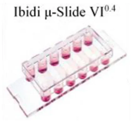 |
| Ibidi μ-slide VI0.4 | No specific coating | HPAECs | 150 s−1 | Mouse/human WB | Platelet activation lethal sepsis | (100) | |
| Ibidi μ-slide VI0.4 | Human Fc-podoplanin fusion protein | LECs /HUVECs | 50–1,350 s−1 | Mouse/human WB | Effect of CLEC-2-podoplanin interactions on Platelet adhesion | (101) |  |
| IbidiÒ μ-slides | Fibrinogen | No ECs | Static | Human WB | Platelet adhesion and thrombo-inflammation | (102) | |
| IbidiÒ μ-slides/Biotechs FCS2 flow chamber | Collagen type 1 | No ECs | 200–1,000 s−1 | Washed platelets | Platelet–monocyte interactions | (103) | |
| Cellix Vena8 Fluoro+ biochip | Fibrillar collagen | No ECs | 200–1,000 s−1 | Washed platelet/Human WB | Effects of β amyloid peptides on platelet | (104) | |
| Cellix Vena8 Fluoro + biochip | Collagen type 1 | No ECs | 90 s−1 | Platelet-rich plasma | Effect of omega-3 fatty acids on platelet aggregation | (105) | |
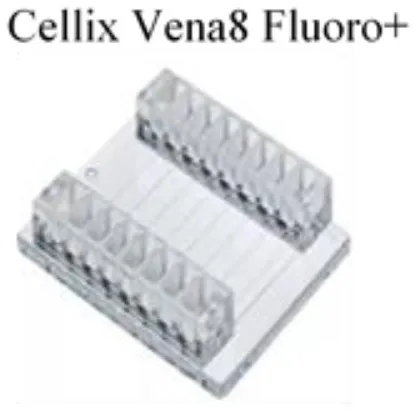 | |||||||
| Cellix Vena8 biochips | Collagen/fibrinogen | No ECs | 0.5 dyne/cm2 | Platelet-rich plasma/human WB | Effect of prostaglandin on platelet aggregation | (106) | 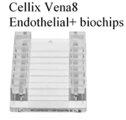 |
| Cellix Vena 8 Endothelial + biochips | Fibronectin | HCAECs | 0.5 dyne/cm2 | Washed platelets | Effect of plasma protein on platelet-endothelium crosstalk | (107) | |
| Cellix Vena8 Fluoro+ biochip | Collagen type 1 | No ECs | 67.5 dyne/cm2 | Human WB | Effect of Gut Microbial Metabolite TMAO on platelet and thrombosis | (99) | |
| Cellix Vena8 biochips | Fibrinogen/collagen | HUVECs w/wo TNF-α stimulation | 0.3 dyne/cm2 | Unwashed platelet-rich plasma/washed platelet | Platelet activation and adhesion to diseased endothelium | (100) | 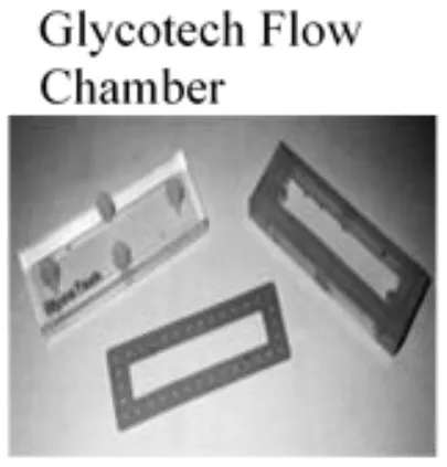 |
| Cellix Vena8 Fluoro+ biochip | Equine tendon collagen | No ECs | 10 dyne/cm2 | Platelet-rich plasma | Effect of legacy perfluoroalkyl substances on platelet dynamics | (101) | |
| Glycotech parallel-plate flow chamber | No specific coating | HUVECs/HUAECs/HMVEC | 2–50 dyne/cm2 | Washed platelets | VWF- platelet interaction in TTP | (102) | 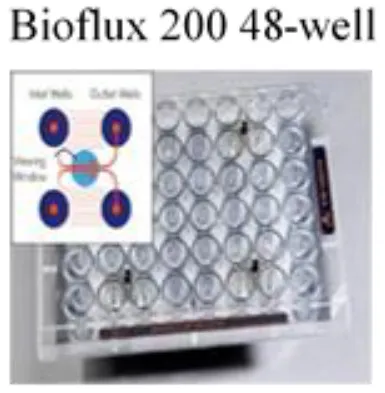 |
| Glycotech Parallel-plate flow chamber | No specific coating | HUVECs | 2.5 dyne/cm2 | Washed platelets | VWF- platelet interaction | (103) | |
| Fluxion Biosciences, high-shear, 48-well Bioflux plates | Fibronectin | HUVECs w/wo TNF-α stimulation | 5 dyne/cm2 | Human WB | TF-driven platelet adhesion and aggregation | (104) | |
| Fluxion Biosciences, 48-well Bioflux plates | Fibronectin | HUVECs | 10 dyne/cm2 | Platelet-rich plasma/human WB | PF4-heparin interaction in HIT | (105) | 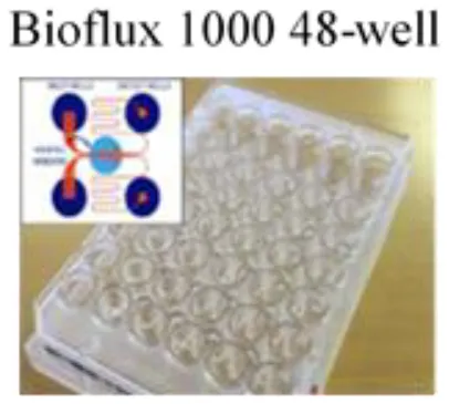 |
| Fluxion Biosciences, 48-well Bioflux plates | Fibronectin | HUVECs | 10 dyne/cm2 | Human WB | PF4-VWF-HIT antibody interaction | (106) |
Commercialized microfluidic slides for laboratory research.
WB, Whole Blood; ECs, Endothelial cells; HUVECs, Human umbilical vein endothelial cells; LECs, Lymphatic endothelial cells; HPAECs, Human Pulmonary Artery Endothelial Cells; HUAECs, Human Umbilical Artery Endothelial Cells; HMVECs, Primary human cardiac microvascular endothelial cells; TNF-α, tumor necrosis factor-alpha; TMAO, Trimethylamine N-oxide; VWF, von Willebrand Factor; TF, Tissue factor; HIT, Heparin-induced thrombocytopenia; PF4, platelet factor 4; CLEC-2, C-type lectin-like type II transmembrane receptor. HCAECs: Human Coronary Aortic Endothelial Cells.
For effective commercialization of more complicated microfluidic structures, the fabrication protocol need to be high-throughput, robust, reproducible and cost-efficient (108). Such fabrication techniques including nanoimprint lithography, 3D printing and anodic aluminum oxidation are promising in supporting rapid-prototyping of highly tailorable complex microfluidics with replicable specifications (90, 108). With increased standardization and advancements in the commercial capacity, biomimetic microfluidic systems will accelerate novel antiplatelet therapeutic development and have great potential in patient screening (109).
Conclusion
Emerging microfluidic technologies have advanced our understanding of platelet mechanobiology and its role in hemostasis and thrombosis. The ease of manufacturing complex geometries, versatility and the high throughput of microfluidic experiments have enabled researchers to investigate how platelets respond to their hemodynamic microenvironment and interact with the circulatory system in a well-controlled biomechanical milieu. The varieties in designs, fabrications, operation procedures and analytical approaches of customized microfluidics have urged the standardization need for reproducibility. The increasing use of commercially available microfluidics accelerates the translation of platelet mechanobiology to pre-clinical and industrial applications. In the next decade, we foresee microfluidic technologies to be used for patient-specific disease management, diagnosis, as well as antiplatelet drug screening.
Funding
This work was supported by the Australian Research Council (ARC) Discovery Project (DP200101970 - LJ), the National Health and Medical Research Council (NHMRC) of Australia Ideas Grant (APP2003904 – LJ), NSW Cardiovascular Capacity Building Program (Early-Mid Career Researcher Grant - LJ), Sydney Research Accelerator prize (SOAR - LJ), and Ramaciotti Foundations Health Investment Grant (2020HIG76 - LJ). LJ is an ARC DECRA fellow (DE190100609) and National Heart Foundation Future Leader fellow (FLF2 105863).
Publisher's Note
All claims expressed in this article are solely those of the authors and do not necessarily represent those of their affiliated organizations, or those of the publisher, the editors and the reviewers. Any product that may be evaluated in this article, or claim that may be made by its manufacturer, is not guaranteed or endorsed by the publisher.
Statements
Author contributions
YZ and LJ conceived the study and wrote the manuscript. VC, RP, and SR co-wrote the manuscript. HL and LJ provided critical comments, suggestions, and text. LJ designed and supervised the study. All authors contributed to the article and approved the submitted version.
Acknowledgments
We thank Yunduo Zhao, Fangyuan Zhou, and Fengtao Jiang for the helpful discussion. We acknowledge Nadia Court and Jacky He at the Research Prototype Foundry, Simon Ringer and Gwenaelle Proust at the Sydney Manufacturing Hub for the support of lab startup.
Conflict of interest
The authors declare that the research was conducted in the absence of any commercial or financial relationships that could be construed as a potential conflict of interest.
References
1.
BagotCNAryaR. Virchow and his triad: a question of attribution. Br J Haematol. (2008) 143:180–90. 10.1111/j.1365-2141.2008.07323.x
2.
ChenYJuLA. Biomechanical thrombosis: the dark side of force and dawn of mechano-medicine. Stroke Vasc Neurol. (2020) 5:185–97. 10.1136/svn-2019-000302
3.
LakshmananHHSShatzelJJOlsonSRMcCartyOJTMaddalaJ. Modeling the effect of blood vessel bifurcation ratio on occlusive thrombus formation. Comput Methods Biomech Biomed Eng. (2019) 22:972–80. 10.1080/10255842.2019.1610744
4.
NesbittWSWesteinETovar-LopezFJToloueiEMitchellAFuJet al. A shear gradient-dependent platelet aggregation mechanism drives thrombus formation. Nat Med. (2009) 15:665–73. 10.1038/nm.1955
5.
ZhangCNeelameghamS. Application of microfluidic devices in studies of thrombosis and hemostasis. Platelets. (2017) 28:434–40. 10.1080/09537104.2017.1319047
6.
WesteinEvan der MeerADKuijpersMJFrimatJPvan den BergAHeemskerkJW. Atherosclerotic geometries exacerbate pathological thrombus formation poststenosis in a von Willebrand factor-dependent manner. Proc Natl Acad Sci USA. (2013) 110:1357–62. 10.1073/pnas.1209905110
7.
VarbleNTrylesinskiGXiangJSnyderKMengH. Identification of vortex structures in a cohort of 204 intracranial aneurysms. J R Soc Interface. (2017) 14:20170021. 10.1098/rsif.2017.0021
8.
HaHZieglerMWelanderMBjarnegårdNCarlhällC-JLindenbergerMet al. Age-related vascular changes affect turbulence in aortic blood flow. Front Physiol. (2018) 9:36. 10.3389/fphys.2018.00036
9.
RuggeriZM. Platelets in atherothrombosis. Nat Med. (2002) 8:1227–34. 10.1038/nm1102-1227
10.
JacksonSP. Arterial thrombosis—insidious, unpredictable and deadly. Nat Med. (2011) 17:1423–36. 10.1038/nm.2515
11.
OsdoitSRosaJP. Fibrin clot retraction by human platelets correlates with alpha(IIb)beta(3) integrin-dependent protein tyrosine dephosphorylation. J Biol Chem. (2001) 276:6703–10. 10.1074/jbc.M008945200
12.
HansenCEQiuYMcCartyOJTLamWA. Platelet mechanotransduction. Annu Rev Biomed Eng. (2018) 20:253–75. 10.1146/annurev-bioeng-062117-121215
13.
McFadyenJDSchaffMPeterK. Current and future antiplatelet therapies: emphasis on preserving haemostasis. Nat Rev Cardiol. (2018) 15:181–91. 10.1038/nrcardio.2017.206
14.
LinJSorrellsMGLamWANeevesKB. Physical forces regulating hemostasis and thrombosis: vessels, cells, and molecules in illustrated review. Res Pract Thromb Haemost. (2021) 5:e12548. 10.1002/rth2.12548
15.
ZhengYChenJLopezJA. Flow-driven assembly of VWF fibres and webs in in vitro microvessels. Nat Commun. (2015) 6:7858. 10.1038/ncomms8858
16.
DupuyAHagimolaLMgaiethNSAHoulahanCBPreketes-TardianiREColemanPRet al. Thromboinflammation model-on-a-chip by whole blood microfluidics on fixed human endothelium. Diagnostics. (2021) 11:203. 10.3390/diagnostics11020203
17.
LamWAChaudhuriOCrowAWebsterKDLiTDKitaAet al. Mechanics and contraction dynamics of single platelets and implications for clot stiffening. Nat Mater. (2011) 10:61–6. 10.1038/nmat2903
18.
ZhangWDengWZhouLXuYYangWLiangXet al. Identification of a juxtamembrane mechanosensitive domain in the platelet mechanosensor glycoprotein Ib-IX complex. Blood. (2015) 125:562–9. 10.1182/blood-2014-07-589507
19.
ChenYJuLAZhouFLiaoJXueLSuQPet al. An integrin alphaIIbbeta3 intermediate affinity state mediates biomechanical platelet aggregation. Nat Mater. (2019) 18:760–69. 10.1038/s41563-019-0323-6
20.
ZhuCChenYJuLA. Dynamic bonds and their roles in mechanosensing. Curr Opin Chem Biol. (2019) 53:88–97. 10.1016/j.cbpa.2019.08.005
21.
ColaceTVTormoenGWMcCartyOJDiamondSL. Microfluidics and coagulation biology. Annu Rev Biomed Eng. (2013) 15:283–303. 10.1146/annurev-bioeng-071812-152406
22.
FurieBFurieBC. Thrombus formation in vivo. J Clin Invest. (2005) 115:3355–62. 10.1172/JCI26987
23.
FestingSWilkinsonR. The ethics of animal research. Talking Point on the use of animals in scientific research. EMBO Rep. (2007) 8:526–30. 10.1038/sj.embor.7400993
24.
BrackenMB. Why animal studies are often poor predictors of human reactions to exposure. J R Soc Med. (2009) 102:120–2. 10.1258/jrsm.2008.08k033
25.
Van KruchtenRCosemansJMHeemskerkJW. Measurement of whole blood thrombus formation using parallel-plate flow chambers - a practical guide. Platelets. (2012) 23:229–42. 10.3109/09537104.2011.630848
26.
GiorgioTDHellumsJD. A cone and plate viscometer for the continuous measurement of blood platelet activation. Biorheology. (1988) 25:605–24. 10.3233/BIR-1988-25402
27.
SolomonCRanucciMHochleitnerGSchöchlHSchlimpCJ. Assessing the methodology for calculating platelet contribution to clot strength (platelet component) in thromboelastometry and thrombelastography. Anesth Analg. (2015) 121:868–78. 10.1213/ANE.0000000000000859
28.
BranchfordBRNgCJNeevesKBDi PaolaJ. Microfluidic technology as an emerging clinical tool to evaluate thrombosis and hemostasis. Thromb Res. (2015) 136:13–9. 10.1016/j.thromres.2015.05.012
29.
HerbigBAYuXDiamondSL. Using microfluidic devices to study thrombosis in pathological blood flows. Biomicrofluidics. (2018) 12:042201. 10.1063/1.5021769
30.
WesteinEde WittSLamersMCosemansJMHeemskerkJW. Monitoring in vitro thrombus formation with novel microfluidic devices. Platelets. (2012) 23:501–9. 10.3109/09537104.2012.709653
31.
Brummel-ZiedinsKEWolbergAS. Global assays of hemostasis. Curr Opin Hematol. (2014) 21:395. 10.1097/MOH.0000000000000074
32.
LipetsENAtaullakhanovFI. Global assays of hemostasis in the diagnostics of hypercoagulation and evaluation of thrombosis risk. Thromb J. (2015) 13:1–15. 10.1186/s12959-015-0038-0
33.
RubensteinDAYinWFrameMD. Biofluid Mechanics : An Introduction to Fluid Mechanics, Macrocirculation, and Microcirculation, 2nd ed. Amsterdam; Boston, MA: Elsevier;Academic Press (2015). p. xii, 531. ISBN: 9780128009444
34.
ZwagingaJJNashGKingMRHeemskerkJWFrojmovicMHoylaertsMFet al. Flow-based assays for global assessment of hemostasis. Part 1: Biorheologic considerations. J Thromb Haemost. (2006) 4:2486–7. 10.1111/j.1538-7836.2006.02177.x
35.
JacksonSPDarboussetRSchoenwaelderSM. Thromboinflammation: challenges of therapeutically targeting coagulation and other host defense mechanisms. Blood. (2019) 133:906–18. 10.1182/blood-2018-11-882993
36.
GandoS. Microvascular thrombosis and multiple organ dysfunction syndrome. Crit Care Med. (2010) 38:S35–42. 10.1097/CCM.0b013e3181c9e31d
37.
McFadyenJDStevensHPeterK. The emerging threat of (micro)thrombosis in covid-19 and its therapeutic implications. Circ Res. (2020) 127:571–87. 10.1161/CIRCRESAHA.120.317447
38.
BrayMASartainSEGollamudiJRumbautRE. Microvascular thrombosis: experimental and clinical implications. Transl Res. (2020) 225:105–30. 10.1016/j.trsl.2020.05.006
39.
TsaiMKitaALeachJRounsevellRHuangJNMoakeJet al. In vitro modeling of the microvascular occlusion and thrombosis that occur in hematologic diseases using microfluidic technology. J Clin Investig. (2012) 122:408–18. 10.1172/JCI58753
40.
TingLHFeghhiSTapariaNSmithAOKarchinALimEet al. Contractile forces in platelet aggregates under microfluidic shear gradients reflect platelet inhibition and bleeding risk. Nat Commun. (2019) 10:1204. 10.1038/s41467-019-09150-9
41.
JainAvan der MeerADPapaALBarrileRLaiASchlechterBLet al. Assessment of whole blood thrombosis in a microfluidic device lined by fixed human endothelium. Biomed Microdev. (2016) 18:73. 10.1007/s10544-016-0095-6
42.
AlbersHJPassierRvan den BergAvan der MeerAD. Automated analysis of platelet aggregation on cultured endothelium in a microfluidic chip perfused with human whole blood. Micromachines. (2019) 10:781. 10.3390/mi10110781
43.
JuLChenYZhouFLuHCruzMAZhuC. Von Willebrand factor-A1 domain binds platelet glycoprotein Ibalpha in multiple states with distinctive force-dependent dissociation kinetics. Thromb Res. (2015) 136:606–12. 10.1016/j.thromres.2015.06.019
44.
de WittSMSwieringaFCavillRLamersMMvan KruchtenRMastenbroekTet al. Identification of platelet function defects by multi-parameter assessment of thrombus formation. Nat Commun. (2014) 5:4257. 10.1038/ncomms5257
45.
JennyLMelmerALaimerMHardyETLamWASchroederV. Diabetes affects endothelial cell function and alters fibrin clot formation in a microvascular flow model: a pilot study. Diab Vasc Dis Res. (2020) 17:1479164120903044. 10.1177/1479164120903044
46.
Tovar-LopezFJRosengartenGWesteinEKhoshmaneshKJacksonSPMitchellAet al. A microfluidics device to monitor platelet aggregation dynamics in response to strain rate micro-gradients in flowing blood. Lab Chip. (2010) 10:291–302. 10.1039/B916757A
47.
Zilberman-RudenkoJSylmanJLLakshmananHHSMcCartyOJTMaddalaJ. Dynamics of blood flow and thrombus formation in a multi-bypass microfluidic ladder network. Cell Mol Bioeng. (2017) 10:16–29. 10.1007/s12195-016-0470-7
48.
GrayKMStrokaKM. Vascular endothelial cell mechanosensing: new insights gained from biomimetic microfluidic models. Semin Cell Dev Biol. (2017) 71:106–17. 10.1016/j.semcdb.2017.06.002
49.
LiangXMHanSJReemsJAGaoDSniadeckiNJ. Platelet retraction force measurements using flexible post force sensors. Lab Chip. (2010) 10:991–8. 10.1039/b918719g
50.
YinWShanmugavelayudamSKRubensteinDA. The effect of physiologically relevant dynamic shear stress on platelet and endothelial cell activation. Thromb Res. (2011) 2011/03/01/;127:235–41. 10.1016/j.thromres.2010.11.021
51.
SheriffJSoaresJSXenosMJestyJBluesteinD. Evaluation of shear-induced platelet activation models under constant and dynamic shear stress loading conditions relevant to devices. Ann Biomed Eng. (2013) 41:1279–96. 10.1007/s10439-013-0758-x
52.
ZhaoYCVatankhahPGohTMichelisRKyanianKZhangYet al. Hemodynamic analysis for stenosis microfluidic model of thrombosis with refined computational fluid dynamics simulation. Sci Rep. (2021) 11:6875. 10.1038/s41598-021-86310-2
53.
CasaLDCDeatonDHKuDN. Role of high shear rate in thrombosis. J Vasc Surg. (2015) 61:1068–80. 10.1016/j.jvs.2014.12.050
54.
Bark DLJrParaANKuDN. Correlation of thrombosis growth rate to pathological wall shear rate during platelet accumulation. Biotechnol Bioeng. (2012) 109:2642–50. 10.1002/bit.24537
55.
SzydzikCBrazilekRJAkbaridoustFde SilvaCMoonMMarusicIet al. Active micropump-mixer for rapid antiplatelet drug screening in whole blood. Anal Chem. (2019) 91:10830–9. 10.1021/acs.analchem.9b02486
56.
CostaPFAlbersHJLinssenJEAMiddelkampHHTvan der HoutLPassierRet al. Mimicking arterial thrombosis in a 3D-printed microfluidic in vitro vascular model based on computed tomography angiography data. Lab Chip. (2017) 17:2785–92. 10.1039/C7LC00202E
57.
MenonNVSuCPangKTPhuaZJTayHMDalanRet al. Recapitulating atherogenic flow disturbances and vascular inflammation in a perfusable 3D stenosis model. Biofabrication. (2020) 12:045009. 10.1088/1758-5090/aba501
58.
LeeEJCinesDBGernsheimerTKesslerCMichelMTarantinoMDet al. Thrombocytopenia following Pfizer and Moderna SARS-CoV-2 vaccination. Am J Hematol. (2021) 96:534–7. 10.1002/ajh.26132
59.
ManninoRGMyersDRAhnBWangYMargoRGoleHet al. Do-it-yourself in vitro vasculature that recapitulates in vivo geometries for investigating endothelial-blood cell interactions. Sci Rep. (2015) 5:12401. 10.1038/srep12401
60.
HeshCAQiuYLamWA. Vascularized microfluidics and the blood-endothelium interface. Micromachines. (2019) 11:18. 10.3390/mi11010018
61.
JainABarrileRvan der MeerADMammotoAMammotoTDe CeunynckKet al. Primary human lung alveolus-on-a-chip model of intravascular thrombosis for assessment of therapeutics. Clin Pharmacol Ther. (2018) 103:332–40. 10.1002/cpt.742
62.
QiuYAhnBSakuraiYHansenCETranRMimchePNet al. Microvasculature-on-a-chip for the long-term study of endothelial barrier dysfunction and microvascular obstruction in disease. Nat Biomed Eng. (2018) 2:453–63. 10.1038/s41551-018-0224-z
63.
LiuXFangJHuangSWuXXieXWangJet al. Tumor-on-a-chip: from bioinspired design to biomedical application. Microsyst Nanoeng. (2021) 7:50. 10.1038/s41378-021-00277-8
64.
Sosa-HernandezJEVillalba-RodriguezAMRomero-CastilloKDAguilar-Aguila-IsaiasMAGarcia-ReyesIEHernandez-AntonioAet al. Organs-on-a-chip module: a review from the development and applications perspective. Micromachines. (2018) 9:536. 10.3390/mi9100536
65.
ZhangYSDavoudiFWalchPManbachiALuoXDell'ErbaVet al. Bioprinted thrombosis-on-a-chip. Lab Chip. (2016) 16:4097–105. 10.1039/C6LC00380J
66.
GutierrezEPetrichBGShattilSJGinsbergMHGroismanAKasirer-FriedeA. Microfluidic devices for studies of shear-dependent platelet adhesion. Lab Chip. (2008) 8:1486–95. 10.1039/b804795b
67.
ZhengYChenJCravenMChoiNWTotoricaSDiaz-SantanaAet al. In vitro microvessels for the study of angiogenesis and thrombosis. Proc Natl Acad Sci USA. (2012) 109:9342–7. 10.1073/pnas.1201240109
68.
MyersDRQiuYFayMETennenbaumMChesterDCuadradoJet al. Single-platelet nanomechanics measured by high-throughput cytometry. Nat Mater. (2017) 16:230–35. 10.1038/nmat4772
69.
SakuraiYHardyETAhnBTranRFayMECicilianoJCet al. A microengineered vascularized bleeding model that integrates the principal components of hemostasis. Nat Commun. (2018) 9:509. 10.1038/s41467-018-02990-x
70.
ConantCGSchwartzMABeecherJERudoffRCIonescu-ZanettiCNevillJT. Well plate microfluidic system for investigation of dynamic platelet behavior under variable shear loads. Biotechnol Bioeng. (2011) 108:2978–87. 10.1002/bit.23243
71.
JainAGravelineAWaterhouseAVernetAFlaumenhaftRIngberDE. A shear gradient-activated microfluidic device for automated monitoring of whole blood haemostasis and platelet function. Nat Commun. (2016) 7:10176. 10.1038/ncomms10176
72.
CicilianoJCSakuraiYMyersDRFayMEHechlerBMeeksSet al. Resolving the multifaceted mechanisms of the ferric chloride thrombosis model using an interdisciplinary microfluidic approach. Blood. (2015) 126:817–24. 10.1182/blood-2015-02-628594
73.
LiMKuDNForestCR. Microfluidic system for simultaneous optical measurement of platelet aggregation at multiple shear rates in whole blood. Lab Chip. (2012) 12:1355–62. 10.1039/c2lc21145a
74.
ChiuJJChienS. Effects of disturbed flow on vascular endothelium: pathophysiological basis and clinical perspectives. Physiol Rev. (2011) 91:327–87. 10.1152/physrev.00047.2009
75.
RanaAWesteinENiegoBHagemeyerCE. Shear-dependent platelet aggregation: mechanisms and therapeutic opportunities. Front Cardiovasc Med. (2019) 6:141. 10.3389/fcvm.2019.00141
76.
LiXJLiPC. Contraction study of a single cardiac muscle cell in a microfluidic chip. Methods Mol Biol. (2006) 321:199–225. 10.1385/1-59259-997-4:199
77.
MuthardRWDiamondSL. Blood clots are rapidly assembled hemodynamic sensors: flow arrest triggers intraluminal thrombus contraction. Arterioscler Thromb Vasc Biol. (2012) 32:2938–45. 10.1161/ATVBAHA.112.300312
78.
MuthardRWDiamondSL. Side view thrombosis microfluidic device with controllable wall shear rate and transthrombus pressure gradient. Lab Chip. (2013) 13:1883–91. 10.1039/c3lc41332b
79.
TingLFeghhiSKarchinATooleyWWhiteNJSniadeckiN. Clot-on-a-chip: a microfluidic device to study platelet aggregation and contractility under shear. Blood. (2013) 122:2363–63. 10.1182/blood.V122.21.2363.2363
80.
ChenZLuJZhangCHsiaIYuXMareckiLet al. Microclot array elastometry for integrated measurement of thrombus formation and clot biomechanics under fluid shear. Nat Commun. (2019) 10:2051. 10.1038/s41467-019-10067-6
81.
van RooijBJMZavodszkyGHoekstraAGKuDN. Biorheology of occlusive thrombi formation under high shear: in vitro growth and shrinkage. Sci Rep. (2020) 10:18604. 10.1038/s41598-020-74518-7
82.
OshinowoOTCopelandRBennettCMLamWAMyersDR. Platelet contraction force as a biophysical biomarker for bleeding risk in patients with immune thrombocytopenia. Blood. (2018) 132(Suppl. 1):517. 10.1182/blood-2018-99-115292
83.
KoupenovaMClancyLCorkreyHAFreedmanJE. Circulating platelets as mediators of immunity, inflammation, and thrombosis. Circ Res. (2018) 122:337–51. 10.1161/CIRCRESAHA.117.310795
84.
PeyvandiFGaragiolaIBaroncianiL. Role of von Willebrand factor in the haemostasis. Blood Transfus. (2011) 9(Suppl. 2):s3–8. 10.2450/2011.002S
85.
DongJFMoakeJLNolascoLBernardoAArceneauxWShrimptonCNet al. ADAMTS-13 rapidly cleaves newly secreted ultralarge von Willebrand factor multimers on the endothelial surface under flowing conditions. Blood. (2002) 100:4033–9. 10.1182/blood-2002-05-1401
86.
PadillaAMoakeJLBernardoABallCWangYAryaMet al. P-selectin anchors newly released ultralarge von Willebrand factor multimers to the endothelial cell surface. Blood. (2004) 103:2150–6. 10.1182/blood-2003-08-2956
87.
KimDAAshworthKJDi PaolaJKuDN. Platelet alpha-granules are required for occlusive high-shear-rate thrombosis. Blood Adv. (2020) 4:3258–267. 10.1182/bloodadvances.2020002117
88.
ChungDWChenJLingMFuXBlevinsTParsonsSet al. High-density lipoprotein modulates thrombosis by preventing von Willebrand factor self-association and subsequent platelet adhesion. Blood. (2016) 127:637–45. 10.1182/blood-2014-09-599530
89.
RoestMReiningerAZwagingaJJKingMRHeemskerkJWBiorheology Subcommittee of the SSCotI. Flow chamber-based assays to measure thrombus formation in vitro: requirements for standardization. J Thromb Haemost. (2011) 9:2322–4. 10.1111/j.1538-7836.2011.04492.x
90.
ChiuDTdeMelloAJDi CarloDDoylePSHansenCMaceiczykRMet al. Small but perfectly formed? Successes, challenges, and opportunities for microfluidics in the chemical and biological sciences. Chem. (2017) 2:201–23. 10.1016/j.chempr.2017.01.009
91.
NeevesKBMcCartyOJReiningerAJSugimotoMKingMRBiorheology Subcommittee of the SSCotI. Flow-dependent thrombin and fibrin generation in vitro: opportunities for standardization: communication from SSC of the ISTH. J Thromb Haemost. (2014) 12:418–20. 10.1111/jth.12482
92.
ZwagingaJJSakariassenKSNashGKingMRHeemskerkJWFrojmovicMet al. Flow-based assays for global assessment of hemostasis. Part 2: current methods and considerations for the future. J Thromb Haemost. (2006) 4:2716–7. 10.1111/j.1538-7836.2006.02178.x
93.
McCartyOJKuDSugimotoMKingMRCosemansJMNeevesKBet al. Dimensional analysis and scaling relevant to flow models of thrombus formation: communication from the SSC of the ISTH. J Thromb Haemost. (2016) 14:619–22. 10.1111/jth.13241
94.
ManginPHGardinerEENesbittWSKerriganSWKorinNLamWAet al. In vitro flow based systems to study platelet function and thrombus formation: recommendations for standardization: communication from the SSC on Biorheology of the ISTH. J Thromb Haemost. (2020) 18:748–52. 10.1111/jth.14717
95.
HeemskerkJWSakariassenKSZwagingaJJBrassLFJacksonSPFarndaleRWet al. Collagen surfaces to measure thrombus formation under flow: possibilities for standardization. J Thromb Haemost. (2011) 9:856–8. 10.1111/j.1538-7836.2011.04230.x
96.
OrvimURoaldHEStephensRWRoosNSakariassenKS. Tissue factor-induced coagulation triggers platelet thrombus formation as efficiently as fibrillar collagen at arterial blood flow conditions. Arterioscler Thromb. (1994) 14:1976–83. 10.1161/01.ATV.14.12.1976
97.
OkorieUMDenneyWSChatterjeeMSNeevesKBDiamondSL. Determination of surface tissue factor thresholds that trigger coagulation at venous and arterial shear rates: amplification of 100 fM circulating tissue factor requires flow. Blood. (2008) 111:3507–13. 10.1182/blood-2007-08-106229
98.
CoenenDMMastenbroekTGCosemansJ. Platelet interaction with activated endothelium: mechanistic insights from microfluidics. Blood. (2017) 130:2819–28. 10.1182/blood-2017-04-780825
99.
MorrowGBWhyteCSMutchNJ. Functional plasminogen activator inhibitor 1 is retained on the activated platelet membrane following platelet activation. Haematologica. (2020) 105:2824–33. 10.3324/haematol.2019.230367
100.
PowersMEBeckerRESailerATurnerJRBubeck WardenburgJ. Synergistic action of Staphylococcus aureus alpha-toxin on platelets and myeloid lineage cells contributes to lethal sepsis. Cell Host Microbe. (2015) 17:775–87. 10.1016/j.chom.2015.05.011
101.
Navarro-NúñezLPollittAYLoweKLatifANashGBWatsonSP. Platelet adhesion to podoplanin under flow is mediated by the receptor CLEC-2 and stabilised by Src/Syk-dependent platelet signalling. Thromb Haemost. (2015) 113:1109–20. 10.1160/TH14-09-0762
102.
PircherJCzermakTEhrlichAEberleCGaitzschEMargrafAet al. Cathelicidins prime platelets to mediate arterial thrombosis and tissue inflammation. Nat Commun. (2018) 9:1523. 10.1038/s41467-018-03925-2
103.
SchulzCVONBRÜHL MLBarockeVCullenPMayerKOkrojekRet al. EMMPRIN (CD147/basigin) mediates platelet–monocyte interactions in vivo and augments monocyte recruitment to the vascular wall. J Thromb Haemost. (2011) 9:1007–019. 10.1111/j.1538-7836.2011.04235.x
104.
AbubakerAAVaraDVisconteCEgglestonITortiMCanobbioIet al. Amyloid peptide β1-42 induces integrin αIIbβ3 activation, platelet adhesion, and thrombus formation in a NADPH oxidase-dependent manner. Oxid Med Cell Longev. (2019) 2019:1–12. 10.1155/2019/1050476
105.
BaggeASchöttUKanderT. High-dose omega-3 fatty acids have no effect on platelet aggregation or coagulation measured with static and flow-based aggregation instruments and Sonoclot; an observational study in healthy volunteers. Scand J Clin Lab Investig. (2018) 78:539–45. 10.1080/00365513.2018.1516477
106.
PhiliposeSKonyaVSreckovicIMarscheGLippeITPeskarBA. The prostaglandin E2 receptor EP4 is expressed by human platelets and potently inhibits platelet aggregation and thrombus formation. Arterioscler Thromb Vasc Biol. (2010) 30:2416–2423. 10.1161/ATVBAHA.110.216374
107.
PasterkLLemeschSLeberBTriebMCurcicSStadlbauerVet al. Oxidized plasma albumin promotes platelet-endothelial crosstalk and endothelial tissue factor expression. Sci Rep. (2016) 6:1–11. 10.1038/srep22104
108.
GaleBKJafekARLambertCJGoennerBLMoghimifamHNzeUCet al. A review of current methods in microfluidic device fabrication and future commercialization prospects. Inventions. (2018) 3:60. 10.3390/inventions3030060
109.
BjornmalmMYanYCarusoF. Engineering and evaluating drug delivery particles in microfluidic devices. J Control Release. (2014) 190:139–49. 10.1016/j.jconrel.2014.04.030
Summary
Keywords
microfluidic, platelet, mechanobiology, thrombosis, hemodynamic, endothelial cells, von Willebrand factor, COVID-19
Citation
Zhang Y, Ramasundara SDZ, Preketes-tardiani RE, Cheng V, Lu H and Ju LA (2021) Emerging Microfluidic Approaches for Platelet Mechanobiology and Interplay With Circulatory Systems. Front. Cardiovasc. Med. 8:766513. doi: 10.3389/fcvm.2021.766513
Received
29 August 2021
Accepted
15 October 2021
Published
25 November 2021
Volume
8 - 2021
Edited by
James David McFadyen, Baker Heart and Diabetes Institute, Australia
Reviewed by
Hirotsugu Tsuchimochi, National Cerebral and Cardiovascular Center, Japan; Mitchell Moon, Baker Heart and Diabetes Institute, Australia
Updates
Copyright
© 2021 Zhang, Ramasundara, Preketes-tardiani, Cheng, Lu and Ju.
This is an open-access article distributed under the terms of the Creative Commons Attribution License (CC BY). The use, distribution or reproduction in other forums is permitted, provided the original author(s) and the copyright owner(s) are credited and that the original publication in this journal is cited, in accordance with accepted academic practice. No use, distribution or reproduction is permitted which does not comply with these terms.
*Correspondence: Lining Arnold Ju arnold.ju@sydney.edu.au
This article was submitted to Cardiovascular Biologics and Regenerative Medicine, a section of the journal Frontiers in Cardiovascular Medicine
Disclaimer
All claims expressed in this article are solely those of the authors and do not necessarily represent those of their affiliated organizations, or those of the publisher, the editors and the reviewers. Any product that may be evaluated in this article or claim that may be made by its manufacturer is not guaranteed or endorsed by the publisher.