- 1Entomology and Insect Science Graduate Interdisciplinary Program, The University of Arizona, Tucson, AZ, United States
- 2Department of Entomology, The University of Arizona, Tucson, AZ, United States
- 3Department of Biology, The University of Texas at Arlington, Arlington, TX, United States
- 4Tropical Biosphere Research Center, The University of the Ryukyus, Nishihara, Japan
The maternally-inherited, intracellular bacterium Lariskella (Alphaproteobacteria: Midichloreaceae) has been widely detected in arthropods including true bugs, beetles, a wasp, a moth, and pathogen-vectoring fleas and ticks. Despite its prevalence, its role in the biology of its hosts has been unknown. We set out to determine the role of this symbiont in the leaffooted bug, Leptoglossus zonatus (Hempitera: Coreidae). To examine the effects of Lariskella on bug performance and reproduction as well as in possible interactions with the bug’s obligate nutritional symbiont, Caballeronia, bugs were reared in a factorial experiment with both Lariskella and Caballeronia positive and negative treatments. Lifetime survival analysis (~120 days) showed significant developmental delays and decrease in survival for bugs that lacked Caballeronia, and Caballeronia-free bugs did not reproduce. However, among the Caballeronia carrying treatments, there were no significant differences in lifetime survival or reproduction in treatments with and without Lariskella, suggesting this symbiont is neutral for overall bug fitness. To test for reproductive manipulation, crossing among Lariskella-positive and negative individuals was performed. When Lariskella-negative females were mated with Lariskella positive males, fewer eggs survived early embryogenesis, consistent with a cytoplasmic incompatibility (CI) phenotype. Wild L. zonatus from California and Arizona showed high but not fixed Lariskella infection rates. Within individuals, Lariskella titer was low during early development (1st–3rd instar), followed by an increase that coincided with development of reproductive tissues. Our results reveal Lariskella to be among a growing number of microbial symbionts that cause CI, a phenotype that increases the relative fitness of females harboring the symbiont. Understanding the mechanism of how Lariskella manipulates reproduction can provide insights into the evolution of reproductive manipulators and may eventually provide tools for management of hosts of Lariskella, including pathogen-vectoring ticks and fleas.
1 Introduction
Arthropods harbor a multitude of microbial symbionts with diverse roles. Among those that are most consequential for the biology of their host, many are maternally (vertically) inherited and have relationships that have endured for remarkably long periods of time (Moran et al., 1997; Janson et al., 2008; Misof et al., 2014). Theory predicts that strictly vertically transmitted symbionts can proliferate through generations if they cause hosts to produce more daughters than do uninfected hosts (Bull, 1983). Some bacterial symbionts achieve this by increasing the total number of offspring, supplying essential nutrients that are deficient in the host’s diet or protecting against parasitism or environmental stress (Sasaki et al., 1991; Montllor et al., 2002; Oliver et al., 2003; Pais et al., 2008; Sabree et al., 2009). However, in some instances, symbionts manipulate host reproduction toward female fitness in ways that solely enhance their own transmission (O’Neill et al., 1997; Hunter et al., 2003; Engelstädter and Hurst, 2009). Reproductive manipulator symbionts can manipulate their host reproduction in many ways, of which the most common is called cytoplasmic incompatibility (CI) (Yen and Barr, 1971; Stouthamer et al., 1999; Shropshire et al., 2020).
CI symbionts modify host males such that reproduction is sabotaged when infected males with modification factors mate with females lacking the CI symbiont, resulting in few or no offspring. The modification is “rescued” when a modified male mates with an infected female carrying rescue factors (Werren, 1997; LePage et al., 2017; Shropshire et al., 2018, 2020). In the well-studied CI-causing symbiont Wolbachia (Hertig and Wolbach, 1924; Werren and Jaenike, 1995; O’Neill et al., 1997; Kozek and Rao, 2007), two cytoplasmic incompatibility genes, cifA and cifB, have been identified (LePage et al., 2017; Shropshire et al., 2018, 2020). While there is ongoing debate about the appropriate nomenclature and CI mechanism (Beckmann et al., 2019a; Shropshire et al., 2019), transgenic studies in Drosophila suggest a “two-by-one” model where both cifA and cifB from the Wolbachia strain wMel must be expressed in males to induce CI, whereas only cifA needs to be expressed in females to rescue CI (Shropshire and Bordenstein, 2019). An alternative hypothesis, known as the toxin-antidote (TA) model, suggests that while both cif factors colocalize in germ cells, cifB travels with mature spermatids and acts as a toxin to developing embryos unless the corresponding antidote cifA factor is present, binding to and neutralizing the toxin’s effects (Beckmann et al., 2019b; Horard et al., 2022).
The exact molecular mechanism behind CI is unknown and may vary depending on the host and symbiont strain (Shropshire and Bordenstein, 2019; Horard et al., 2022). However, the consequence of CI is a clear fitness decrease for symbiont-free females, which are only able to mate successfully with other symbiont-free males, and a relative fitness benefit for females that harbor the CI symbiont and can successfully mate with both symbiotic and aposymbiotic males. Reproductive manipulation can wield significant influence on host ecology and evolution due to rapid changes in host population structure and potentially contribute to speciation by altering mating outcomes and reproductive isolation mechanisms (Shoemaker et al., 1999; Ferrari and Vavre, 2011; Correa and Ballard, 2016; Gebiola et al., 2016). Leveraging these manipulation mechanisms can also lead to development of novel pest control strategies (Dobson et al., 2002; Zabalou et al., 2004; Bourtzis, 2008; Zhou and Li, 2016) as well as techniques to limit the spread of deadly disease vectors (Laven, 1967; Braig et al., 1994; Hoffmann et al., 2011; Walker et al., 2011).
The focal symbiont of this study, Lariskella, is a maternally inherited alphaproteobacterium belonging to the recently characterized family of uncultivable intracellular symbionts in the order Rickettsiales, Midichloriaceae (Montagna et al., 2013; Giannotti et al., 2022). Midichloriaceae is an ancestrally aquatic clade of endosymbionts that includes the tick-associated clade Midichloria and the arthropod associated clade Lariskella, along with other lineages found in amoebae, corals, sponges, and aquatic invertebrates (Giannotti et al., 2022). However, Midichloriaceae has received considerably less attention relative to the other lineages of Rickettsiales.
Lariskella, provisionally named “Montezuma” was first detected in the southern Khabarovsk Territory in Russia from blood and tissue samples from humans experiencing acute fever following tick bites (Mediannikov et al., 2004). Phylogenetic analysis placed the bacterial 16S rRNA of Lariskella on a distinct branch within the Rickettsiales. Sequencing showed 97% of Ixodes persulcatus and 5% of Haemophysalis concinnae carried Lariskella (Mediannikov et al., 2004). In I. persulcatus, Lariskella is highly prevalent in females (up to 90%) but less so in males (around 30%). This sex-specific distribution is consistent across Russian and Japanese tick populations, suggesting that Lariskella may influence reproductive processes or fitness in ticks (Duron et al., 2017; Becker et al., 2023). Lariskella was found at varying abundances within flea-associated bacterial communities (Jones et al., 2015), and in the hen flea Ceratophyllus gallinae, Lariskella is among the dominant bacterial associates (Aivelo and Tschirren, 2020).
“Montezuma” was later found and characterized in seed bugs of the genus Nysius (Hemiptera: Lygaeidae) and formally proposed as Ca. Lariskella arthropodarum (Matsuura et al., 2012). In a survey of 191 species of bugs in the infraorder Pentatomomorpha, Lariskella was found in 16 host species, with the highest infection frequencies found in Nysius spp. (Lygaeidae), at 77–100% (Matsuura et al., 2012). Since then, Lariskella has been identified in several other insect orders. In Hemiptera, Lariskella was found sporadically in microbiome sequencing data from the lygaeid bug, Henestaris halophilus (Santos-Garcia et al., 2017) and was found in Macrosteles maculosus leafhoppers, where it may contribute to nutrition or host fitness (Mulio et al., 2024). In Coleoptera, Lariskella was detected in the myrmecophile beetle of the genus Cephaloplectus (Ptiliidae) (Valdivia et al., 2023). In weevils in the genus Curculio, Lariskella exhibits a complex evolutionary history, as their sequences do not align with host phylogenies nor form a monophyletic group, indicating likely horizontal transmission events (Toju et al., 2013). Additionally, Lariskella has also been identified in the tortricid moth, Epinotia ramella (NCBI: 3066224) and the chrysidid wasp, Hedychridium roseum (NCBI: 3077949) through metagenome sequencing (Schoch, 2020). The function of Lariskella in all these hosts is unknown. Its potential role as a nutritional endosymbiont in ticks, aiding in the synthesis of essential nutrients deficient in their blood-based diet has been considered (Buysse and Duron, 2021), but incomplete vitamin biosynthesis pathways observed in genomic analyses suggest a more complex role that requires further investigation (Buysse and Duron, 2021).
Here we investigated the role of Lariskella in the seed-feeding leaffooted bug Leptoglossus zonatus (Hemiptera: Coreidae). First, we determined the effects of Lariskella on the lifetime fitness of its host in a factorial design, comparing bugs with and without both Lariskella and the primary symbiont in this system, the obligate, environmentally-acquired symbiotic gut bacterium Caballeronia (Betaproteobacteria: Burkholderiaceae) (Hunter et al., 2022). We tested the hypothesis that Lariskella might provide nutritional benefits and rescue host development when Caballeronia was absent. In a second experiment, crosses between Lariskella-positive and negative adults were performed to investigate whether Lariskella caused CI. We found virtually no fitness costs or benefits of Lariskella, nor any interaction of Lariskella with Caballeronia. We also found a pattern of offspring production that suggests Lariskella causes CI, providing evidence to add Lariskella to the growing list of bacterial symbionts that cause this reproductive manipulation. Lastly, we found high frequencies of Lariskella in field populations of L. zonatus, as would be expected for a bacterium that causes moderate CI, has a near-perfect rate of maternal transmission, and imposes no fitness costs over the lifetime of the bug.
Leptoglossus zonatus is a polyphagous agricultural pest widely distributed in the southern and southwestern United States and in South America (Tollerup, 2019; Joyce et al., 2021). In the Southwest, L. zonatus is arboreal and commonly feeds on pomegranate, almonds, pistachio and oranges (Ingels and Haviland, 2014; Daane et al., 2019). As in Riptortus pedestris (Alydidae), the model system for bug-Caballeronia interactions, second instar Leptoglossus zonatus nymphs acquire their obligate symbiont, Caballeronia, orally from a complex assemblage of soil microbes (Kikuchi et al., 2011; Kikuchi and Fukatsu, 2014). In these insects, a very narrow tube (the “constricted region” (CR)) joins the midgut 3rd (M3) and 4th (M4) sections. The CR acts as a sorting organ that allows only specific lineages of bacteria to pass and colonize the M4, which then functions as a symbiotic organ (Ohbayashi et al., 2015; Itoh et al., 2019). In L. zonatus, Caballeronia acquisition appears to be obligate for normal development (Hunter et al., 2022). Nymphs that failed to acquire Caballeronia experienced developmental delay, high rates of juvenile mortality, and were half the weight of their symbiotic counterparts (Hunter et al., 2022). In R. pedestris, genomic and transcriptomic analyses revealed that Caballeronia can provide essential amino acids and B vitamins and help recycle metabolic waste, while receiving diverse sugars and sulfur compounds from the host (Ohbayashi et al., 2019).
2 Methods
2.1 Laboratory cultures
2.1.1 Leptoglossus zonatus culture
Leptoglossus zonatus adults were collected at the West Campus Agricultural Center pomegranate orchard maintained by the University of Arizona (Tucson, AZ, USA) in 2018 and established in the laboratory in large, screened plexiglass cages (30 × 30 × 30 cm) in a walk-in incubator set at 27°C, 16 L:8D. The cages contained whole cowpea plants (Vigna unguiculata) potted in PRO-MIX MP potting mix in 15 cm pots with raw Spanish peanuts glued to index cards for food.
2.1.2 Generating Lariskella-free cultures
First and 2nd instar nymphs were fed 75 μL of rifampicin-saturated EtOH in 1 mL H2O. They were fed the antibiotic for 3 days, then given deionized water with 0.05% ascorbic acid (DWA) for 3 days before the nymphs were fed with Caballeronia for 2 days. Once these individuals reached adulthood and reproduced, a portion of the newly hatched offspring was sacrificed and screened for Lariskella 16S rRNA via diagnostic PCR using the Duron et al. (2017) primer set (Forward: MIDF2: CCTTGGGCTYAACCYAAGAAT) and (Reverse: LARISR2: TTCCCAGCTTTACCTGATGGCAAC). For the first generation, pairs chosen to produce the next generation had between 75 and 100% Lariskella negative progeny (among tested siblings) and were kept in separate containers. These F1 1st and 2nd instar nymphs were then treated with a higher dose (150 μL) rifampicin-saturated EtOH added to 1 mL H2O, then fed Caballeronia as before and reared to adulthood and allowed to mate and lay eggs. Again, a portion of neonates from several egg clutches were tested and only individuals with siblings that were 100% negative for Lariskella were kept. These F2 1st and 2nd instar individuals were treated for one more generation with the higher F1 dose of rifampicin. After these three generations of antibiotic treatment, nymphs were tested from different clutches. Nymphs from clutches in which 100% of the tested individuals were found to be Lariskella negative were combined to produce the final Lariskella negative (L−) culture. The L− culture was maintained without additional antibiotics for >50 generations in the same rearing room as the Lariskella positive (L+) culture and is periodically checked with diagnostic PCR to confirm symbiont status.
2.1.3 Maternal transmission rate of Lariskella
To test for the maternal transmission efficiency of Lariskella, 113 eggs from 11 different females were collected, frozen at −20°C and individually extracted using the Qiagen DNeasy Blood and Tissue kit. Infection status was confirmed via diagnostic PCR and gel electrophoresis, with extractions from known Lariskella-free bugs included as negative controls for both the DNA extraction and PCR steps.
2.2 Absolute Lariskella quantification throughout development and in reproductive tissue
2.2.1 DNA extraction
To estimate Lariskella titer throughout development and in reproductive tissue, we reared a clutch of eggs from a single female and collected 4–6 individuals 2–4 days after hatching, as well as in each subsequent developmental stage. Collected individuals were stored at −80°C. Additionally, we isolated 8 pairs of testes and 8 pairs of ovaries from adult bugs within 48 h after eclosion and stored them at −80°C. Each set of testes and ovaries were snap-frozen with liquid-nitrogen, pulverized with a disposable pestle and DNA extracted using the Qiagen DNeasy Blood and Tissue kit. For whole-body insect DNA extractions, we followed the same method, but for 4th instar to adult stages, we split individuals among 2–5 spin columns and combined the extractions after elution. This ensured that a maximum of 50 mg of tissue homogenate was used per column as per manufacturer’s instructions. Total DNA was quantified using the Qubit dsDNA assay and all extractions were kept at −20°C.
2.2.2 Absolute quantification
To quantify exact Lariskella genome copies in L. zonatus individuals, a 1.3 kb of the single copy dnaA gene of Lariskella was amplified by TaKaRa ExTaq DNA polymerase by using Lariskella specific-primers designed by the available genome sequences of Lariskella, namely LardnaA_23F: TAGTTGATGTTGAGTCTCAT and LardnaA_1355R ACACTAGAATTATCGCTAAT, and the products were sub-cloned into a pt7Blue T-vector (Novagen). DNA sequences of sub-cloned fragments were further determined by BigDye terminator v3.1 cycle sequencing kit and ABI 3130xl genetic analyzer. Then, quantitative PCR was performed using L. zonatus Lariskella specific primers targeting the dnaA gene (Forward: LzLar_1113F: ACCTTCTATTACTGCAATAC) and (Reverse: LzLar_1216R: GCCTAGCAAGCACAGACTTTCC). A 3-step qPCR was performed with an annealing temperature of 54°C for 40 cycles with an additional melt curve step using the Bio-Rad CFX Connect system using the Maxima SYBR Green Master Mix. The absolute Lariskella abundance was estimated using 10-fold serial dilution standards (108–103) made directly from the pT7-blue vector containing the cloned Lariskella dnaA gene. All standards, unknown DNA samples and negative controls were run in triplicate.
2.3 Reproductive tissue and whole-body 16S rRNA gene Illumina sequencing
2.3.1 Illumina sequencing
To verify that other reproductive manipulators were not present in L. zonatus (e.g., Wolbachia, Cardinium), 16S Illumina amplicon sequencing was performed on 5 whole-body 4th instar nymphs, and 8 pairs of testes and ovaries. (These were the same samples collected for qPCR, described above.) Briefly, we followed Illumina’s two-step amplification protocol (Illumina, 2013). In an initial PCR, we amplified the V3-V4 hypervariable regions of the 16S rRNA gene using primers 341F/785R (Klindworth et al., 2013). In a second PCR we added 8 bp barcodes to the forward and reverse ends of the amplicons; these uniquely identified each sample, allowing multiplexing. We sequenced an equal mass of each sample’s PCR product on a 600 cycle paired-end Illumina MiSeq run at the University of Texas Arlington’s Life Science Core Facility. A DNA extraction blank was included with and processed identically to the samples, including sequencing.
2.3.2 Sequence data analysis
Adapters and primers were trimmed with cutadapt (Martin, 2011). Poor quality reads were removed and bacterial amplicon sequence variants (ASVs, which approximate bacterial strains) were inferred using the R DADA2 package (Callahan et al., 2016). We performed de novo chimera checking and removal. Taxonomy was assigned using the RDP classifier with the SILVA nr99 v138 database as the training set (Wang et al., 2007; Quast et al., 2013). We removed mitochondria, chloroplasts, and reads that did not fall within the expected length of the amplicon (398–445 bp). Contaminants were identified via the R decontam package’s isContaminant function, using a stringent threshold of 0.5 (Davis et al., 2018). This resulted in removal of four contaminants belonging to the genera Acinetobacter, Ralstonia, Rahnella, and Micrococcus. To control for differences in sequencing depth among samples, data were rarefied to 17,875 reads per sample. This was the minimum per-sample read depth. All sample rarefaction curves had plateaued at this depth, indicating that bacterial diversity was fully characterized (Supplementary Figure 1).
2.4 Lariskella fitness effects and interaction with Caballeronia
2.4.1 Insects rearing
Insects were reared with and without Lariskella and with and without Caballeronia in a factorial design to test the effects of Lariskella on fitness, as well as the possibility of an interaction between the intracellular Lariskella and nutritional gut symbiont Caballeronia. We measured insect developmental mortality, development time, weight at adulthood, lifespan, lifetime fecundity, and egg viability (hatch rate). We reasoned that if Lariskella had a nutritional role we might expect greater fitness of Caballeronia-negative bugs when Lariskella was present.
2.4.2 Symbiont feeding
Eggs were collected from both Lariskella positive (L+) and Lariskella negative (L−) L. zonatus cultures. The eggs were transferred to Petri dishes supplied with water tubes (containing DWA). After confirming Lariskella status, early 2nd instar nymphs (the first feeding stage) were distributed into 16 L+ boxes and 16 L− plexiglass boxes (11.33 cm × 11.33 cm × 4 cm) with mesh lids. Eight nymphs were placed into each box and provided with raw peanuts for food, but initially, no water. Twenty-four hours later, the nymphs in eight of the L+ boxes and eight of the L− boxes were fed an aqueous suspension of Caballeronia cells (10,000 cfu/μL) once per day for 3 days. The remaining 8 L+ and 8 L− boxes were fed water alone and served as Caballeronia-negative treatments. After the third day of Caballeronia or water-only feeding, water vials were returned to all boxes and a single cowpea (Vigna unguiculata) seedling in a tube with water agar was added to each box. Seedlings were replaced as needed and water vials refilled until the bugs reached adulthood.
2.4.3 Development and adult fecundity
Nymphal development and mortality were tracked daily until adulthood. Bugs that died before reaching adulthood were removed from the rearing box and the date of death and development stage was recorded. The fresh weight of each adult was measured within 48 h after eclosion and then adults were paired within each of the four treatments (L−C−, L−C+, L+C−, L+C+). The pairs were placed in small cages (transparent, lidded plastic 500 mL drink cups), each with peanuts, a water vial, and a single cowpea seedling. The pairs were monitored daily until the female died. When males died, they were replaced by other males from the same treatment (8 males replaced in total). Each day, egg clutches were collected from the cups and transferred to individual Petri dishes where eggs were counted, and hatching success was measured.
2.4.4 Survival, development, and weight analysis
To assess the effect that infection status (L−C−, L−C+, L+C−, L+C+) had on L. zonatus lifespan, survivorship was analyzed using a mixed-effects Cox regression model using the coxme R package (Therneau, 2024) with modified R code from Duarte et al. (2021) and cage as a random effect. The effect of treatment on time to reach each developmental stage was analyzed using a mixed-effects generalized linear model, with time (number of days to reach each stage post-Caballeronia feeding), and the presence or absence of Lariskella and Caballeronia as explanatory variables and cage as a random effect using the R package lme4 (Bates et al., 2015). The effect of infection status on adult weight was also analyzed using the same mixed-effects model for males and females separately with adult weight as the response variable. Post-hoc multiple comparisons were done with the emmeans package (Lenth, 2018) for survivorship, development time and weight resulting in adjusted p-values for these analyses.
2.4.5 Lifetime fecundity analysis
We analyzed the effect of Lariskella on lifetime fecundity of females. The few Caballeronia negative females that survived to adulthood failed to produce any eggs and were therefore excluded from analysis. The effect of Lariskella on lifetime reproduction of females was analyzed for the response variables total egg number, clutch size, and hatch rate, using a multiple linear regression model in R using the base stats package (v4.2.3, R Core Team, 2023) with time (days from pairing) and the presence or absence of Lariskella as explanatory variables. The effect of Lariskella on total egg production was determined using a one-way analysis of variance (ANOVA) (v4.2.3, R Core Team, 2023). Data involving bugs in six of the 32 boxes (4 C−, 2 C+) were excluded from analysis because diagnostic PCR indicated Caballeronia was either acquired from contamination sometime during the experiment (4 boxes in C− treatments) or was not acquired during exposure to Caballeronia (2 boxes in C+ treatments) using the same methods used in Hunter et al. (2022). Data from these boxes were excluded because lack of Caballeronia acquisition has severe negative fitness effects (Hunter et al., 2022), and late Caballeronia acquisition in boxes that were not supposed to have it would have had unknown effects on fitness.
2.5 Cytoplasmic incompatibility (CI)
2.5.1 CI crosses
When no apparent effects of Lariskella on L. zonatus fitness were found, the possibility of Lariskella causing cytoplasmic incompatibility was evaluated. If Lariskella induces CI, we would expect few or no eggs to hatch (due to early embryonic mortality) in the cross in which L+ males were mated with L− females. Leptoglossus zonatus bugs were reared and fed using the same protocol described above, but in this experiment, all adults were Caballeronia positive. They were paired in all four possible crosses among Lariskella infected and uninfected bugs (L+female/L+male (n = 7), L−female/L−male (n = 7), L+female/L−male (n = 6), L−female/L+male (n = 6)). Eggs were collected from each pair at daily intervals for 2 weeks and held in Petri dishes for 2 weeks to monitor hatching.
2.5.2 CI analysis
Careful observation of eggs showed two types of hatching failure. Unhatched pale, homogeneously-colored eggs appeared to have died early in embryogenesis (“early-death”; Figures 1a,b), while dark eggs often showed a well-developed embryo through the semi-transparent chorion that failed to eclose or died during emergence (“late-death”; Figures 1a–c). Eggs were categorized into successful hatch, early-death and late-death embryos (Figure 1). Late mortality of eggs appears to be common; we noticed these dark eggs in every treatment in the fitness experiment. We also hypothesized that CI would cause early embryonic mortality based on observations from other CI− inducing bacteria (Duron and Weill, 2006; Shropshire et al., 2020). To test for CI, we therefore compared exclusively early egg death among treatments, using the Kruskal-Wallis one-way analysis of variance and the Dunn pairwise test for pairwise comparisons between groups.
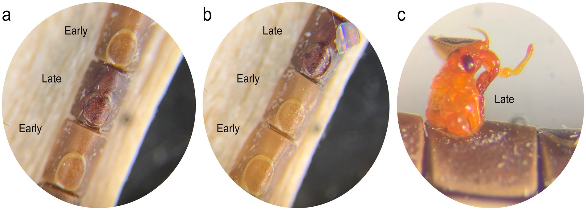
Figure 1. (a,b) Unhatched L. zonatus eggs showing the pale homogenous color of eggs that died early in development (“early death”) and the dark brown color of eggs in which the embryo is well developed (“late death.”), (c) Nymphs that died during emergence were included in the “late death” category.
2.6 Survey for Lariskella in field-collected Leptoglossus zonatus
2.6.1 Collection of Leptoglossus zonatus
The lack of a performance or fecundity cost for L. zonatus bearing Lariskella, coupled with near perfect maternal transmission and moderate CI would all lead to a prediction that Lariskella in field populations of L. zonatus should be at high frequencies, but not likely fixed (Turelli and Hoffmann, 1995; Turelli et al., 2022). To test this prediction, we surveyed L. zonatus adults collected from two locations in California, USA (Fresno and Bakersfield) and in Tucson, Arizona in 2018–2019 (Ravenscraft et al., 2024). All samples were stored in 95% ethanol. We also examined the frequency of Lariskella over time at one location, a University of Arizona pomegranate orchard adjacent to the Arizona Veterinary Diagnostics Laboratory, Tucson, AZ. A sample of adults was collected at approximately 6-week intervals from April to October in both 2019 and 2020.
2.6.2 DNA extraction and diagnostic PCR
Adult and nymphal bugs from the CA and 2018–2019 AZ samples were dissected, and DNA was extracted from a small portion of the M4 midgut region and surrounding tissue using the Qiagen DNeasy Blood and Tissue kit. DNA from the Tucson, AZ pomegranate orchard samples of 2019–2020 was extracted using an alternative but equivalent method: abdomens were removed from bugs, and entire abdomens were homogenized via bead beating. DNA extractions were performed with a small amount of the homogenate (5 μL) and a Chelex extraction protocol (Hunter et al., 2022). All the extractions were kept at −20°C until diagnostic PCR was performed using Lariskella specific primers (Duron et al., 2017).
3 Results
3.1 Lariskella maternal transmission rate and abundance through development
Lariskella was maternally transmitted with >99% efficiency. All the eggs tested (113) were positive for Lariskella, while the negative control extrtactions from the Lariskella-free culture consistently tested negative. The titer of Lariskella remained similar throughout the first three developmental stages (1st–3rd instar), with an average of 1.95 × 10⁴ copies per nymph. Lariskella titer increased after the third instar (Figure 2a) and was also high in reproductive tissue (testes and ovaries), with an average total abundance similar to the whole-body 4th instar nymph (Figure 2b). On average, Lariskella was similarly abundant in ovaries and testes, although titers of Lariskella in ovaries had higher variance (Figure 2b).
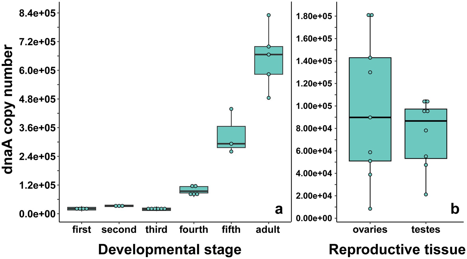
Figure 2. (a) Absolute Lariskella dnaA copy number per individual throughout development (1st instar nymph—adult) and (b) in reproductive tissue of adults. Each point in panel (b) represents the paired ovaries or testes from one individual.
3.2 Reproductive tissue and whole-body 16S Illumina sequencing
Amplicon sequencing with universal 16S rRNA primers of whole-body 4th instar nymphs and reproductive tissues was used to characterize the bacteria associated with L. zonatus. Unsurprisingly, the dominant sequence variant (SV) in whole-body samples was the obligate nutritional gut symbiont Caballeronia (Figure 3). Lariskella reads occurred in low abundance in 3/5 of 4th instar nymphs, with the common gut bacterium, Enterococcus, also being abundant in samples without Lariskella. In contrast, Lariskella was abundant in both ovaries and testes, consistent with a CI-causing phenotype. Lariskella was the most consistently present and abundant intracellular taxon found. Importantly, symbionts known to cause CI (Wolbachia, Cardinium, Rickettsiella, Spiroplasma, Mesenetia and Rickettsia) were all absent. Enterococcus was also abundant in several reproductive tissue samples. This bacterium is a common gut inhabitant (Engel and Moran, 2013; Lebreton et al., 2014) and was likely a contaminant from gut disruption during dissections. The genus Serratia includes opportunistic pathogens and intracellular symbionts, but this lineage was found in a minority of reproductive tissue samples (43%) (Slatten and Larson, 1967; Sikorowski et al., 2001; Perreau et al., 2021).
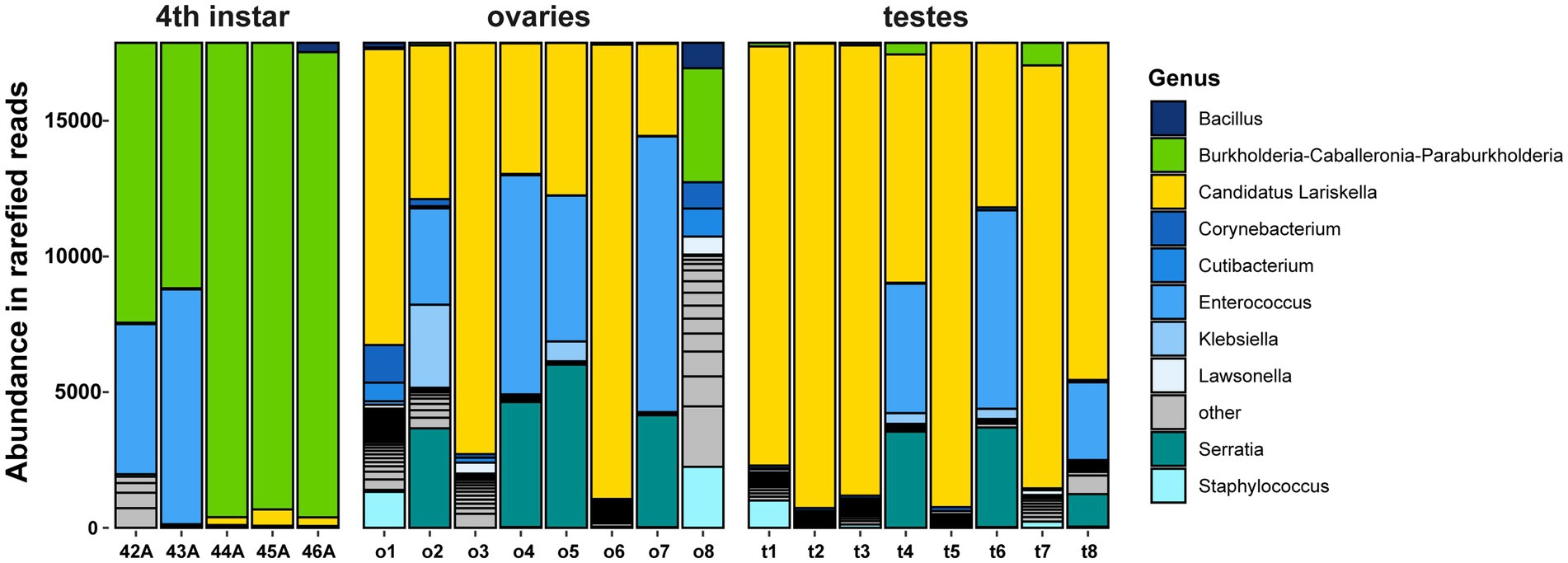
Figure 3. Leptoglossus zonatus amplicon 16S rRNA sequences from whole-body 4th instar nymphs and reproductive tissue (ovaries and testes). Each bar represents an individual, and the colors represent reads of bacterial taxa denoted in the caption. In whole-body samples, the gut-associated bacterium Caballeronia was the most abundant followed by Enterococcus and Lariskella. Lariskella was the most abundant bacterium in the reproductive tissue followed by Enterococcus and Serratia.
3.3 Caballeronia effects on performance and fitness of Leptoglossus zonatus
Pairwise comparisons of insect performance and fitness were conducted among Lariskella negative (L−) and positive (L+) and Caballeronia negative (C−) and positive (C+) bugs. The individuals in both Caballeronia negative treatments showed the major fitness deficits we expected from a previous study (Hunter et al., 2022). Survival analysis showed a significant decrease in lifetime survival for bugs that did not receive Caballeronia (L−C−/L−C+, z = 6.60, adjusted p < 0.001 and L+C−/L+C+, z = 4.07, adjusted p < 0.001; Figure 4). The few survivors of the Caballeronia-negative treatments showed significantly longer development times (L−C−/L−C+, t = 6.55, df = 16.3, adjusted p < 0.0001; L+C−/L+C+, t = 7.42, df = 19.2, adjusted p < 0.001; Figure 5). There was also no evidence that Lariskella was able to rescue bugs that lacked Caballeronia; the lengthened development times were equivalent in Caballeronia negative bugs with and without Lariskella (L−C−/L+C−, t = 1.557, df = 17.8, adjusted p = 0.42; Figure 5). Although a few Caballeronia negative bugs did eclose as adults, females weighed significantly less than Caballeronia positive bugs (L+C+/L+C, t = 3.33, df = 27.3, adjusted p = 0.01 and L−C+/L−C−, t = 3.20, df = 27.7, adjusted p = 0.017; Figure 6a). Similarly, Caballeronia negative males weighed significantly less than their Caballeronia positive counterparts, (L+C+/L+C−, t = 5.573, df = 27.58, adjusted p < 0.0001 and L−C+/L−C−, t = 3.72, df = 24.15, adjusted p = 0.0054; Figure 6b). Lastly, when Caballeronia negative females were paired with mates, no female produced any eggs, indicating that Caballeronia is required for L. zonatus reproduction.
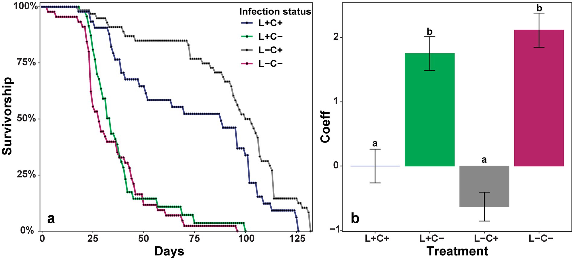
Figure 4. (a) Kaplan–Meier survival curves showing the total lifespan of individuals from the 2nd instar nymphal stage (when Caballeronia was acquired) based on the presence or absence of the primary symbiont Caballeronia (C+ or C−) and the presence or absence of the secondary symbiont Lariskella (L+ or L−). (b) Coefficients of a mixed-effects Cox regression model in which higher coefficients indicate lower survivorship. The model shows a significant decrease in survivorship for bugs that lack Caballeronia regardless of Lariskella infection status. It also shows that Lariskella presence or absence does not significantly influence survivorship (L−C−/L+C−, adjusted p = 0.36 and L−C+/L+C+, adjusted p = 0.21, all other pairwise comparisons, adjusted p < 0.001).
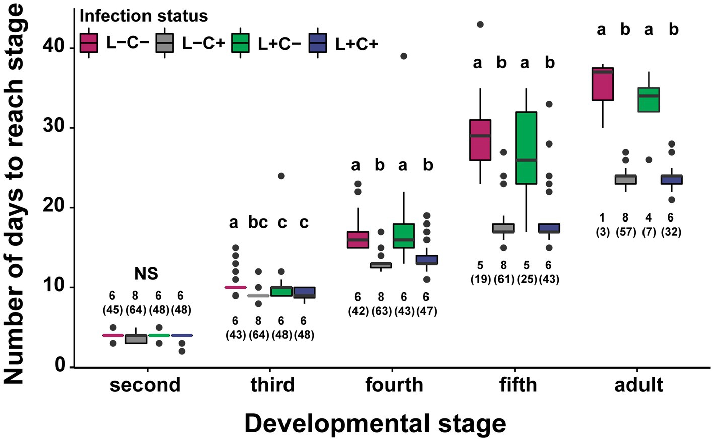
Figure 5. Development times for four treatments across developmental stages starting at the molt into the 2nd instar when the bugs were fed either Caballeronia (C+) or deionized water (C−). Development time lagged significantly for the Caballeronia negative treatments but was not significantly different between Lariskella positive and negative bugs (adjusted p > 0.4 for Lariskella comparisons while keeping Caballeronia status constant). Bars with different letters reflect statistically significant differences. Numbers next to the bars indicate the number of replicates analyzed, with total numbers of individuals measured in parentheses.
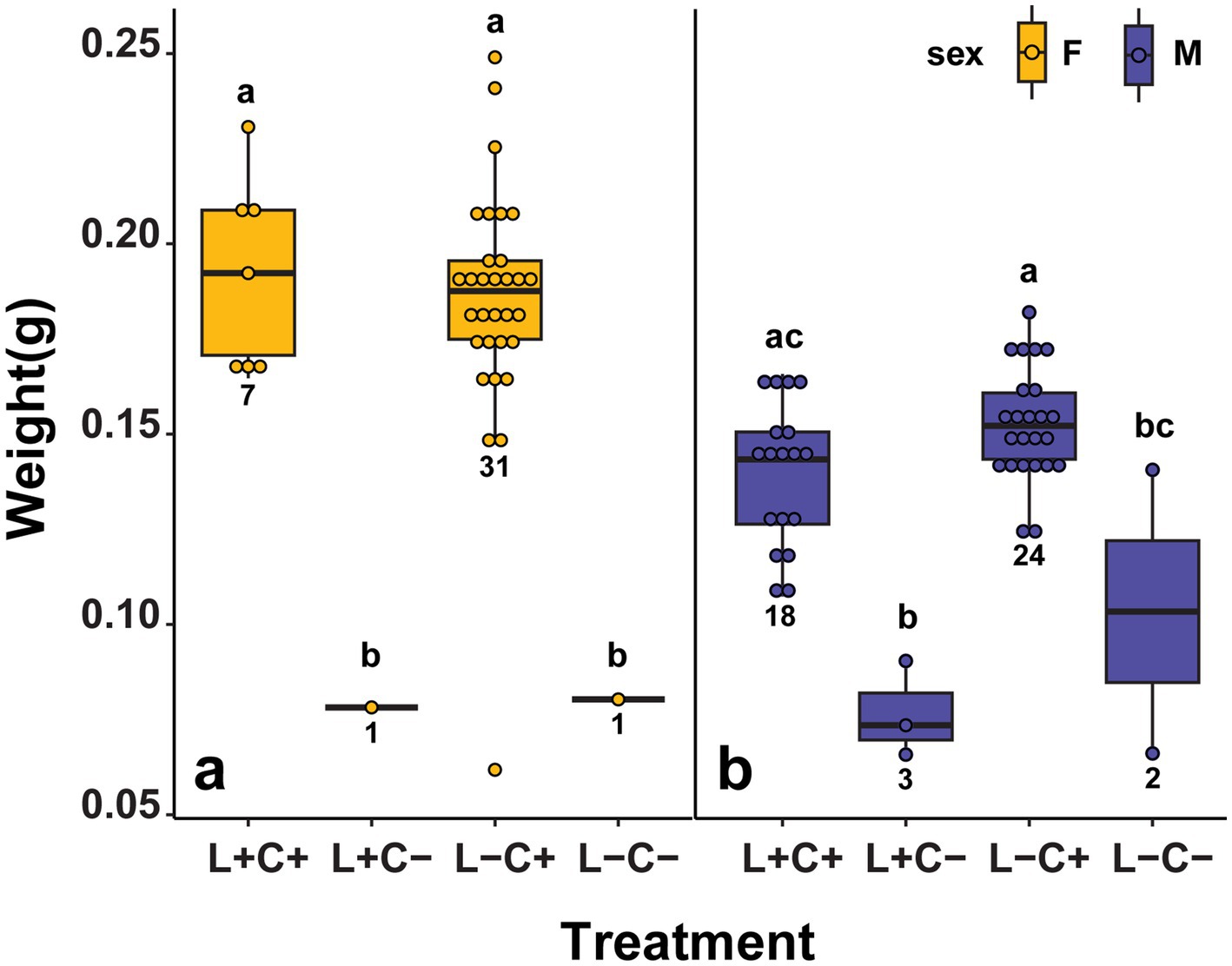
Figure 6. (a) Mean weights of adults from the fitness experiment, showing a significant difference in weight between Caballeronia positive and negative females (C+, C−) regardless of Lariskella status (adjusted p > 0.9 for Lariskella comparisons while keeping Caballeronia status constant and all other pairwise comparisons, adjusted p < 0.018). (b) Although there was a significant difference in weights among C+/C− males when controlling for Lariskella status (adjusted p < 0.01), there was no significant difference between L−C−/L+C+ males (adjusted p = 0.0571).
3.4 Lariskella effects on performance and fitness of Leptoglossus zonatus
In contrast to the findings for Caballeronia, there were no significant differences in lifetime survival between the treatments with and without Lariskella (adjusted p-value of all possible Lariskella comparisons >0.21; Figure 4). Similarly, adults that were Lariskella positive developed at equivalent rates to individuals that lacked Lariskella (L−C+/L+C+, t = −0.226, df = 13.9, adjusted p < 0.99; Figure 5). Finally, adult female weight was not influenced by the presence of Lariskella (L+C+/L−C+, t = 0.55, df = 14.0, adjusted p = 0.945 and L+C−/L−C−, t = −0.047, df = 29.4, adjusted p = 1.0; Figure 6a). Caballeronia positive males with and without Lariskella were also similar weights (L+C+/L−C+, t = −2.288, df = 9.93, adjusted p = 0.17; Figure 6b).
3.5 Lifetime fitness
Of the egg laying adults in the treatments with Caballeronia, paired adult females had a long reproductive period of about 100 days. Both clutch size and egg hatch rates declined throughout the life of the female, so time was a significant factor for both (Fclutch size = 25.08, df = 258, p < 0.001; Figure 7a), (F hatch rate = 7.757, df = 258, p < 0.001; Figure 7b). However, the fecundity of reproducing females was not influenced by the presence of Lariskella (t = 0.757, p = 0.45) with females producing approximately 300 eggs over their lifetime whether Lariskella was present or absent (Figure 8). Similarly, the presence of Lariskella did not influence egg hatch rate (t = −0.389, p = 0.69; Figure 7b).
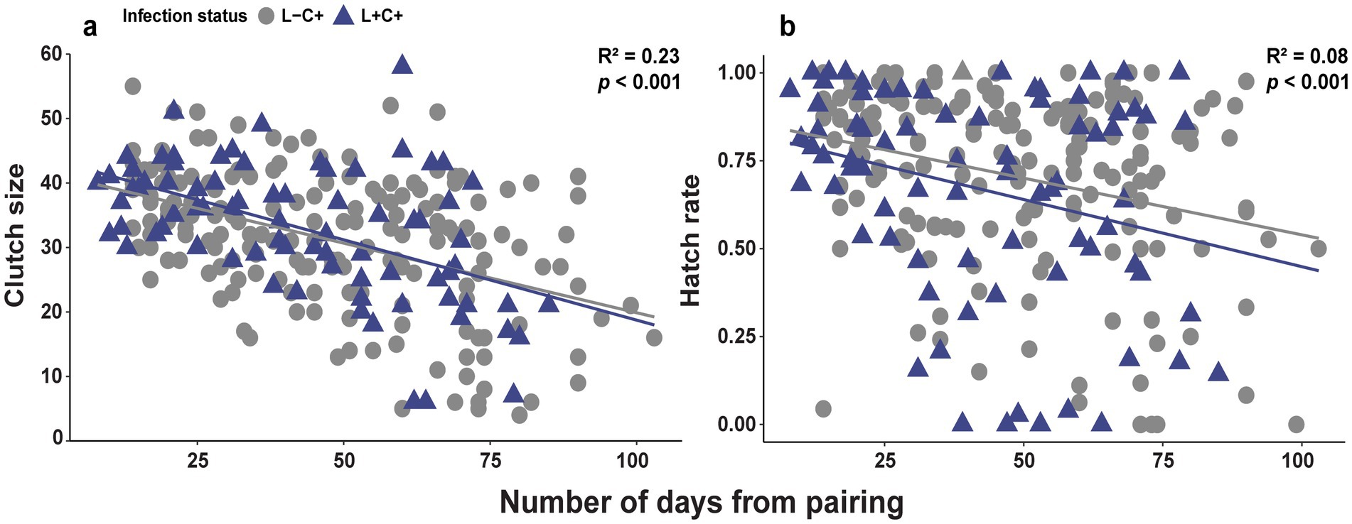
Figure 7. Scatterplots of (a) clutch size and (b) egg hatch rate of adult female L. zonatus with and without Lariskella over the lifetime reproductive period of ~100 days. The fecundity of females (Caballeronia-positive treatments only) was not influenced by the presence of Lariskella (p = 0.45 and p = 0.70 for clutch size and hatch rate respectively). Both clutch size and egg viability (hatch rate) declined significantly throughout life.
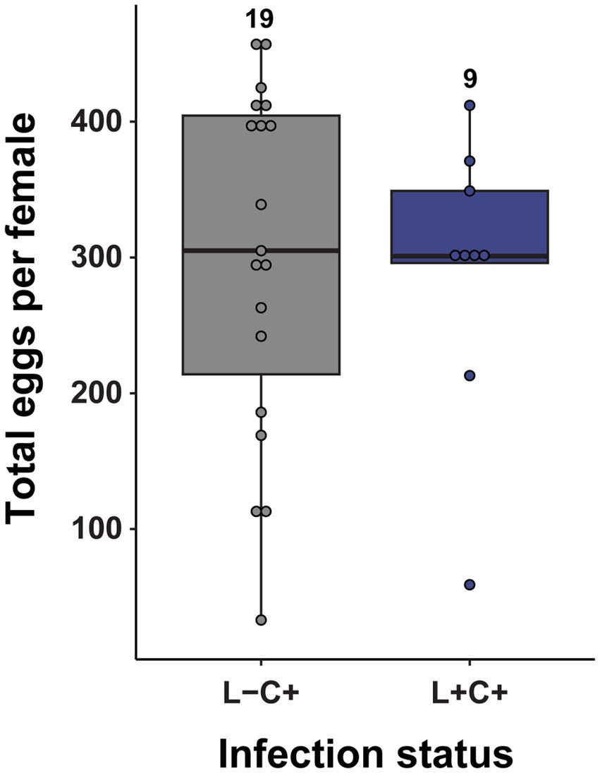
Figure 8. The presence of Lariskella did not influence the lifetime number of eggs produced by Caballeronia positive females during their lifetime (t = −0.23, p = 0.82). No Caballeronia negative adult females laid eggs, so those two treatments are absent from this figure.
3.6 CI crosses
In a second experiment, males and females with and without Lariskella were crossed in all four possible combinations to evaluate the possibility that Lariskella caused cytoplasmic incompatibility. In evaluating egg mortality in these crosses, we distinguished between early embryonic mortality, which appeared to be uncommon in all crosses except the putative CI cross, and the more frequent late embryonic mortality observed in offspring of aging females (e.g., Figure 7b, and see Methods, Figure 1). In the putative CI cross, with L+ males mated with L− females, there was a significant decrease in early embryonic survival of offspring relative to the other three crosses (c2 = 23.44, df = 3, p > 0.0001, Figure 9), consistent with the pattern expected in a CI phenotype. Cytoplasmic incompatibility is not complete, but survival of eggs laid by females in the CI cross was less than two thirds that of eggs in the other three crosses.
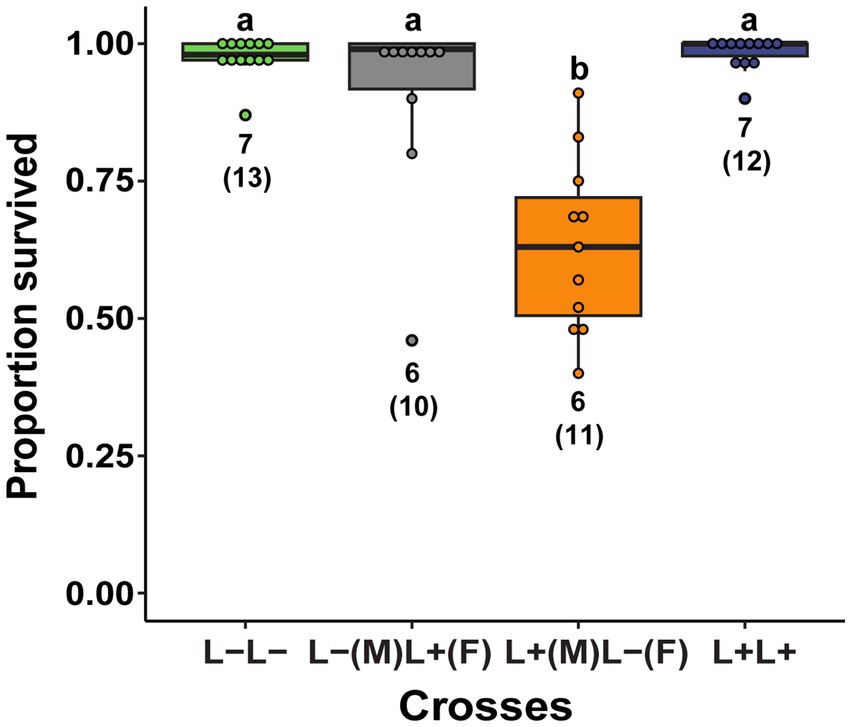
Figure 9. The proportion of a female’s eggs that survived early embryonic development among crosses of Lariskella-infected (L+) and uninfected (L−) adults in Caballeronia + L. zonatus. Significantly fewer eggs survived early embryogenesis in the putative CI cross than in any of the other crosses (p < 0.001), suggesting that Lariskella causes CI in L. zonatus. Numbers under bars refer to the number of replicates. Bars with different letters reflect statistically significant differences. Numbers next to the bars indicate the number of replicates analyzed, with total numbers of egg clutches measured in parentheses.
3.7 Lariskella frequency in the field
The proportion of bugs infected with Lariskella was high but not fixed for L. zonatus samples collected in California and Arizona, USA (Figure 10a). In a Tucson pomegranate orchard sampled repeatedly over two seasons, the proportion of Lariskella positive bugs in samples was variable and ranged from a low of 66% in July and August of 2019 to a high of 100% in June and July of 2020, but showed no clear seasonal pattern (Figure 10b).
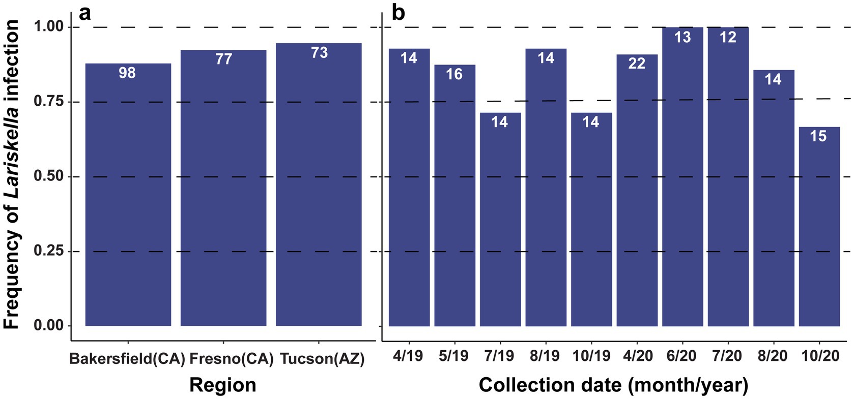
Figure 10. (a) Lariskella infection frequencies in three L. zonatus populations in USA on pomegranates: two populations in California (Bakersfield and Fresno) and one from Tucson, Arizona. Lariskella frequencies were high (0.88 Bakersfield, 0.92 Fresno, and 0.94 Tucson), but were not fixed in any population. (b) Lariskella infection frequency in L. zonatus over time in a single pomegranate orchard in Tucson, AZ, USA. Bugs were sampled at approximately 6-week intervals over the bugs’ active period from April to October in 2019 and 2020.
4 Discussion
We examined the role of the intracellular symbiont Lariskella on Leptoglossus zonatus fitness when the obligate nutritional symbiont, Caballeronia was present or absent. We found no fitness costs or benefits to Lariskella throughout the lifetime of L. zonatus, nor did we find evidence that Lariskella provides any benefits in the absence of Caballeronia. However, crossing experiments revealed that Lariskella causes incomplete cytoplasmic incompatibility (CI), characterized by a dramatic increase in early embryonic mortality, observed only in the putative CI cross. Additionally, high but not fixed frequencies of Lariskella were found in field populations. The high frequencies would be predicted for a symbiont that spreads via CI, has near-perfect maternal transmission and an absence of fitness costs (Fine, 1978; Turelli et al., 1992; O’Neill et al., 1997; Hurst and Frost, 2015).
In the current study, bugs with and without Lariskella showed equivalent development times and lifetime survivorship under laboratory conditions. In contrast, most individuals lacking the primary symbiont, Caballeronia, died before reaching adulthood. Although a few Caballeronia-negative individuals survived for several months, nearly all failed to reach adulthood, and none were able to reproduce. This result provides even more support for an earlier conclusion that Caballeronia is obligate for L. zonatus (Hunter et al., 2022). While the survivorship of Lariskella-positive bugs (L+C+) appeared lower than that of Lariskella-negative bugs (L−C+), the difference was not statistically significant. However, this finding is based on laboratory conditions, and bugs in the field likely face more environmental stressors. Under such conditions, the costs and benefits of harboring Lariskella are likely to differ.
CI Wolbachia titer and effects have been shown to be influenced by stressful conditions. Some strains have shown reduced densities and CI strength when exposed to increased rearing and nutritional stress (Sinkins et al., 1995; Yamada et al., 2007). It is not entirely clear what influence this has on host fitness; reduced symbiont titers might even be beneficial to the host under stress. Notably, other Wolbachia strains do not show such declines in CI strength under stress (Dutton and Sinkins, 2004), suggesting that CI-symbiont interactions vary depending on both symbiont strain and environmental context.
Our results further suggest that Lariskella cannot offset the severe fitness costs experienced by bugs lacking Caballeronia, at least under the laboratory conditions tested. This does not rule out a possible nutritional role for Lariskella, however. If signaling by the presence of Caballeronia in the gut is necessary for basic functions like gut development as has been found in R. pedestris (Jang et al., 2023), it could be that even if Lariskella synthesized all the nutrients limiting for L. zonatus, it would not compensate for the absence of the development regulating function of Caballeronia. Further, Caballeronia alone may provide all limiting nutrients in abundance such that any nutrient biosynthesis of Lariskella is entirely redundant. Nevertheless, it remains possible (though, we speculate unlikely) that Lariskella could confer a nutritional benefit that becomes evident when L. zonatus is paired with a suboptimal Caballeronia strain or close relative.
Previous work showed that some Caballeronia strains are more beneficial to L. zonatus development and adult weight than others (Hunter et al., 2022), and allied genera in the Burkholderiacae can colonize the R. pedestris gut and provide some benefits but are inferior to Caballeronia for bug fitness (Itoh et al., 2019). A field survey of L. zonatus revealed 26 distinct Caballeronia lineages, with three lineages present in two-thirds of the individuals sampled. It was also not uncommon for multiple lineages to be present simultaneously in the midgut (Ravenscraft et al., 2024). Considering these findings, perhaps a better test of a nutritional role for Lariskella would be to introduce a suboptimal Caballeronia that triggers the normal developmental program of the bug but falls short of providing complete nutrition for L. zonatus. In this situation, a nutritional role of Lariskella could benefit L. zonatus. Analysis of the genome of the CI-causing Lariskella in L. zonatus, when available, will allow us to predict whether Lariskella could complement the nutrition provided by a suboptimal Caballeronia or other Burkholderiaceae strain.
The recent emergence of Lariskella as a relatively common symbiont of arthropods underscores the mystery of the family to which it belongs, the Midichloriaceae. This family is the most diverse yet least understood within the intracellular bacterial order Rickettsiales, an order of alphaproteobacteria that includes significant human and livestock pathogens, and is hypothesized to have given rise to mitochondria (Andersson et al., 1998; Fitzpatrick et al., 2006; Salje, 2021; Giannotti et al., 2022; Schön et al., 2022). Members of Midichloriaceae are intracellular symbionts found in a wide array of hosts and habitats, primarily aquatic, and have been detected in protists (e.g., amoebas, ciliates) and invertebrates (e.g., ticks, corals, and arthropods), reflecting a complex ecological distribution. Despite a shared intracellular lifestyle, their genomes exhibit considerable variation in size and gene content, including key metabolic pathways, even among closely related genera, evidence of rampant horizontal gene transfer and recent host shifts (Giannotti et al., 2022; Castelli et al., 2024). The type genus Midichloria has been found exclusively in ticks, especially Ixodes spp., where it can inhabit mitochondria (Lo et al., 2006; Sassera et al., 2006; Duron, 2024) and appears to function as a nutritional symbiont by synthesizing folate, biotin, and B vitamins essential to its blood-feeding hosts (Duron, 2024; Leclerc et al., 2024). Given the frequent host shifts within Midichloriaceae and the similarity in B vitamin deficiencies between blood and plant sap diets (Moran et al., 2003; Douglas, 2017), it is plausible that some Lariskella strains may provide nutritional benefits in both blood and sap-feeding arthropods. The current study adds cytoplasmic incompatibility (CI) as another phenotype associated with Midichloriaceae.
Wolbachia, in the Anaplasmataceae family of Rickettsiales, was thought to be unique in causing CI for several decades after the phenomenon was first documented (Yen and Barr, 1971; Werren and Jaenike, 1995; Stouthamer et al., 1999). Since then, representatives of five other bacterial lineages have been shown to cause CI, including two other Alphaproteobacteria (Mesenetia & Rickettsia), Ricketsiella (Gammaproteobacteria), Cardinium (Bacteroidota), and Spiroplasma (Mollicutes) (Hunter et al., 2003; Takano et al., 2017; Rosenwald et al., 2020; Pollmann et al., 2022; Owashi et al., 2024). The current work may be the first characterization of the functional role of Lariskella in any host, and places this bacterium among a group of now seven lineages that cause CI.
We do not know whether Lariskella causes CI in other hosts, but here the frequency of hosts carrying Lariskella may give some hints. Theory predicts that the invasion of a CI symbiont with a near perfect maternal transmission rate coupled with a lack of fitness costs should result in high frequencies or fixation in a population (Fine, 1978; Turelli et al., 1992; O’Neill et al., 1997; Engelstädter and Telschow, 2009; Hurst and Frost, 2015). Although Lariskella infection in L. zonatus was not fixed in any of the California or Tucson populations surveyed, all three sites had a similarly high frequency of Lariskella infection (>85%), similar to the numbers previously observed for Nysius seed bug species in Japan (Matsuura et al., 2012) and Ixodes ticks in Russia and Japan (Mediannikov et al., 2004; Duron et al., 2017; Becker et al., 2023). These high frequency infections would be consistent with either a mutualistic or parasitic association, such as a nutritional role or reproductive manipulation. Within L. zonatus populations, several factors could explain the lack of fixation in the field, including an incomplete CI phenotype and low symbiont titer early in host development that may render the symbiont vulnerable to environmental stressors like heat or environmental antibiotic exposure (Corbin et al., 2017; Endersby-Harshman et al., 2019; Martins et al., 2023). The diversity of Lariskella strains in L. zonatus is currently unknown, but it is possible that multiple strains exist in natural populations. Notably, the mitochondrial population structure of L. zonatus in California shows signs of a selective sweep or bottleneck, with only three mitochondrial haplotypes compared to 17 in its congener L. clypealis (Joyce et al., 2017). In the context of our findings, this reduced haplotype diversity may suggest a recent spread of a single Lariskella strain along with a co-inherited mitochondrial haplotype (Raychoudhury et al., 2010). Additionally, the long reproductive period of L. zonatus may reduce CI strength, as CI Wolbachia have been shown to decline with male age in some systems (Reynolds and Hoffmann, 2002). While low prevalence of Lariskella in other arthropod species could suggest a number of scenarios including a relatively recent, horizontally acquired association, or one that is asymptomatic and slowly declining, it may also indicate a conditional role other than CI such as defense or temperature stress mediation (Oliver et al., 2003, 2010).
Understanding Lariskella’s role in L. zonatus could also inform pest management strategies for tree crops and other hosts and may also provide insight into management of blood-feeding arthropods that vector human pathogens. Although the current study observed only a mild CI phenotype (with 40% of eggs affected), more examples of Lariskella CI strains and host backgrounds are needed to determine the range of CI strength that can be caused by this lineage. In Wolbachia and Mesenetia, CI can cause complete (100%) offspring mortality (Hoffmann et al., 1986; Merçot and Charlat, 2004; Takano et al., 2017; Shropshire et al., 2021), but CI strength in Wolbachia varies tremendously depending on both the symbiont and the host genotype (Merçot and Charlat, 2004; Sicard et al., 2021). Recent work shows that incomplete CI Wolbachia strength in Culex pipiens can be explained by the divergence of CI gene repertoires relative to strains that induce complete CI (Sicard et al., 2021). Conversely, in Drosophila, Wolbachia CI strength can vary from 30 to 100% mortality, depending on host species (Merçot and Charlat, 2004). The identification of a new CI lineage, Lariskella, that also infects human disease vectors (e.g., ticks and fleas) is notable given the pathogen-blocking effects of CI Wolbachia in mosquitoes (McMeniman et al., 2009; Moreira et al., 2009; Walker et al., 2011). CI Wolbachia pathogen-blocking has spurred a global program deploying Wolbachia in mosquitoes to combat RNA viruses responsible for deadly diseases (Utarini et al., 2021).
Future research should focus on comprehensive screening of Lariskella across a broad range of arthropods and comparative genome sequencing to characterize its metabolic pathways, potential nutritional roles, and capacity to manipulate host reproduction via homologs of known cytoplasmic incompatibility (CI) genes. For example, Mesenetia, another alphaproteobacterium within Rickettsiales, carries homologs to Wolbachia CI genes (Takano et al., 2017). Both Mesenetia and Wolbachia belong to the family Anaplasmataceae, which phylogenetic analyses often identify as a sister group to Midichloriaceae. Given the widespread occurrence of horizontal gene transfer and host shifts, Lariskella may have evolved CI independently, similar to Cardinium, which lacks Wolbachia CI genes (Mann et al., 2017; Lindsey et al., 2018). Alternatively, Lariskella could represent a novel mechanistic model of CI that diverges from the toxin-antidote, and two-by-one models observed in Wolbachia. Understanding the prevalence of Lariskella, its evolutionary trajectory, and its interactions within arthropod hosts will advance our knowledge of symbiont-driven reproductive manipulation and vector ecology. This research could also provide insights for developing new control strategies for pest and pathogen vectors.
Data availability statement
The datasets generated for this study can be found in the Dryad repository, doi: 10.5061/dryad.bvq83bkkp. The raw amplicon sequences have been submitted to the Sequence Read Archive (SRA) under the accession number PRJNA1266735.
Ethics statement
The manuscript presents research on animals that do not require ethical approval for their study.
Author contributions
EU: Conceptualization, Data curation, Formal analysis, Investigation, Methodology, Validation, Visualization, Writing – original draft, Writing – review & editing. SK: Conceptualization, Data curation, Investigation, Methodology, Project administration, Writing – original draft, Writing – review & editing. AR: Data curation, Formal analysis, Investigation, Methodology, Visualization, Writing – original draft, Writing – review & editing. YM: Conceptualization, Investigation, Methodology, Resources, Validation, Writing – original draft, Writing – review & editing. MH: Conceptualization, Funding acquisition, Investigation, Methodology, Project administration, Resources, Supervision, Validation, Writing – original draft, Writing – review & editing.
Funding
The author(s) declare that financial support was received for the research and/or publication of this article. This work was supported by the Foundational and Applied Science Program, project award nos. 2019-67013-29407 to MH, AR and 2023-67013-39897 to MH and AR, from the U.S. Department of Agriculture’s National Institute of Food and Agriculture and by NSF IOS 2426306 to MH. EU was supported by JSPS Pre-doctoral Fellowship, JSPS Summer Program and the Collaborative Research of Tropical Biosphere Research Center, Univ. Ryukyus. This study was also supported by JSPS Grant-in-Aid KAKENHI Grants No. 18KK0211, and 19H03275.
Acknowledgments
We sincerely thank David Haviland for substantial advice and help in the field, the Kern County UC Cooperative Extension for use of their lab facilities, and Johnathan Adamson, Reiko Sekine, Chiaki Matsuura, and Alex Lombard for laboratory assistance. We also thank White Forest Nursery (Bakersfield, CA), the Kearney Agricultural Research and Extension Center (Fresno, CA), the Mission Garden (Tucson, AZ), Mesquite Valley Growers (Tucson, CA), and Ursula Schuch (University of Arizona West Campus Agricultural Center pomegranate orchard) for permission to sample insects. The authors also thank Dave Baltrus who contributed to the USDA NIFA proposal that funded this research.
Conflict of interest
The authors declare that the research was conducted in the absence of any commercial or financial relationships that could be construed as a potential conflict of interest.
Generative AI statement
The authors declare that no Gen AI was used in the creation of this manuscript.
Publisher’s note
All claims expressed in this article are solely those of the authors and do not necessarily represent those of their affiliated organizations, or those of the publisher, the editors and the reviewers. Any product that may be evaluated in this article, or claim that may be made by its manufacturer, is not guaranteed or endorsed by the publisher.
Supplementary material
The Supplementary material for this article can be found online at: https://www.frontiersin.org/articles/10.3389/fmicb.2025.1595917/full#supplementary-material
References
Aivelo, T., and Tschirren, B. (2020). Bacterial microbiota composition of a common ectoparasite of cavity-breeding birds, the hen flea Ceratophyllus gallinae. Ibis 162, 1088–1092. doi: 10.1111/ibi.12811
Andersson, S. G., Zomorodipour, A., Andersson, J. O., Sicheritz-Pontén, T., Alsmark, U. C., Podowski, R. M., et al. (1998). The genome sequence of Rickettsia prowazekii and the origin of mitochondria. Nature 396, 133–140. doi: 10.1038/24094
Bates, D., Mächler, M., Bolker, B., and Walker, S. (2015). Fitting linear mixed-effects models using lme4. J. Stat. Softw. 67, 1–48. doi: 10.18637/jss.v067.i01
Becker, N. S., Rollins, R. E., Stephens, R., Sato, K., Brachmann, A., Nakao, M., et al. (2023). Candidatus Lariskella arthopodarum endosymbiont is the main factor differentiating the microbiome communities of female and male Borrelia-positive Ixodes persulcatus ticks. Ticks Tick Borne Dis. 14:102183. doi: 10.1016/j.ttbdis.2023.102183
Beckmann, J. F., Bonneau, M., Chen, H., Hochstrasser, M., Poinsot, D., Merçot, H., et al. (2019a). Caution does not preclude predictive and testable models of cytoplasmic incompatibility: a reply to Shropshire et al. Trends Genet. 35, 399–400. doi: 10.1016/j.tig.2019.03.002
Beckmann, J. F., Bonneau, M., Chen, H., Hochstrasser, M., Poinsot, D., Merçot, H., et al. (2019b). The toxin-antidote model of cytoplasmic incompatibility: genetics and evolutionary implications. Trends Genet. 35, 175–185. doi: 10.1016/j.tig.2018.12.004
Bourtzis, K. (2008). Wolbachia-based technologies for insect pest population control. Adv. Exp. Med. Biol. 627, 104–113. doi: 10.1007/978-0-387-78225-6_9
Braig, H. R., Guzman, H., Tesh, R. B., and O’Neill, S. L. (1994). Replacement of the natural Wolbachia symbiont of Drosophila simulans with a mosquito counterpart. Nature 367, 453–455. doi: 10.1038/367453a0
Bull, J. J. (1983). Sex determining mechanisms: an evolutionary perspective. Experientia 41, 1285–1296. doi: 10.1007/BF01952071
Buysse, M., and Duron, O. (2021). Evidence that microbes identified as tick-borne pathogens are nutritional endosymbionts. Cell 184, 2259–2260. doi: 10.1016/j.cell.2021.03.053
Callahan, B. J., McMurdie, P. J., Rosen, M. J., Han, A. W., Johnson, A. J. A., and Holmes, S. P. (2016). DADA2: high-resolution sample inference from Illumina amplicon data. Nat. Methods 13, 581–583. doi: 10.1038/nmeth.3869
Castelli, M., Nardi, T., Gammuto, L., Bellinzona, G., Sabaneyeva, E., Potekhin, A., et al. (2024). Host association and intracellularity evolved multiple times independently in the Rickettsiales. Nat. Commun. 15:1093. doi: 10.1038/s41467-024-45351-7
Corbin, C., Heyworth, E. R., Ferrari, J., and Hurst, G. D. D. (2017). Heritable symbionts in a world of varying temperature. Heredity 118, 10–20. doi: 10.1038/hdy.2016.71
Correa, C. C., and Ballard, J. W. O. (2016). Wolbachia associations with insects: winning or losing against a master manipulator. Front. Ecol. Evol. 3:153. doi: 10.3389/fevo.2015.00153
Daane, K. M., Yokota, G. Y., and Wilson, H. (2019). Seasonal dynamics of the Leaffooted bug Leptoglossus zonatus and its implications for control in almonds and pistachios. Insects 10:255. doi: 10.3390/insects10080255
Davis, N. M., Proctor, D. M., Holmes, S. P., Relman, D. A., and Callahan, B. J. (2018). Simple statistical identification and removal of contaminant sequences in marker-gene and metagenomics data. Microbiome 6:226. doi: 10.1186/s40168-018-0605-2
Dobson, S. L., Fox, C. W., and Jiggins, F. M. (2002). The effect of Wolbachia-induced cytoplasmic incompatibility on host population size in natural and manipulated systems. Proc. R. Soc. Lond. B Biol. Sci. 269, 437–445. doi: 10.1098/rspb.2001.1876
Douglas, A. E. (2017). The B vitamin nutrition of insects: the contributions of diet, microbiome and horizontally acquired genes. Curr. Opin. Insect Sci. 23, 65–69. doi: 10.1016/j.cois.2017.07.012
Duarte, E. H., Carvalho, A., López-Madrigal, S., Costa, J., and Teixeira, L. (2021). Forward genetics in Wolbachia: regulation of Wolbachia proliferation by the amplification and deletion of an addictive genomic island. PLoS Genet. 17:e1009612. doi: 10.1371/journal.pgen.1009612
Duron, O. (2024). Nutritional symbiosis in ticks: singularities of the genus Ixodes. Trends Parasitol. 40, 696–706. doi: 10.1016/j.pt.2024.06.006
Duron, O., Binetruy, F., Noël, V., Cremaschi, J., McCoy, K. D., Arnathau, C., et al. (2017). Evolutionary changes in symbiont community structure in ticks. Mol. Ecol. 26, 2905–2921. doi: 10.1111/mec.14094
Duron, O., and Weill, M. (2006). Wolbachia infection influences the development of Culex pipiens embryo in incompatible crosses. Heredity 96, 493–500. doi: 10.1038/sj.hdy.6800831
Dutton, T. J., and Sinkins, S. P. (2004). Strain-specific quantification of Wolbachia density in Aedes albopictus and effects of larval rearing conditions. Insect Mol. Biol. 13, 317–322. doi: 10.1111/j.0962-1075.2004.00490.x
Endersby-Harshman, N. M., Axford, J. K., and Hoffmann, A. A. (2019). Environmental concentrations of antibiotics may diminish Wolbachia infections in Aedes aegypti (Diptera: Culicidae). J. Med. Entomol. 56, 1078–1086. doi: 10.1093/jme/tjz023
Engel, P., and Moran, N. A. (2013). The gut microbiota of insects – diversity in structure and function. FEMS Microbiol. Rev. 37, 699–735. doi: 10.1111/1574-6976.12025
Engelstädter, J., and Hurst, G. D. D. (2009). The ecology and evolution of microbes that manipulate host reproduction. Annu. Rev. Ecol. Evol. Syst. 40, 127–149. doi: 10.1146/annurev.ecolsys.110308.120206
Engelstädter, J., and Telschow, A. (2009). Cytoplasmic incompatibility and host population structure. Heredity 103, 196–207. doi: 10.1038/hdy.2009.53
Ferrari, J., and Vavre, F. (2011). Bacterial symbionts in insects or the story of communities affecting communities. Philos. Trans. R. Soc. Lond. Ser. B Biol. Sci. 366, 1389–1400. doi: 10.1098/rstb.2010.0226
Fine, P. E. M. (1978). On the dynamics of symbiote-dependent cytoplasmic incompatibility in culicine mosquitoes. J. Invertebr. Pathol. 31, 10–18. doi: 10.1016/0022-2011(78)90102-7
Fitzpatrick, D. A., Creevey, C. J., and McInerney, J. O. (2006). Genome phylogenies indicate a meaningful α-Proteobacterial phylogeny and support a grouping of the mitochondria with the Rickettsiales. Mol. Biol. Evol. 23, 74–85. doi: 10.1093/molbev/msj009
Gebiola, M., White, J. A., Cass, B. N., Kozuch, A., Harris, L. R., Kelly, S. E., et al. (2016). Cryptic diversity, reproductive isolation and cytoplasmic incompatibility in a classic biological control success story. Biol. J. Linn. Soc. 117, 217–230. doi: 10.1111/bij.12648
Giannotti, D., Boscaro, V., Husnik, F., Vannini, C., and Keeling, P. J. (2022). The “other” Rickettsiales: an overview of the family “Candidatus Midichloriaceae”. Appl. Environ. Microbiol. 88:e02432-21. doi: 10.1128/aem.02432-21
Hertig, M., and Wolbach, S. B. (1924). Studies on Rickettsia-like micro-organisms in insects. J. Med. Res. 44, 329–374. 7
Hoffmann, A. A., Montgomery, B. L., Popovici, J., Iturbe-Ormaetxe, I., Johnson, P. H., Muzzi, F., et al. (2011). Successful establishment of Wolbachia in Aedes populations to suppress dengue transmission. Nature 476, 454–457. doi: 10.1038/nature10356
Hoffmann, A. A., Turelli, M., and Simmons, G. M. (1986). Unidirectional incompatibility between populations of Drosophila simulans. Evolution 40, 692–701. doi: 10.1111/j.1558-5646.1986.tb00531.x
Horard, B., Terretaz, K., Gosselin-Grenet, A.-S., Sobry, H., Sicard, M., Landmann, F., et al. (2022). Paternal transmission of the Wolbachia CidB toxin underlies cytoplasmic incompatibility. Curr. Biol. 32, 1319–1331.e5. doi: 10.1016/j.cub.2022.01.052
Hunter, M. S., Perlman, S. J., and Kelly, S. E. (2003). A bacterial symbiont in the Bacteroidetes induces cytoplasmic incompatibility in the parasitoid wasp Encarsia pergandiella. Proc. R. Soc. B Biol. Sci. 270, 2185–2190. doi: 10.1098/rspb.2003.2475
Hunter, M. S., Umanzor, E. F., Kelly, S. E., Whitaker, S. M., and Ravenscraft, A. (2022). Development of common leaf-footed bug pests depends on the presence and identity of their environmentally acquired symbionts. Appl. Environ. Microbiol. 88:e0177821. doi: 10.1128/AEM.01778-21
Hurst, G. D. D., and Frost, C. L. (2015). Reproductive parasitism: maternally inherited symbionts in a Biparental world. Cold Spring Harb. Perspect. Biol. 7:a017699. doi: 10.1101/cshperspect.a017699
Illumina (2013). Illumina 16S metagenomic sequencing library preparation (Illumina technical note 15044223). Available online at: http://support.illumina.com/documents/documentation/chemistry_documentation/16s/16s-metagenomic-library-prep-guide-15044223-b.pdf (Accessed July 18, 2024).
Ingels, C., and Haviland, D. (2014). Leaffooted bug management guidelines--UC IPM. Available online at: https://ipm.ucanr.edu/PMG/PESTNOTES/pn74168.html?src=302-www&fr=4503 (Accessed July 28, 2024).
Itoh, H., Jang, S., Takeshita, K., Ohbayashi, T., Ohnishi, N., Meng, X.-Y., et al. (2019). Host–symbiont specificity determined by microbe–microbe competition in an insect gut. Proc. Natl. Acad. Sci. 116, 22673–22682. doi: 10.1073/pnas.1912397116
Jang, S., Matsuura, Y., Ishigami, K., Mergaert, P., and Kikuchi, Y. (2023). Symbiont coordinates stem cell proliferation, apoptosis, and morphogenesis of gut symbiotic organ in the stinkbug-Caballeronia symbiosis. Front. Physiol. 13:1071987. doi: 10.3389/fphys.2022.1071987
Janson, E. M., Stireman, J. O. III, Singer, M. S., and Abbot, P. (2008). Phytophagous insect–microbe mutualisms and adaptive evolutionary diversification. Evolution 62, 997–1012. doi: 10.1111/j.1558-5646.2008.00348.x
Jones, R. T., Borchert, J., Eisen, R., MacMillan, K., Boegler, K., and Gage, K. L. (2015). Flea-associated bacterial communities across an environmental transect in a Plague-endemic region of Uganda. PLoS One 10:e0141057. doi: 10.1371/journal.pone.0141057
Joyce, A. L., Higbee, B. S., Haviland, D. R., and Brailovsky, H. (2017). Genetic variability of two Leaffooted bugs, Leptoglossus clypealis and Leptoglossus zonatus (Hemiptera: Coreidae) in the Central Valley of California. J. Econ. Entomol. 110, 2576–2589. doi: 10.1093/jee/tox222
Joyce, A. L., Parolini, H., and Brailovsky, H. (2021). Distribution of two strains of Leptoglossus zonatus (Dallas) (Hemiptera: Coreidae) in the Western hemisphere: is L. zonatus a potential invasive species in California? Insects 12:1094. doi: 10.3390/insects12121094
Kikuchi, Y., and Fukatsu, T. (2014). Live imaging of symbiosis: spatiotemporal infection dynamics of a GFP-labelled Burkholderia symbiont in the bean bug Riptortus pedestris. Mol. Ecol. 23, 1445–1456. doi: 10.1111/mec.12479
Kikuchi, Y., Hosokawa, T., and Fukatsu, T. (2011). An ancient but promiscuous host–symbiont association between Burkholderia gut symbionts and their heteropteran hosts. ISME J. 5, 446–460. doi: 10.1038/ismej.2010.150
Klindworth, A., Pruesse, E., Schweer, T., Peplies, J., Quast, C., Horn, M., et al. (2013). Evaluation of general 16S ribosomal RNA gene PCR primers for classical and next-generation sequencing-based diversity studies. Nucleic Acids Res. 41:e1. doi: 10.1093/nar/gks808
Kozek, W. J., and Rao, R. U. (2007). The discovery of Wolbachia in arthropods and nematodes – a historical perspective. Wolbachia Bugs Life Another Bug 5, 1–14. doi: 10.1159/000104228
Laven, H. (1967). A possible model for speciation by cytoplasmic isolation in the Culex pipiens complex. Bull. World Health Organ. 37, 263–266.
Lebreton, F., Willems, R. J. L., and Gilmore, M. S. (2014). “Enterococcus diversity, origins in nature, and gut colonization” in Enterococci: from commensals to leading causes of drug resistant infection. eds. M. S. Gilmore, D. B. Clewell, Y. Ike, and N. Shankar (Boston: Massachusetts Eye and Ear Infirmary).
Leclerc, L., Mattick, J., Burns, B. P., Sassera, D., Hotopp, J. D., and Lo, N. (2024). Metatranscriptomics provide insights into the role of the symbiont Midichloria mitochondrii in Ixodes ticks. FEMS Microbiol. Ecol. 100:fiae133. doi: 10.1093/femsec/fiae133
Lenth, R. (2018). Emmeans: estimated marginal means, aka least-squares means. Available online at: https://rvlenth.github.io/emmeans/authors.html (Accessed September 17, 2024).
LePage, D. P., Metcalf, J. A., Bordenstein, S. R., On, J., Perlmutter, J. I., Shropshire, J. D., et al. (2017). Prophage WO genes recapitulate and enhance Wolbachia-induced cytoplasmic incompatibility. Nature 543, 243–247. doi: 10.1038/nature21391
Lindsey, A. R. I., Rice, D. W., Bordenstein, S. R., Brooks, A. W., Bordenstein, S. R., and Newton, I. L. G. (2018). Evolutionary genetics of cytoplasmic incompatibility genes cifA and cifB in prophage WO of Wolbachia. Genome Biol. Evol. 10, 434–451. doi: 10.1093/gbe/evy012
Lo, N., Beninati, T., Sassera, D., Bouman, E. a. P., Santagati, S., Gern, L., et al. (2006). Widespread distribution and high prevalence of an alpha-proteobacterial symbiont in the tick Ixodes ricinus. Environ. Microbiol. 8, 1280–1287. doi: 10.1111/j.1462-2920.2006.01024.x
Mann, E., Stouthamer, C. M., Kelly, S. E., Dzieciol, M., Hunter, M. S., and Schmitz-Esser, S. (2017). Transcriptome sequencing reveals novel candidate genes for Cardinium hertigii-caused cytoplasmic incompatibility and host-cell interaction. mSystems 2:e00141-17. doi: 10.1128/mSystems.00141-17
Martin, M. (2011). Cutadapt removes adapter sequences from high-throughput sequencing reads. EMBnet. J. 17, 10–12. doi: 10.14806/ej.17.1.200
Martins, M., César, C. S., and Cogni, R. (2023). The effects of temperature on prevalence of facultative insect heritable symbionts across spatial and seasonal scales. Front. Microbiol. 14:1321341. doi: 10.3389/fmicb.2023.1321341
Matsuura, Y., Kikuchi, Y., Meng, X. Y., Koga, R., and Fukatsu, T. (2012). Novel clade of Alphaproteobacterial endosymbionts associated with stinkbugs and other arthropods. Appl. Environ. Microbiol. 78, 4149–4156. doi: 10.1128/AEM.00673-12
McMeniman, C. J., Lane, R. V., Cass, B. N., Fong, A. W. C., Sidhu, M., Wang, Y.-F., et al. (2009). Stable introduction of a life-shortening Wolbachia infection into the mosquito Aedes aegypti. Science 323, 141–144. doi: 10.1126/science.1165326
Mediannikov, O. I., Ivanov, L. I., Nishikawa, M., Saito, R., Sidel’nikov, I. N., Zdanovskaia, N. I., et al. (2004). Microorganism “Montezuma” of the order Rickettsiales: the potential causative agent of tick-borne disease in the Far East of Russia. Zh. Mikrobiol. Epidemiol. Immunobiol. 1, 7–13.
Merçot, H., and Charlat, S. (2004). Wolbachia infections in Drosophila melanogaster and D. simulans: polymorphism and levels of cytoplasmic incompatibility. Genetica 120, 51–59. doi: 10.1023/B:GENE.0000017629.31383.8f
Misof, B., Liu, S., Meusemann, K., Peters, R. S., Donath, A., Mayer, C., et al. (2014). Phylogenomics resolves the timing and pattern of insect evolution. Science 346, 763–767. doi: 10.1126/science.1257570
Montagna, M., Sassera, D., Epis, S., Bazzocchi, C., Vannini, C., Lo, N., et al. (2013). “Candidatus Midichloriaceae” fam. nov. (Rickettsiales), an ecologically widespread clade of intracellular Alphaproteobacteria. Appl. Environ. Microbiol. 79, 3241–3248. doi: 10.1128/AEM.03971-12
Montllor, C. B., Maxmen, A., and Purcell, A. H. (2002). Facultative bacterial endosymbionts benefit pea aphids Acyrthosiphon pisum under heat stress. Ecol. Entomol. 27, 189–195. doi: 10.1046/j.1365-2311.2002.00393.x
Moran, N. A., Munson, M. A., Baumann, P., and Ishikawa, H. (1997). A molecular clock in endosymbiotic bacteria is calibrated using the insect hosts. Proc. R. Soc. Lond. B Biol. Sci. 253, 167–171. doi: 10.1098/rspb.1993.0098
Moran, N. A., Plague, G. R., Sandström, J. P., and Wilcox, J. L. (2003). A genomic perspective on nutrient provisioning by bacterial symbionts of insects. Proc. Natl. Acad. Sci. 100, 14543–14548. doi: 10.1073/pnas.2135345100
Moreira, L. A., Iturbe-Ormaetxe, I., Jeffery, J. A., Lu, G., Pyke, A. T., Hedges, L. M., et al. (2009). A Wolbachia symbiont in Aedes aegypti limits infection with dengue, chikungunya, and plasmodium. Cell 139, 1268–1278. doi: 10.1016/j.cell.2009.11.042
Mulio, S. Å., Zwolińska, A., Klejdysz, T., Prus-Frankowska, M., Michalik, A., Kolasa, M., et al. (2024). Limited variation in microbial communities across populations of macrosteles leafhoppers (Hemiptera: Cicadellidae). Environ. Microbiol. Rep. 16:e13279. doi: 10.1111/1758-2229.13279
O’Neill, S. L., Hoffmann, A. A., and Werren, J. H. (1997). Influential passengers: inherited microorganisms and arthropod reproduction. Oxford, UK: Oxford University Press.
Ohbayashi, T., Futahashi, R., Terashima, M., Barrière, Q., Lamouche, F., Takeshita, K., et al. (2019). Comparative cytology, physiology and transcriptomics of Burkholderia insecticola in symbiosis with the bean bug Riptortus pedestris and in culture. ISME J. 13, 1469–1483. doi: 10.1038/s41396-019-0361-8
Ohbayashi, T., Takeshita, K., Kitagawa, W., Nikoh, N., Koga, R., Meng, X.-Y., et al. (2015). Insect’s intestinal organ for symbiont sorting. Proc. Natl. Acad. Sci. 112, E5179–E5188. doi: 10.1073/pnas.1511454112
Oliver, K. M., Degnan, P. H., Burke, G. R., and Moran, N. A. (2010). Facultative symbionts in aphids and the horizontal transfer of ecologically important traits. Annu. Rev. Entomol. 55, 247–266. doi: 10.1146/annurev-ento-112408-085305
Oliver, K. M., Russell, J. A., Moran, N. A., and Hunter, M. S. (2003). Facultative bacterial symbionts in aphids confer resistance to parasitic wasps. Proc. Natl. Acad. Sci. 100, 1803–1807. doi: 10.1073/pnas.0335320100
Owashi, Y., Arai, H., Adachi-Hagimori, T., and Kageyama, D. (2024). Rickettsia induces strong cytoplasmic incompatibility in a predatory insect. Proc. R. Soc. B Biol. Sci. 291:20240680. doi: 10.1098/rspb.2024.0680
Pais, R., Lohs, C., Wu, Y., Wang, J., and Aksoy, S. (2008). The obligate mutualist Wigglesworthia glossinidia influences reproduction, digestion, and immunity processes of its host, the tsetse Fly. Appl. Environ. Microbiol. 74, 5965–5974. doi: 10.1128/AEM.00741-08
Perreau, J., Patel, D. J., Anderson, H., Maeda, G. P., Elston, K. M., Barrick, J. E., et al. (2021). Vertical transmission at the pathogen-symbiont interface: Serratia symbiotica and aphids. MBio 12:e00359-21. doi: 10.1128/mBio.00359-21
Pollmann, M., Moore, L. D., Krimmer, E., D’Alvise, P., Hasselmann, M., Perlman, S. J., et al. (2022). Highly transmissible cytoplasmic incompatibility by the extracellular insect symbiont Spiroplasma. IScience 25:104335. doi: 10.1016/j.isci.2022.104335
Quast, C., Pruesse, E., Yilmaz, P., Gerken, J., Schweer, T., Yarza, P., et al. (2013). The SILVA ribosomal RNA gene database project: improved data processing and web-based tools. Nucleic Acids Res. 41, D590–D596. doi: 10.1093/nar/gks1219
Ravenscraft, A., Kelly, S., Haviland, D., Adamson, J., and Hunter, M. (2024). Spatial and temporal structure of environmentally-acquired Caballeronia symbionts of a leaffooted bug. Available online at: https://www.authorea.com/users/826008/articles/1221365-spatial-and-temporal-structure-of-environmentally-acquired-caballeronia-symbionts-of-a-leaffooted-bug?commit=668081d8ba6b32cb64422ac4a606c0ad36954ca9 (Accessed May 14, 2025).
Raychoudhury, R., Grillenberger, B. K., Gadau, J., Bijlsma, R., van de Zande, L., Werren, J. H., et al. (2010). Phylogeography of Nasonia vitripennis (Hymenoptera) indicates a mitochondrial–Wolbachia sweep in North America. Heredity 104, 318–326. doi: 10.1038/hdy.2009.160
R Core Team (2023). R: A Language and Environment for Statistical Computing. Vienna, Austria: R Foundation for Statistical Computing. Available at: https://www.R-project.org/
Reynolds, K. T., and Hoffmann, A. A. (2002). Male age, host effects and the weak expression or non-expression of cytoplasmic incompatibility in Drosophila strains infected by maternally transmitted Wolbachia. Genet. Res. 80, 79–87. doi: 10.1017/S0016672302005827
Rosenwald, L. C., Sitvarin, M. I., and White, J. A. (2020). Endosymbiotic Rickettsiella causes cytoplasmic incompatibility in a spider host. Proc. R. Soc. B Biol. Sci. 287:20201107. doi: 10.1098/rspb.2020.1107
Sabree, Z. L., Kambhampati, S., and Moran, N. A. (2009). Nitrogen recycling and nutritional provisioning by Blattabacterium, the cockroach endosymbiont. Proc. Natl. Acad. Sci. USA 106, 19521–19526. doi: 10.1073/pnas.0907504106
Salje, J. (2021). Cells within cells: Rickettsiales and the obligate intracellular bacterial lifestyle. Nat. Rev. Microbiol. 19, 375–390. doi: 10.1038/s41579-020-00507-2
Santos-Garcia, D., Silva, F. J., Morin, S., Dettner, K., and Kuechler, S. M. (2017). The all-rounder Sodalis: A new Bacteriome-associated endosymbiont of the Lygaeoid bug Henestaris halophilus (Heteroptera: Henestarinae) and a critical examination of its evolution. Genome Biol. Evol. 9, 2893–2910. doi: 10.1093/gbe/evx202
Sasaki, T., Hayashi, H., and Ishikawa, H. (1991). Growth and reproduction of the symbiotic and aposymbiotic pea aphids, Acyrthosiphon pisum maintained on artificial diets. J. Insect Physiol. 37, 749–756. doi: 10.1016/0022-1910(91)90109-D
Sassera, D., Beninati, T., Bandi, C., Bouman, E. A. P., Sacchi, L., Fabbi, M., et al. (2006). “Candidatus Midichloria mitochondrii”, an endosymbiont of the tick Ixodes ricinus with a unique intramitochondrial lifestyle. Int. J. Syst. Evol. Microbiol. 56, 2535–2540. doi: 10.1099/ijs.0.64386-0
Schoch, C. L. (2020). NCBI taxonomy: a comprehensive update on curation, resources and tools. Database 2020. doi: 10.1093/database/baaa062
Schön, M. E., Martijn, J., Vosseberg, J., Köstlbacher, S., and Ettema, T. J. G. (2022). The evolutionary origin of host association in the Rickettsiales. Nat. Microbiol. 7, 1189–1199. doi: 10.1038/s41564-022-01169-x
Shoemaker, D. D., Katju, V., and Jaenike, J. (1999). Wolbachia and the evolution of reproductive isolation between Drosophila recens and Drosophila subquinaria. Evolution 53, 1157–1164. doi: 10.2307/2640819
Shropshire, J. D., and Bordenstein, S. R. (2019). Two-by-one model of cytoplasmic incompatibility: synthetic recapitulation by transgenic expression of cifA and cifB in Drosophila. PLoS Genet. 15:e1008221. doi: 10.1371/journal.pgen.1008221
Shropshire, J. D., Hamant, E., and Cooper, B. S. (2021). Male age and Wolbachia dynamics: investigating how fast and why bacterial densities and cytoplasmic incompatibility strengths vary. MBio 12:e02998-21. doi: 10.1128/mbio.02998-21
Shropshire, J. D., Leigh, B., and Bordenstein, S. R. (2020). Symbiont-mediated cytoplasmic incompatibility: what have we learned in 50 years? eLife 9:e61989. doi: 10.7554/eLife.61989
Shropshire, J. D., Leigh, B., Bordenstein, S. R., Duplouy, A., Riegler, M., Brownlie, J. C., et al. (2019). Models and nomenclature for cytoplasmic incompatibility: caution over premature conclusions – A response to Beckmann et al. Trends Genet. 35, 397–399. doi: 10.1016/j.tig.2019.03.004
Shropshire, J. D., On, J., Layton, E. M., Zhou, H., and Bordenstein, S. R. (2018). One prophage WO gene rescues cytoplasmic incompatibility in Drosophila melanogaster. Proc. Natl. Acad. Sci. USA 115, 4987–4991. doi: 10.1073/pnas.1800650115
Sicard, M., Namias, A., Perriat-Sanguinet, M., Carron, E., Unal, S., Altinli, M., et al. (2021). Cytoplasmic incompatibility variations in relation with Wolbachia cid genes divergence in Culex pipiens. MBio 12:e02797-20. doi: 10.1128/mBio.02797-20
Sikorowski, P. P., Lawrence, A. M., and Inglis, G. D. (2001). Effects of Serratia marcescens on rearing of the tobacco budworm (Lepidoptera: Noctuidae). Am. Entomol. 47, 51–60. doi: 10.1093/ae/47.1.51
Sinkins, S. P., Braig, H. R., and O’Neill, S. L. (1995). Wolbachia superinfections and the expression of cytoplasmic incompatibility. Proc. R. Soc. Lond. B Biol. Sci. 261, 325–330. doi: 10.1098/rspb.1995.0154
Slatten, B. H., and Larson, A. D. (1967). Mechanism of pathogenicity of Serratia marcescens. J. Invertebr. Pathol. 9, 78–81. doi: 10.1016/0022-2011(67)90046-8
Stouthamer, R., Breeuwer, J. A., and Hurst, G. D. (1999). Wolbachia pipientis: microbial manipulator of arthropod reproduction. Ann. Rev. Microbiol. 53, 71–102. doi: 10.1146/annurev.micro.53.1.71
Takano, S.-I., Tuda, M., Takasu, K., Furuya, N., Imamura, Y., Kim, S., et al. (2017). Unique clade of alphaproteobacterial endosymbionts induces complete cytoplasmic incompatibility in the coconut beetle. Proc. Natl. Acad. Sci. USA 114, 6110–6115. doi: 10.1073/pnas.1618094114
Therneau, T. (2024). A package for survival analysis in R. Available online at: https://cran.r-project.org/web/packages/survival/citation.html (Accessed September 17, 2024).
Toju, H., Tanabe, A. S., Notsu, Y., Sota, T., and Fukatsu, T. (2013). Diversification of endosymbiosis: replacements, co-speciation and promiscuity of bacteriocyte symbionts in weevils. ISME J. 7, 1378–1390. doi: 10.1038/ismej.2013.27
Tollerup, K. E. (2019). Cold tolerance and population dynamics of Leptoglossus zonatus (Hemiptera: Coreidae). Insects 10:351. doi: 10.3390/insects10100351
Turelli, M., and Hoffmann, A. A. (1995). Cytoplasmic incompatibility in Drosophila simulans: dynamics and parameter estimates from natural populations. Genetics 140, 1319–1338. doi: 10.1093/genetics/140.4.1319
Turelli, M., Hoffmann, A. A., and McKechnie, S. W. (1992). Dynamics of cytoplasmic incompatibility and mtDNA variation in natural Drosophila simulans populations. Genetics 132, 713–723. doi: 10.1093/genetics/132.3.713
Turelli, M., Katznelson, A., and Ginsberg, P. S. (2022). Why Wolbachia-induced cytoplasmic incompatibility is so common. Proc. Natl. Acad. Sci. 119:e2211637119. doi: 10.1073/pnas.2211637119
Utarini, A., Indriani, C., Ahmad, R. A., Tantowijoyo, W., Arguni, E., Ansari, M. R., et al. (2021). Efficacy of Wolbachia-infected mosquito deployments for the control of dengue. N. Engl. J. Med. 384, 2177–2186. doi: 10.1056/NEJMoa2030243
Valdivia, C., Newton, J. A., von Beeren, C., O’Donnell, S., Kronauer, D. J. C., Russell, J. A., et al. (2023). Microbial symbionts are shared between ants and their associated beetles. Environ. Microbiol. 25, 3466–3483. doi: 10.1111/1462-2920.16544
Walker, T., Johnson, P. H., Moreira, L. A., Iturbe-Ormaetxe, I., Frentiu, F. D., McMeniman, C. J., et al. (2011). The wMel Wolbachia strain blocks dengue and invades caged Aedes aegypti populations. Nature 476, 450–453. doi: 10.1038/nature10355
Wang, Q., Garrity, G. M., Tiedje, J. M., and Cole, J. R. (2007). Naïve Bayesian classifier for rapid assignment of rRNA sequences into the new bacterial taxonomy. Appl. Environ. Microbiol. 73, 5261–5267. doi: 10.1128/AEM.00062-07
Werren, J. H. (1997). Biology of Wolbachia. Annu. Rev. Entomol. 42, 587–609. doi: 10.1146/annurev.ento.42.1.587
Werren, J. H., and Jaenike, J. (1995). Wolbachia and cytoplasmic incompatibility in mycophagous Drosophila and their relatives. Heredity 75, 320–326. doi: 10.1038/hdy.1995.140
Yamada, R., Floate, K. D., Riegler, M., and O’Neill, S. L. (2007). Male development time influences the strength of Wolbachia-induced cytoplasmic incompatibility expression in Drosophila melanogaster. Genetics 177, 801–808. doi: 10.1534/genetics.106.068486
Yen, J. H., and Barr, A. R. (1971). New hypothesis of the cause of cytoplasmic incompatibility in Culex pipiens L. Nature 232, 657–658. doi: 10.1038/232657a0
Zabalou, S., Riegler, M., Theodorakopoulou, M., Stauffer, C., Savakis, C., and Bourtzis, K. (2004). Wolbachia-induced cytoplasmic incompatibility as a means for insect pest population control. Proc. Natl. Acad. Sci. 101, 15042–15045. doi: 10.1073/pnas.0403853101
Keywords: symbiosis, host–microbe interactions, reproductive manipulation, Midichloreaceae, Caballeronia , Wolbachia , Cardinium
Citation: Umanzor EF, Kelly SE, Ravenscraft A, Matsuura Y and Hunter MS (2025) The facultative intracellular symbiont Lariskella is neutral for lifetime fitness and spreads through cytoplasmic incompatibility in the leaffooted bug, Leptoglossus zonatus. Front. Microbiol. 16:1595917. doi: 10.3389/fmicb.2025.1595917
Edited by:
Mariana Mateos, Texas A and M University, United StatesReviewed by:
Monica Rosenblueth, National Autonomous University of Mexico, MexicoJürgen Wierz, Max Planck Institute for Chemical Ecology, Germany
Copyright © 2025 Umanzor, Kelly, Ravenscraft, Matsuura and Hunter. This is an open-access article distributed under the terms of the Creative Commons Attribution License (CC BY). The use, distribution or reproduction in other forums is permitted, provided the original author(s) and the copyright owner(s) are credited and that the original publication in this journal is cited, in accordance with accepted academic practice. No use, distribution or reproduction is permitted which does not comply with these terms.
*Correspondence: Edwin F. Umanzor, ZWR3aW5uem9yQGFyaXpvbmEuZWR1; Martha S. Hunter, bWh1bnRlckBhZy5hcml6b25hLmVkdQ==
 Edwin F. Umanzor
 Suzanne E. Kelly
 Alison Ravenscraft
 Yu Matsuura
 Martha S. Hunter