- Cancer Center, Renmin Hospital of Wuhan University, Wuhan, China
Background: To assess the role of nodal involvement in stage III renal cell carcinoma (RCC) according to the American Joint Committee on Cancer (AJCC) 8th staging system. We compared the survival outcomes of RCC patients with pT1−3N1M0 disease and those with pT3N0M0 or stage IV (stratified as pT4NanyM0 and pTanyNanyM1) disease in a large population-based cohort.
Methods: A cohort of 3,112 eligible patients with RCC was identified from the Surveillance, Epidemiology, and End Results (SEER) database, registered between January 2004 and December 2015. Kaplan-Meier and Cox proportional hazards models were used to evaluate the overall survival (OS), and cancer-specific survival (CSS). The prognostic value of the modified stage for pT1−3N1M0 disease was assessed by nomogram-based analyses. Propensity score matching (PSM) was used to adjust for potential baseline confounding.
Results: Patients with pT1−3N1M0 disease showed similar survival outcomes (median OS 41.0 vs. 38.0 months, P = 0.77; CSS 45.0 vs. 39.0 months, P = 0.59) to pT4NanyM0 patients, whereas the significantly better survival outcome was found for pT3N0M0 patients. After PSM, comparable survival rates were observed between pT1−3N1M0 group and pT4NanyM0 group, which were still significantly worse than the survival of pT3N0M0 patients. The modified stage IIIA (pT3N0M0), IIIB (pT1−3N1M0, pT4NanyM0), and IV (pTanyNanyM1) showed higher predictive accuracy than AJCC stage system in the nomogram-based analyses (concordance index: 0.70 vs. 0.68, P < 0.001 for OS; 0.71 vs. 0.69, P < 0.001 for CSS).
Conclusions: The pT1−3N1M0 RCC might be reclassified as stage IIIB together with pT4NanyM0 disease for better prediction of prognosis, further examination and validation are warranted.
Introduction
Renal cell carcinoma (RCC) ranks the third most common genitourinary malignancy in men and fourth among women, with an estimated 403,262 new cases and 175,098 deaths worldwide (1). Lymph node (LN) involvement accounts for 6% to 20% in patients diagnosed with RCC (2, 3). The 5-years overall survival (OS) was significantly worse in node-positive patients ranging from 11 to 38% compared to 65 to 87% relative to those without nodal disease (2, 4, 5). Positive node disease has been frequently shown to have an independent adverse effect on survival, regardless of other prognostic factors (6, 7). However, even though the determinant prognostic role of LN involvement might exist in the survival of RCC patients, the current 8th version of the American Joint Committee on Cancer (AJCC) staging manual classifies both the pT1−3N1M0 disease and pT3N0M0 disease as stage III disease (8).
Several studies suggested that RCC patients with pT1−3N1M0 disease were associated with poor survival compared with RCC patients with pT3N0M0 disease (9, 10). Furthermore, the survival between RCC patients with M1 disease (N0M1) and patients with node-positive only (N+M0) disease was similar (11). The recent MD Anderson Cancer Center (MDACC) study reported that RCC patients with pT1−3N1M0 disease by the AJCC 8th staging should be reclassified as having stage IV disease (12). Meanwhile, another large cohort from China also suggested that T1−3N1M0 disease should be reclassified to be combined with T4N0M0 rather than T3N0M0 disease (13).
Therefore, given the heterogeneity of survival outcomes using the current AJCC staging system, we sought to analyze the RCC cohorts of patients from the Surveillance, Epidemiology, and End Results (SEER) registry to improve stratification of survival outcomes, including pT3N0M0, pT1−3N1M0, pT4NanyM0, and pTanyNanyM1 patient populations.
Methods
Study Populations and Data Sources
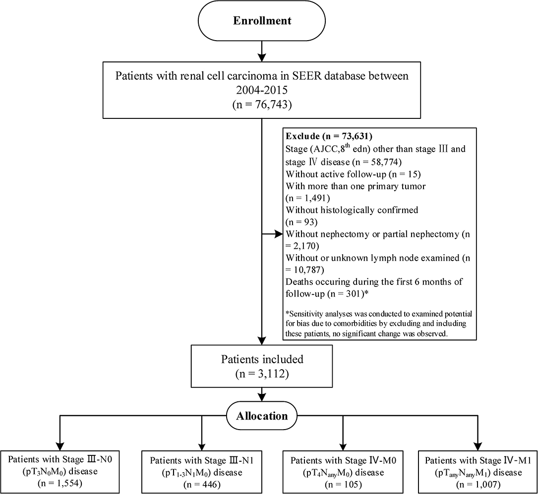
Patient consent was not required because the study was a retrospective database research in nature, there was no direct patient contact. Institutional Review Board approval was not required according to our institution policy. The SEER program of the National Cancer Institute is an authoritative source on cancer incidence and survival in the US covering approximately 34.6% of the US population, which routinely collects basic demographics and some clinical characteristics (14). SEER*Stat software (version 8.3.5) was queried to identify patients from SEER-18 database if they were diagnosed with RCC (International Classification of Disease for Oncology, 3rd edition [ICD-O-3], C67.0–C67.9) between January 2004 and December 2015 (n = 76,743). The study cohort was then limited to patients with stage III disease or pTanyNanyM1 and pT4NanyM0 disease according to the AJCC 8th Staging Manual using the collaborative stage information (n = 17,969). The eligible criteria included in the study were participants with one primary only, pathologically confirmed RCC, and contained complete data of age, race, gender, surgery records, pathological information, and with active follow up; and all patients underwent radical or partial nephrectomy with LN dissection which guaranteed the specimen and harvested LNs were sent for pathology in SEER (15). As a result, the study included a total of 3,112 patients—1,554 patients with pT3N0M0 disease, 446 patients with pT1−3N1M0 disease, 105 patients with pT4NanyM0 disease, and 1,007 patients with pTanyNanyM1 disease (Figure 1).
Description of Covariates
Baseline characteristics include age (continuous variable), sex (male, female), race (white, non-white), tumor site (left, right), and histological subtypes (clear cell, papillary, and chromophobe). Grade in SEER is coded according to the ICD-O-3. The number of examined and positive LNs is queried from collaborative stage information and presented as a continuous variable. Since information of invasion beyond capsule, fuhrman nuclear grade, and sarcomatoid features are only applicable after 2010, these covariates are not examined in χ2-test (16).
Propensity Score Matching (PSM)
PSM is a tool to reduce selection bias in observational studies (17). PSM with 1:1 matching was performed in our study to reduce the selection bias and ensure baseline balance among study groups. The covariates selected for matching were based on prior literature reports, known clinical prognostic factors, and statistical differences in the multivariate analysis. The variables were first forced into univariate analysis based on the prior literature reports and clinical significance. Then, variables that remained significant in the univariate analysis entered into multivariate Cox regression models. Variables left in the final multivariate analysis were used to generate the propensity score. Selected variables included age (18, 19) stage (8), grade (20), and histology (21) (Figure S1).
Statistical Analysis
Overall survival (OS) was defined as the time from diagnosis to death from any cause; cancer-specific survival (CSS) was defined as the time from diagnosis to death related to RCC. Frequencies and proportions, as well as means were reported for categorical and continuous variables, respectively. General linear models or χ2-test were performed to compare the distribution of baseline characteristics. The survival curves were estimated by the Kaplan-Meier method and compared by the log-rank test. The univariate and multivariate analyses and hazard ratios (HR) were evaluated by Cox proportional hazards regression model to find its independent prognostic risks. Significant variables in univariate analysis were entered into a multivariate model, and variables that remained significant were further entered into the final multivariate regression model. P ≤ 0.05 (2-sided probability) was considered statistically significant.
A nomogram consisted of modified stage based on the multivariate analyses was established using the rms packages in R (22). The concordance index (C-index) and calibration curve were performed to assess the discriminatory powers of the nomogram. The larger the C-index was, the more accurate the prognostic prediction demonstrated (23). Comparisons between the nomogram and current AJCC stage were performed with the survcomp packages in R (24). The predictive performance of model was further examined in the randomly 1 to 1 ratio selected internal validation cohort (25). Although validated in an internal cohort, the predictive accuracy of the modified staging system might be still suffer from some unmeasured confounding. All analyses were conducted using R software (version 3.5.3). Sensitivity analyses were conducted to investigate the potential for bias due to the existence of comorbidities by including and excluding deaths occurring during the first 6 months of follow-up.
Results
Between January 2004 and December 2015, 3,112 eligible patients were identified and categorized into pT3N0M0 group (n = 1,554), pT1−3N1M0 group (n = 446), pT4NanyM0 group (n = 105), and pTanyNanyM1 group (n = 1,007). Tables 1, 2 summarize the baseline characteristics for study groups before and after matching (for more information, please see Table S1). Before matching, slightly more non-white patients were included in pT1−3N1M0 group (19.5 vs. 12.9%, P < 0.001), patients in pT1−3N1M0 group are more likely to have undifferentiated tumors (22.9 vs. 17.7%, P < 0.001) and papillary adenocarcinoma (31.2 vs. 4.3%, P < 0.001) than those in pT3N0M0 group. After PSM, all variables between the study groups were balanced, except for those variables that were only applicable after 2010.
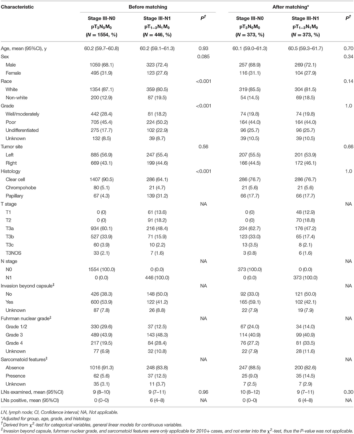
Table 1. Baseline characteristics of pT3N0M0 and pT1−3N1M0 groups before and after matching, SEER 2004-2015.
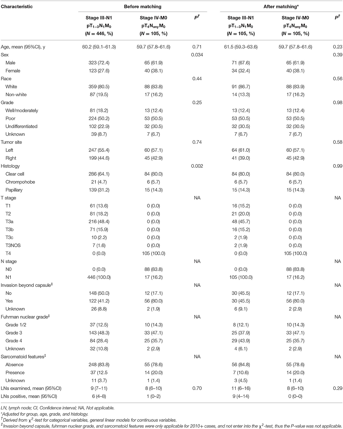
Table 2. Baseline characteristics of pT1−3N1M0 and pT4NanyM0 groups before and after matching, SEER 2004-2015.
Table 3 summarizes the independent risk factors for survival of stage III RCC patients. Patients with higher tumor grade (P < 0.001), higher fuhrman grade (P < 0.001), presence of sarcomatoid features (P < 0.001), and presence of node disease (P < 0.001) were associated with remarkably worse OS and CSS among stage III RCC patients before matching. After matching, similar outcomes were observed for stage III RCC patients (Table S2). Moreover, patients with the chrompohobe type RCC was associated with better OS (P = 0.002) and CSS (P = 0.001) compared to patients with clear cell type RCC, which indicated the potential survival benefit of chrompohobe type RCC (Figure S2). In the multivariate analysis, pT1−3N1M0 group was found to be associated with poor outcome compared to the pT3N0M0 group both before (HR = 2.5, 95% CI = 2.1–2.9 for OS, HR = 2.6, 95% CI = 2.2–3.2 for CSS) and after matching (HR = 2.5, 95% CI = 1.9–3.1 for OS, HR =2.5, 95% CI = 2.0–3.3 for CSS).
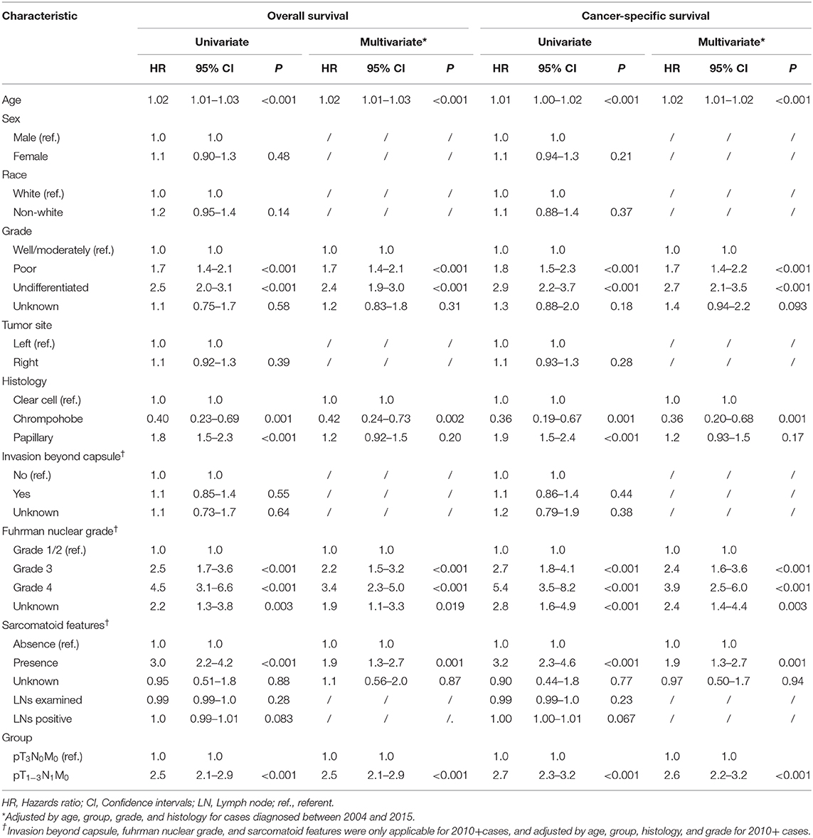
Table 3. Univariate and multivariate analyses of overall survival and cause-specific survival to pT3N0M0 and pT1−3N1M0 groups before matching, SEER 2004-2015.
We further used the Kaplan-Meier curves to demonstrate that the nodal involvement was associated with worse survival. The highest OS and CSS was observed in pT3N0M0 group (median OS = 102.0 months, median CSS = 110.0 months, P < 0.001 compared with other groups), whereas the similar survival was found for pT1−3N1M0 group and pT4NanyM0 group (P = 0.91 for OS, P = 0.82 for CSS). Moreover, significantly worse survival (median OS = 27.0 months, median CSS = 28.0 months, P < 0.05 compared with other groups) were found for pTanyNanyM1 group (Figure 2). After matching, significantly better survival was found for pT3N0M0 patients (median OS = 91.0 months, median CSS = 119.0 months, P < 0.001) compared with those of pT1−3N1M0 group (Figure 3). In addition, the survival of pT1−3N1M0 group didn't show remarkable difference compared to pT4NanyM0 group (Figure S3). However, both pT1−3N1M0 group and pT4NanyM0 group were associated with better survival than pTanyNanyM1 group (Figure S4). Moreover, for those patients diagnosed after 2010, the survival for pT1−3N1M0 group and pT4NanyM0 group remained stable both before and after matching (Figure S5).
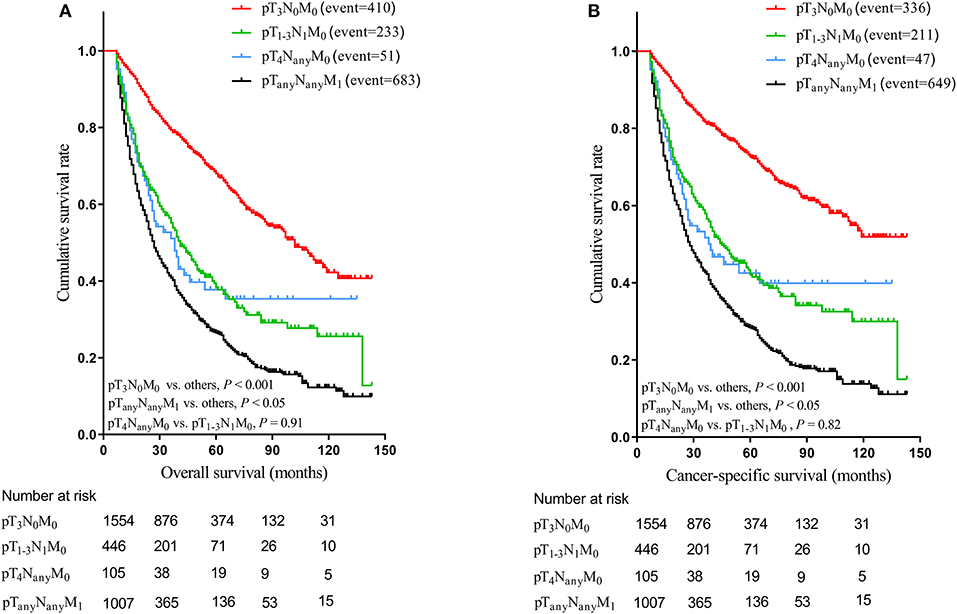
Figure 2. Kaplan-Meier estimation of overall survival curves (A) and cancer-specific survival curves (B) relative to each group before matching.
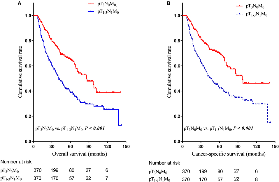
Figure 3. Kaplan-Meier estimation of overall survival curves (A) and cancer-specific survival curves (B) relative to stage III-N0 (pT3N0M0) and stage III-N1 (pT1−3N1M0) groups after matching.
Given the above results, pT1−3N1M0 and pT4NanyM0 disease were regrouped into stage IIIB according to the OS and CSS of each subgroup without changing the definition of TNM. pT3N0M0 disease was classified as stage IIIA and pTanyNanyM1 were classified as stage IV. The prognostic nomogram for modified stage group that integrated significant independent factors for CSS in the primary cohort was shown in Figure 4 (OS shown in Figure S6). The observed probability of 1-, 3-, and 5-years CSS in the primary cohort and 1-, 3-, and 5-years CSS in the validation cohort showed optimal consistency with the nomogram-predicted CSS (Figures 4B–G), the similar results for nomogram-predicted OS were also observed. The C-index for the modified stage group were improved significantly (0.70, 95% CI: 0.68–0.71 vs. 0.68, 95% CI: 0.66–0.69, P < 0.001 for OS; 0.71, 95% CI: 0.69–0.73 vs. 0.69, 95% CI: 0.68–0.72, P < 0.001 for CSS) compared to the AJCC 8th stage group in the primary cohort as well as the validation cohort (0.71, 95% CI: 0.69–0.73 vs. 0.70, 95% CI: 0.68–0.72, P = 0.002 for OS; 0.73, 95% CI: 0.71–0.74 vs. 0.70, 95% CI: 0.69–0.72, P < 0.001 for CSS), which indicated the higher discriminatory power of the modified stage.
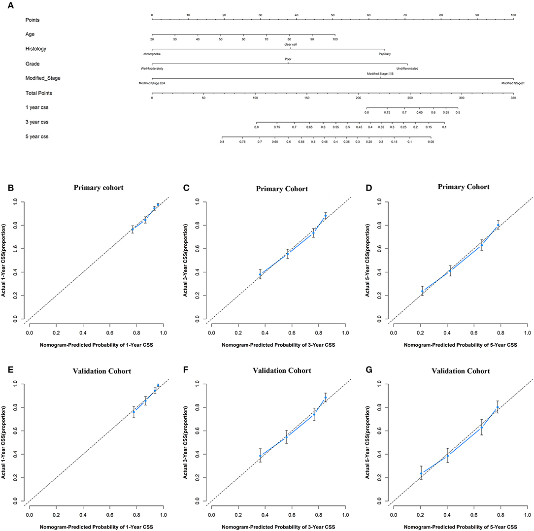
Figure 4. Prognostic nomogram, calibration curves of cancer-specific survival (CSS) for the modified stage of study patients. (A) The nomogram predicts 1-, 3-, and 5-years CSS in patients with renal cell carcinoma (RCC). The calibration curves predict CSS at 1-year (B), 3-years (C), and 5-years (D) in the primary cohort and at 1-year (E), 3-years (F), and 5-years (G) in the validation cohort.
Discussion
To the best of our knowledge, the present study is the first population-based analysis using the PSM method to assess the impact of LN involvement on survival in stage III RCC patients. In the current study, LN involvement, in general, was associated with remarkably poor survival in patients with stage III disease in the AJCC 8th staging system in both multivariate regression and PSM analyses. We found that RCC patients with pT3N0M0 disease could be reclassified as stage IIIA, while pT1−3N1M0 RCC together with pT4NanyM0 disease could be classified as stage IIIB for better prediction of prognosis.
LN involvement in RCC patients has been decreasing over years, from 30% in historical series to approximately 6%-20% in more recent studies (2, 11, 26–28). Despite the early detection of nodal disease, survival is still poor for those node-positive RCC patients (4, 29, 30). Previous studies consistently showed that node-positive disease was associated with worse survival compared to node-negative disease (4, 6, 7). Capitanio et al. examined the stage-specific effect of nodal involvement in non-metastatic RCC patients based on the 12 institutional databases (4). Of the 2,023 patients enrolled, 165 (4.7%) patients presented with nodal disease. The 5-years CSS rate was 87% for node-negative patients and 38% for node-positive patients (HR = 7.1, P < 0.001). Srivastava et al. found the consistent outcomes for node-negative and positive group in their NCDB cohort (median OS, 79.5 vs. 18.6 months) (31). A more recent population-based analysis compared the survival between node-positive disease and node-negative disease patients in the absence of distant metastasis, the 5-years OS rate was 72.7% for pN0 disease patients and 38.1% for pN1 disease patients (P < 0.001) (13). Comparable outcomes were observed in our study (5-years OS rate was 78.1, 40.8% for pT3N0M0 groups and pT1−3N1M0 groups, respectively), and thus reinforcing these findings.
LN involvement as well as distant metastasis contributed to the recurrence and progression of disease in RCC patients, resulting in poor survival among advanced RCC patients (11, 21). Pantuck et al. directly compared the survival between 43 (4.8%) patients with pN0M1 disease and 236 (26%) with pN+M0 disease, among the 900 study patients (11). No difference was found for OS between the two groups (P = 0.59). MDACC study compared the pT1−3N1M0 disease with pT1−3N0M1 disease in OS and CSS, and found that pT1−3N1M0 disease was not different in both OS (median OS, 2.4y vs. 2.4y, P = 0.62) and CSS (median CSS, 2.8y vs. 2.4y, P = 0.10) relative to pT1−3N0M1 diseases (12). Meanwhile, in a large cohort analysis of 2,120 patients with RCC from China (13), Shao et al. suggested that T1−3N1M0 disease should be separated from T3N0M0 disease and reclassified as same stage disease with T4N0M0 disease in the AJCC 8th staging system. T4N0M0 disease accounted for almost 79.3% for T4M0 groups in this study, only 85 T4N1M0 patients were included, while 10,382 patients with M1 disease were enrolled, it is not convincing that T4N1M0 groups should be combined with M1 disease. However, these studies including this current one indicated that node-positive stage III RCC might need to be separately classified from node-negative stage III disease. In our study, we found the survival of pT1−3N1M0 patients was similar to that of pT4NanyM0 patients, and the modified stage that combined pT1−3N1M0 and pT4NanyM0 showed improved discriminatory power than 8th AJCC stage. These findings suggest that these two groups might be re-staged together as the same stage.
Pathologic staging of RCC is essential for guiding clinical treatment decision-making process and selecting patients for potential adjuvant therapy (32). High-risk stage II and stage III RCC patients with clear cell histology are considered candidates for adjuvant therapy according to the National Comprehensive Cancer Network (NCCN) guidelines (33). However, two randomized controlled trials compared the adjuvant therapy for non-metastatic RCC patients, and contrary results were found. ASSURE trial showed no differences between sunitinib group and placebo group for stage III/IV patients in disease-free survival (34). Whereas the S-TRAC trial found favorable disease-free survival for sunitinib group in stage III/IV patients (35). In the subset analyses of S-TRAC trial (36), no survival benefit was found in both the low-risk group and high-risk group of T3N0M0 patients by receiving sunitinib. On the contrary, even combined with high-risk group of T3N0M0 patients, T1−3N1M0 patients and T4NanyM0 patients were associated with improved survival (HR = 0.74, 95% CI = 0.55–0.99, P = 0.04). Given the better survival of T3N0M0 disease in the current study, these controversial findings indicated that T3N0M0 may not significantly benefit from adjuvant therapy similar to T1−3N1M0 and T4NanyM0 disease. The poor survival observed for pT1−3N1M0 and pT4NanyM0 disease in our study might partly support the necessity of adjuvant therapy. Thus, it might be appropriate to regroup the pT1−3N1M0 disease together with pT4NanyM0 disease for better guiding adjuvant therapy.
Over years, the AJCC staging system has aimed to improve its prognostic accuracy by publishing revisions. As a heterogeneous group, locally advanced RCC underwent several staging revisions. The AJCC 7th staging manual reclassified the ipsilateral adrenal invasion into pT4 disease for the similar CSS between ipsilateral adrenal invasion and pT4 disease (37). Moreover, Anderson et al. (38) showed the similar survival between RCC patients with urinary collecting system invasion (UCSI) and patients with pT4 disease, but tumor with UCSI was classified as pT3a in the AJCC 8th staging system. Patel et al. discussed multiple proposed classification systems, and suggested that the understanding and re-evaluation for stage III and IV RCC should be further discussed and validated (39). In the current AJCC 8th cancer staging manual, node-negative disease and node-positive disease in stage III were integrated together into the same stage III disease without further categorization such as stage IIIA and stage IIIB. The higher discriminatory power for our modified stage group in this SEER cohort might also indicate that the current staging system still has room for improve the RCC staging. Nevertheless, the consistency between MDACC results and the current study, and the validation from the large Chinese cohorts and other studies warrants a reexamination of the heterogeneity of the stage III disease defined by the current AJCC 8th cancer staging system in prospective studies.
Our findings must be interpreted within the context of several known limitations. Firstly, as previously described (40), information of invasion beyond capsule, fuhrman nuclear grade, and sarcomatoid features were only applicable after 2010, and not examined in the initial analysis. Nevertheless, subsequent analyses were conducted among cases after 2010, similar results were found among the study groups (Figure S5). Secondly, some important information is not available in SEER database such as performance status (e.g., Eastern Cooperative Oncology Group or Karnofsky performance status), staging methods for patients, comorbidities, and treatment details. These may introduce biases. However, such limitations were also observed in the previous SEER-based analyses (41, 42). Thirdly, there were still some small sample groups in our study, although we could not rule out the biases from unmeasured factors, using PSM methods, well-balanced comparisons might somewhat reduce the biases in our results. It is preferable to obtain more details, however, the remarkable survival difference by nodal status and consistent findings from other cohorts highlighted the future direction for RCC re-staging. We sought to include only the pathologically staged patients and used the pTNM stage throughout the study. Obviously, this may introduce selection bias because we confined our study population to these highly selected patients. However, these highly selected cases represented patients with high-quality medical documentation. It is unlikely the quality of documentation might differ among patients with different stages; therefore, confining our study population to those patients with explicit information should not significantly bias our conclusion. Nevertheless, the present results should be interpreted as preliminary. Further studies, especially large prospective studies, are required to clarify the heterogeneity of the current stage III RCCs classified by AJCC 8th staging system.
In conclusion, the present study suggested that the current AJCC 8th staging system for stage III RCC should be further discussed and validated for the better prediction of prognosis. If validated, patients with pT1−3N1M0 disease might be separated from pT3N0M0 disease (which might be classified as stage IIIA) and classified as stage IIIB together with pT4NanyM0 disease. Further researches and independent cohort validations are warranted to address the heterogeneity of the stage III RCCs.
Data Availability Statement
The datasets generated for this study are available on request to the corresponding author.
Ethics Statement
Patient consents were not required because the study is a retrospective database research in nature, there was no direct patient contact. Institutional Review Board approval was not required according to our institution policy.
Author Contributions
ZF: guarantor of the article. ZF and JH: conception/design. JH, QL, PL, SW, YQ, and RZ: collection and/or assembly of data. JH, PL, QL, RZ, SW, YQ, and QS: data analysis and interpretation. JH, QL, PL, SW, RZ, YQ, QS, and ZF: manuscript writing. ZF, JH, PL, SW, QL, RZ, YQ, and QS: final approval of manuscript.
Funding
This study was partially supported by the National Natural Science Foundation of China [grants 81472971 and 81773555], the funders had no role in the design of the study or the interpretation of the findings.
Conflict of Interest
The authors declare that the research was conducted in the absence of any commercial or financial relationships that could be construed as a potential conflict of interest.
Acknowledgments
We sincerely thank the National Cancer Institute and the SEER staff for providing this invaluable database.
Supplementary Material
The Supplementary Material for this article can be found online at: https://www.frontiersin.org/articles/10.3389/fonc.2020.00365/full#supplementary-material
References
1. GLOBOCAN 2018: renal cell carcinoma: estimated cancer incidence, mortality and prevalence worldwide in 2018. International Agency for Research on Cancer: World Health Organization. (2018). Available online at: http://gco.iarc.fr/today/fact-sheets-populations (accessed December 23, 2018).
2. Capitanio U, Suardi N, Matloob R, Roscigno M, Abdollah F, Di Trapani E, et al. Extent of lymph node dissection at nephrectomy affects cancer-specific survival and metastatic progression in specific sub-categories of patients with renal cell carcinoma (RCC). BJU Int. (2014) 114:210–5. doi: 10.1111/bju.12508
3. Tilki D, Chandrasekar T, Capitanio U, Ciancio G, Daneshmand S, Gontero P, et al. Impact of lymph node dissection at the time of radical nephrectomy with tumor thrombectomy on oncological outcomes: results from the international renal cell carcinoma-venous thrombus consortium (IRCC-VTC). Urol Oncol. (2018) 36:79e11–17. doi: 10.1016/j.urolonc.2017.10.008
4. Capitanio U, Jeldres C, Patard JJ, Perrotte P, Zini L, de La Taille A, et al. Stage-specific effect of nodal metastases on survival in patients with non-metastatic renal cell carcinoma. BJU Int. (2009) 103:33–7. doi: 10.1111/j.1464-410X.2008.08014.x
5. Terrone C, Cracco C, Porpiglia F, Bollito E, Scoffone C, Poggio M, et al. Reassessing the current TNM lymph node staging for renal cell carcinoma. Eur Urol. (2006) 49:324–31. doi: 10.1016/j.eururo.2005.11.014
6. Lughezzani G, Capitanio U, Jeldres C, Isbarn H, Shariat SF, Arjane P, et al. Prognostic significance of lymph node invasion in patients with metastatic renal cell carcinoma: a population-based perspective. Cancer. (2009) 115:5680–7. doi: 10.1002/cncr.24682
7. Blute ML, Leibovich BC, Cheville JC, Lohse CM, Zincke H. A protocol for performing extended lymph node dissection using primary tumor pathological features for patients treated with radical nephrectomy for clear cell renal cell carcinoma. J Urol. (2004) 172:465–9. doi: 10.1097/01.ju.0000129815.91927.85
9. Canfield SE, Kamat AM, Sánchez-Ortiz RF, Detry M, Swanson DA, Wood CG. Renal cell carcinoma with nodal metastases in the absence of distant metastatic disease (clinical stage TxN1-2M0): the impact of aggressive surgical resection on patient outcome. J Urol. (2006) 175:864–9. doi: 10.1016/S0022-5347(05)00334-4
10. Trinh QD, Schmitges J, Bianchi M, Sun M, Shariat SF, Sammon J, et al. Node-positive renal cell carcinoma in the absence of distant metastases: predictors of cancer-specific mortality in a population-based cohort. BJU Int. (2012) 110:E21–7. doi: 10.1111/j.1464-410X.2011.10701.x
11. Pantuck AJ, Zisman A, Dorey F, Chao DH, Han KR, Said J, et al. Renal cell carcinoma with retroperitoneal lymph nodes: role of lymph node dissection. J Urol. (2003) 169:2076–83. doi: 10.1097/01.ju.0000066130.27119.1c
12. Yu KJ, Keskin SK, Meissner MA, Petros FG, Wang X, Borregales LD, et al. Renal cell carcinoma and pathologic nodal disease: implications for American Joint Committee on cancer staging. Cancer. (2018) 124:4023–31. doi: 10.1002/cncr.31661
13. Shao N, Wang HK, Zhu Y, Ye DW. Modification of American Joint Committee on cancer prognostic groups for renal cell carcinoma. Cancer Med. (2018) 7:5431–8. doi: 10.1002/cam4.1790
14. NCI. Overview of the SEER program. (2018). Available online at: https://seer.cancer.gov/about/overview.html (accessed December 23, 2018).
15. Surgery Codes of Kidney. (2018). Available online at: https://seer.cancer.gov/manuals/2018/AppendixC/Surgery_Codes_Kidney_2018.pdf (accessed December 23, 2018).
16. NCI. Collaborative Stage Site-Specific Factors (CS SSF). (2018). Available online at: https://seer.cancer.gov/seerstat/databases/ssf/ (accessed December 23, 2018).
17. Rosenbaum PR, Rubin DB. The central role of the propensity score in observational studies for causal effects. Biometrika. (1983) 70:41–55. doi: 10.1093/biomet/70.1.41
18. Hollingsworth JM, Miller DC, Daignault S, Hollenbeck BK. Five-year survival after surgical treatment for kidney cancer: a population-based competing risk analysis. Cancer. (2007) 109:1763–8. doi: 10.1002/cncr.22600
19. Verhoest G, Veillard D, Guille F, De La Taille A, Salomon L, Abbou CC, et al. Relationship between age at diagnosis and clinicopathologic features of renal cell carcinoma. Eur Urol. (2007) 51:1298–304. doi: 10.1016/j.eururo.2006.11.056
20. Minervini A, Lilas L, Minervini R, Selli C. Prognostic value of nuclear grading in patients with intracapsular (pT1-pT2) renal cell carcinoma. Long-term analysis in 213 patients. Cancer. (2002) 94:2590–5. doi: 10.1002/cncr.10510
21. Sun M, Bianchi M, Hansen J, Abdollah F, Trinh QD, Lughezzani G, et al. Nodal involvement at nephrectomy is associated with worse survival: a stage-for-stage and grade-for-grade analysis. Int J Urol. (2013) 20:372–80. doi: 10.1111/j.1442-2042.2012.03170.x
22. RMS: Regression Modeling Strategies. R package version 3.5.1. Available online at: https://CRAN.R-project.org/package=rms (accessed December 23, 2018).
23. Wang Y, Li J, Xia Y, Gong R, Wang K, Yan Z, et al. Prognostic nomogram for intrahepatic cholangiocarcinoma after partial hepatectomy. J Clin Oncol. (2013) 31:1188–95. doi: 10.1200/JCO.2012.41.5984
24. Schroeder M, Culhane A, Quackenbush J, Haibe-Kains B. survcomp: an R/Bioconductor package for performance assessment and comparison of survival models. Bioinformatics. (2011) 27:3206–8. doi: 10.1093/bioinformatics/btr511
25. Pu N, Li J, Xu Y, Lee W, Fang Y, Han X, et al. Comparison of prognostic prediction between nomogram based on lymph node ratio and AJCC 8th staging system for patients with resected pancreatic head carcinoma: a SEER analysis. Cancer Manag Res. (2018) 10:227–38. doi: 10.2147/CMAR.S157940
26. Blom JH, van Poppel H, Marechal JM, Jacqmin D, Schroder FH, de Prijck L, et al. Radical nephrectomy with and without lymph-node dissection: final results of European organization for research and treatment of cancer (EORTC) randomized phase 3 trial 30881. Eur Urol. (2009) 55:28–34. doi: 10.1016/j.eururo.2008.09.052
27. Pizzocaro G, Piva L. Pros and cons of retroperitoneal lymphadenectomy in operable renal cell carcinoma. Eur Urol. (1990) 18(Suppl.2):22–3. doi: 10.1159/000463953
28. Giberti C, Oneto F, Martorana G, Rovida S, Carmignani G. Radical nephrectomy for renal cell carcinoma: long-term results and prognostic factors on a series of 328 cases. Eur Urol. (1997) 31:40–8. doi: 10.1159/000474416
29. Antonelli A, Cozzoli A, Zani D, Zanotelli T, Nicolai M, Cunico SC, et al. The follow-up management of non-metastatic renal cell carcinoma: definition of a surveillance protocol. BJU Int. (2007) 99:296–300. doi: 10.1111/j.1464-410X.2006.06616.x
30. Lam JS, Shvarts O, Pantuck AJ. Changing concepts in the surgical management of renal cell carcinoma. Eur Urol. (2004) 45:692–705. doi: 10.1016/j.eururo.2004.02.002
31. Srivastava A, Rivera-Nunez Z, Kim S, Sterling J, Farber N, Radadia K, et al. mp14-20 Impact of pathologic node positive renal cell carcinoma on survival in patients without Metastasis: evidence in support of expanding the definition of stage IV kidney cancer. J Urol. (2019) 201(Supp.l4):e195. doi: 10.1097/01.JU.0000555316.15591.47
32. Miller LA, Stemkowski S, Saverno K, Lane DC, Tao Z, Hackshaw MD, et al. Patterns of care in patients with metastatic renal cell carcinoma among a U.S. payer population with commercial or medicare advantage membership. J Manag Care Spec Pharm. (2016) 22:219–26. doi: 10.18553/jmcp.2016.22.3.219
33. Kidney cancer, Version 2. NCCN clinical practice guidelines in oncology. National Comprehensive Cancer Network. (2018). Available online at: https://www.nccn.org/professionals/physician_gls/default.aspx (accessed January 21, 2019).
34. Haas NB, Manola J, Uzzo RG, Flaherty KT, Wood CG, Kane C, et al. Adjuvant sunitinib or sorafenib for high-risk, non-metastatic renal-cell carcinoma (ECOG-ACRIN E2805): a double-blind, placebo-controlled, randomised, phase 3 trial. Lancet. (2016) 387:2008–16. doi: 10.1016/S0140-6736(16)00559-6
35. Ravaud A, Motzer RJ, Pandha HS, George DJ, Pantuck AJ, Patel A, et al. Adjuvant sunitinib in high-risk renal-cell carcinoma after nephrectomy. N Engl J Med. (2016) 375:2246–54. doi: 10.1056/NEJMoa1611406
36. Motzer RJ, Ravaud A, Patard JJ, Pandha HS, George DJ, Patel A, et al. Adjuvant sunitinib for high-risk renal cell carcinoma after nephrectomy: subgroup analyses and updated overall survival results. Eur Urol. (2018) 73:62–8. doi: 10.1016/j.eururo.2017.09.008
37. Edge S, Byrd D, Compton C, Fritz A, Greene F, Trotti A. AJCC Cancer Staging Manual. 7th ed. New York, NY: Springer (2010).
38. Anderson CB, Clark PE, Morgan TM, Stratton KL, Herrell SD, Davis R, et al. Urinary collecting system invasion is a predictor for overall and disease-specific survival in locally invasive renal cell carcinoma. Urology. (2011) 78:99–104. doi: 10.1016/j.urology.2011.02.039
39. Patel HV, Srivastava A, Shinder B, Sadimin E, Singer EA. Strengthening the foundation of kidney cancer treatment and research: revising the AJCC staging system. Ann Transl Med. (2019) 7(Suppl. 1):S33–37 doi: 10.21037/atm.2019.02.19
40. Altekruse SF, Dickie L, Wu XC, Hsieh MC, Wu M, Lee R, et al. Clinical and prognostic factors for renal parenchymal, pelvis, and ureter cancers in SEER registries: collaborative stage data collection system, version 2. Cancer. (2014) 120(Suppl.23):3826–35. doi: 10.1002/cncr.29051
41. Joslyn SA, Sirintrapun SJ, Konety BR. Impact of lymphadenectomy and nodal burden in renal cell carcinoma: retrospective analysis of the national surveillance, epidemiology, and end results database. Urology. (2005) 65:675–80. doi: 10.1016/j.urology.2004.10.068
Keywords: renal cell carcinoma, lymph node metastases, SEER, staging, nomogram, propensity score matching
Citation: Han J, Li Q, Li P, Wang S, Zhang R, Qiao Y, Song Q and Fu Z (2020) Reassessment of American Joint Committee on Cancer Staging for Stage III Renal Cell Carcinoma With Nodal Involvement: Propensity Score Matched Analyses of a Large Population-Based Study. Front. Oncol. 10:365. doi: 10.3389/fonc.2020.00365
Received: 16 June 2019; Accepted: 02 March 2020;
Published: 19 March 2020.
Edited by:
Rosa M. Nadal, National Heart, Lung, and Blood Institute (NHLBI), United StatesReviewed by:
Felix K. H. Chun, University Medical Center Hamburg-Eppendorf, GermanyEric A. Singer, Rutgers Cancer Institute of New Jersey, United States
Copyright © 2020 Han, Li, Li, Wang, Zhang, Qiao, Song and Fu. This is an open-access article distributed under the terms of the Creative Commons Attribution License (CC BY). The use, distribution or reproduction in other forums is permitted, provided the original author(s) and the copyright owner(s) are credited and that the original publication in this journal is cited, in accordance with accepted academic practice. No use, distribution or reproduction is permitted which does not comply with these terms.
*Correspondence: Zhenming Fu, ZGF2aWRmdXptaW5nQHdodS5lZHUuY24=
†These authors have contributed equally to this work
 Jianglong Han
 Qin Li†
 Zhenming Fu