- 1Department of Pathology, Xiangya Hospital, Central South University, Changsha, China
- 2Department of Pathology, School of Basic Medicine, Central South University, Changsha, China
- 3National Clinical Research Center for Geriatric Disorders, Xiangya Hospital, Central South University, Changsha, China
- 4Department of Pathology, Hunan Provincial People’s Hospital, The First Affiliated Hospital of Hunan Normal University, Changsha, China
Intravascular large B-cell lymphoma (IVLBCL) is a rare and highly malignant non-Hodgkin B-cell lymphoma with uncommon clinical presentation and poor prognosis. The diagnostic pitfall of IVLBCL is mainly due to the fact that subtle histological changes could be easily overlooked, in addition to its rare occurrence, non-specific and variable clinical presentations, and the absence of significant mass lesions. The purpose of this study is to further explore the clinicopathologic and molecular features of IVLBCL to ensure an accurate diagnosis of this entity. Here, we retrospectively present the data of the four new cases and the literature cases. The age ranged from 23 to 92, with a medium age of 67 and a male-to-female ratio of 1:1. The clinical manifestations are extremely variable, including fever, night sweats, weight loss, anemia, thrombocytopenia, unexplained hypoxemia, impaired consciousness, and skin lesions, as well as the extremely low levels of serum albumin, high levels of serum lactate dehydrogenase (LDH), soluble interleukin-2 receptor (sIL2R), and ferritin. Morphologically, 99.9% of cases showed a selective growth pattern with large, atypical lymphocytes within the lumen of small blood vessels. In addition, vast majority of cases were positive for CD20, CD79a, PAX5, MUM1, and BCL6, and a subset of cases expressed BCL2 and CD5, whereas CD3 and CD10 were typically negative. Ki-67 proliferative index ranged from 20% to 100%. To sum up, we have conducted comprehensive case reports, to the best of our knowledge, this is the largest reported cohort of IVLBCL cases. Comprehensive assessments and more IVLBCL cases are required for early diagnosis and prompt treatment.
Introduction
Intravascular large B-cell lymphoma (IVLBCL) is a rare and distinct subtype of extranidal diffuse large B-cell lymphoma (DLBCL) and was first described in 1959 (1), and since then, more than 300 cases have been reported in the literature, mostly case reports and small series. It is characterized by tumor cells accumulation in the lumens of small and medium vessels, but there are rare cases characterized by proliferation in large blood vessels (2). Clinical presentations of IVLBCL are extremely various, including unexplained fever, night sweat, anemia, weight loss, impaired consciousness, and skin lesions. Diagnostic awareness deficiency and variable clinical behavior of IVLBCL potentially lead to delayed treatment and poor prognosis. In this study, we retrospectively analyzed the comprehensive clinical, laboratory, imaging, morphologic, immunophenotypic, and molecular characteristics of IVLBCL using data on four cases diagnosed at our hospital and additional 311 cases from the literature, to further improve the full recognition of the clinicopathologic characteristics of IVLBCL to ensure accurate diagnosis of this entity and facilitate appropriate treatment.
Materials and Methods
Cases Selection
We retrospectively reviewed the records of four patients with IVLBCL from December 2011 to July 2019 at Xiangya Hospital of Central South University. The clinical history, laboratory data, imaging abnormalities, treatment regimens, and outcomes were collected. The diagnosis of each case was confirmed with morphologic examination, immunohistochemical stains, or molecular studies. We also conducted a comprehensive literature review for the cases of IVLBCL published in the last 20 years (between 1999 and 2021) in PubMed (http://www.ncbi.nlm.nih.gov/pubmed/) using combinations of keywords in the title/abstract field, including “intravascular large b-cell lymphoma and IVLBCL”. The cases in the literature were carefully reviewed to extract necessary clinicopathologic data and combined with the cases repeatedly studied in different papers. A total of 311 cases of IVLBCL were retrieved from the literature and were included in our research (3–91).
Specimen Processing and Immunohistochemical Staining
Computed tomography (CT)–guided percutaneous liver, lung, and adrenal glands biopsies were performed. All biopsied specimens of four cases were formalin-fixed and paraffin-embedded (FFPE) and subsequently sectioned at 4.0 μm for hematoxylin-eosin staining and immunohistochemical study. Appropriate negative and positive controls were performed with satisfactory staining.
In Situ Hybridization
Digoxigenin-labeled probe (Dako) was used to detect Epstein–Barr virus–encoded small RNA. The in situ hybridization (ISH) assays were performed on a Ventana Benchmark XT platform with an iViewBlue detection kit according to the manufacture’s manual (Ventana Medical Systems Inc., Tucson, AZ, USA).
Fluorescence In Situ Hybridization
Fluorescence ISH (FISH) studies were performed on two cases on FFPE tissue using C-MYC, BCL2, and BCL6 dual-colored break-apart rearrangement probes (Vysis Inc., IL, USA). Hybridization signals were assessed from 100 nuclei of tumor cells with an established cutoff of 15% for rearrangement of the gene locus. When the proportion of tumor cells with separation signals was less than 15%, the gene was interpreted as not rearranged. The FISH images were obtained using an Olympus BX51 fluorescence microscope and were captured on the Olympus Image DP2-BSW Software (Olympus Soft Imaging GmbH, Hamburg, DE).
Results
Clinical Characteristics
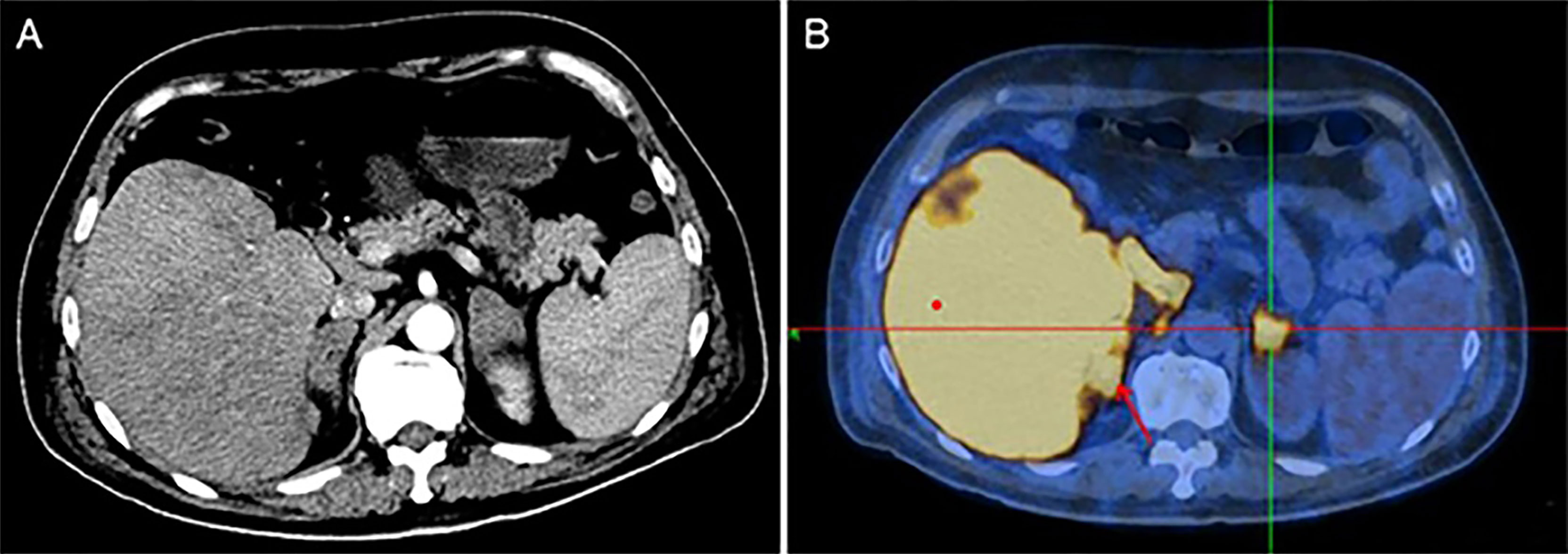
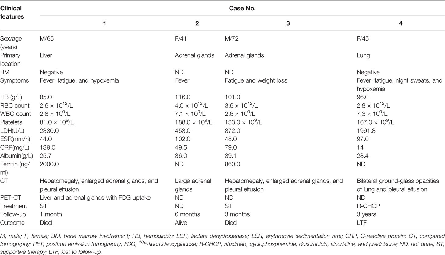
The major clinical features of the four cases of IVLBCL are presented in Table 1. Patients were middle-aged and elderly with an average age of 56 years at diagnosis ranging from 41 to 72 years and a male-to-female ratio of 1:1. The sites of initial diagnosis of IVLBCL were in the adrenal glands (two cases) and the liver and lung (one each case). All patients presented with B symptoms, including fever, night sweats, weight loss, and fatigue. Laboratory examination results revealed anemia (three of four), hypoalbuminemia (four of four), thrombocytopenia (one of four), and remarkably elevated levels of serum LDH, erythrocyte sedimentation rate (ESR), and C-reactive protein (CRP). Abdominal CT with contrast enhancement showed enlargement of bilateral adrenal glands (three of four) and hepatosplenomegaly (two of four). CT scanning of the chest revealed pleural effusion (three of four), and one case showed ground glass opacities in bilateral lung fields (84). Two patients presented enlarged retroperitoneal lymph nodes at CT scan, which were not biopsied. Fluorine-18 fluorodeoxyglucose (18-FDG) positron emission tomography–CT (PET-CT) study showed high uptake in the liver and bilateral adrenal glands (Figure 1). Bone marrow biopsies were performed in two patients, showing an active proliferation of granulocyte with obvious toxic reaction, but no evidence of lymphoma infiltration was detected. However, a bone marrow biopsy in one of the patients showed hemophagocytosis. In addition, the other two patients without bone marrow biopsy showed no hemophagocytic syndrome in clinical symptoms and serological results. All patients were diagnosed as IVLBCL by percutaneous organ biopsy. Only one patient received systemic chemotherapy consisting of rituximab, cyclophosphamide, doxorubicin, vincristine, and prednisone (R-CHOP) and had a complete remission at least initially; two patients received supportive care; and one patient refused any treatment. A total of two patients died of disease during the short period of follow-up.

Figure 1 (A) Abdominal CT with contrast enhancement image showed diffuse enlargement of the liver, spleen, and bilateral adrenal glands. (B) PET-CT showed high uptake in liver and bilateral adrenal glands.
Morphologic Features
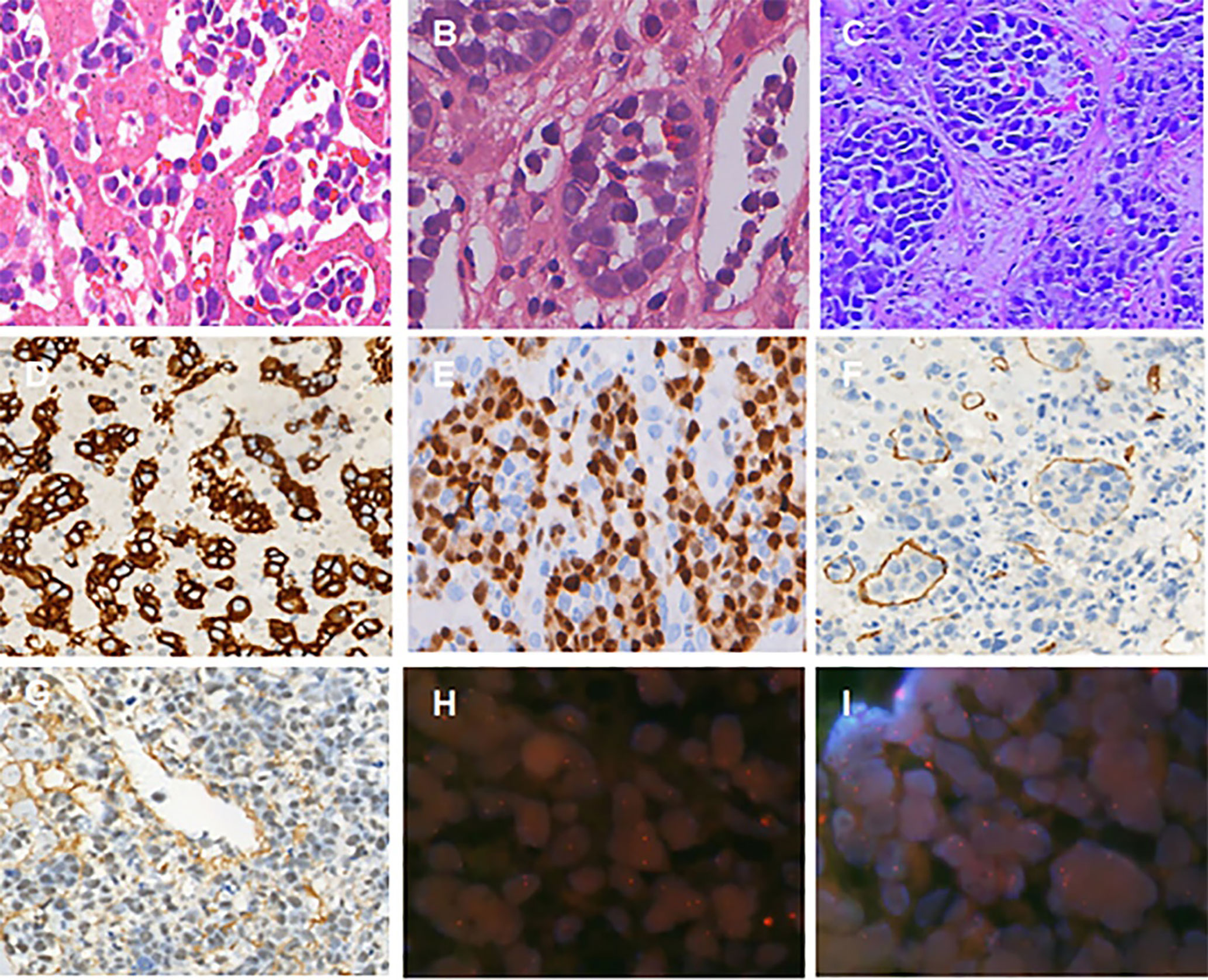
All cases showed selective growth of large, atypical lymphocytes within the lumen of small blood vessels. Various degrees of cellulose thrombosis were observed in two cases. The neoplastic lymphocytes were large in size with one or more nucleoli, scant cytoplasm, and frequent mitotic figures (Figure 2).

Figure 2 Histologic features of IVLBCL from the percutaneous organ biopsy specimens. (A) A liver biopsy demonstrated intermediate to large cells within sinusoids with irregular nuclei, dispersed chromatin, occasional nucleoli, and scant cytoplasm (case 1). (B) Cellulose thrombosis could be found (case 2). (C) Lymphoma cells were large, with scant cytoplasm, prominent nucleus, and frequent mitotic figures (case 3). (D–G) Immunophenotypic features of IVLBCL. The neoplastic cells were positive for CD20, MUM1, CD31, and EBER (original magnification D-G, ×200); FISH study showed non-rearrangement of C-MYC, BCL2, or BCL6 gene on the IVLBCL cells. Non-rearranged gene is represented by orange/green fusion signal (H, I).
Immunohistochemistry and ISH
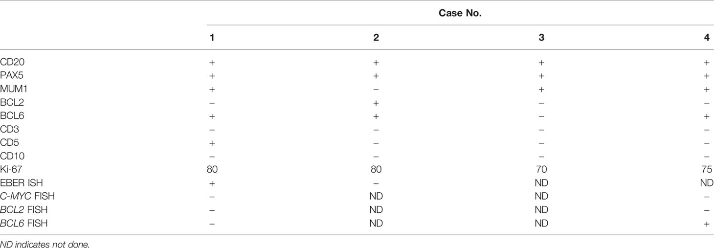
The results of immunohistochemistry and EBER-ISH are summarized in Table 2. Tumor cells were positive for CD20 (four of four), PAX5 (four of four), MUM1 (four of four), BCL6 (three of four), BCL2 (one of four), and CD5 (one of four), but negative for CD3 and CD10. The Ki-67 proliferative index ranged from 70% to 80%. ISH for EBER (Epstein–Barr virus-encoded small RNA) was positive in one patient (Figure 2).
ISH Study
The C-MYC, BCL2, and BCL6 gene translocation were assessed using the dual-colored break-apart rearrangement probes in two cases (Table 2). In our study, rearrangement of BCL6 gene on the IVLBCL cells was detected in case 4, which had been reported previously (83). However, FISH study did not show any rearrangement of C-MYC, BCL2, and BCL6 genes in case 1 (Figure 2).
Discussion
IVLBCL is a rare large B-cell lymphoma characterized by multifocal and selective growth of capillaries rather than arteries and veins. It primarily affects elderly individuals and patients usually without lymphadenopathy. Symptoms and signs are related to organs that block small blood vessels. The most commonly involved organs are the skin, central nervous system (CNS), and bone marrow (2). Tumor cell are positive for commonly used B-cell markers (CD20, CD79a, and PAX5), and aberrant expression of the T-cell marker CD5 is detected in a subset of cases.
There are some drawbacks in this study including the following: The number of cases is inadequate and clinical examination items were requested non-uniformly. Furthermore, the patient treatment information summarized from the literature is incomplete, and the follow-up duration is not enough. However, all the cases that we reported had typical clinicopathological and immunohistochemical features of IVLBCL. Notably, we noticed important findings in four cases. EBER-positive nuclear signals were detected by ISH in our case. Although Epstein-Barr virus (EBV)-positive DLBCL was listed as a distinct subtype of DLBCL in the revised World Health Organization classification (89). Cases reporting EBV-positivity in the liver are very rare. Few studies focusing on EBV expression in IVLBCL have been reported.
In the past, the majority of IVLBCL cases were diagnosed by autopsy. With the improvement of awareness of this entity, most patients were diagnosed by bone marrow biopsy and skin biopsy from positive skin lesions or random skin biopsy (RSB) (90). The liquid biopsy has also been reported as a promising method for diagnosing IVLBCL (85). In our study, no evidence of lymphoma infiltration was detected by bone marrow biopsy. However, we used CT-guided percutaneous organ biopsy as our diagnostic method of choice, including the adrenal gland biopsy (two cases) and the liver and lung biopsies (one each case). CT-guided percutaneous biopsy is a safe and accurate diagnostic procedure in IVLBCL.
Imaging study is helpful to the diagnosis of IVLBCL. A few cases reported PET-CT had been used for making initial diagnosis of IVLBCL by identifying appropriate target sites for biopsy, as these patients usually show high FDG-uptake in involved organs (85). In addition, the distribution and intensity of FDG uptake lesions on PET/CT can be used to predict outcomes and survival rates (86). In this study, two patients’ lesions were detected by PET-CT in the liver and adrenals. Moreover, CT showed ground-glass opacities of lung, hepatomegaly, and enlargement of adrenals. Some previous studies showed high frequency of neurological abnormalities on brain magnetic resonance imaging (MRI), and these abnormal findings appeared to relate to ischemic changes of small brain capillaries due to neoplastic lymphoma cells (27–35). However all patients in four new cases did not present neurological symptoms, and thus, brain MRI was not performed.
With standard chemotherapy of R-CHOP or autologous stem cell transplantation (SCT), about 60% of patients with IVLBCL was possible survived more than 5 years (90). In the present study, all patients were clinically suspected of and diagnosed with IVLBCL by two experts and received follow-up at our hospital. Importantly, a patient treated with R-CHOP has a survival time of at least 3 years.
Conclusion
We described a comprehensive approach for the diagnosis and treatment of patients with IVLBCL. We also described peculiar clinical, laboratory, imaging, morphologic, and molecular features of this disease. We point out that this study provides valuable observations for recognizing the clinicopathologic features of IVLBCL and contributes to the early diagnosis of IVLBCL, which will increase the possibility of prompt treatments and ultimately lead to a better survival rate.
Review of 311 Cases of IVLBCL
After an extensive search of the literature, we found 311 cases of IVLBCL. The detailed clinicopathologic data of the 311 cases from the literature are included in Supplementary Tables (Supplemental Digital Content 1, http://links.lww.com/PAS/A438).
IVLBCL mostly affected elderly patients with a male-to-female ratio of 1:1. The average age and median age were 65.2 and 67 years, respectively, ranging from 23 to 92 years. Diagnosis was established in vivo in 258 of 311 (83.0%) patients and postmortem in 51 patients (16.4%). Patients with IVLBCL were diagnosed via RSB (81 of 311, 26.0%), bone marrow biopsy (46 of 311, 14.8%), lung biopsy, and adrenal biopsy. IVLBCL lesions were found in almost all organs including the thyroid, liver, pancreas, prostate, and brain. Bone marrow lymphoma infiltration was detected in 92 patients (92 of 204, 45.1%). Characteristic features included persistent fever (190o of 199, 95.6%), hypoxemia (91 of 92, 99.0%), impaired consciousness (48 of 48, 100%), hypoalbuminemia (41 of 45, 91.1%). The clinical manifestations are mainly divided into two subtypes: one is the classic type and the other is the Asian variant. In Western countries, patients with IVLBCL usually show symptoms of CNS and skin involvement in 311 literature cases. In contrast, patients with IVLBCL in Asian countries usually present hemophagocytic syndrome, mainly including pancytopenia and hepatosplenomegaly. Among the laboratory abnormalities, anemia, and thrombocytopenia were the most frequently observed findings. Most patients exhibited extremely low levels of serum albumin, high levels of serum LDH, ESR, CRP, sIL2R, and ferritin. The increased level of serum LDH was present in almost 97% of cases, whereas anemia was present in two-thirds of cases.
Baseline imaging studies included whole body CT scan, brain MRI, and PET-CT in 180, 73, and 75 patients, respectively (Table S2). Prominent lymphadenopathy was not detected in the majority patients, but small groin or intra-peritoneal lymphadenopathy (<2 cm in diameter) was often observed. Splenomegaly and adrenal tumors were detected in 66 (66 of 180, 36.7%) and 11 (11 of 180, 6.1%) patients, respectively. Chest CT detected pleural effusion in 17 (17 of 180, 9.4%) and ground glass opacity of lung field in 35(35 of 180, 19.4%) patients. Brain MRI showed abnormal findings in 55 patients (55 of 73, 75.3%), with hyperintense lesions in the pons in three (3of 73, 4.1%), non-specific white matter lesions in eight (eight of 73, 11.0%), infarct-like lesions in four (four of 73, 5.5%), and meningeal enhancement in five (five of 73, 6.8%) patients. PET-CT revealed a high incidence of bone marrow (three of 75, 4%), bone (16 of 75, 21.3%), spleen (21 of 75, 28%), lymph node (two o 75, 2.7%), and adrenal gland (11 of 75, 14.7%) involvement.
The results of immunohistochemical analysis of markers for most patients remained consistent. In these statistics, most cases expressed CD20 (273 of 276, 99.0%), CD79a (112 of 114, 98.2%), and other whole B cell markers and expressed MUMI (85 of 100, 85%), BCL2 (62 of 76, 81.6%), CD5 (64 of 142, 45.1%), CD10 (16 of 135, 11.9%), and BCL6 (40 of 90, 44.4%). T cell marker CD3 (2 of 124, 1.6%) is negative. The expression of CD20 is negative, and the expression of CD3 is positive in a very small number of patients, which brings difficulties to the correct diagnosis. The proliferation activity of Ki-67 ranges from 20% to 100%. It has been reported in the literature that the proliferative activity of Ki-67 is an independent risk factor for the prognosis (survival time) of patients with IVL (88). The immunophenotype of most patients (68 of 83, 81.9%) is of non-germinal center origin (non-GCB). Studies have shown that the simultaneous expression of C-MYC and BCL2 may be a useful prognostic indicator. Compared with non-dual expressions, C-MYC/BCL2 dual expressions have a significantly higher mortality rate (88). Positive EBER expression accounts for only four (four of 52, 7.7%) patients.
Molecularly, chromosomal abnormalities were detected in bone marrow samples from the 34 patients in whom karyotype analysis was successful. A common pattern of abnormality could not be defined due to the heterogeneous and complex karyotype pattern of the disease and the small sample size (90). Noticeable, abnormality of chromosome 18 (four of 24, 16.7%) accounts for the largest proportion of chromosome abnormalities in patients with successful karyotype analysis in this study.
Therapeutic management of 220 patients is summarized in Table S2. A total of 220 patients were treated in hospital. Among them, 146 patients received targeted therapy based on the CHOP regimen, and most of them have received different doses of drug adjuvant therapy. Ten patients received at least one course of R-CHOP (median, six cycles), and then, methotrexate (MTX) was injected intrathecally during treatment for each course. In a few cases, hematopoietic SCT, surgery, and radiotherapy were selected as the treatment plan, and the treatment effect was also significantly improved. Because most of the cases did not mention the follow-up time and prognosis of patients after treatment, the overall survival rate of patients could not be calculated in this paper.
This review is the largest to date on clinical presentation forms, pathological features, therapeutic management, and outcome in patients with IVLBCL from the literature.
Data Availability Statement
The original contributions presented in the study are included in the article/Supplementary Material. Further inquiries can be directed to the corresponding authors.
Ethics Statement
Written informed consent was obtained from the minor(s)’ legal guardian/next of kin for the publication of any potentially identifiable images or data included in this article.
Author Contributions
QL and YH designed the study, performed the statistical analysis, and wrote the manuscript. All authors contributed to the article and approved the submitted version.
Funding
This work was partially supported by the National Natural Science Foundation of China (project no. 81602167), the Hunan Provincial Natural Science Foundation of China (project nos. 2017JJ3494 and 2021JJ31100), and the Science and Technology Program Foundation of Changsha City (project no. kq2004085).
Conflict of Interest
The authors declare that the research was conducted in the absence of any commercial or financial relationships that could be construed as a potential conflict of interest.
Publisher’s Note
All claims expressed in this article are solely those of the authors and do not necessarily represent those of their affiliated organizations, or those of the publisher, the editors and the reviewers. Any product that may be evaluated in this article, or claim that may be made by its manufacturer, is not guaranteed or endorsed by the publisher.
Acknowledgments
We would like to thank the doctors of Department of Radiology and all technicians of the Pathology Department at Xiangya Hospital of Central South University.
Supplementary Material
The Supplementary Material for this article can be found online at: https://www.frontiersin.org/articles/10.3389/fonc.2022.883141/full#supplementary-material
References
1. Pfleger L, Tappeiner J. On the Recognition of Systematized Endotheliomatosis of the Cutaneous Blood Vessels Reticuloendotheliosis?. Der Hautarzt Z Fur Dermatol Venerol und Verwandte Gebiete (1959) 10:359–63.
2. Ferreri AJ, Campo E, Seymour JF, Willemze R, Ilariucci F, Ambrosetti A, et al. Intravascular Lymphoma: Clinical Presentation, Natural History, Management and Prognostic Factors in a Series of 38 Cases, With Special Emphasis on the 'Cutaneous Variant'. Br J Haematol (2004) 127(2):173–83. doi: 10.1111/j.1365-2141.2004.05177.x
3. Sato K, Motokura E, Deguchi K, Takemoto M, Hishikawa N, Ohta Y, et al. An Autopsy Case of Intravascular Large B-Cell Lymphoma With Subcortical U-Fiber Sparing and Unique Lymphocyte Markers. J Neurol Sci (2016) 369:273–5. doi: 10.1016/j.jns.2016.08.051
4. Peng M, Shi J, Liu H, Li G. Intravascular Large B Cell Lymphoma as a Rare Cause of Reversed Halo Sign: A Case Report. Med (Baltimore) (2016) 95(12):e3138. doi: 10.1097/MD.0000000000003138
5. Nishii-Ito S, Izumi H, Touge H, Takeda K, Hosoda Y, Yamasaki A, et al. Pulmonary Intravascular Large B-Cell Lymphoma Successfully Treated With Rituximab, Cyclophosphamide, Vincristine, Doxorubicin and Prednisolone Immunochemotherapy: Report of a Patient Surviving for Over 1 Year. Mol Clin Oncol (2016) 5(6):689–92. doi: 10.3892/mco.2016.1063
6. Moling O, Piccin A, Tauber M, Marinello P, Canova M, Casini M, et al. Intravascular Large B-Cell Lymphoma Associated With Silicone Breast Implant, HLA-DRB1*11:01, and HLA-DQB1*03:01 Manifesting as Macrophage Activation Syndrome and With Severe Neurological Symptoms: A Case Report. J Med Case Rep (2016) 10(1):254. doi: 10.1186/s13256-016-0993-5
7. Kikuchi J, Kaneko Y, Kasahara H, Emoto K, Kubo A, Okamoto S, et al. Methotrexate-Associated Intravascular Large B-Cell Lymphoma in a Patient With Rheumatoid Arthritis. Intern Med (2016) 55(12):1661–5. doi: 10.2169/internalmedicine.55.6943
8. Kaneyuki D, Komeno Y, Yoshimoto H, Yoshimura N, Iihara K, Ryu T. Intestinal Intravascular Large B-Cell Lymphoma Mimicking Ulcerative Colitis With Secondary Membranoproliferative Glomerulonephritis. Intern Med (2016) 55(17):2475–81. doi: 10.2169/internalmedicine.55.6737
9. Hall JM, Meyers N, Andrews J. Hemophagocytosis-Related (Asian Variant) Intravascular Large B-Cell Lymphoma in a Hispanic Patient: A Case Report Highlighting a Micronodular Pattern in the Spleen. Am J Clin Pathol (2016) 145(5):727–35. doi: 10.1093/ajcp/aqw027
10. Arai T, Kato Y, Funaki M, Shimamura S, Yokogawa N, Sugii S, et al. Three Cases of Intravascular Large B-Cell Lymphoma Detected in a Biopsy of Skin Lesions. Dermatology (2016) 232(2):185–8. doi: 10.1159/000437363
11. Zhang F, Luo X, Chen Y, Liu Y. Intravascular Large B-Cell Lymphoma Involving Gastrointestinal Stromal Tumor: A Case Report and Literature Review. Diagn Pathol (2015) 10:214. doi: 10.1186/s13000-015-0446-2
12. Yamazaki H, Imai K, Hamanaka M, Yamada T, Tsuto K, Yamamoto A, et al. [A Case of Acute Progressive Myelopathy Due to Intravascular Large B Cell Lymphoma Diagnosed With Only Random Skin Biopsy]. Rinsho Shinkeigaku (2015) 55(2):115–8. doi: 10.5692/clinicalneurol.55.115
13. Yamakawa H, Yoshida M, Yabe M, Baba E, Ishikawa T, Takagi M, et al. Useful Strategy of Pulmonary Microvascular Cytology in the Early Diagnosis of Intravascular Large B-Cell Lymphoma in a Patient With Hypoxemia: A Case Report and Literature Review. Intern Med (2015) 54(11):1403–6. doi: 10.2169/internalmedicine.54.4379
14. Tsuda M, Nakashima Y, Ikeda M, Shimada S, Nomura M, Matsushima T, et al. : Intravascular Large B-Cell Lymphoma Complicated by Anti-Neutrophil Cytoplasmic Antibody-Associated Vasculitis That was Successfully Treated With Rituximab-Containing Chemotherapy. J Clin Exp Hematop (2015) 55(1):39–43. doi: 10.3960/jslrt.55.39
15. Tanaka Y, Kobayashi Y, Maeshima AM, Oh SY, Nomoto J, Fukuhara S, et al. Intravascular Large B-Cell Lymphoma Secondary to Lymphoplasmacytic Lymphoma: A Case Report and Review of Literature With Clonality Analysis. Int J Clin Exp Pathol (2015) 8(3):3339–43.
16. Takahashi T, Ikejiri F, Takami S, Okada T, Kumanomidou S, Adachi K, et al. Spontaneous Regression of Intravascular Large B-Cell Lymphoma and Apoptosis of Lymphoma Cells: A Case Report. J Clin Exp Hematop (2015) 55(3):151–6. doi: 10.3960/jslrt.55.151
17. Mitsutake A, Kanemoto T, Suzuki Y, Sakai N, Kuriki K. [A Case of Intravascular Large B-Cell Lymphoma That Presented With Recurrent Multiple Cerebral Infarctions and Followed an Indolent Course]. Rinsho Shinkeigaku (2015) 55(2):101–6. doi: 10.5692/clinicalneurol.55.101
18. Katayama K, Tomoda K, Ohya T, Asada H, Ohbayashi C, Kimura H. Ground-Glass Opacities and a Solitary Nodule on Chest in Intravascular Large B-Cell Lymphoma. Respirol Case Rep (2015) 3(3):108–11. doi: 10.1002/rcr2.116
19. Hasegawa J, Hoshino J, Suwabe T, Hayami N, Sumida K, Mise K, et al. : Characteristics of Intravascular Large B-Cell Lymphoma Limited to the Glomerular Capillaries: A Case Report. Case Rep Nephrol Dial (2015) 5(2):173–9. doi: 10.1159/000437296
20. Bae J, Lim HK, Park HY. Imaging Findings for Intravascular Large B-Cell Lymphoma of the Liver. Clin Mol Hepatol (2015) 21(3):295–9. doi: 10.3350/cmh.2015.21.3.295
21. Yadav S, Chisti MM, Rosenbaum L, Barnes MA. Intravascular Large B Cell Lymphoma Presenting as Cholecystitis: Diagnostic Challenges Persist. Ann Hematol (2014) 93(7):1259–60. doi: 10.1007/s00277-013-1963-2
22. Takamine Y, Ikumi N, Onoe H, Hayase M, Nagasawa Y, Sakagami M, et al. [Diagnostic Value of Brain Biopsy in Intravascular Large B-Cell Lymphoma Mimicking Progressive Multifocal Leukoencephalopathy: Case Report]. Nihon Rinsho Meneki Gakkai Kaishi (2014) 37(2):111–5. doi: 10.2177/jsci.37.111
23. Tajima S, Waki M, Yamazaki H, Nagata Y, Fukano H, Hossen MA, et al. Intravascular Large B-Cell Lymphoma Manifesting as Cholecystitis: Report of an Asian Variant Showing Gain of Chromosome 18 With Concurrent Deletion of Chromosome 6q. Int J Clin Exp Pathol (2014) 7(11):8181–9.
24. Stonecypher M, Yan Z, Wasik MA, LiVolsi V. Intravascular Large B Cell Lymphoma Presenting as a Thyroid Mass. Endocr Pathol (2014) 25(3):359–60. doi: 10.1007/s12022-013-9266-7
25. Shiiba M, Izutsu K, Ishihara M. Early Detection of Intravascular Large B-Cell Lymphoma by (18)FDG-PET/CT With Diffuse FDG Uptake in the Lung Without Respiratory Symptoms or Chest CT Abnormalities. Asia Ocean J Nucl Med Biol (2014) 2(1):65–8.
26. Sekiguchi Y, Shimada A, Imai H, Wakabayashi M, Sugimoto K, Nakamura N, et al. Intravascular Large B-Cell Lymphoma With Pontine Involvement Successfully Treated With R-CHOP Therapy and Intrathecal Administration: A Case Report and Review of Literature. Int J Clin Exp Pathol (2014) 7(6):3363–9.
27. Sawada T, Omuro Y, Kobayashi T, Hishima T, Koizumi F, Kanemasa Y, et al. Long-Term Complete Remission in a Patient With Intravascular Large B-Cell Lymphoma With Central Nervous System Involvement. Onco Targets Ther (2014) 7:2133–6. doi: 10.2147/OTT.S72596
28. Patel SS, Aasen GA, Dolan MM, Linden MA, McKenna RW, Rudrapatna VK, et al. Early Diagnosis of Intravascular Large B-Cell Lymphoma: Clues From Routine Blood Smear Morphologic Findings. Lab Med (2014) 45(3):248–52:e293. doi: 10.1309/LMSVEOKLN18M5XTV
29. Ozsan N, Sarsik B, Yilmaz AF, Simsir A, Donmez A. Intravascular Large B-Cell Lymphoma Diagnosed on Prostate Biopsy: A Case Report. Turk J Haematol (2014) 31(4):403–7. doi: 10.4274/tjh.2013.0090
30. Mahasneh T, Harrington Z, Williamson J, Alkhawaja D, Duflou J, Shin JS. Intravascular Large B-Cell Lymphoma Complicated by Invasive Pulmonary Aspergillosis: A Rare Presentation. Respirol Case Rep (2014) 2(2):67–9. doi: 10.1002/rcr2.51
31. Liu C, Lai N, Zhou Y, Li S, Chen R, Zhang N. Intravascular Large B-Cell Lymphoma Confirmed by Lung Biopsy. Int J Clin Exp Pathol (2014) 7(9):6301–6.
32. Lee J, Tan SY, Tan LH, Lee HY, Chuah KL, Tang T, et al. : One Patient, Two Uncommon B-Cell Neoplasms: Solitary Plasmacytoma Following Complete Remission From Intravascular Large B-Cell Lymphoma Involving Central Nervous System. Case Rep Med (2014) 2014:620423. doi: 10.1155/2014/620423
33. Khojeini EV, Song JY. Intravascular Large B-Cell Lymphoma Presenting as Interstitial Lung Disease. Case Rep Pathol (2014) 2014:928065. doi: 10.1155/2014/928065
34. Khan MS, McCubbin M, Nand S. Intravascular Large B-Cell Lymphoma: A Difficult Diagnostic Challenge. J Investig Med High Impact Case Rep (2014) 2(1):2324709614526702. doi: 10.1177/2324709614526702
35. Kanazawa Y, Hagiwara N, Matsuo R, Arakawa S, Ago T, Kitazono T. [Case of Intravascular Large B-Cell Lymphoma (IVLBCL) With Central Nervous System Symptoms Diagnosed by Renal Biopsy]. Rinsho Shinkeigaku (2014) 54(6):484–8. doi: 10.5692/clinicalneurol.54.484
36. Ho CW, Mantoo S, Lim CH, Wong CY. Synchronous Invasive Ductal Carcinoma and Intravascular Large B-Cell Lymphoma of the Breast: A Case Report and Review of the Literature. World J Surg Oncol (2014) 12:88. doi: 10.1186/1477-7819-12-88
37. Ha JM, Kim E, Lee WJ, Hwang JW, Yune S, Ko YH, et al. Unusual Manifestation of Intravascular Large B-Cell Lymphoma: Severe Hypercalcemia With Parathyroid Hormone-Related Protein. Cancer Res Treat (2014) 46(3):307–11. doi: 10.4143/crt.2014.46.3.307
38. Garcia-Munoz R, Rubio-Mediavilla S, Robles-de-Castro D, Munoz A, Herrera-Perez P, Rabasa P. Intravascular Large B Cell Lymphoma. Leuk Res Rep (2014) 3(1):21–3. doi: 10.1016/j.lrr.2013.12.002
39. Cui J, Liu Q, Cheng Y, Chen S, Sun Q. An Intravascular Large B-Cell Lymphoma With a T(3;14)(Q27;Q32) Translocation. J Clin Pathol (2014) 67(3):279–81. doi: 10.1136/jclinpath-2013-201980
40. Chen Y, Ding C, Lin Q, Yang K, Li Y, Chen S. Primary Intravascular Large B-Cell Lymphoma of the Lung: A Review and Case Report. J Thorac Dis (2014) 6(10):E242–245. doi: 10.1186/1746-1596-7-70
41. Arima H, Inoue D, Tabata S, Matsushita A, Imai Y, Ishikawa T, et al. Simultaneous Thrombosis of the Mesenteric Artery and Vein as a Novel Clinical Manifestation of Intravascular Large B-Cell Lymphoma. Acta Haematol (2014) 132(1):108–11. doi: 10.1159/000356682
42. Adachi Y, Kosami K, Mizuta N, Ito M, Matsuoka Y, Kanata M, et al. Benefits of Skin Biopsy of Senile Hemangioma in Intravascular Large B-Cell Lymphoma: A Case Report and Review of the Literature. Oncol Lett (2014) 7(6):2003–6. doi: 10.3892/ol.2014.2017
43. Yamamoto K, Yakushijin K, Okamura A, Hayashi Y, Matsuoka H, Minami H. Gain of 11q by an Additional Ring Chromosome 11 and Trisomy 18 in CD5-Positive Intravascular Large B-Cell Lymphoma. J Clin Exp Hematop (2013) 53(2):161–5. doi: 10.3960/jslrt.53.161
44. Wang T, Ghaffar H, Grin A. Intravascular Large B-Cell Lymphoma Presenting With Fulminant Pseudomembranous Colitis. Pathol Res Pract (2013) 209(5):323–6. doi: 10.1016/j.prp.2013.03.006
45. Sekiguchi N, Joshita S, Yoshida T, Kurozumi M, Sano K, Nakagawa M, et al. : Liver Dysfunction and Thrombocytopenia Diagnosed as Intravascular Large B-Cell Lymphoma Using a Timely and Accurate Transjugular Liver Biopsy. Intern Med (2013) 52(17):1903–8. doi: 10.2169/internalmedicine.52.0278
46. Renjen PN, Khan NI, Gujrati Y, Kumar S. Intravascular Large B-Cell Lymphoma Confirmed by Brain Biopsy: A Case Report. BMJ Case Rep (2013) 34(1):127–2. doi: 10.1136/bcr-2012-007990
47. Aasfara J, Guessous F, Al Bouzidi A, Ouhabi H, Schiff D. Encephalomyeloradiculoneuropathy Revealing a Rare Case of Intravascular Large B-Cell Lymphoma. Cureus (2021) 13(1):749–3. doi: 10.7759/cureus.12749
48. Bayçelebi D, Yıldız L, Şentürk N. A Case Report and Literature Review of Cutaneous Intravascular Large B-Cell Lymphoma Presenting Clinically as Panniculitis: A Difficult Diagnosis, But a Good Prognosis. Anais Brasileiros Dermatol (2021) 96(1):72–5. doi: 10.1016/j.abd.2020.08.004
49. Chen R, Singh G, McNally JS, Palmer CA, De Havenon A. Intravascular Lymphoma With Progressive CNS Hemorrhage and Multiple Dissections. Case Rep Neurol Med (2020) 2020:1–7. doi: 10.1155/2020/6134830
50. Englert P, Levy S, Vekemans M, De Wilde V. Intravascular Lymphoma Presenting With Hypoxaemia, Platypnoea and Lactic Acidosis. BMJ Case Rep (2021) 14(6):1067–71. doi: 10.1136/bcr-2020-241067
51. Enzan N, Kitadate A, Suzuki H, Yoshioka M, Narita K, Tanaka A, et al. Intravascular Large B-Cell Lymphoma Diagnosed by Incisional Random Skin Biopsy After Failure of Diagnosis Using Punch Biopsy: A Case Report. J Cutaneous Pathol (2020) 48(4):589–91. doi: 10.1111/cup.13877
52. Fukami Y, Koike H, Iijima M, Hagita J, Niwa H, Nishi R, et al. Demyelinating Neuropathy Due to Intravascular Large B-Cell Lymphoma. Internal Med (2020) 59(3):435–8. doi: 10.2169/internalmedicine.3228-19
53. Gwiti P, Jenkins M, Sutak J, Melegh Z. Two Cases of Rare HHV8-Driven Intravascular Lymphoma With Synchronous Kaposi Sarcoma, Both Diagnosed at Autopsy in Renal Transplant Recipients. Autopsy Case Rep (2020) 10(4):206–303. doi: 10.4322/acr.2020.206
54. Herrscher H, Blind A, Freysz M, Cribier B, Mahé A. Lymphome Intravasculaire Simulant Une Récidive De Cancer Du Sein: Une Présentation Clinique Originale. Annales Dermatol Vénéréol (2019) 146(4):292–6. doi: 10.1016/j.annder.2019.01.012
55. Itami A, Miyagaki T, Miyano K, Takeuchi S, Uemura Y, Kadono T. Intravascular Large B-Cell Lymphoma Mimicking Erythema Nodosum: A Case Report and Review of Published Works. J Dermatol (2021) 48(4):184–5. doi: 10.1111/1346-8138.15792
56. Iwami E, Ito F, Sasahara K, Kuroda A, Matsuzaki T, Nakajima T, et al. Pulmonary Intravascular Large B-Cell Lymphoma in a Patient Administered Methotrexate for Rheumatoid Arthritis. Internal Med (2020) 59(3):429–33. doi: 10.2169/internalmedicine.3216-19
57. Kaito S, Muto H, Miyawaki S, Ohashi K, Tomiyama J. Timely Diagnosis and Successful Treatment of Intravascular Large B-Cell Lymphoma in an Elderly Patient With Poor Performance Status. Am J Case Rep (2019) 20:780–4. doi: 10.12659/AJCR.915189
58. Kim M, Chung H, Yang WI, Jeong HJ. Renal Intravascular Large B Cell Lymphoma: The First Case Report in Korea and a Review of the Literature. J Pathol Trans Med (2020) 54(5):426–31. doi: 10.4132/jptm.2020.06.18
59. Kiriakopoulos A, Linos D. Intravascular B-Large Cell Lymphoma: An Unexpected Diagnosis of an Incidental Adrenal Mass. J Surg Case Rep (2019) 45(2):48–52. doi: 10.1093/jscr/rjz048
60. Komeno Y, Akiyama M, Okochi Y, Tokuda H, Abe K, Iihara K, et al. Hemophagocytic Syndrome-Associated Variant of Methotrexate-Associated Intravascular Large B-Cell Lymphoma in a Rheumatoid Arthritis Patient. Case Rep Hematol (2019) 2019:1–6. doi: 10.1155/2019/8947616
61. Kyle R, Mount G, Li SS, Thain J. Severe Peripheral Oedema as the Only Presenting Symptom of Intravascular Large B-Cell Lymphoma: A Diagnosis Too Frequently Made on Autopsy. BMJ Case Rep (2019) 12(5):802–5. doi: 10.1136/bcr-2018-228802
62. Minish JM, Kelkar AH, Mehta AR, Gaffar M, Dang NH. Intravascular Large B-Cell Lymphoma Presenting as Acute Axonal Polyneuropathy: A Case Report and Literature Review. J Invest Med High Impact Case Rep (2020) 8(1):237–42. doi: 10.1177/2324709620959997
63. Miyake Z, Tomidokoro Y, Tsurubuchi T, Matsumura A, Sakamoto N, Noguchi M, et al. Intravascular Large B-Cell Lymphoma Presenting With Hearing Loss and Dizziness. Medicine (2019) 98(7):470–4. doi: 10.1097/MD.0000000000014470
64. Oba R, Koike K, Okabe M, Matsumoto K, Tsuboi N, Yokoo T. A Case of Intravascular Large B-Cell Lymphoma With Renal Involvement Presenting With Elevated Serum ANCA Titers. CEN Case Rep (2020) 10(1):59–63. doi: 10.1007/s13730-020-00515-4
65. Ong Y-C, Kao H-W, Chuang W-Y, Hung Y-S. Intravascular Large B-Cell Lymphoma: A Case Series and Review of Literatures. Biomed J (2021) 44(4):479–88. doi: 10.1016/j.bj.2020.04.005
66. Piccaluga PP, Paolini S, Campidelli C, Vianelli N, Bolondi L. A Western Case of Intravascular Large B-Cell Lymphoma as Unusual Cause of Persistent Fever. Case Rep Hematol (2019) 2019:1–6. doi: 10.1155/2019/1480710
67. Pothen L, Aydin S, Camboni A, Hainaut P. Nephrotic Syndrome Without Kidney Injury Revealing Intravascular Large B Cell Lymphoma. BMJ Case Rep (2019) 12(6):359–62. doi: 10.1136/bcr-2019-229359
68. Prabhakaran N, Sheikh H, Zhang X, Sheikh-Fayyaz S. Intravascular Large B Cell Lymphoma of the Breast: A Rare Entity. Breast Cancer: Basic Clin Res (2021) 15(3). doi: 10.1177/11782234211050728
69. Rayfield C, Mertz L, Kelemen K, Aslam F. Vasculitis on Brain Angiography is Not Always Vasculitis: Intravascular Large B-Cell Lymphoma Mimicking Central Nervous System Vasculitis. BMJ Case Rep (2019) 69(12). doi: 10.1136/bcr-2019-230753
70. Şahin Ö, Kaya B, Serdengeçti M, Çizmecioğlu HA, Şen AE. “Hot Lung” Sign in Pulmonary Intravascular Large B-Cell Lymphoma on 18F-FDG PET/Ct. Clin Nucl Med (2020) 45(4):e211–2. doi: 10.1097/RLU.0000000000002939
71. Sakakibara A, Inagaki Y, Imaoka E, Sakai Y, Ito M, Ishikawa E, et al. Divergence and Heterogeneity of Neoplastic PD-L1 Expression: Two Autopsy Case Reports of Intravascular Large B-Cell Lymphoma. Pathol Int (2019) 69(3):148–54. doi: 10.1111/pin.12757
72. Sato S, Teshima S, Nakamura N, Ohtake T, Kikuchi J, Kishi H, et al. Intravascular Large B-Cell Lymphoma Involving Large Blood Vessels, Three Autopsy Cases. Pathol Int (2019) 69(2):97–103. doi: 10.1111/pin.12751
73. Satoh T, Arai E, Kayano H, Sakaguchi H, Takahashi N, Tsukasaki K, et al. Pulmonary Intravascular Large B-Cell Lymphoma Accompanying Synchronous Primary Pulmonary Adenocarcinoma and Benign Interstitial Lesions. J Clin Exp Hematopathol (2019) 59(3):140–4. doi: 10.3960/jslrt.19012
74. Serrano AG, Elsner B, Cabral Lorenzo MC, Morales Clavijo FA. Intravascular Large B-Cell Lymphoma and Renal Clear Cell Carcinoma as Collision Tumor: A Case Report and Review of the Literature. Int J Surg Pathol (2020) 29(6):653–7. doi: 10.1177/1066896920981633
75. Shinkawa Y, Hatachi S, Yagita M. Intravascular Large B-Cell Lymphoma With a High Titer of Proteinase-3-Anti-Neutrophil Cytoplasmic Antibody Mimicking Granulomatosis With Polyangiitis. Modern Rheumatol (2016) 29(1):195–7. doi: 10.1080/14397595.2016.1205798
76. Spencer J, Dusing R, Yap W, Hill J, Walter C. Intravascular Large B-Cell Lymphoma Presenting With Diffusely Increased Pulmonary Fluorodeoxyglucose Uptake Without Corresponding CT Abnormality. Radiol Case Rep (2019) 14(2):260–4. doi: 10.1016/j.radcr.2018.10.035
77. Storandt MH, Koponen MA. Intravascular Large B-Cell Lymphoma Presenting With Fever and Refractory Acidosis. Autopsy Case Rep (2021) 11(1):324–7. doi: 10.4322/acr.2021.324
78. Tokushima M, Tago M, Katsuki NE, Yamashita S-I. Intravascular Large B-Cell Lymphoma Presenting With Reticular Telangiectasia on the Trunk and Panhypopituitarism: An Autopsy Case. BMJ Case Rep (2021) 14(3). doi: 10.1136/bcr-2020-239422
79. Veloza L, Tsai C-Y, Bisig B, Pantet O, Alberio L, Sempoux C, et al. EBV-Positive Large B-Cell Lymphoma With an Unusual Intravascular Presentation and Associated Haemophagocytic Syndrome in an HIV-Positive Patient: Report of a Case Expanding the Spectrum of EBV-Positive Immunodeficiency-Associated Lymphoproliferative Disorders. Virchows Archiv (2021) 480(3). doi: 10.1007/s00428-021-03142-1
80. Yu Y, Govindarajan R. Intravascular Large B-Cell Lymphoma Presenting as an Isolated Cauda Equina–Conus Medullaris Syndrome – A Case Report. J Spinal Cord Med (2018) 43(4):556–9. doi: 10.1080/10790268.2018.1527083
81. Yuan Y, Wang E-H, Wang L. The First Case of a Collision Tumor of Gastrointestinal Stromal Tumor and Intravascular Large B Cell Lymphoma. Ann Hematol (2021) 10(1). doi: 10.1007/s00277-021-04418-x
82. Zhu F, Pan H, Xiao Y, Li Q, Liu T, Liu X, et al. A Case Report of Primary Prostate Intravascular Large B Cell Lymphoma Presenting as Prostatic Hyperplasia. Medicine (2019) 98(50):384–8. doi: 10.1097/MD.0000000000018384
83. Xiao D, Fu C, Long X, Liu W, Chen C, Zhou J, et al. Lung Intravascular Large B-Cell Lymphoma With Ground Glass Opacities on Chest Computed Tomography: A Case Report. Int J Clin Exp Pathol (2014) 7(8):5285–90. doi: 10.21037/tcr-21-769
84. Tokushima M, Katsuki NE, Tago M, Yamashita SI. Intravascular Large B-Cell Lymphoma Presenting With Hypoxemia Without Any Abnormalities on Standard Imaging Studies. Am J Case Rep (2019) 20:1199–204. doi: 10.12659/AJCR.916877
85. Suehara Y, Sakata-Yanagimoto M, Hattori K, Nanmoku T, Itoh T, Kaji D, et al. Liquid Biopsy for the Identification of Intravascular Large B-Cell Lymphoma. Haematologica (2018) 103(6):241–4. doi: 10.3324/haematol.2017.178830
86. Lim CH, Yoon SE, Kim WS, Lee KH, Kim SJ. Imaging Features and Prognostic Value of FDG PET/CT in Patients With Intravascular Large B-Cell Lymphoma. Cancer Manag Res (2021) 13:7289–97. doi: 10.2147/CMAR.S330308
87. Liu Y, Ma Y, Zhou H, Zhou X, Shao J. Analysis of Clinicopathological Features and Prognostic Factors of non-Hodgkin's Intravascular Large B-Cell Lymphoma. Oncol Lett (2020) 20(4):43. doi: 10.3892/ol.2020.11908
88. Boonsakan P, Iamsumang W, Chantrathammachart P, Chayavichitsilp P, Suchonwanit P, Rutnin S. Prognostic Value of Concurrent Expression of C-MYC and BCL2 in Intravascular Large B-Cell Lymphoma: A 10-Year Retrospective Study. BioMed Res Int (2020) 2020:1350820. doi: 10.1155/2020/1350820
89. Sukswai N, Lyapichev K, Khoury JD, Medeiros LJ. Diffuse Large B-Cell Lymphoma Variants: An Update. Pathology (2020) 52(1):53–67. doi: 10.1016/j.pathol.2019.08.013
90. Matsue K, Abe Y, Narita K, Kobayashi H, Kitadate A, Takeuchi M, et al. Diagnosis of Intravascular Large B Cell Lymphoma: Novel Insights Into Clinicopathological Features From 42 Patients at a Single Institution Over 20 Years. Br J Haematol (2019) 187(3):328–36. doi: 10.1111/bjh.16081
Keywords: Intravascular large B-cell lymphoma (IVLBCL), clinicopathologic feature, hemopathy, 331 cases, case report
Citation: Han Y, Li Q, Wang D, Peng L, Huang T, Ou C, Yang K and Wang J (2022) Case Report: Intravascular Large B-Cell Lymphoma: A Clinicopathologic Study of Four Cases With Review of Additional 331 Cases in the Literature. Front. Oncol. 12:883141. doi: 10.3389/fonc.2022.883141
Received: 24 February 2022; Accepted: 28 March 2022;
Published: 13 May 2022.
Edited by:
Mohamed A. Yassin, Hamad Medical Corporation, QatarCopyright © 2022 Han, Li, Wang, Peng, Huang, Ou, Yang and Wang. This is an open-access article distributed under the terms of the Creative Commons Attribution License (CC BY). The use, distribution or reproduction in other forums is permitted, provided the original author(s) and the copyright owner(s) are credited and that the original publication in this journal is cited, in accordance with accepted academic practice. No use, distribution or reproduction is permitted which does not comply with these terms.
*Correspondence: Junpu Wang, d2FuZy1qcDIwMTNAY3N1LmVkdS5jbg==; Keda Yang, MzQ4NzA5NDU0QHFxLmNvbQ==
†These authors share first authorship
 Yingying Han1,2,3†
 Lushan Peng
 Tao Huang
 Junpu Wang
