- 1Department of Medical Affairs, 3D Medicines Inc., Shanghai, China
- 2Department of Medical Oncology, First Affiliated Hospital of Nanchang University, Nanchang, China
- 3National Cancer Center/National Clinical Research Center for Cancer/Cancer Hospital, Chinese Academy of Medical Sciences and Peking Union Medical College, Beijing, China
- 4Department of Gastrointestinal Oncology, Key Laboratory of Carcinogenesis and Translational Research, Ministry of Education, Peking University Cancer Hospital & Institute, Beijing, China
BRCA1/2 mutation is a biomarker for guiding multiple solid tumor treatment. However, the prevalence of BRCA1/2 large genomic rearrangements (LGRs) in Chinese cancer patients has not been well revealed partially due to technical difficulties in LGR detection. This study utilized next-generation sequencing (NGS) to analyze the BRCA1/2 mutation profile, including LGR, in 56126 Chinese cancer patients. We also reported that two ovarian and breast cancer patients with NGS-determined BRCA1/2 LGR benefited from PARP inhibitors (PARPi). DNA sequencing identified BRCA1/2 variants (including LGR, pathogenic and likely-pathogenic variants) in 2108 individuals. Seventy patients were discovered to harbor germline LGRs in BRCA1 and 14 had germline LGRs in BRCA2. Among the LGRs detected, exon 1-2 deletion was the predominant LGR (14/70) in BRCA1, and exon 22-24 deletion was the most frequent LGR (3/14) in BRCA2. Notably, the BRCA1 exon 7 deletion was a novel LGR and was identified in six patients, suggesting a specific LGR in Chinese cancer patients. The prevalence analysis of BRCA1 and BRCA2 LGRs across multiple cancers revealed that BRCA1 LGR more frequently occurred in ovarian cancer (1.31%, 33/2526), and BRCA2 LGR was more commonly seen in cholangiocarcinoma (0.47%, 2/425). Two ovarian and breast cancer patients with BRCA1/2 LGR benefited from PARPi therapy. This is the first study to reveal the BRCA1/2 LGR profile of a Chinese pan-cancer cohort by using an NGS-based assay. Two breast and ovarian cancer patients harboring NGS-determined BRCA1/2 LGR benefited from PARPi, indicating that NGS-based detection of BRCA1/2 LGR has the potential to guide PARPi treatment.
Introduction
BRCA1 and BRCA2 are tumor suppressor genes that protect genomic stability via homologous recombination repair (HRR). Germline mutations in BRCA1/2 are associated with a high risk of cancers, particularly breast and ovarian cancers. PARP inhibitors (PARPi), such as olaparib and niraparib, induce synthetic lethality in tumors with HRR-deficiency and are currently available in the clinic (1, 2). Accurately characterizing BRCA1/2 mutational status, including BRCA1/2 somatic and germline mutations, can guide treatment for patients with these gene alterations and help assess the risk of developing hereditary breast and ovarian cancer (HBOC). Different from the well-studied single nucleotide variations (SNVs) and small insertions or deletions (InDels) of BRCA1/2 genes, large genomic rearrangements (LGRs) refer to the genomic reconstruction of large DNA fragments that affect one or more exons. Most of these chromosomal changes are pathogenic. In particular, BRCA1/2 germline LGR can be inherited, which may lead to an accumulation of HBOC in a familial manner (3). Thus, accurate characterization of LGR is of great clinical significance.
Indeed, LGR cannot be well identified using conventional methods but can be detected with copy-number sensitive assays such as multiplex ligation-dependent probe amplification (MLPA) and quantitative multiplex PCR of short fluorescent fragments (QMPSF). Their shared limitations are the requirements of high-purity DNA samples and high-order multiplex PCR to guarantee wide coverage. The next-generation sequencing (NGS)-based method is a newcomer with high-throughput that has the potential to detect various types of mutations in a one-set test (4). Given the increasing awareness of LGR significance, the LGR profile of different racial populations has been studied in recent years. According to data from Caucasian populations, LGRs account for approximately 4%-28% of BRCA1/2 mutations and 10% of BRCA1/2 germline mutations (4, 5). Partially due to the technical difficulties in detecting LGR, the profile of BRCA1/2 LGR in Asian cancer patients has not been well elucidated (6–8). Data from the Chinese mainland are extremely limited (9, 10). This study deciphered the BRCA1/2 variant profile, including LGR, in a large population of 56126 pan-cancer patients from the Chinese mainland; the largest Chinese cohort analyzed using NGS for BRCA1/2 LGR cancers to date. We also explored the utility of NGS-based detection of BRCA1/2 LGR in guiding the treatment of breast and ovarian cancer patients.
Method
Data source and patients
We analyzed the sequencing data of tumor specimens from 56126 pan-cancer patients in the 3DMed database. All breast cancer and ovarian cancer patients included in this study had both FFPE tissues/plasma samples and paired white blood cell samples for screening paired somatic and germline variants in BRCA1/2. Ovarian and breast cancer patients reported in the case presentation section were identified according to their electronic medical records at First Affiliated Hospital of Nanchang University and National Cancer Center/National Clinical Research Center for Cancer/Cancer Hospital. This study was approved by each site’s ethics committee.
Sample preparation and next-generation sequencing
Formalin-fixed paraffin-embedded (FFPE) tissue or blood samples were obtained from patients and subjected to somatic mutation profiling using NGS. Paired white blood cells were also collected to identify germline mutations. NGS was performed at 3D Medicines Inc., a College of American Pathologists (CAP) and Clinical Laboratory Improvement Amendments (CLIA)-certified laboratory. The sequencing coverage and quality statistics of each sample are summarized in Supplementary Table S1. The details of sample processing, library preparation, and sequencing data analysis are shown in the Supplementary Methods. Pathogenic and likely-pathogenic germline variants were identified according to the American College of Medical Genetics and Genomics (ACMG)/Association for Molecular Pathology (AMP) guidelines (11–13). Pathogenic and likely-pathogenic somatic variants were identified as per AMP/American Society of Clinical Oncology (ASCO)/College of American Pathologists (CAP) guidelines (14).
Development of the LGR detection assay
Germline LGRs in BRCA1 and BRCA2 were detected using a capture-based NGS method developed by 3D Medicines Inc. The details of LGR detection method have been submitted for a Chinese patent (Patent NO. CN111534579A; https://pss-system.cponline.cnipa.gov.cn/). The capture probes of the invention comprise exon probes and single nucleotide polymorphism (SNP) probes. The exon probes comprise at least three probes aiming at each exon of the targeted gene and 60 bp regions at two ends of the exon. The SNP probes comprise SNP loci with a population occurrence frequency of 30%-70% in the gene intron region and the upstream and downstream within the 5000 bp ranges of the gene. Meanwhile, the invention further provides an algorithm formed by integrating a read depth algorithm module, an allelic imbalance algorithm module, and a discordant sequence algorithm module. The algorithm is used for carrying out large genomic rearrangement analyses on the NGS sequencing data. False positives caused by single-base variation were reduced, and high detection performance was achieved for exon amplification. (Supplementary Figures S1, S2). A total of 38 patients with solid tumors who had FFPE tissues/plasma samples and paired white blood cell samples were included for technical validation. NGS was performed on FFPE tissues/plasma samples and paired white blood cell samples, and MLPA, the gold standard for LGR detection, was performed on white blood cell samples. The NGS-based LGR detection assay had 100% concordance with MLPA in detecting germline LGRs (Supplementary Tables S2, S3).
Results
Sequencing data of tumor samples from Chinese cancer patients
A total of 56126 cancer patients were screened for variants in BRCA1/2 genes using NGS. DNA sequencing analysis identified BRCA1/2 variants (including pathogenic and likely pathogenic variants in BRCA1/2 and germline LGR in BRCA1/2) in 2108 individuals (BRCA1, n=888; BRCA2, n=1284). Sixty-four (64/2108, 3.0%) patients had both BRCA1 and BRCA2 mutations. Seventy patients were discovered to harbor germline LGRs in BRCA1 and 14 in BRCA2. Both in ovarian cancer and breast cancer, the BRCA1/2 germline variant was more frequently observed than the BRCA1/2 somatic variant (ovarian cancer, BRCA1/2 germline, 369/2526, 14.6%; BRCA1/2 somatic, 127/2526, 5.0%; breast cancer, BRCA1/2 germline, 170/1788, 9.5%; BRCA1/2 somatic, 43/1788, 2.4%), which were comparable to the findings of previous studies (15–18). The prevalence of BRCA1/2 variants in different cancers is summarized in Supplementary Tables S4–S6.
Characterization of BRCA1/2 LGR
The median age of patients with BRCA1 and BRCA2 LGR was 54 and 63 years, respectively. Female was the predominant subpopulation in patients with BRCA1 LGR and BRCA2 LGR. No significant difference was found in age or sex distribution between patients with BRCA1 LGR and BRCA2 LGR (Supplementary Table 7).
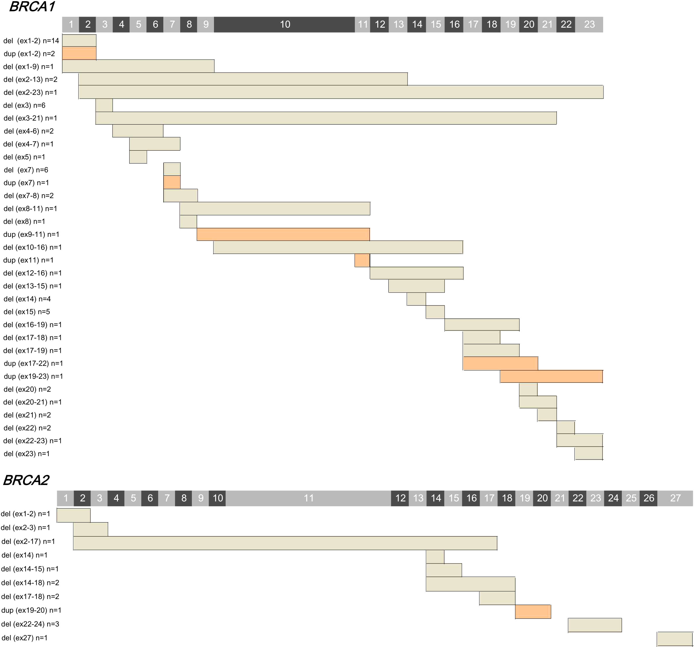
Among the 84 LGRs detected in BRCA1/2, exon 1-2 deletion was the predominant mutation type (20.0%, 14/70), followed by exon 3 deletion (8.6%, 6/70), exon 7 deletion (8.6%, 6/70), exon 15 deletion (7.1%, 5/70), and exon 14 deletion (5.7%, 4/70). Among the LGRs identified in BRCA2, exon 22-24 deletion was more common (21.4%, 3/14) (Figure 1). A total of 43 identical LGRs were identified, of which six were novel, and the remaining 37 have been previously reported. Exon 7 deletion in BRCA1 was identified in six patients and was not reported previously in cohorts from other countries, which indicated a novel or specific LGR type in Chinese cancer patients. Five of 6 cases who had exon 7 deletion in BRCA1 were subjected to MLPA test, and all five cases were turned out to be BRCA1 LGR-positive by MLPA. In addition, exon 10−16 deletion (n=1) in BRCA1 and exon 2−3 (n=1), exon 2−17 (n=1), exon 14 (n=1), and exon 27 (n=1) deletions in BRCA2 were other novel LGRs. Deletion was the primary LGR subtype among both BRCA1 (deletion. vs. duplication; 90.0% vs. 10.0%) and BRCA2 LGRs (deletion. vs. duplication. 92.9% vs. 7.1%) (Supplementary Table S8). The characteristics of BRCA1 LGR across multiple cancers are summarized (Supplementary Table S9, Table S10).

Figure 1 The spectrum of large genetic rearrangements (LGRs) detected in BRCA1 and BRCA2 genes. Exon numbers are indicated on the top. The characteristics of the detected LGRs are listed on the left. The affected exons are shown as bars, among which, grey represents deletion, and orange indicates duplication. Actual locations of breakpoint are not implied.
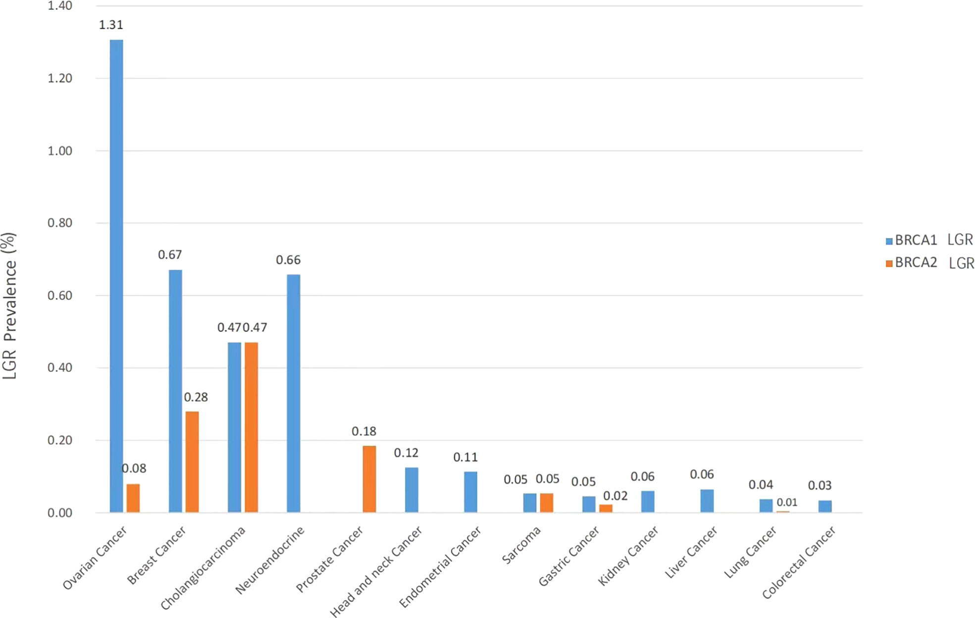
The prevalence of BRCA1 and BRCA2 germline LGRs across multiple cancer types is shown in Figure 2. BRCA1 LGR more frequently occurred in ovarian cancer (1.31%, 33/2526), followed by breast cancer (0.67%, 12/1788) and neuroendocrine cancer (0.66%, 1/152). BRCA2 LGR more commonly occurred in cholangiocarcinoma (0.47%, 2/425), breast cancer (0.28%, 5/1788), and prostate cancer (0.18%, 2/1083) (Figure 2). Among the 70 patients with BRCA1 LGR, patients with ovarian cancer accounted for the largest proportion (33/70, 47.1%), followed by breast cancer (12/70, 17.1%) and lung cancer (9/70, 12.9%). Of the 14 patients with BRCA2 LGR, 5 (5/14, 35.7%) had breast cancers, and two had ovarian cancers (2/14, 14.3%) (Supplementary Table S11).

Figure 2 Prevalence of BRCA1 (orange) and BRCA2 (blue) LGR across multiple cancer types. Cancer types included ovarian cancer (n = 2526), breast cancer (n = 1788), cholangiocarcinoma (n = 425), neuroendocrine (n = 152), prostate cancer (n = 1083), head and neck cancer (n = 801), endometrial cancer (n = 880), sarcoma (n = 1863), gastric cancer (n = 4427), kidney cancer (n = 1545), liver cancer (n = 4973), lung cancer (n = 23920) and colorectal cancer (n = 11743).
Ovarian cancer case presentation
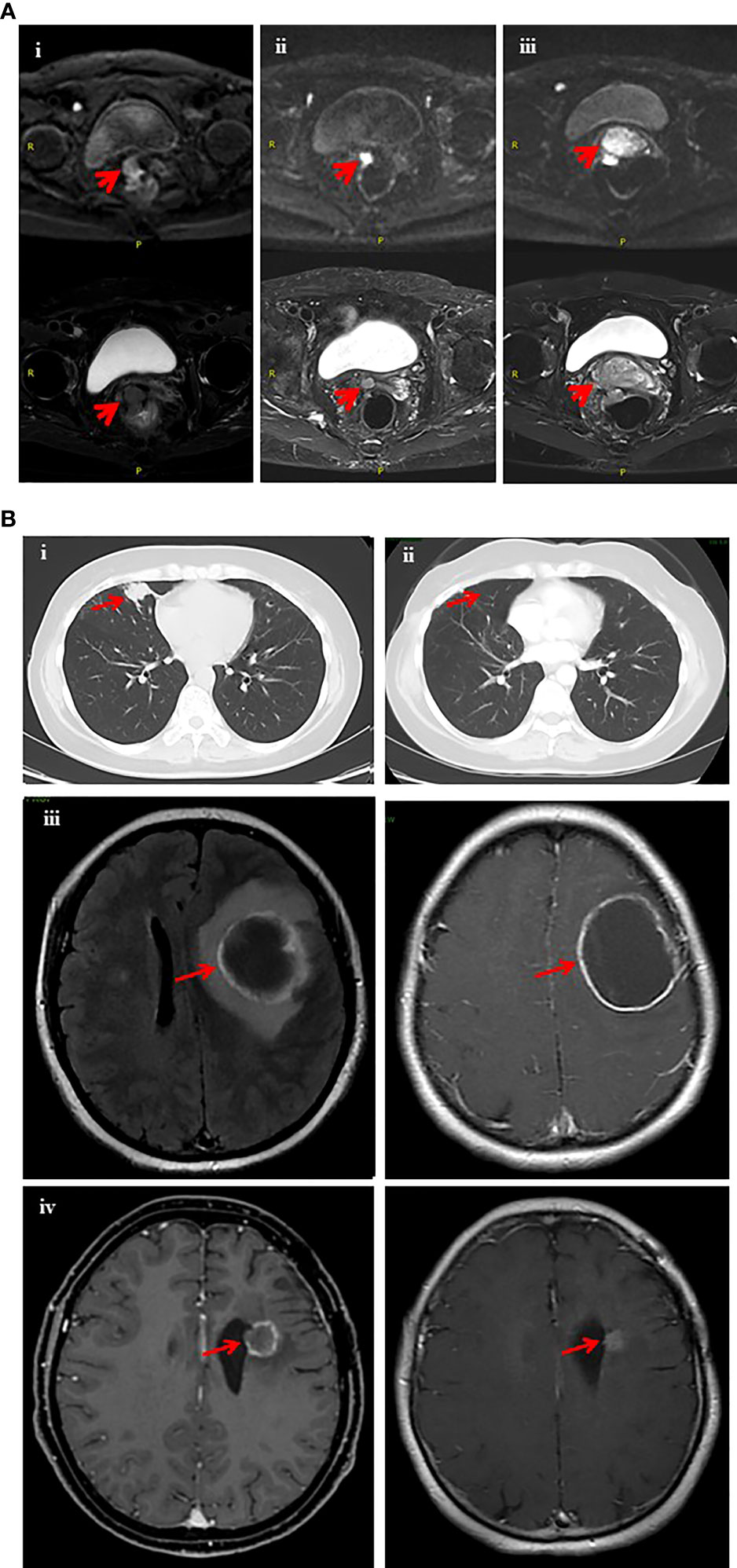
On June 5, 2019, a 69-year-old woman with ovarian cancer underwent cytoreductive surgery consisting of bilateral salpingo-oophorectomy, hysterectomy, omentectomy, partial rectectomy, and removal of adhesion between the ovary and pelvic/peritoneum. She was considered to have FIGO stage II disease according to the pathologic results and findings observed during surgery. The pathological results revealed high-grade serous carcinoma in the right adnexa. Tumor lesions were also involved in the epiploic appendix and the rectum anterior wall. She received six cycles of adjuvant intravenous chemotherapy (120 mg paclitaxel + 50 mg lobaplatin) after surgery. On July 16, 2020, the patient visited our clinic and presented with a high level of CA125 (3775 U/mL) and a high ROMA index. Enhanced computed tomography (CT) revealed multiple nodular soft-tissue density shadows, with obviously non-uniform enhancement. She was considered to have disease recurrence and was therefore treated with adjuvant intravenous chemotherapy (50 mg doxorubicin hydrochloride + 50 mg lobaplatin) for six months. On February 17, 2021, pathological examination of the vaginal stump revealed ovarian-derived high-grade serous cystadenocarcinoma vaginal metastasis. Magnetic resonance imaging (MRI) on March 5, 2021 showed a soft-tissue density mass shadow in the anastomotic stoma of a previous rectal resection, suggesting that the disease relapsed (Figure 3A). Molecular profiling of vaginal stump biopsy identified a BRCA1 germline exon 8-11 deletion. Accordingly, the patient was administered the PARPi fluzoparib, which led to a drastic decrease in CA125 (decreased to a normal level with a reduction of 85%) and a shrinkage in the pelvic lesion (Figure 3A), achieving partial response (PR). After six weeks of fluzoparib treatment, the patient experienced a grade 3 decrease in platelet number in peripheral blood, for which she discontinued fluzoparib administration. Upon discontinuation of fluzoparib, MRI revealed an enlarged irregular soft-tissue nodular shadow in the anastomotic stoma of previous rectal resection and a drastic increase in the level of serum CA125, which was nine folds higher than the normal level.

Figure 3 Imaging records of two patients who had BRCA1/2 LGRs and benefited from PARP inhibitors. (A) Magnetic resonance imaging (MRI) scan of the pelvic before and after fluzoparib treatment (ovarian cancer case). The red arrows indicate tumor lesions. (i) The pelvic MRI scan on March 5, 2021 revealed tumor status before fluzoparib treatment (5.5*3.4 cm). (ii) Pelvic MRI on June 9, 2021 indicated a significant decrease in tumor size (1.9 *1.6 cm) after fluzoparib treatment. (iii) The pelvic MRI scan on August 10, 2021 revealed progression of tumor lesions (3.0*4.2 cm) after 3 months of discontinuation of fluzoparib. (B) Computed tomography CT scan of the chest and magnetic resonance imaging (MRI) scan of the brain (breast cancer case). The red arrows indicate tumor lesions. (i) Chest CT in May 2017 revealed a metastatic lesion (approximately 2.3*1.8 cm) in the middle lobe of the right lung. (ii) The patient received surgery in September 2017 and maintenance therapy with olaparib. Chest CT in October 2021 indicated pulmonary metastasis disappeared during maintenance therapy with olaparib. (iii) Brain MRI in August 2017 revealed a metastatic lesion with ring-like enhancement in the left frontal lobe with peri-tumoral edema. (iv) Brain MRI in October 2021 indicated a significant shrinkage in brain metastasis lesions during the maintenance therapy with olaparib.
Breast cancer case presentation
On July 11, 2011, a 40-year-old woman was diagnosed with stage IIA (pT2N0M0) triple-negative breast cancer (HER2-, ER-, and PR-negative). She received chemo-radiotherapy after resection of her right breast mass. In June 2014, a nodule biopsy of the left breast revealed invasive breast carcinoma. The patient underwent extensive resection of the left breast mass and lymph node dissection. Between August 21, 2014 and December 16, 2014, she participated in a clinical trial of paclitaxel liposomes combined with carboplatin (Clinical trial NCT01150513). The patient suffered from grade 3 gastrointestinal reactions and leukocytosis upon six cycles of treatment. The patient had stable disease until metastases occurred on the lung in May 2017 (Figure 3B). She underwent right-lung middle lobe resection and mediastinal and hilar lymph node dissection. Immunohistochemical staining of the lung lesion showed negative results for NapsinA, TTF1, ER, PR, HER2, CK5/6 and GATA3, and Ki67 nuclear positivity in 70% stromal cells. The radiotherapist considered the effect of radiotherapy to be poor for intracranial hypertension and obvious peripheral edema. To explore other potential therapeutic opportunities, NGS was performed on the retrieved right lung and left breast tumor lesions collected in July 2017 and June 2014, respectively. Molecular profiling identified potentially pathogenic mutations, including germline exon 2 deletion in BRCA1 and somatic gene arrangement in BRCA2-STARD13 from the right lung lesion. The patient refused to receive chemotherapy, and according to the NGS results she started to receive the PARPi olaparib at a dose of 200 mg twice per day on September 20, 2017. Disease had remained stable as of the time of the submission of this manuscript (Figure 3B).
Discussion
In clinical practice, MLPA is the most widely used method to detect BRCA1/2 LGRs (19, 20). The primary weaknesses of MLPA are as follows: a high requirement for the purity of nucleic acid samples, due to which alcohol, metal ions, phenol, and TRIzol all affect the performance of MLPA; false-positive result tends to happen when SNV occurs at the probe binding site; poor performance to detect duplication. A wide range of other methods are available for LGR detection, but no one has overwhelming advantages. Southern blotting, real-time PCR, dual-color fluorescence in situ hybridization (FISH), comparative genomic hybridization, and multiplex amplifiable quantification all have disadvantages such as being time-consuming, having a high false-positive rate, and having low sensitivity (21). In addition, BRCA1/2 has a long gene sequence and multiple pathogenic mutation types, including missense, frameshift, nonsense, and LGR, rendering it challenging to comprehensively detect BRCA1/2 mutations. Herein, we utilized a capture-based NGS method to reveal the profile of BRCA1/2 mutation, making it accurate and time-saving to characterize point mutations and small or large insert-deletions in a one-set test. The well-designed and strictly validated NGS method has full coverage for all exons and sufficient sequencing depth, demonstrating 100% concordance with MLPA.
Ovarian and breast cancers are the leading causes of gynecologic cancer-related death. BRCA1/2-mutated cancer patients represent a typical molecular subset. For ovarian cancer patients, germline and somatic BRCA1/2 mutations have been utilized as biomarkers for the use of PAPRi (1). For breast cancers, National Comprehensive Cancer Network (NCCN) guidelines recommend that recurrent or metastatic patients with germline BRCA1/2 mutations receive PARPi therapy (2). In this study, we presented two cases with BRCA1/2 LGR as determined by NGS (ovarian cancer case: BRCA1 germline exon 8-11 deletion; breast cancer case: BRCA1 germline exon 2 deletion), and both patients benefited from PARPi therapy, providing evidence for the feasibility and significance of NGS-based LGR detection in guiding treatment. Further studies are warranted to confirm the findings.
In this study, the frequency of co-occurring variants in both the BRCA1 and BRCA2 genes in the pan-cancer cohort was approximately 3%, which was somehow higher than that of most previous studies (22–25). The possible reason for the relatively high frequency in our study might be the fact that except for pathogenic variants, we also included likely-pathogenic variants and LGRs, which were not included in some studies. Another possibility was that we analyzed the sequencing data in a pan-tumor cohort, which might have led to the discrepancy among studies.
One of the limitations of this study was that the NGS platform employed in our study could not identify the accurate breakpoints of LGR. Breakpoints frequently occur in introns, the length of which can be thousands of bp. The NGS assay applied for LGR detection was designed with capture probes covering the whole exons and extending 60 bp into intron regions at both ends of every exon, which was incapable of identifying accurate breakpoints in introns. Due to a lack of breakpoint information for these LGRs, predicting the impact of these LGRs on BRCA1/2 proteins was theoretically not feasible. Based on previous reports, deletions in BRCA1/2 exons could result in premature termination of the BRCA1 protein, an in-frame deletion, and prevention of transcription. Rearrangements involving different exons in distinct domains may have distinct effects on protein functions (26).
Most of previous studies regarding BRCA1/2 LGR have primarily focused on ovarian and breast cancer. This study examined the prevalence of BRCA1/2 germline LGRs in multiple solid tumors. Of note, the prevalence of BRCA1/2 germline LGR in other cancers, except for ovarian and breast cancers, might be underestimated because a few cases were screened only with tissue specimens. To the best of our knowledge, this was the first and largest study to depict the BRCA1/2 LGR profile of Chinese pan-cancer patients by using an NGS-based assay. We also reported two cases who had BRCA1/2 LGR benefited from PARPi, supporting the feasibility of NGS-based detection of BRCA1/2 LGRs to guide treatment.
Data availability statement
The VCF files of 84 LGR cases were uploaded to the GVM database (Accession NO. GVM000365, Project NO. PRJCA010855, https://ngdc.cncb.ac.cn/gvm/). Other datasets generated and/or analyzed during the current study are available from the corresponding author on reasonable request.
Ethics statement
The studies involving human participants were reviewed and approved by The ethics committees of the First Affiliated Hospital of Nanchang University and National Cancer Center/National Clinical Research Center for Cancer/Cancer Hospital. The patients/participants provided their written informed consent to participate in this study. Written informed consent was obtained from the individual(s) for the publication of any potentially identifiable images or data included in this article.
Author contributions
XZ, DH, and PY conceptualized the study; DH, QT, and XW, BZ, YB, and CB designed the Methods; DH, QT, XW, and LC collected, analyzed, and interpreted the patient data regarding the discovery and validation cohorts. TB and DH were major contributors in writing the manuscript. TB, XZ, and PY conducted critical revision of the manuscript for important intellectual content. DH, QT, and XW contributed equally and served as co-first authors. All authors read and approved the final manuscript.
Funding
This study was funded by the Beijing Hope Run Special Fund of Cancer Foundation of China (LC2019B16 to Xue Wang).
Acknowledgments
We would like to thank all coordinators at the First Affiliated Hospital of Nanchang University, National Cancer Center/National Clinical Research Center for Cancer/Cancer Hospital, Chinese Academy of Medical Sciences and Peking Union Medical College and 3D Medicines Inc. for supporting this study.
Conflict of interest
D Hua, T Bei, L Cui, B Zhang, C Bao, Y Bai, and X Zhao are employees of 3D Medicines Inc.
The remaining authors declare that the research was conducted in the absence of any commercial or financial relationships that could be constructed as a potential conflict of interest.
The reviewer (JL) declared a shared affiliation, with no collaboration, with the authors (XW, PY) to the handling editor at the time of review
Publisher’s note
All claims expressed in this article are solely those of the authors and do not necessarily represent those of their affiliated organizations, or those of the publisher, the editors and the reviewers. Any product that may be evaluated in this article, or claim that may be made by its manufacturer, is not guaranteed or endorsed by the publisher.
Supplementary material
The Supplementary Material for this article can be found online at: https://www.frontiersin.org/articles/10.3389/fonc.2022.898916/full#supplementary-material
References
1. (NCCN). N.C.C.N., NCCN clinical practice guidelines in oncology. In: Ovarian cancer. version 1. 2021. 3025 Chemical Road, Suite 100, Plymouth Meeting, PA 19462, USA: National Comprehensive Cancer Network (NCCN (2021).
2. (NCCN). N.C.C.N., NCCN clinical practice guidelines in oncology. In: Breast cancer. version 4. 2021. 3025 Chemical Road, Suite 100, Plymouth Meeting, PA 19462, USA: National Comprehensive Cancer Network (NCCN (2021).
3. Ewald IP, Ribeiro PLI, Palmero EI, Cossio SL, Giugliani R, Ashton-Prolla P. Genomic rearrangements in BRCA1 and BRCA2: A literature review. Genet Mol Biol (2009) 32(3):437–46. doi: 10.1590/S1415-47572009005000049
4. Bozsik A, Pocza T, Papp J, Vaszko T, Butz H, Patocs A, et al. Complex characterization of germline Large genomic rearrangements of the BRCA1 and BRCA2 genes in high-risk breast cancer patients-novel variants from a Large national center. Int J Mol Sci (2020) 21(13):4605. doi: 10.3390/ijms21134650
5. Germani A, Libi F, Maggi S, Stanzani G, Lombardi A, Pellegrini P, et al. Rapid detection of copy number variations and point mutations in BRCA1/2 genes using a single workflow by ion semiconductor sequencing pipeline. Oncotarget (2018) 9(72):33648–55. doi: 10.18632/oncotarget.26000
6. Kwong A, Chen J, Shin VY, Ho JCW, Law FBF, Au CH, et al. The importance of analysis of long-range rearrangement of BRCA1 and BRCA2 in genetic diagnosis of familial breast cancer. Cancer Genet (2015) 208(9):448–54. doi: 10.1016/j.cancergen.2015.05.031
7. Lim YK, Iau PTC, Ali AB, Lee SC, Wong J-L, Putti TC, et al. Identification of novel BRCA large genomic rearrangements in Singapore Asian breast and ovarian patients with cancer. Clin Genet (2007) 71(4):331–42. doi: 10.1111/j.1399-0004.2007.00773.x
8. Kang P, Mariapun S, Phuah SY, Lim LS, Liu J, Yoon S-Y, et al. Large BRCA1 and BRCA2 genomic rearrangements in Malaysian high risk breast-ovarian cancer families. Breast Cancer Res Treat (2010) 124(2):579–84. doi: 10.1007/s10549-010-1018-5
9. Cao W-M, Zheng Y-B, Gao Y, Ding X-W, Sun Y, Huang Y, et al. Comprehensive mutation detection of BRCA1/2 genes reveals large genomic rearrangements contribute to hereditary breast and ovarian cancer in Chinese women. BMC Cancer (2019) 19(1):551–1. doi: 10.1186/s12885-019-5765-3
10. Ji G, Bao L, Yao Q, Zhang J, Zhu X, Bai Q, et al. Germline and tumor BRCA1/2 pathogenic variants in Chinese triple-negative breast carcinomas. J Cancer Res Clin Oncol (2021) 147(10):2935–44. doi: 10.1007/s00432-021-03696-2
11. Richards S, Aziz N, Bale S, Bick D, Das S, Gastier-Foster J, et al. Standards and guidelines for the interpretation of sequence variants: a joint consensus recommendation of the American college of medical genetics and genomics and the association for molecular pathology. Genet Med Off J Am Coll Med Genet (2015) 17(5):405–24. doi: 10.1038/gim.2015.30
12. Brandt T, Sack LM, Arjona D, Tan D, Mei H, Cui H, et al. Adapting ACMG/AMP sequence variant classification guidelines for single-gene copy number variants. Genet Med (2020) 22(2):336–44. doi: 10.1038/s41436-019-0655-2
13. Riggs ER, Andersen EF, Cherry AM, Kantarci S, Kearney H, Patel A, et al. Technical standards for the interpretation and reporting of constitutional copy-number variants: A joint consensus recommendation of the American college of medical genetics and genomics (ACMG) and the clinical genome resource (ClinGen). Genet Med (2020) 22(2):245–57. doi: 10.1038/s41436-019-0686-8
14. Li MM, Datto M, Duncavage EJ, Kulkarni S, Lindeman NI, Roy S, et al. Standards and guidelines for the interpretation and reporting of sequence variants in cancer: A joint consensus recommendation of the association for molecular pathology, American society of clinical oncology, and college of American pathologists. J Mol Diagn (2017) 19(1):4–23. doi: 10.1016/j.jmoldx.2016.10.002
15. Chao A, Chang T-C, Lapke N, Jung S-M, Chi P, Chen C-H, et al. Prevalence and clinical significance of BRCA1/2 germline and somatic mutations in Taiwanese patients with ovarian cancer. Oncotarget (2016) 7(51):85529–41. doi: 10.18632/oncotarget.13456
16. Sánchez-Lorenzo L, Salas-Benito D, Villamayor J, Patiño-García A, González-Martín A. The BRCA gene in epithelial ovarian cancer. Cancers (2022) 14(5):1235. doi: 10.3390/cancers14051235
17. Zhong X, Dong Z, Dong H, Li J, Peng Z, Deng L, et al. Prevalence and prognostic role of BRCA1/2 variants in unselected Chinese breast cancer patients. PloS One (2016) 11(6):e0156789–e0156789. doi: 10.1371/journal.pone.0156789
18. Winter C, Nilsson MP, Olsson E, George AM, Chen Y, Kvist A, et al. Targeted sequencing of BRCA1 and BRCA2 across a large unselected breast cancer cohort suggests that one-third of mutations are somatic. Ann Oncol Off J Eur Soc Med Oncol (2016) 27(8):1532–8. doi: 10.1093/annonc/mdw209
19. Hogervorst FB, Nederlof PM, Gille JJ, McElgunn CJ, Grippeling M, Pruntel R, et al. Large Genomic deletions and duplications in the BRCA1 gene identified by a novel quantitative method. Cancer Res (2003) 63(7):1449–53. Available at: http://aacrjournals.org/cancerres/article-pdf/63/7/1449/2513026/ch0703001449.pdf
20. Schouten JP, McElgunn CJ, Waaijer R, Zwijnenburg D, Diepvens F, Pals G. Relative quantification of 40 nucleic acid sequences by multiplex ligation-dependent probe amplification. Nucleic Acids Res (2002) 30(12):e57. doi: 10.1093/nar/gnf056
21. Armour JA, Barton DE, Cockburn DJ, Taylor GR. The detection of large deletions or duplications in genomic DNA. Hum Mutat (2002) 20(5):325–37. doi: 10.1002/humu.10133
22. Mansfield CA, Metcalfe KA, Snyder C, Lindeman GJ, Posner J, Friedman S, et al. Preferences for breast cancer prevention among women with a BRCA1 or BRCA2 mutation. Hereditary Cancer Clin Pract (2020) 18:20–0. doi: 10.1186/s13053-020-00152-z
23. Kim J, Turan V, Oktay K. Long-term safety of letrozole and gonadotropin stimulation for fertility preservation in women with breast cancer. J Clin Endocrinol Metab (2016) 101(4):1364–71. doi: 10.1210/jc.2015-3878
24. Sun P, Li Y, Chao X, Li J, Luo R, Li M, et al. Clinical characteristics and prognostic implications of BRCA-associated tumors in males: A pan-tumor survey. BMC Cancer (2020) 20(1):994–4. doi: 10.1186/s12885-020-07481-1
25. Gupta S, Rajappa S, Advani S, Agarwal A, Aggarwal S, Goswami C, et al. Prevalence of BRCA1 and BRCA2 mutations among patients with ovarian, primary peritoneal, and fallopian tube cancer in India: A multicenter cross-sectional study. JCO Global Oncol (2021) 7:849–61. doi: 10.1200/GO.21.00051
Keywords: BRCA, large genomic rearrangements, breast cancer, ovarian cancer, pan-cancer, next-generation sequencing
Citation: Hua D, Tian Q, Wang X, Bei T, Cui L, Zhang B, Bao C, Bai Y, Zhao X and Yuan P (2022) Next-generation sequencing based detection of BRCA1 and BRCA2 large genomic rearrangements in Chinese cancer patients. Front. Oncol. 12:898916. doi: 10.3389/fonc.2022.898916
Received: 18 March 2022; Accepted: 08 August 2022;
Published: 06 September 2022.
Edited by:
Ian Campbell, Peter MacCallum Cancer Centre, AustraliaReviewed by:
Jiaqi Liu, National Cancer Center of China, ChinaMuhammad Usman Rashid, Shaukat Khanum Memorial Cancer Hospital and Research Center, Pakistan
Gerburg Wulf, Harvard Medical School, United States
Copyright © 2022 Hua, Tian, Wang, Bei, Cui, Zhang, Bao, Bai, Zhao and Yuan. This is an open-access article distributed under the terms of the Creative Commons Attribution License (CC BY). The use, distribution or reproduction in other forums is permitted, provided the original author(s) and the copyright owner(s) are credited and that the original publication in this journal is cited, in accordance with accepted academic practice. No use, distribution or reproduction is permitted which does not comply with these terms.
*Correspondence: Xiaochen Zhao, eGlhb2NoZW4uemhhb0AzZG1lZGNhcmUuY29t; Peng Yuan, eXVhbnBlbmcwMUBjaWNhbXMuYWMuY24=
†These authors have contributed equally to this work and share first authorship
 Dingchao Hua1†
 Xue Wang
 Xiaochen Zhao