- 1Department of Gastrointestinal Surgery, Xinghua People’s Hospital Affiliated to Yangzhou University, Xinghua, China
- 2School of Public Health, Shandong Second Medical University, Weifang, China
- 3Department of Cardiology, Xinghua People’s Hospital Affiliated to Yangzhou University, Xinghua, China
Background: This systematic review and meta-analysis aimed to assess the impact of visceral obesity (VO) on postoperative outcomes in colorectal cancer (CRC) patients.
Methods: Primary studies were obtained from sources like Embase, PubMed, and Web of Science during the search, which ran until October 2024. Patients with colorectal cancer who had visceral obesity (VO) and those who did not were compared in terms of intraoperative conditions, postoperative outcomes, postoperative complications, and long-term prognoses, including overall survival (OS) and disease-free survival (DFS).
Results: 5,756 individuals with VO and 5,373 patients without VO were among the 11,129 patients who had colorectal cancer resected. Patients with VO had higher conversion rates (p = 0.03), fewer lymph nodes removed (p = 0.05), and longer recovery times for bowel movements (p = 0.009). Furthermore, patients with VO had a considerably greater overall incidence of sequelae than those without (p = 0.0003), including anastomotic leaks (p = 0.01), intestinal obstruction (p = 0.0003), intra-abdominal abscesses (p = 0.004), wound infections (p < 0.00001), and pulmonary problems (p = 0.0003). OS and DFS, however, did not differ between the two groups (p > 0.05).
Conclusions: Colorectal cancer patients with VO who have surgery tend to have fewer lymph nodes taken, more problems after surgery, and a higher rate of switching to open surgery.
1 Introduction
Colorectal cancer (CRC) is one of the most common gastrointestinal malignancies globally, ranking third in incidence and second in mortality (1). Although curative surgery remains the most effective treatment for colorectal cancer, the incidence of postoperative complications remains high, reaching up to 35% (2). These complications are not only related to the surgery itself but are also influenced by the patient’s overall health and other non-surgical factors.
After potentially curative surgery for colorectal cancer, prognosis and treatment decisions are typically based on tumor pathology. However, an increasing body of research indicates that tumor staging is not the only factor determining patient outcomes. Body composition has increasingly been recognized as playing an important role in prognosis. Body composition parameters, such as obesity, are strongly linked to the prognosis of colorectal cancer. Epidemiological studies have shown that obesity is a significant risk factor for colorectal cancer (3). Furthermore, obesity is considered a risk factor for postoperative complications in various abdominal surgeries (4). However, the impact of obesity on colorectal cancer prognosis remains controversial. Some studies report worse outcomes in both obese (5, 6) and underweight patients (7), while others have not observed such associations (8).
Visceral obesity (VO) refers to the excessive accumulation of intra-abdominal adipose tissue and is generally considered a more accurate indicator of obesity than body mass index (BMI) (9, 10). Due to its central role in metabolism and its potential impact on postoperative recovery, accurately measuring visceral fat may have significant clinical value in the preoperative risk assessment of colorectal cancer patients. Compared to traditional BMI, computed tomography (CT) provides a more precise and reproducible method for measuring visceral fat area (VFA) (11). In the preoperative evaluation of colorectal cancer patients, abdominal CT imaging is routinely used not only to screen for metastatic disease but also to assess visceral fat accumulation.
This study aims to conduct a systematic review and meta-analysis to explore the impact of VO, measured exclusively by CT imaging, on the prognosis of colorectal cancer patients. Specifically, the study will evaluate the predictive value of visceral obesity for both short-term outcomes, such as postoperative complications, and long-term outcomes, including overall survival (OS) and disease-free survival (DFS), following elective colorectal cancer surgery.
2 Materials and methods
2.1 Search strategy
The search was conducted until October 2024, and primary studies were retrieved from databases such as Embase, PubMed, and Web of Science. “Colorectal cancer,” “visceral obesity,” and “visceral fat area” were among the search terms. The comprehensive search approach is available in Supplementary Appendix 1.
2.2 Inclusion and exclusion criteria
The following requirements had to be fulfilled for a study to be included: (1) all patients had been diagnosed with colorectal cancer and treated surgically; (2) patients had a CT scan prior to surgery, and VFA was measured; (3) the article includes intraoperative information or short-term or long-term postoperative outcomes. The study with more recent data collection or more extensive data was preferred among several papers written by the same researcher from a certain university.
Studies that satisfied any of the following requirements were disqualified: (1) did not employ CT scans to evaluate VFA for diagnosing VO; (2) did not compare patients with and without VO; (3) was not published in English; and (4) was a conference abstract, case report, review article, or letter.
2.3 Data extraction
Detailed information, including the names of the authors, study duration, year of publication, type of surgery performed, sample size, mean age of participants, gender distribution, study location, diagnostic criteria, length of hospital stay, operative time, intraoperative blood loss, number of lymph nodes retrieved, conversion rates, reoperation rates, postoperative readmission and mortality rates, overall and severe complication rates, the incidence of specific complications, and OS and DFS outcomes, was included in the compiled data.
2.4 Study quality
Using the Newcastle-Ottawa Scale (NOS), we assessed the calibre of the research that were part of our review. The maximum score on this scale is nine points. Research is considered methodologically sound if it receives a score of six or higher.
2.5 Statistical analysis
RevMan 5.4 and Stata 12.0 software were used for the data analysis. Weighted mean differences (WMD) were applied to continuous variables, odds ratios (OR) to binary variables, and hazard ratios (HR) to survival data. Heterogeneity between studies was evaluated using the I2 statistic. For I2 values of 50% or less, a fixed-effects model was used; for I2 values more than 50%, a random-effects model was used. To evaluate publication bias, funnel plots were employed in conjunction with Begg’s and Egger’s tests. Statistical significance was defined as a P-value of less than 0.05.
3 Results
3.1 Selected articles
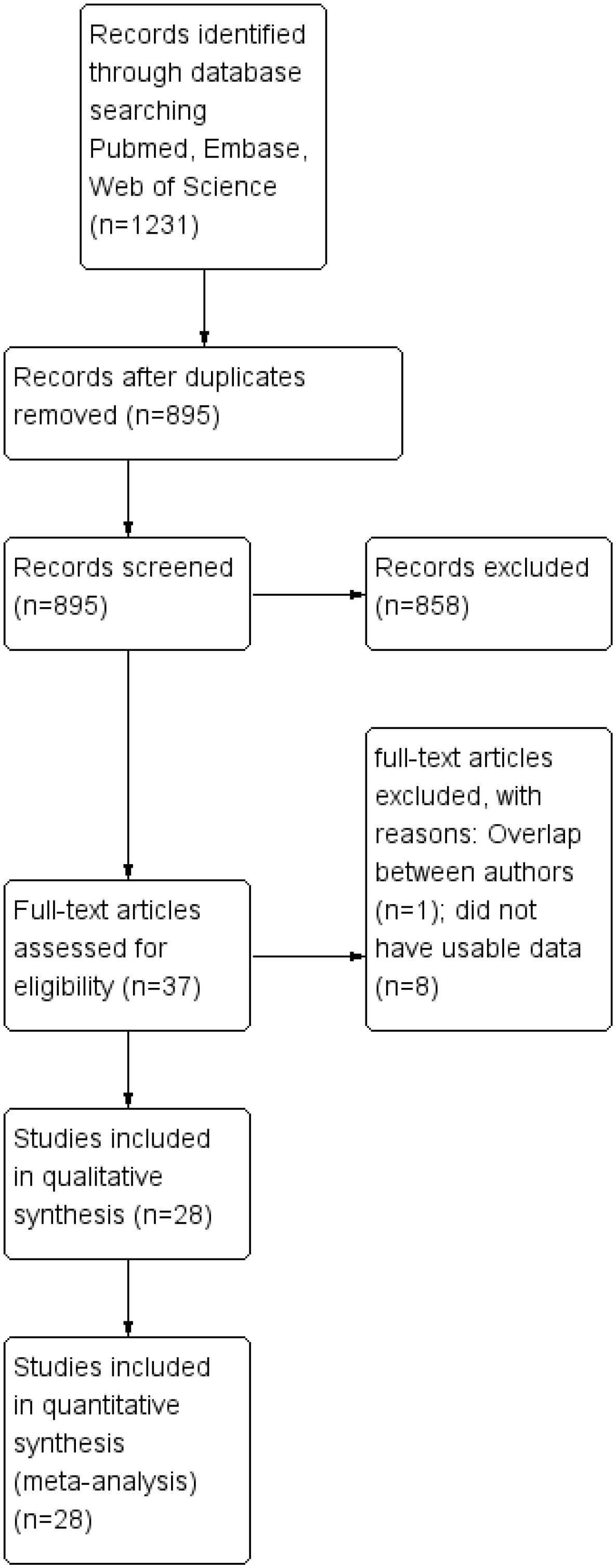
1231 publications were selected during the first search phase. 28 (12–39) researches were eventually included in the meta-analysis after these publications were reviewed (Figure 1).
3.2 Study characteristics and study quality
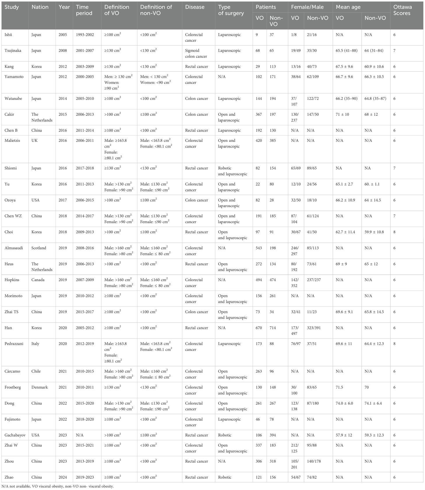
The characteristics of the studies that were part of the analysis are compiled in Table 1. From 2005 to 2024, these studies were carried out in Japan, Korea, China, the Netherlands, the United Kingdom, the United States, Scotland, Canada, Italy, Chile, and Denmark. 5,756 individuals with VO and 5,373 patients without VO were among the 11,129 patients who had colorectal cancer resected. All of the publications that were part of this investigation had NOS values between 6 and 9, which meant that the study quality was adequate.
3.3 Length of hospital stay
The average length of stay for the VO group ranged from 6 to 15.21 days, whereas for the non-VO group, it ranged from 6 to 13.2 days (Table 2). The length of hospital stay for the VO and non-VO groups did not differ significantly, as seen by Figure 2a (WMD, 0.33 days; 95% confidence interval [CI], -0.25 to 0.91; p = 0.26).
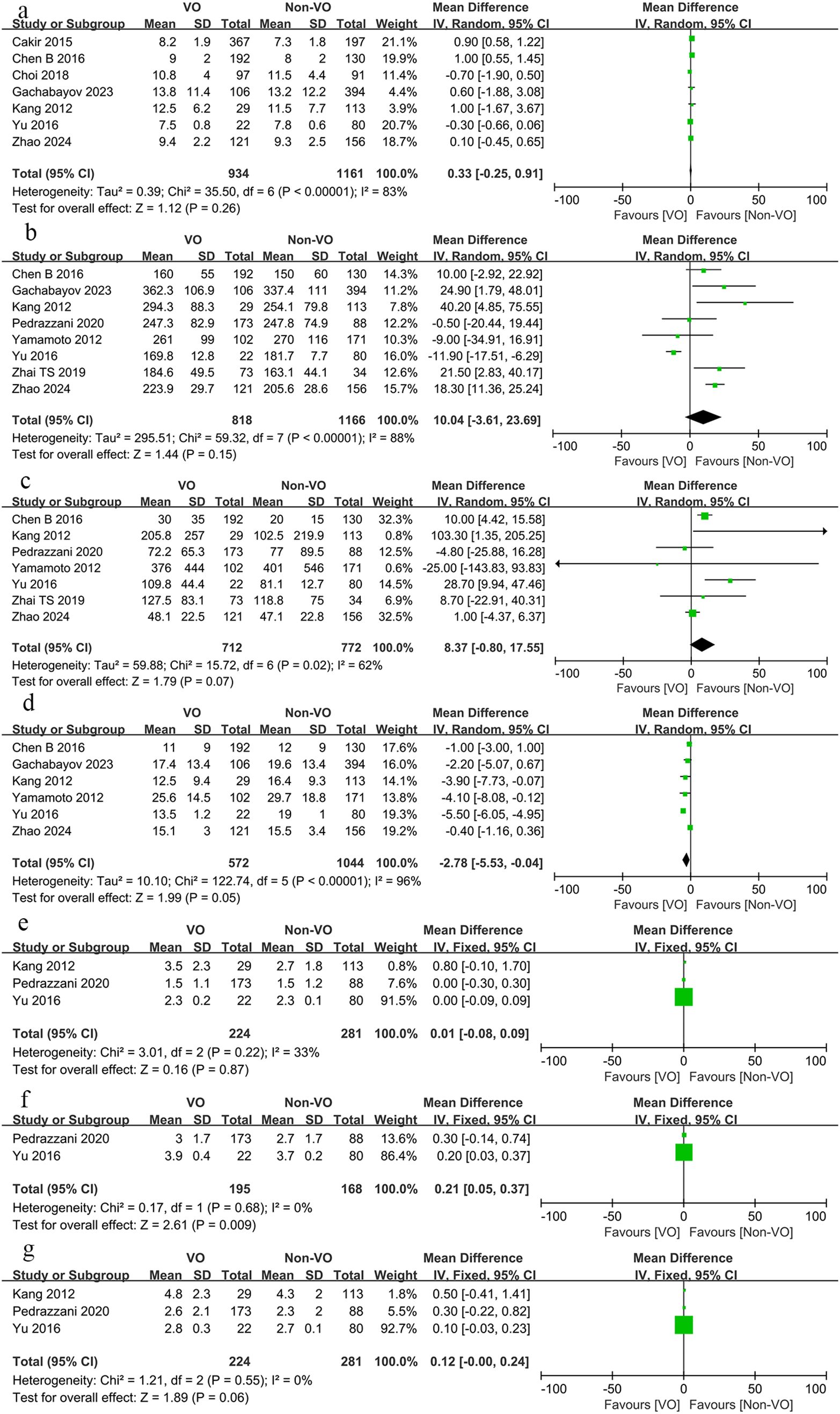
Figure 2. Forest plot describing the differences in (a) length of hospital stay, (b) operative time, (c) blood loss, (d) number of lymph nodes retrieved, (e) time to first flatus, (f) time to first bowel movement and (g) time to first food intake between VO and non-VO.
3.4 Operative time
The average operative time for the VO group ranged from 160 to 377 minutes, while for the non-VO group, it spanned from 149 to 337.4 minutes (Table 2). The operative duration for the VO and non-VO groups was not substantially different, as illustrated in Figure 2b (WMD, 0.15 minutes; 95% CI, -3.61 to 23.69; p = 0.15).
3.5 Blood loss
In the VO group, the average intraoperative blood loss was between 20 and 431 ml, whereas in the non-VO group, it was between 10 and 401 ml (Table 2). As Figure 2c illustrates, the difference was not substantial (WMD, 8.37 ml; 95% CI, -0.8 to 17.55; p = 0.07).
3.6 Number of lymph nodes retrieved
Compared to the non-VO group, fewer lymph nodes were recovered from the VO group (WMD, -2.87; 95% CI, -5.53 to -0.04; p = 0.05, Figure 2d).
3.7 Time to first flatus, bowel movement, and intake of food
There was no difference in the time to first flatus between the VO and non-VO groups (WMD, 0.01 days; 95% CI, -0.08 to 0.09; p = 0.87, Figure 2e). Although the time to first bowel movement was significantly shorter in the non-VO group compared to the VO group (WMD, 0.21 days; 95% CI, 0.05 to 0.37; p = 0.009, Figure 2f), the difference in the time to first food intake was not significant (WMD, 0.12 days; 95% CI, -0.00 to 0.24; p = 0.06, Figure 2g).
3.8 Conversion, reoperation, postoperative readmission and mortality rates
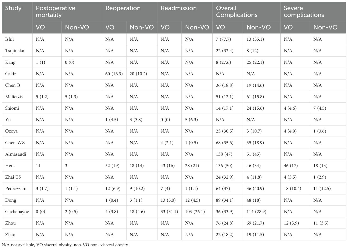
Reoperation (OR, 1.3; 95% CI, 0.94 to 1.81; p = 0.12; Figure 3a), postoperative readmission (OR, 1.07 95% CI, 0.78 to 1.46; p = 0.67; Figure 3b), and mortality rates (OR, 1.47; 95% CI, 0.16 to 2.53; p = 0.32; Figure 3c) did not significantly differ between the two groups, however the conversion rate in the VO group was significantly higher than that in the non-VO group (OR, 1.85; 95% CI, 1.05 to 3.27; p = 0.03; Figure 3d) (Table 3).
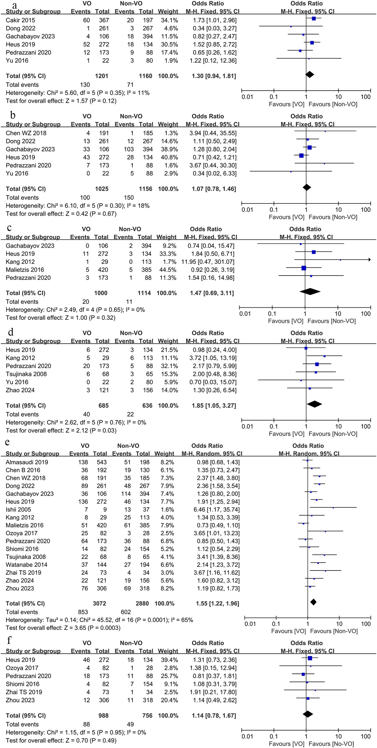
Figure 3. Forest plot describing the differences in (a) reoperation rate, (b) readmission rate, (c) mortality, (d) conversion rate, (e) total complications and (f) severe complications between VO and non-VO.
3.9 Complications
The total complication rate in the VO group exceeded that of the non-VO group (OR, 1.55; 95% CI, 1.22 to 1.96; p = 0.0003; Figure 3e), although there was no significant difference in the occurrence of severe complications between the two groups (OR, 1.14; 95% CI, 0.78 to 1.67; p = 0.49; Figure 3f) (Table 3).
We subsequently compared complications associated with surgery, including anastomotic leak, hemorrhage, intestinal obstruction, intra-abdominal abscess, wound infection, and gastrointestinal dysfunction, alongside other complications such as pulmonary issues, cardiac complications, urinary tract infections, and urinary retention. The findings indicated that the occurrence of anastomotic leak (OR, 1.38; 95% CI, 1.07 to 1.78; p = 0.01; Figure 4a), intestinal obstruction (OR, 1.56; 95% CI, 1.16 to 2.11; p = 0.0003; Figure 4b), intra-abdominal abscess (OR, 2.08; 95% CI, 1.26 to 3.44; p = 0.004, Figure 4c), and wound infection (OR, 2.57; 95% CI, 1.92 to 3.47; p < 0.00001; Figure 4d) was markedly elevated in the VO group relative to the non-VO group, whereas no statistically significant difference was observed between the two groups regarding bleeding (p = 0.74; Figure 4e) and gastrointestinal dysfunction (p = 0.74; Figure 4f). The occurrence of pulmonary problems (OR, 1.79; 95% CI, 1.31 to 2.45; p = 0.0003; Figure 5a) was elevated in the VO group, although no significant disparity was observed in the frequencies of cardiac issues (p = 0.25; Figure 5b), urinary tract infections (p = 0.69; Figure 5c), and urine retention (p = 0.53; Figure 5d) between the two groups.
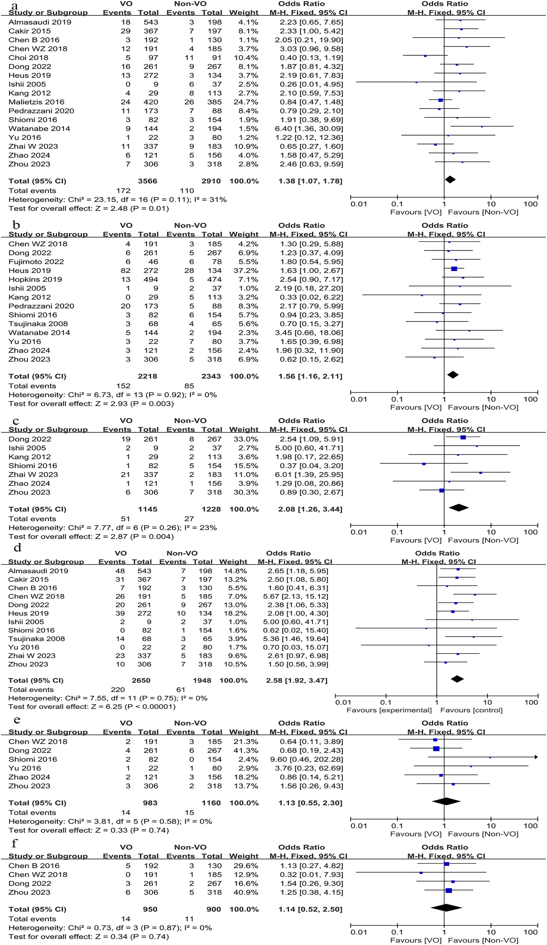
Figure 4. Forest plot describing the differences in (a) anastomotic leak, (b) intestinal obstruction, (c) intra-abdominal abscess, (d) wound infection, (e) bleeding and (f) gastrointestinal dysfunction between VO and non-VO.
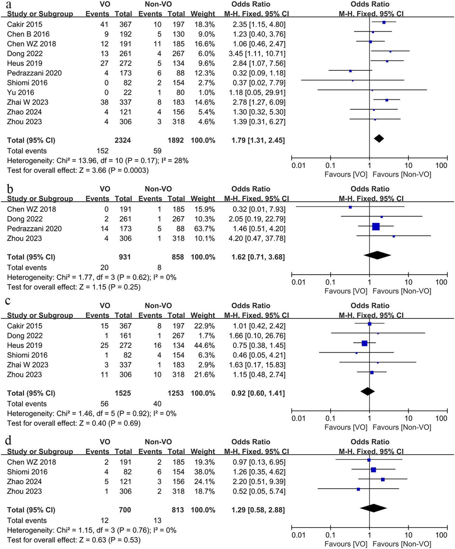
Figure 5. Forest plot describing the differences in (a) pulmonary problems, (b) cardiac issues, (c) urinary tract infections and (d) urine retention between VO and non-VO.
3.10 Prognosis
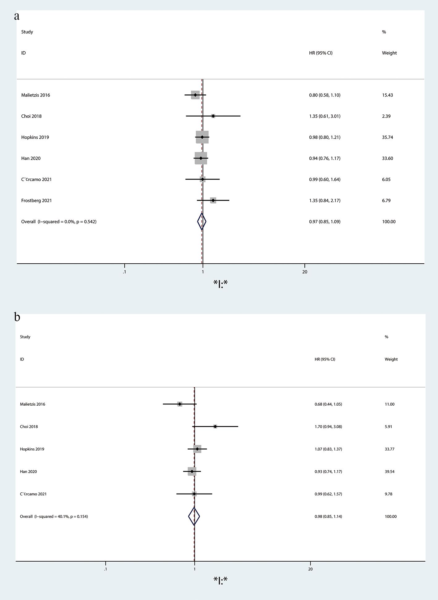
OS (HR = 0.97, 95%CI = 0.85-1.09, p = 0.524; Figure 6a) and DFS (HR = 0.98, 95%CI = 0.85-1.14, p = 0.154; Figure 6a) did not differ statistically significantly between the VO and non-VO groups.
3.11 Publication bias
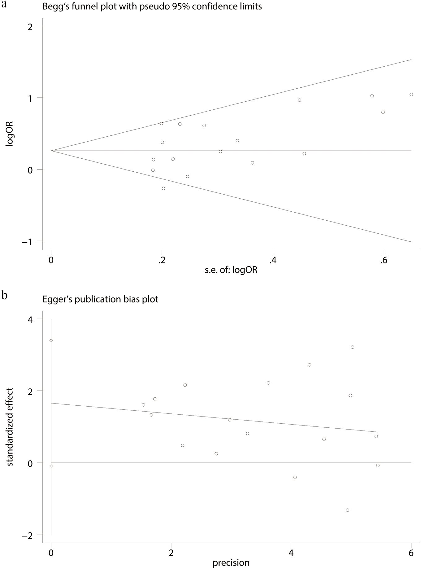
Using the comprehensive complication data, Begg’s (p = 0.174; Figure 7a) and Egger’s tests (p = 0.061; Figure 7b) were performed, and no discernible publication bias was found.
4 Discussion
Obesity, being directly linked to several chronic illnesses and cancers, is a major global public health concern (40). Although BMI has long been the most commonly used metric for diagnosing obesity, it does not adequately reflect the distribution of fat in obese individuals. This is because obesity is characterized by an uneven distribution of fat tissue throughout the body, along with localized disruptions in fat and glucose metabolism (41). Obesity can be classified into two categories based on the location of fat accumulation: visceral obesity and peripheral obesity. The assessment of VFA through CT scanning is considered the gold standard for diagnosing visceral obesity, which is predominantly found in the abdominal region (42). Early studies have linked visceral fat to the incidence of CRC, the second most common cancer worldwide (43). However, the impact of VO on survival and surgical complications in colorectal cancer patients remains inconclusive (44, 45). Therefore, we conducted this meta-analysis to evaluate the effect of visceral obesity on postoperative outcomes in colorectal cancer.
We compared the data of colorectal cancer surgery patients with and without VO. We found that patients with VO experienced a longer recovery time for bowel movements, had fewer lymph nodes harvested, and had higher conversion rates. Additionally, the overall incidence of complications-including anastomotic leaks, intestinal obstruction, intra-abdominal abscesses, wound infections, and pulmonary complications-was significantly higher in patients with VO compared to those without. However, there was no difference in OS and DFS between the two groups.
The quantity of lymph nodes removed during tumor excision is a crucial predictor of oncological results in addition to demonstrating the surgeon’s ability. VFA influences the number of lymph nodes harvested, either because visceral obese patients have thicker mesenteries, which can make it difficult to see anatomical planes during surgery, making the surgical field more difficult and making it more difficult to dissect lymph nodes and identify blood vessels, which leads to fewer lymph nodes being harvested (14), or because fat tissue adheres to the mesentery, making it more difficult to identify lymph nodes in visceral obese patients (16). Despite the difficulty of dissecting lymph nodes, the operating time did not increase appreciably. The findings of meta-analyses that have already been published contradict this (46). We think that even with obese patients, the operational time can be reduced after a long learning period and overcoming the learning curve, even though the process is more complex.
In general, obesity, tumor size, and pelvic anatomical characteristics are the primary factors that contribute to the transition to open surgery (47, 48). Visceral obesity can further complicate the surgical field and operability, in addition to the challenges posed by limited anatomical features (49). This is specifically verified by our research.
Postoperative problems serve as critical indicators for evaluating short-term surgical outcomes, particularly in colorectal cancer surgery, where anastomotic leakage is a significant postoperative complication. Thickened mesentery, oversized omentum, and excessive visceral fat are frequently seen in patients with VO. VO is a significant predictor of surgical difficulties and patient outcomes because of these characteristics (50). Anastomotic leaking is more common in VO patients due to the increased technical difficulties of surgery caused by the growth in intra-abdominal fat. Furthermore, obesity raises intra-abdominal pressure for an extended period of time, which may hinder anastomotic site microcirculation and recovery (51). Metabolic problems are strongly associated with obesity, and the inflammatory state that these abnormalities cause may have a detrimental effect on anastomotic healing and tissue repair (52). Excess belly fat increases the risk of bowel blockage because it can impede or disrupt normal intestinal movement, which in turn increases the mechanical pressure on the intestines. Patients who are obese usually have thicker mesentery, which increases the risk of obstruction by causing insufficient blood flow to the intestines. According to research, inflammatory factors such IL-1α, IL-1β, and IL-1 receptor antagonists are more prevalent in human visceral fat tissue than subcutaneous fat tissue (53–55). Bowel obstruction is more likely to occur when the intestines are altered intraoperatively since this causes the release of these inflammatory substances. This also explains the delayed recovery of bowel function in the VO group, as evidenced by the prolonged time to first bowel movement after surgery. Furthermore, too much fat raises the danger of fat liquefaction and infection, which explains why wound and intra-abdominal infections are more common (56, 57).
Some negative outcomes can be improved with thorough preoperative evaluation and therapy. According to studies, visceral obesity can be successfully decreased by pharmaceutical therapies, dietary changes, exercise, and behavioral counselling (58). In addition to greatly reducing the risk of problems from surgery, preoperative reduction of visceral fat may also be taken into consideration for patients who have pneumonia or other pulmonary risk factors, especially if they are unable to bear additional respiratory harm. However, it takes time to reduce visceral fat, which may have negative consequences including tumor growth. As a result, it is crucial to perform a comprehensive evaluation of the patient’s general health and carefully consider the risks and timing of the intervention.
There is ongoing discussion over the effect of VFA on the long-term prognosis following colorectal cancer surgery. According to some research, VFA is a significant predictor of DFS following surgery for colorectal cancer (59) and is regarded as a separate risk factor for OS in patients undergoing adjuvant chemotherapy for the disease (60). Contrary to the above findings, new research indicates that patients with greater VFA scores actually have longer OS (44). Notably, our study’s findings indicate that there were no appreciable variations in long-term outcomes (OS and DFS) between the VO and non-VO groups among patients who had colon cancer surgery. There are several factors that affect the association between visceral fat and the long-term prognosis of colorectal cancer, some of which are not well supported by clinical data. We think that this association may be significantly influenced by variables including hormone levels (61), patient gender (62), and tumor stage (9). Visceral fat’s endocrine effects may encourage tumor growth and recurrence in patients with early and intermediate-stage cancer, which would lower OS and DFS. On the other hand, since cancer patients frequently have a negative energy balance and a larger visceral fat content may be advantageous for survival, a higher visceral fat content typically suggests superior nutritional reserves for patients with advanced disease. The long-term prognosis of patients with colorectal cancer may also be impacted by the notable variations in steroid hormone levels (such as testosterone and estradiol) between women before and after menopause. These hormones are strongly linked to fat deposition. Nevertheless, there is currently little clinical evidence to validate these putative determinants, and the study’s data set is small. To evaluate the effect of these factors on the long-term prognosis of patients with colorectal cancer, more extensive prospective studies are therefore required.
Considering the potential impact of our findings on clinical practice for colorectal cancer patients, this study highlights the importance of distinguishing between visceral and non-visceral obesity, which may lead to more targeted and effective interventions. By identifying visceral obesity as a key metabolic risk factor in colorectal cancer patients, clinicians can prioritize interventions such as lifestyle modifications or pharmacotherapy, especially for those with higher visceral fat accumulation. Furthermore, our findings may assist in the early identification of high-risk colorectal cancer patients for obesity-related comorbidities such as diabetes, cardiovascular diseases, and fatty liver, enabling more timely interventions and improving long-term outcomes. Moreover, incorporating the measurement of visceral fat into routine screening for colorectal cancer patients can help healthcare providers better stratify patient risks and personalize treatment strategies. Overall, this study emphasizes the importance of personalized obesity management, which could significantly improve clinical decision-making, patient outcomes, and public health interventions for colorectal cancer patients.
Among the limitations of this study are the following: (1) the definition of visceral obesity differed among the included studies (some defined it as VFA ≥ 100 cm2, others as VFA ≥ 130 cm2, and some studies classified it based on gender), (2) the surgical approaches used in the included studies varied (open surgery, laparoscopic surgery, and robotic surgery were used, which is another source of high heterogeneity and potential bias), and (3) there may be publication bias because the studies only included patients who had completed preoperative CT scans, while those without CT scans were excluded.
5 Conclusions
In summary, patients with VO who have colorectal cancer and have surgery tend to have fewer lymph nodes removed, higher conversion rates to open surgery, and more postoperative problems. Nevertheless, OS nor DFS seem to be substantially impacted by these factors. Large-scale, prospective research from other nations and areas are still required to confirm these findings and investigate the underlying mechanisms, though, because of sample size limits and regional diversity.
Data availability statement
The original contributions presented in the study are included in the article/Supplementary Material. Further inquiries can be directed to the corresponding author.
Author contributions
YW: Conceptualization, Data curation, Formal Analysis, Supervision, Writing – original draft, Writing – review & editing. XL: Investigation, Methodology, Software, Writing – original draft, Writing – review & editing. XF: Validation, Writing – original draft, Writing – review & editing. XJ: Formal Analysis, Writing – original draft, Writing – review & editing. LH: Conceptualization, Data curation, Visualization, Writing – original draft, Writing – review & editing.
Funding
The author(s) declare that no financial support was received for the research and/or publication of this article.
Conflict of interest
The authors declare that the research was conducted in the absence of any commercial or financial relationships that could be construed as a potential conflict of interest.
Generative AI statement
The author(s) declare that no Generative AI was used in the creation of this manuscript.
Publisher’s note
All claims expressed in this article are solely those of the authors and do not necessarily represent those of their affiliated organizations, or those of the publisher, the editors and the reviewers. Any product that may be evaluated in this article, or claim that may be made by its manufacturer, is not guaranteed or endorsed by the publisher.
Supplementary material
The Supplementary Material for this article can be found online at: https://www.frontiersin.org/articles/10.3389/fonc.2025.1538073/full#supplementary-material
References
1. Bray F, Laversanne M, Sung H, Ferlay J, Siegel RL, Soerjomataram I, et al. Global cancer statistics 2022: GLOBOCAN estimates of incidence and mortality worldwide for 36 cancers in 185 countries. CA: A Cancer J clinicians. (2024) 74:229–63. doi: 10.3322/caac.21834
2. Pallan A, Dedelaite M, Mirajkar N, Newman PA, Plowright J, Ashraf S. Postoperative complications of colorectal cancer. Clin Radiol. (2021) 76:896–907. doi: 10.1016/j.crad.2021.06.002
3. Ma Y, Yang Y, Wang F, Zhang P, Shi C, Zou Y, et al. Obesity and risk of colorectal cancer: a systematic review of prospective studies. PloS One. (2013) 8:e53916. doi: 10.1371/journal.pone.0053916
4. Scheidbach H, Benedix F, Hügel O, Kose D, Köckerling F, Lippert H. Laparoscopic approach to colorectal procedures in the obese patient: risk factor or benefit? Obesity Surg. (2008) 18:66–70. doi: 10.1007/s11695-007-9266-0
5. Dignam JJ, Polite BN, Yothers G, Raich P, Colangelo L, O'Connell MJ, et al. Body mass index and outcomes in patients who receive adjuvant chemotherapy for colon cancer. J Natl Cancer Institute. (2006) 98:1647–54. doi: 10.1093/jnci/djj442
6. Basile D, Bartoletti M, Polano M, Bortot L, Gerratana L, Di Nardo P, et al. Prognostic role of visceral fat for overall survival in metastatic colorectal cancer: A pilot study. Clin Nutr. (2021) 40:286–94. doi: 10.1016/j.clnu.2020.05.019
7. Baade PD, Meng X, Youl PH, Aitken JF, Dunn J, Chambers SK. The impact of body mass index and physical activity on mortality among patients with colorectal cancer in Queensland, Australia. Cancer epidemiology Biomarkers prevention: A Publ Am Assoc Cancer Research cosponsored by Am Soc Preventive Oncol. (2011) 20:1410–20. doi: 10.1158/1055-9965.Epi-11-0079
8. Alipour S, Kennecke HF, Woods R, Lim HJ, Speers C, Brown CJ, et al. Body mass index and body surface area and their associations with outcomes in stage II and III colon cancer. J gastrointestinal Cancer. (2013) 44:203–10. doi: 10.1007/s12029-012-9472-4
9. Rickles AS, Iannuzzi JC, Mironov O, Deeb AP, Sharma A, Fleming FJ, et al. Visceral obesity and colorectal cancer: are we missing the boat with BMI? J gastrointestinal surgery: Off J Soc Surg Alimentary Tract. (2013) 17:133–43. doi: 10.1007/s11605-012-2045-9
10. Aquina CT, Rickles AS, Probst CP, Kelly KN, Deeb AP, Monson JR, et al. Visceral obesity, not elevated BMI, is strongly associated with incisional hernia after colorectal surgery. Dis colon rectum. (2015) 58:220–7. doi: 10.1097/dcr.0000000000000261
11. Shuster A, Patlas M, Pinthus JH, Mourtzakis M. The clinical importance of visceral adiposity: a critical review of methods for visceral adipose tissue analysis. Br J Radiol. (2012) 85:1–10. doi: 10.1259/bjr/38447238
12. Ishii Y, Hasegawa H, Nishibori H, Watanabe M, Kitajima M. Impact of visceral obesity on surgical outcome after laparoscopic surgery for rectal cancer. Br J Surg. (2005) 92:1261–2. doi: 10.1002/bjs.5069
13. Tsujinaka S, Konishi F, Kawamura YJ, Saito M, Tajima N, Tanaka O, et al. Visceral obesity predicts surgical outcomes after laparoscopic colectomy for sigmoid colon cancer. Dis colon rectum. (2008) 51:1757–65. doi: 10.1007/s10350-008-9395-0
14. Kang J, Baek SE, Kim T, Hur H, Min BS, Lim JS, et al. Impact of fat obesity on laparoscopic total mesorectal excision: more reliable indicator than body mass index. Int J colorectal Dis. (2012) 27:497–505. doi: 10.1007/s00384-011-1333-2
15. Yamamoto N, Fujii S, Sato T, Oshima T, Rino Y, Kunisaki C, et al. Impact of body mass index and visceral adiposity on outcomes in colorectal cancer. Asia-Pacific J Clin Oncol. (2012) 8:337–45. doi: 10.1111/j.1743-7563.2011.01512.x
16. Watanabe J, Tatsumi K, Ota M, Suwa Y, Suzuki S, Watanabe A, et al. The impact of visceral obesity on surgical outcomes of laparoscopic surgery for colon cancer. Int J colorectal Dis. (2014) 29:343–51. doi: 10.1007/s00384-013-1803-9
17. Cakir H, Heus C, Verduin WM, Lak A, Doodeman HJ, Bemelman WA, et al. Visceral obesity, body mass index and risk of complications after colon cancer resection: A retrospective cohort study. Surgery. (2015) 157:909–15. doi: 10.1016/j.surg.2014.12.012
18. Chen B, Zhang Y, Zhao S, Yang T, Wu Q, Jin C, et al. The impact of general/visceral obesity on completion of mesorectum and perioperative outcomes of laparoscopic TME for rectal cancer: A STARD-compliant article. Medicine. (2016) 95:e4462. doi: 10.1097/md.0000000000004462
19. Malietzis G, Currie AC, Athanasiou T, Johns N, Anyamene N, Glynne-Jones R, et al. Influence of body composition profile on outcomes following colorectal cancer surgery. Br J Surg. (2016) 103:572–80. doi: 10.1002/bjs.10075
20. Shiomi A, Kinugasa Y, Yamaguchi T, Kagawa H, Yamakawa Y. Robot-assisted versus laparoscopic surgery for lower rectal cancer: the impact of visceral obesity on surgical outcomes. Int J colorectal Dis. (2016) 31:1701–10. doi: 10.1007/s00384-016-2653-z
21. Yu H, Joh YG, Son GM, Kim HS, Jo HJ, Kim HY. Distribution and impact of the visceral fat area in patients with colorectal cancer. Ann coloproctology. (2016) 32:20–6. doi: 10.3393/ac.2016.32.1.20
22. Ozoya OO, Siegel EM, Srikumar T, Bloomer AM, DeRenzis A, Shibata D. Quantitative assessment of visceral obesity and postoperative colon cancer outcomes. J gastrointestinal surgery: Off J Soc Surg Alimentary Tract. (2017) 21:534–42. doi: 10.1007/s11605-017-3362-9
23. Chen WZ, Chen XD, Ma LL, Zhang FM, Lin J, Zhuang CL, et al. Impact of visceral obesity and sarcopenia on short-term outcomes after colorectal cancer surgery. Digestive Dis Sci. (2018) 63:1620–30. doi: 10.1007/s10620-018-5019-2
24. Choi MH, Oh SN, Lee IK, Oh ST, Won DD. Sarcopenia is negatively associated with long-term outcomes in locally advanced rectal cancer. J cachexia sarcopenia Muscle. (2018) 9:53–9. doi: 10.1002/jcsm.12234
25. Almasaudi AS, Dolan RD, McSorley ST, Horgan PG, Edwards C, McMillan DC. Relationship between computed tomography-derived body composition, sex, and post-operative complications in patients with colorectal cancer. Eur J Clin Nutr. (2019) 73:1450–7. doi: 10.1038/s41430-019-0414-0
26. Heus C, Bakker N, Verduin WM, Doodeman HJ, Houdijk APJ. Impact of body composition on surgical outcome in rectal cancer patients, a retrospective cohort study. World J Surg. (2019) 43:1370–6. doi: 10.1007/s00268-019-04925-z
27. Hopkins JJ, Reif RL, Bigam DL, Baracos VE, Eurich DT, Sawyer MB. The impact of muscle and adipose tissue on long-term survival in patients with stage I to III colorectal cancer. Dis Colon Rectum. (2019) 62:549–60. doi: 10.1097/dcr.0000000000001352
28. Morimoto Y, Takahashi H, Fujii M, Miyoshi N, Uemura M, Matsuda C, et al. Visceral obesity is a preoperative risk factor for postoperative ileus after surgery for colorectal cancer: Single-institution retrospective analysis. Ann gastroenterological Surg. (2019) 3:657–66. doi: 10.1002/ags3.12291
29. Zhai TS, Kang Y, Ren WH, Liu Q, Liu C, Mao WZ. Elevated visceral fat area is associated with adverse postoperative outcome of radical colectomy for colon adenocarcinoma patients. ANZ J Surg. (2019) 89:E368–72. doi: 10.1111/ans.15283
30. Han JS, Ryu H, Park IJ, Kim KW, Shin Y, Kim SO, et al. Association of body composition with long-term survival in non-metastatic rectal cancer patients. Cancer Res Treat. (2020) 52:563–72. doi: 10.4143/crt.2019.249
31. Pedrazzani C, Conti C, Zamboni GA, Chincarini M, Turri G, Valdegamberi A, et al. Impact of visceral obesity and sarcobesity on surgical outcomes and recovery after laparoscopic resection for colorectal cancer. Clin Nutr (Edinburgh Scotland). (2020) 39:3763–70. doi: 10.1016/j.clnu.2020.04.004
32. Cárcamo L, Peñailillo E, Bellolio F, Miguieles R, Urrejola G, Zúñiga A, et al. Computed tomography-measured body composition parameters do not influence survival in non-metastatic colorectal cancer. ANZ J Surg. (2021) 91:E298–e306. doi: 10.1111/ans.16708
33. Frostberg E, Pedersen MR, Manhoobi Y, Rahr HB, Rafaelsen SR. Three different computed tomography obesity indices, two standard methods, and one novel measurement, and their association with outcomes after colorectal cancer surgery. Acta radiologica (Stockholm Sweden: 1987). (2021) 62:182–9. doi: 10.1177/0284185120918373
34. Dong Q, Song H, Chen W, Wang W, Ruan X, Xie T, et al. The association between visceral obesity and postoperative outcomes in elderly patients with colorectal cancer. Front Surg. (2022) 9:827481. doi: 10.3389/fsurg.2022.827481
35. Fujimoto T, Manabe T, Yukimoto K, Tsuru Y, Kitagawa H, Okuyama K, et al. Risk factors for postoperative paralytic ileus in advanced-age patients after laparoscopic colorectal surgery: A retrospective study of 124 consecutive patients. J Anus Rectum Colon. (2023) 7:30–7. doi: 10.23922/jarc.2022-044
36. Gachabayov M, Felsenreich D, Bhatti S, Bergamaschi R, Kim SH, Piozzi GN, et al. Does the visceral fat area impact the histopathology specimen metrics after total mesorectal excision for distal rectal cancer? Langenbeck’s Arch Surg. (2023) 408. doi: 10.1007/s00423-023-02981-7
37. Zhai W, Yang Y, Zhang K, Sun L, Luo M, Han X, et al. Impact of visceral obesity on infectious complications after resection for colorectal cancer: a retrospective cohort study. Lipids Health Dis. (2023) 22:139. doi: 10.1186/s12944-023-01890-4
38. Zhou CJ, Lin Y, Liu JY, Wang ZL, Chen XY, Zheng CG. Malnutrition and visceral obesity predicted adverse short-term and long-term outcomes in patients undergoing proctectomy for rectal cancer. BMC Cancer. (2023) 23:576. doi: 10.1186/s12885-023-11083-y
39. Zhao S, Ma Y, Li R, Zhou J, Sun L, Sun Q, et al. Impact of visceral fat area on short-term outcomes in robotic surgery for mid and low rectal cancer. J robotic Surg. (2024) 18:59. doi: 10.1007/s11701-023-01814-5
40. Avgerinos KI, Spyrou N, Mantzoros CS, Dalamaga M. Obesity and cancer risk: Emerging biological mechanisms and perspectives. Metabolism: Clin Exp. (2019) 92:121–35. doi: 10.1016/j.metabol.2018.11.001
41. Yang SJ, Li HR, Zhang WH, Liu K, Zhang DY, Sun LF, et al. Visceral fat area (VFA) superior to BMI for predicting postoperative complications after radical gastrectomy: a prospective cohort study. J gastrointestinal surgery: Off J Soc Surg Alimentary Tract. (2020) 24:1298–306. doi: 10.1007/s11605-019-04259-0
42. Gradmark AM, Rydh A, Renström F, De Lucia-Rolfe E, Sleigh A, Nordström P, et al. Computed tomography-based validation of abdominal adiposity measurements from ultrasonography, dual-energy X-ray absorptiometry and anthropometry. Br J Nutr. (2010) 104:582–8. doi: 10.1017/s0007114510000796
43. Lauby-Secretan B, Scoccianti C, Loomis D, Grosse Y, Bianchini F, Straif K. Body fatness and cancer–viewpoint of the IARC working group. New Engl J Med. (2016) 375:794–8. doi: 10.1056/NEJMsr1606602
44. Park SW, Lee HL, Doo EY, Lee KN, Jun DW, Lee OY, et al. Visceral obesity predicts fewer lymph node metastases and better overall survival in colon cancer. J gastrointestinal surgery: Off J Soc Surg Alimentary Tract. (2015) 19:1513–21. doi: 10.1007/s11605-015-2834-z
45. Guiu B, Petit JM, Bonnetain F, Ladoire S, Guiu S, Cercueil JP, et al. Visceral fat area is an independent predictive biomarker of outcome after first-line bevacizumab-based treatment in metastatic colorectal cancer. Gut. (2010) 59:341–7. doi: 10.1136/gut.2009.188946
46. Yang TH, Wei MT, He YZ, Deng XB, Wang ZQ. Impact of visceral obesity on outcomes of laparoscopic colorectal surgery: a meta-analysis. ANZ J Surg. (2015) 85:507–13. doi: 10.1111/ans.13132
47. Zhang GD, Zhi XT, Zhang JL, Bu GB, Ma G, Wang KL. Preoperative prediction of conversion from laparoscopic rectal resection to open surgery: a clinical study of conversion scoring of laparoscopic rectal resection to open surgery. Int J colorectal Dis. (2015) 30:1209–16. doi: 10.1007/s00384-015-2275-x
48. Targarona EM, Balague C, Pernas JC, Martinez C, Berindoague R, Gich I, et al. Can we predict immediate outcome after laparoscopic rectal surgery? Multivariate analysis of clinical, anatomic, and pathologic features after 3-dimensional reconstruction of the pelvic anatomy. Ann Surg. (2008) 247:642–9. doi: 10.1097/SLA.0b013e3181612c6a
49. Baastrup NN, Christensen JK, Jensen KK, Jorgensen LN. Visceral obesity and short-term outcomes after laparoscopic rectal cancer resection. Surg Endoscopy Other Interventional Techniques. (2020) 34:177–85. doi: 10.1007/s00464-019-06748-4
50. Seki Y, Ohue M, Sekimoto M, Takiguchi S, Takemasa I, Ikeda M, et al. Evaluation of the technical difficulty performing laparoscopic resection of a rectosigmoid carcinoma: visceral fat reflects technical difficulty more accurately than body mass index. Surg endoscopy. (2007) 21:929–34. doi: 10.1007/s00464-006-9084-9
51. Nguyen NT, Wolfe BM. The physiologic effects of pneumoperitoneum in the morbidly obese. Ann Surg. (2005) 241:219–26. doi: 10.1097/01.sla.0000151791.93571.70
52. Sciuto A, Merola G, De Palma GD, Sodo M, Pirozzi F, Bracale UM, et al. Predictive factors for anastomotic leakage after laparoscopic colorectal surgery. World J gastroenterology. (2018) 24:2247–60. doi: 10.3748/wjg.v24.i21.2247
53. Koenen TB, Stienstra R, van Tits LJ, Joosten LA, van Velzen JF, Hijmans A, et al. The inflammasome and caspase-1 activation: a new mechanism underlying increased inflammatory activity in human visceral adipose tissue. Endocrinology. (2011) 152:3769–78. doi: 10.1210/en.2010-1480
54. Moschen AR, Molnar C, Enrich B, Geiger S, Ebenbichler CF, Tilg H. Adipose and liver expression of interleukin (IL)-1 family members in morbid obesity and effects of weight loss. Mol Med (Cambridge Mass). (2011) 17:840–5. doi: 10.2119/molmed.2010.00108
55. Ballak DB, Stienstra R, Tack CJ, Dinarello CA, van Diepen JA. IL-1 family members in the pathogenesis and treatment of metabolic disease: Focus on adipose tissue inflammation and insulin resistance. Cytokine. (2015) 75:280–90. doi: 10.1016/j.cyto.2015.05.005
56. Shi Z, Ma L, Wang H, Yang Y, Li X, Schreiber A, et al. Insulin and hypertonic glucose in the management of aseptic fat liquefaction of post-surgical incision: a meta-analysis and systematic review. Int Wound J. (2013) 10:91–7. doi: 10.1111/j.1742-481X.2012.00949.x
57. Winfield RD, Reese S, Bochicchio K, Mazuski JE, Bochicchio GV. Obesity and the risk for surgical site infection in abdominal surgery. Am surgeon. (2016) 82:331–6. doi: 10.1177/000313481608200418
58. Kesztyüs D, Erhardt J, Schönsteiner D, Kesztyüs T. Therapeutic treatment for abdominal obesity in adults. Deutsches Arzteblatt Int. (2018) 115:487–93. doi: 10.3238/arztebl.2018.0487
59. Moon HG, Ju YT, Jeong CY, Jung EJ, Lee YJ, Hong SC, et al. Visceral obesity may affect oncologic outcome in patients with colorectal cancer. Ann Surg Oncol. (2008) 15:1918–22. doi: 10.1245/s10434-008-9891-4
60. Lee CS, Murphy DJ, McMahon C, Nolan B, Cullen G, Mulcahy H, et al. Visceral adiposity is a risk factor for poor prognosis in colorectal cancer patients receiving adjuvant chemotherapy. Gastroenterology. (2014) 146:S–689. doi: 10.1016/S0016-5085(14)62500-2
61. Tokunaga R, Nakagawa S, Miyamoto Y, Ohuchi M, Izumi D, Kosumi K, et al. The clinical impact of preoperative body composition differs between male and female colorectal cancer patients. Colorectal disease: Off J Assoc Coloproctology Great Britain Ireland. (2020) 22:62–70. doi: 10.1111/codi.14793
Keywords: colorectal cancer, visceral obesity, visceral fat area, surgery, complications
Citation: Wang Y, Liu X, Feng X, Jiang X and Huang L (2025) Impact of visceral obesity on postoperative outcomes in colorectal cancer: a systematic review and meta-analysis. Front. Oncol. 15:1538073. doi: 10.3389/fonc.2025.1538073
Received: 02 December 2024; Accepted: 11 April 2025;
Published: 06 May 2025.
Edited by:
Liang Qiao, The University of Sydney, AustraliaReviewed by:
Huseyin Kemal Rasa, Anadolu Medcal Center Hospital, TürkiyeRahul Gupta, Synergy Institute of Medical Sciences, India
Copyright © 2025 Wang, Liu, Feng, Jiang and Huang. This is an open-access article distributed under the terms of the Creative Commons Attribution License (CC BY). The use, distribution or reproduction in other forums is permitted, provided the original author(s) and the copyright owner(s) are credited and that the original publication in this journal is cited, in accordance with accepted academic practice. No use, distribution or reproduction is permitted which does not comply with these terms.
*Correspondence: Lili Huang, aGxsY2FyZWZyZWVAMTYzLmNvbQ==
 Yulong Wang1
 Lili Huang