- Radiation Oncology Center, Chongqing University Cancer Hospital, School of Medicine, Chongqing University, Chongqing, China
Background and purpose: The relationship between peripheral blood monocyte count and primary gross tumor volume with survival prognosis in newly diagnosed nasopharyngeal carcinoma(NPC) patients who received radiotherapy remains unclear. Therefore, We conducted a cohort study to assess the association of peripheral blood monocyte count and primary gross tumor volume with survival outcomes in newly diagnosed non-metastatic NPC patients who received radiotherapy.
Materials/methods: We included newly diagnosed non-metastatic NPC patients who underwent radiotherapy in our hospital from January 2013 to December 2015. General clinical characteristics such as age, gender, ECOG score and tumor stage, peripheral blood monocyte count, lymphocyte count, white blood cell count (WBC), neutrophil count, radiotherapy technology, total radiotherapy days, gross tumor volume of nasopharyngeal carcinoma (GTVnx) and gross tumor volume of cervix node (GTVnd) of patients before radiotherapy, and whether chemotherapy was induced were recorded. The primary endpoint was overall survival, the secondary endpoint was progression-free survival. Univariate and multivariate COX regression were used to analyze the relationship among peripheral blood monocyte count, GTVnx, and survival outcomes. Spearman correlation analysis was used to analyze the correlation between risk factors. Based on the independent risk factors for OS, we further divide patients into three different risk groups, and the differences in clinical and therapeutic indicators and survival outcomes between the three groups were analyzed using a one-way analysis of variance.
Results: A total of 448 participants were included in the study, the median follow-up time was 74.3 months. Of these, 97 (21.7%) died. In the univariate and multivariate Cox regression analyses, peripheral blood monocyte count and GTVnx were independently associated with OS. The high monocyte count and GTVnx were associated with the poor OS and PFS. Correlation analysis showed that monocyte count was positively correlated with WBC, platelet, and neutrophil. GTVnx was positively correlated with platelet, neutrophil, and Epstein-Barr virus before treatment. Survival curves significantly differed among patients in different risk groups for OS (p = 0.0008) and PFS (p = 0.0007). Besides, For every increase in monocyte unit count, the OS and PFS risks of patients in the low GTVnx group increased by 2.64 and 2.31 folds, respectively.
Conclusions: Peripheral blood monocyte count combined with GTVnx is an independent predictor for overall survival and progression free-survival in newly diagnosed non-metastatic NPC patients who received radiotherapy. The benefit of patients with GTVnx< 28.5cm3 could be remarkably attenuated by the high monocyte count.
Introduction
Nasopharyngeal carcinoma (NPC) is one of the head and neck malignancies, which arises in the upper lining epithelium of the nasopharynx and is often observed at the pharyngeal recess (1). According to the International Agency for Research on Cancer, in 2020, there were about 133,354 new cases and 80,008 new deaths of nasopharyngeal carcinoma, accounting for only 0.7% of all cancers diagnosed and 0.8% of all cancers deaths (2). Although the incidence and mortality of nasopharyngeal carcinoma are not high compared with other cancers, it has a remarkable geographical distribution, >70% of new cases are in East and Southeast Asia. It is well known that radiotherapy is the standard treatment for this malignancy, because of the high sensitivity to ionizing radiation and anatomical location (1). Combined with the current standard chemoradiotherapy regimens, the survival of nasopharyngeal carcinoma has been greatly improved, according to reports, the estimated 5-year overall survival (OS) after radiotherapy for stage I-II NPC is 98% and 92%, local failure-free survival (FFS) is 98% and 94%, and distant FFS is 98% and 91% (3).
Unfortunately, although the use of concurrent chemo-radiotherapy (CCRT) as the standard treatment for locally advanced nasopharyngeal carcinoma has significantly improved survival, the results for locally advanced disease are not satisfactory (4). Therefore, an efficient, inexpensive, and valuable biochemical index is needed to predict the prognosis of patients with NPC and to promote individualized treatment to provide a better prognosis for patients. Inflammatory response plays an important role in the occurrence and development of cancer, and it has even been reported that inflammatory cells have better prognostic importance for tumors compared with histopathological results (5). In previous studies, many inflammatory markers such as lymphocytes, neutrophils, neutrophil to lymphocyte ratio (NLR), and lymphocyte to monocyte ratio (LMR) are associated with cancer prognosis (6, 7).
Monocytes, which can differentiate into macrophages and myeloid dendritic cells, are key immune cells in the inflammatory response (8). The roles of monocytes are complicated, they act as a bridge between innate and adaptive immune responses, influencing the tumor microenvironment by inducing increased immune tolerance, angiogenesis, and tumor cell dissemination through multiple mechanisms (9). It has been reported to be independently associated with the prognosis of various tumors, such as non-small cell lung cancer, squamous cell carcinoma of the head and neck, prostate cancer, and metastatic nasopharyngeal carcinoma (10–13).
Gross tumor volume(GTV) is an intuitive quantitative reflection of tumor burden, the longer the malignancy occurs, the larger the GTV, the wider the anatomical range of possible invasion, and the worse the therapeutic effect. Several studies have shown that GTV is an important prognostic indicator of malignant tumors (14, 15). GTV included gross tumor volume of nasopharyngeal carcinoma (GTVnx) and gross tumor volume of cervix node(GTVnd). Sze WM et al. (16) confirmed that every 1 cm3 increase in nasopharyngeal carcinoma volume caused a 1% increased risk of local recurrence.
The purpose of the study is to determine the prognostic significance of pre-treatment peripheral blood monocyte count and GTVnx in newly diagnosed non-metastatic NPC patients treated with radiotherapy.
Materials and methods
Study design and participants
We included 448 newly diagnosed non-metastatic nasopharyngeal carcinoma(NPC) patients who received radiotherapy at Radiation Oncology Center, Chongqing University Cancer Hospital from January 2013 to December 2015 in this study.
Inclusion criteria: (1) Patients who are first diagnosed as NPC by pathology and without distant metastasis. (2) Age Over 18 and under 80. (3) ECOG Score ≤ 2. (4) Routine blood test was performed one week before radiotherapy. (5) No complications that seriously affect peripheral blood cells (i.e., acute or chronic infection, hematological system diseases, any immune deficiency disease, and diseases requiring treatment with glucocorticoid replacement). (6) The radiotherapy plan was clearly filled out, the total duration of radiotherapy did not exceed 70 days, and the total radiotherapy dose in the nasopharyngeal region was≥ 66 Gy.
Informed consent was obtained from all participants at baseline and all procedures were performed in accordance with our local guidelines and clinical regulations. The study was conducted in accordance with the Declaration of Helsinki, and the protocol was approved by the Ethics Committee of The Chongqing University Cancer Hospital (No. CZLLYJ0252).
Data collection and laboratory measurements
Medical and social history were collected and recorded. Include age, gender, smoking, drinking, pre-treatment Epstein-Barr virus DNA, Charlson Comorbidity Index(CCI), Eastern Cooperative Oncology Group (ECOG) score, TNM stage (based on the American Joint Committee on Cancer (AJCC) 7th edition), induction chemotherapy (no chemotherapy was performed within 3 weeks before the peripheral blood cells counts were recorded), radiotherapy parameters (total duration of radiotherapy, GTVnx and GTVnd (cm3), body Dmean(Gy), bone Dmean(Gy), and radiation technology). After fasting for at least 8 hours overnight, all patients underwent fasting vein blood collection the next morning and were sent to the laboratory within one hour after blood collection. The blood routine was tested by an automatic hematology analyzer. Baseline peripheral blood cell counts including peripheral blood monocyte count, lymphocyte count (LC), white blood cell count (WBC), hemoglobin (Hgb), and neutrophil count were measured and recorded. LMR and NLR are defined as the ratio of lymphocyte count to monocyte count and neutrophil count to lymphocyte count. Post-treatment peripheral blood monocyte count were also be recorded after one month, two months and three months of the radiotherapy.
Outcome and follow-up
The primary endpoint of this study was the overall survival (OS), which was defined as the time from the first day of radiotherapy to all-cause death or the last follow-up visit. The secondary endpoint was progression-free survival (PFS), which calculated the time from the start of treatment to the first failure in any part or death for any reason or final follow-up. Patients were examined every 3 months for the first 2 years after completing treatment, every 6 months for the third to fifth years, and annually after 5 years. Survival information were collected from patients’ hospital records and periodic telephone follow-up records.
Statistical analysis
In this study, SPSS 22.0 statistical software was used for data analysis. The measurement data were represented by mean ± standard deviation (SD), and the counting data were represented by number and percentage. An Independent sample t-test or Pearson Chi-square test was used for the comparison between the two groups. Comparison among three groups using one-way analysis of variance. Then Kaplan-Meier method was used to draw the survival curves. Univariate Cox regression analyses were used to evaluate the correlation between parameters and OS. Multivariate Cox regression analyses were used for the evaluation of OS and monocyte count, GTVnx. Spearman correlation analysis was used to analyze the correlation between risk factors. Two-tailed p values <0.05 were considered statistically significant.
Results
A total of 448 NPC patients were included in the final analysis. Among them, 319 (71.2%) were males, 416 (92.9%) patients underwent intensity-modulated radiation therapy (IMRT) and 32 (7.1%) patients underwent three-dimensional conformal radiation therapy (3D-CRT). There were 428 (95.5%) patients with ECOG score 0-1 and 20 (4.5%) patients with ECOG score 2. The mean age of all NPC patients was 51 years, 425 of whom underwent induction chemotherapy before, and 110 (24.6%), 233 (52%), and 105 (23.4%) patients with comprehensive stage II, III, and IV, respectively. The median follow-up time was 74.3 months (interquartile range 24.7-87.8). At the last follow-up, 97 people (21.7%) had died from all causes. Radiotherapy parameters for all patients were as follows: mean total radiotherapy days were 47 days, mean total radiotherapy dose was 71Gy, mean body Dmean (Gy) radiotherapy dose was 19.5Gy, mean GTVnx was 37.6cc, GTVnd was 28.6cc, and mean bone dose was 21.3Gy. Baseline routine blood parameters were as follows: mean blood white blood cell count of 5.71, mean monocyte count of 0.31, mean lymphocyte count of 1.51, mean lymphocyte count to monocyte count ratio of 13.5, mean Hemoglobin is 135, mean neutrophil count is 3.77 (Table 1).
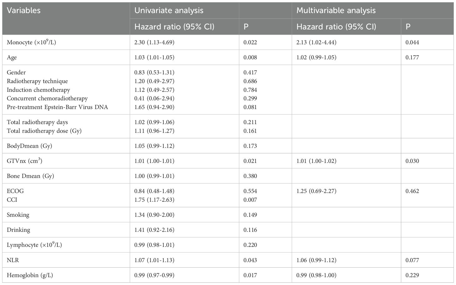
In the univariate Cox regression analysis (Table 2) gender, radiotherapy technique, induction chemotherapy, concurrent chemoradiotherapy, EBV DNA, ECOG, CCI, smoking, and drinking are categorical variable), parameters such as monocyte count [HR 2.30, 95%CI (1.13-4.69)], age[1.03(1.01-1.05)], GTVnx[1.01(1.00-1.01)], CCI[1.75(1.17-2.63)], NLR[1.07(1.01-1.13)], Hgb[0.99(0.97-0.99)] were shown to be significantly associated with OS. In the multivariate Cox regression analyses (Table 2), monocyte count[2.13(1.02-4.44)] and GTVnx[1.01(1.00-1.02)] were independently associated with OS.
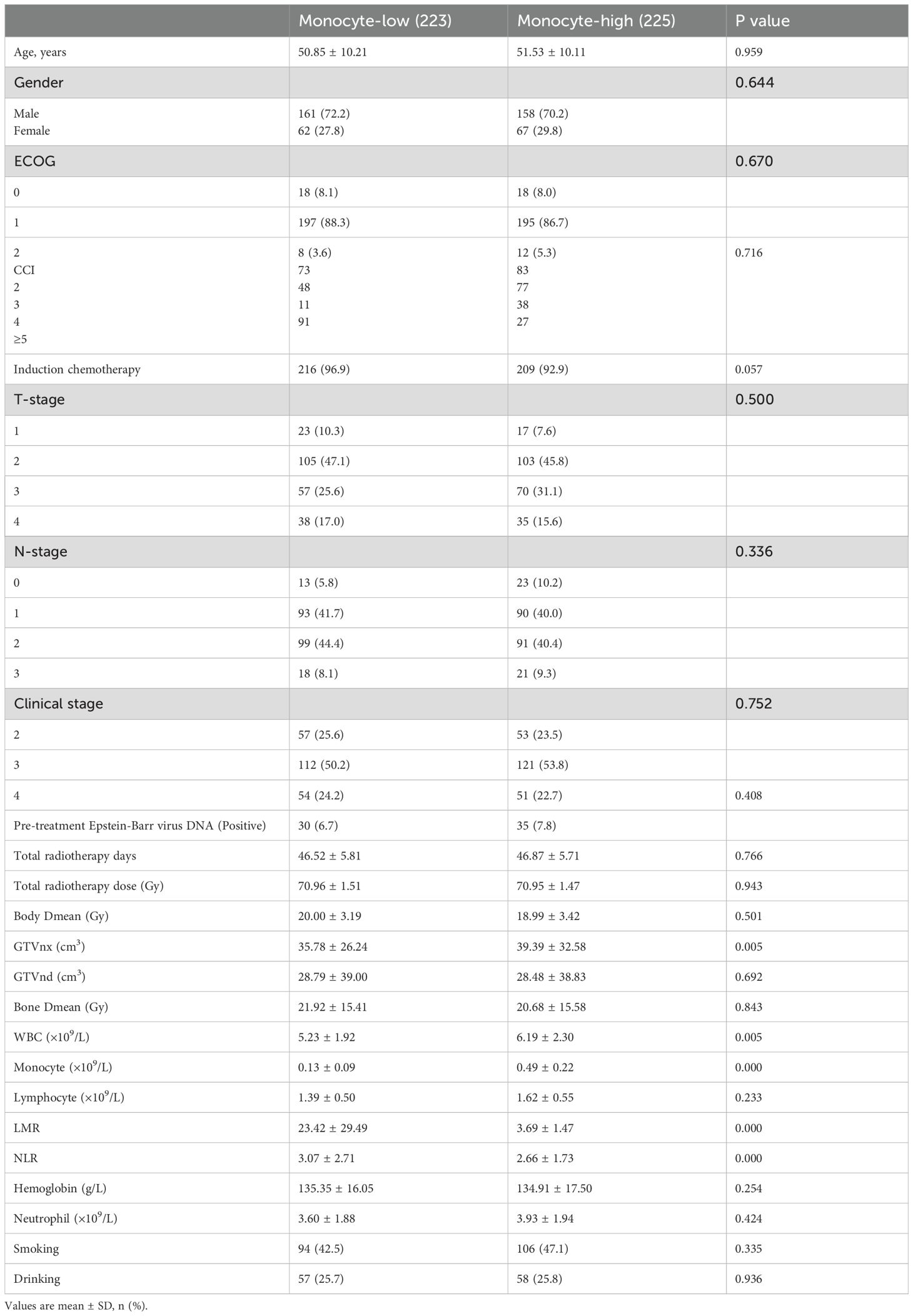
To further confirm the predictive effect of monocyte count and GTVnx on OS and PFS, we divided patients into low and high groups based on baseline median monocyte count and GTVnx. As shown in Figure 1, the high monocyte count group and high GTVnx group were associated with poor OS and PFS. In the two-independent sample T-test, there were no statistical differences in other clinical parameters between the two groups except baseline monocyte count, GTVnx, WBC, LMR, and NLR (Table 3). The clinical characteristics between the high monocyte count group and low monocyte count group were compared, and there were no statistical differences in age, sex, pre-treatment Epstein-Barr virus DNA, ECOG score, tumor stage, smoking history, drinking history, CCI, radiotherapy method, total radiotherapy days, whether chemotherapy was induced, radiotherapy and hematology indexes between the two groups.
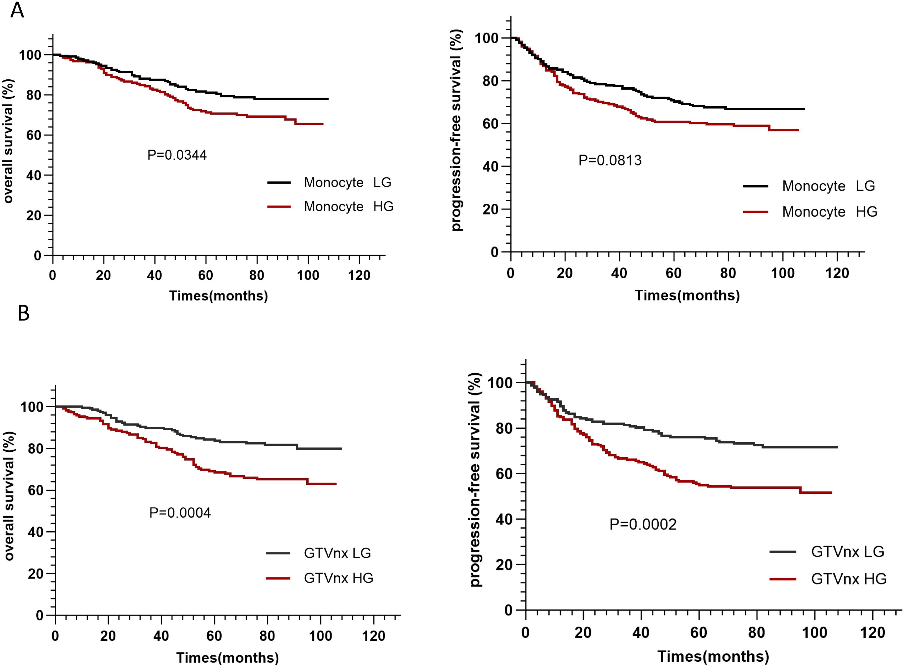
Figure 1. The Kaplan–Meier survival curves of monocyte count (A), GTVnx (B) based on the overall survival (OS) and progression-free survival (PFS) in NPC patients. (Monocyte LG: monocyte count <0.29×10⁹/L, Monocyte HG: monocyte count ≥0.29×10⁹/L, GTVnx LG: GTVnx <28.5cm3, GTVnx HG: GTVnx ≥28.5cm3.).
The correlation analysis of Figure 2 showed that peripheral blood monocyte count was positively correlated with WBC, platelet, and neutrophil (P<0.05). GTVnx was positively correlated with platelet, neutrophil, and Epstein-Barr virus before treatment (P<0.05). As shown in Supplementary Figure S1, the higher the peripheral blood monocyte count at 1 month, 2 months, and 3 months after radiotherapy, the worse the OS and PFS, which supports our previous conclusion.
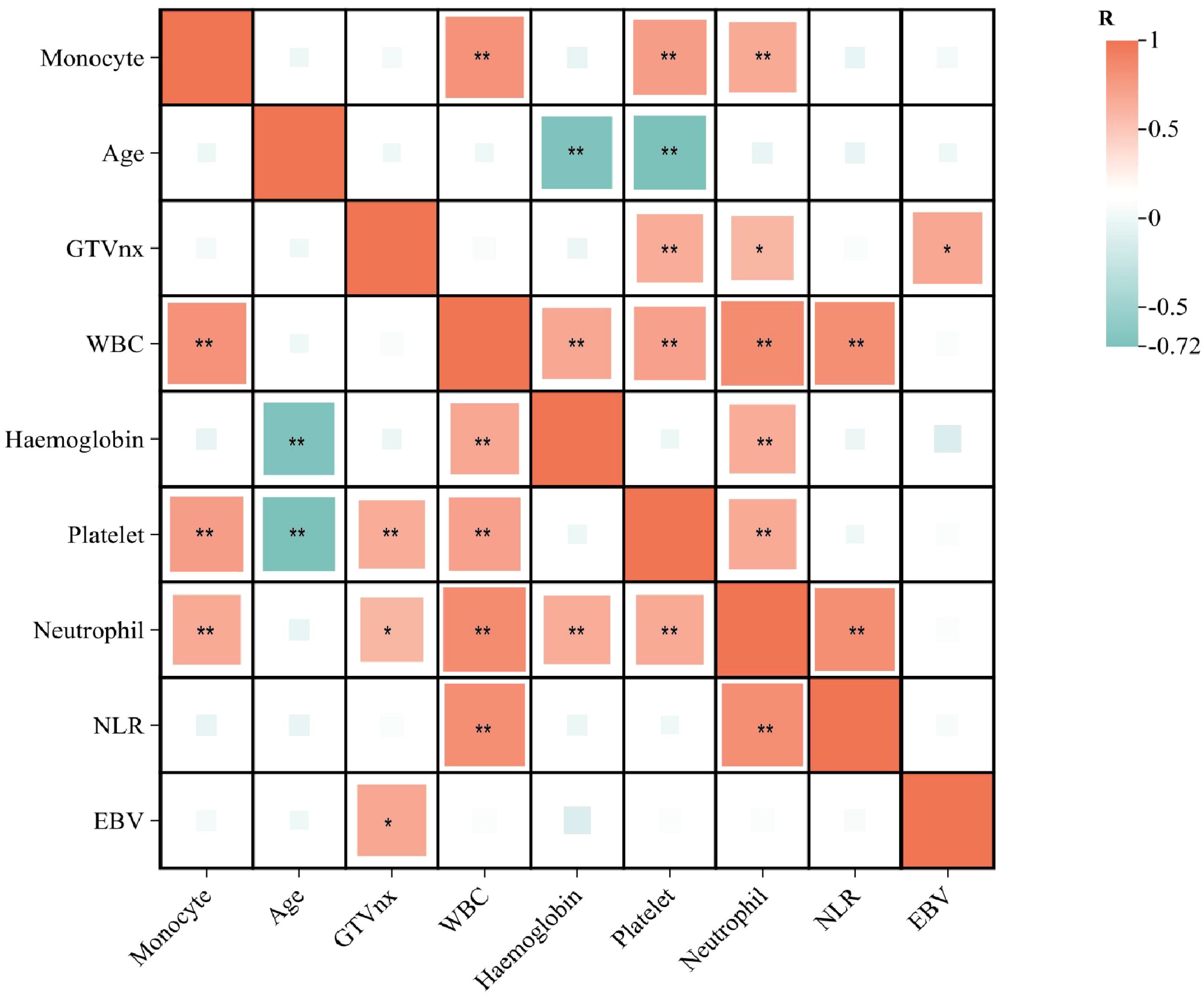
Figure 2. Risk factor correlation matrix diagram (*=P<0.05; **=P<0.01). This figure was drawn using ChiPlot (https://www.chiplot.online/) (accessed on March 2025).
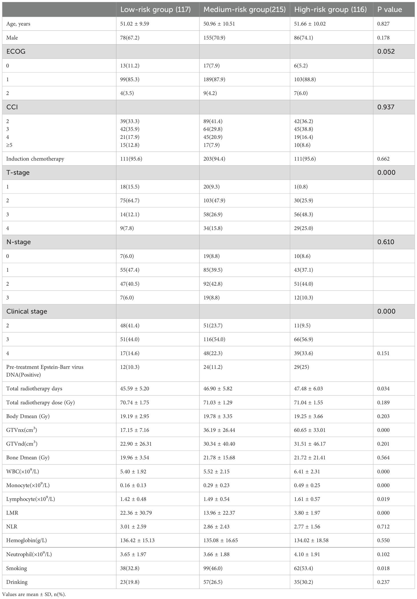
Based on the independent risk factors (monocyte count ≥ 0.29×10⁹/L (median), GTVnx ≥ 28.5cm3 (median)) for OS, we further divide patients into three different risk groups: low-risk group (with <1 risk factor), medium-risk group (with 1 risk factors), and high-risk group (with 2 risk factors). Survival curves were significantly different among patients in different risk groups for OS (p = 0.0008) and PFS (p = 0.0007) (Figure 3). The characteristics of patients in the different risk groups are shown in Table 4. As shown in Table 5, patients in the GTVnx low group had a 2.64-fold and 2.31-fold increased risk of OS and PFS for every increase in the unit count of monocytes, and in the GTVnx high group had a 1.94-fold and 1.09-fold increase, respectively. In the low monocyte count group, the risk of OS and PFS increased by 1.01 times per unit volume increase of GTVnx, and in the high monocyte count group, GTVnx increases the risk of OS and PFS by 1.00 times per unit volume increase (Supplementary Table 1). Altogether, the prognostic benefit of patients with GTVnx< 28.5cm3 could be remarkably attenuated by the high monocyte count.
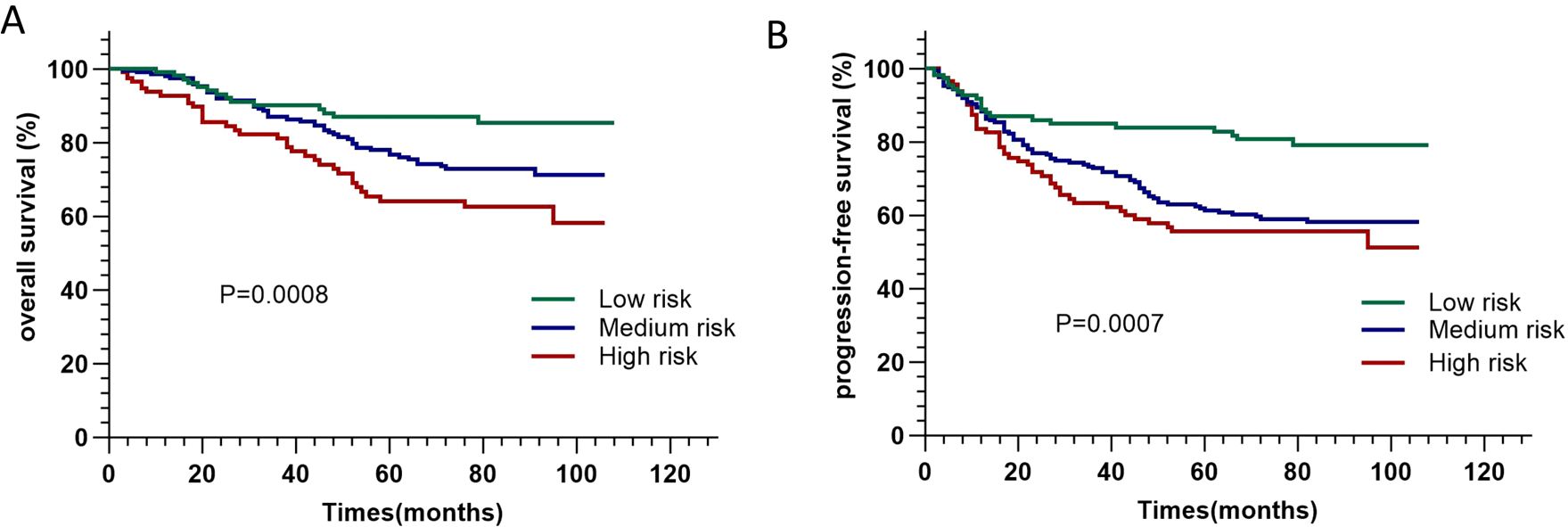
Figure 3. Kaplan-Meier curves of overall survival (A) and progression-free survival (B) of patients in different risk groups.
Discussion
Although the role of systemic inflammation in the development of cancer has been investigated in many studies (17), the main mechanism is still unclear. When tumors or inflammation occur in the body, monocytes in the peripheral circulation are recruited into the tumor microenvironment and further differentiate into macrophages (8) to play a functional or programmed death. Macrophages then play a role in stimulating angiogenesis, enhancing tumor cell migration and invasion, and inhibiting anti-tumor immunity in the development of tumors (18). GTV is generally considered a part of the tumor microenvironment. Still, more specifically, it represents the tumor volume visible on imaging, is a potential biological reflection of tumor burden, and is associated with the survival of tumor patients (19). Understanding the relationship between different monocyte counts and the prognosis of tumors after radiotherapy and chemotherapy under different tumor loads is conducive to the formulation of individualized treatment decisions.
In this study, it’s confirmed that pre-treatment monocyte count and GTVnx are independent predictors of survival in newly diagnosed non-metastatic nasopharyngeal carcinoma patients undergoing radiotherapy. The high monocyte count (≥ 0.29×10⁹/L) and high GTVnx (GTVnx≥ 28.5cm3) were associated with poor OS and PFS. At the same time, the monocyte data at 1 month, 2 months, and 3 months after radiotherapy also supported this conclusion. Moreover, correlation analysis showed that monocyte count was positively correlated with WBC, platelet, and neutrophil. GTVnx was positively correlated with platelet, neutrophil, and Epstein-Barr virus before treatment. Elevated WBC, platelets, and neutrophils are often associated with poor prognosis and may reflect systemic inflammatory responses or immune system disorders that promote tumor growth and metastasis.
After adjusting for a variety of confounding factors, for every increase in unit count of monocytes, the risk of death increases by 2.30 times. Besides, for every increase in monocyte unit count, the OS and PFS risks of patients in the low and high GTVnx groups increased by 2.64 times, 2.31 times, 1.94 times, and 1.09 times, respectively. A large number of previous studies have shown that the increase of peripheral blood monocytes is associated with poor survival of various cancers (10–13, 20–22). This is consistent with the results of our study in which NPC patients with higher baseline peripheral blood monocyte counts had worse survival outcomes than those with lower baseline peripheral blood monocyte counts. This study further verified that GTVnx and systemic inflammation, including monocytes, are important factors affecting the prognosis of NPC patients.
Monocytes can secrete various proinflammatory cytokines, which have been associated with shorter survival and worse prognosis in malignancies (9). Moreover, monocytes can also promote tumor growth and angiogenesis by releasing VEGF, epidermal growth factor (EGF), and tumor necrosis factor-α (TNF-α) (23). Peripheral blood monocyte count is closely related to tumor-associated macrophages (TAM), which differentiate into TAM through tumor microenvironment (24), and produce various cytokines and growth factors to induce tumor progression. These cytokines and growth factors can cause angiogenesis and anti-immune response. In the tumor or inflammatory site, differentiated macrophages can polarize to form different subtypes with the change of the environment, mainly divided into M1 macrophages and M2 macrophages (25). However, there is evidence that TAM in the tumor microenvironment can no longer be simply classified by the M1/M2 macrophage polarization model (26). They often have the common characteristics of both. As the tumor progresses, TAM will eventually shift to an immunosuppressive phenotype, with the characteristics of stimulating angiogenesis, inhibiting adaptive immunity, and promoting tumor growth and metastasis (27).
During radiotherapy, there is evidence that monocytes are recruited into the tumor to promote angiogenesis during radiotherapy, counteracting the therapeutic effect and ultimately negatively affecting the treatment outcome (28). When the tumor volume is large, it may also lead to changes in the tumor microenvironment, and the blood supply is difficult to reach the center of the tumor, resulting in obvious hypoxic tissue in this area, which is less responsive to radiotherapy and chemotherapy. In addition, the anatomical structure of nasopharyngeal carcinoma is complex, the wider the range of tumor invasion, the closer the distance between the tumor and the optic nerve, spinal cord, and brainstem, the greater the difficulty of radiotherapy planning, resulting in decreased efficacy and poor prognosis. A better understanding of the process of monocyte recruitment may lead to the development of new therapies to control the number and distribution of monocytes, thereby enhancing the therapeutic efficacy of malignancies and improving survival.
As one of the types of leukocyte, monocytes can be combined with GTVnx as a prognostic indicator of nasopharyngeal carcinoma due to its convenient, rapid, and low-cost detection. The results of this study showed that GTVnx ≥ 28.5cm3 and monocyte count ≥ 0.29×10⁹/L were adverse factors for PFS and OS. In patients with GTVnx < 28.5cm3, increasing monocyte count will significantly increase the risk of poor survival and disease progression. Therefore, it is imperative to develop drugs that regulate monocyte infiltration and reshape the function of TAM for clinical application. At the same time, Clinicians should be alert to the risk of local recurrence or distant metastasis and develop individualized treatment plans (such as increasing adjuvant chemotherapy, Combined immunotherapy with the targeting of tumor-infiltrating myeloid cells, etc.) for patients with nasopharyngeal carcinoma who meet these requirements according to specific conditions.
Some limitations need to be mentioned. First of all, this is a retrospective study, and inevitably there may be some bias. Furthermore, this study was conducted in one medical center, so the selection bias was inevitable. Therefore, multi-center, large-sample prospective studies are needed to further confirm our findings.
Conclusions
In conclusion, peripheral blood monocyte count combined with gross tumor volume of nasopharyngeal carcinoma can independently predict the overall survival and progression free-survival in newly diagnosed non-metastatic NPC patients who received radiotherapy. The benefit of patients with GTVnx< 28.5cm3 could be remarkably attenuated by the high monocyte count. These findings should be verified in more prospective studies conducted among different populations.
Data availability statement
The raw data supporting the conclusions of this article will be made available by the authors, without undue reservation.
Ethics statement
The studies involving humans were approved by Ethics Committee of Chongqing University Affiliated Tumor Hospital. The studies were conducted in accordance with the local legislation and institutional requirements. The participants provided their written informed consent to participate in this study.
Author contributions
YS: Data curation, Writing – original draft. HZ: Writing – review & editing, Formal analysis. LY: Conceptualization, Methodology, Writing – original draft. DL: Software, Writing – original draft. LT: Funding acquisition, Resources, Writing – review & editing. ML: Writing – review & editing. DL: Writing – review & editing. YX: Writing – review & editing. YW: Writing – review & editing. JS: Writing – review & editing.
Funding
The author(s) declare that financial support was received for the research and/or publication of this article. The current study was supported by grants from the National Natural Science Foundation of China (Grant No. 82203821); the Natural Science Foundation of Chongqing, China (Grant No. cstc2021jcyj-msxmX0587); the research program for PHD personnel in Chongqing, China (Grant No. CSTB2022BSXM-JCX0079).
Conflict of interest
The authors declare that the research was conducted in the absence of any commercial or financial relationships that could be construed as a potential conflict of interest.
Correction note
A correction has been made to this article. Details can be found at: 10.3389/fonc.2025.1637309.
Generative AI statement
The author(s) declare that no Generative AI was used in the creation of this manuscript.
Publisher’s note
All claims expressed in this article are solely those of the authors and do not necessarily represent those of their affiliated organizations, or those of the publisher, the editors and the reviewers. Any product that may be evaluated in this article, or claim that may be made by its manufacturer, is not guaranteed or endorsed by the publisher.
Supplementary material
The Supplementary Material for this article can be found online at: https://www.frontiersin.org/articles/10.3389/fonc.2025.1541212/full#supplementary-material
Supplementary Figure 1 | The Kaplan–Meier survival curves of monocyte count one month (A), two months (B) and three months (C) after radiotherapy based on the overall survival (OS) and progression-free survival (PFS) in NPC patients. (A: Monocyte LG: monocyte count <0.28×10⁹/L, Monocyte HG: monocyte count ≥0.28×10⁹/L; B: Monocyte LG: monocyte count <0.27×10⁹/L, Monocyte HG: monocyte count ≥0.27×10⁹/L; C: Monocyte LG: monocyte count <0.26×10⁹/L, Monocyte HG: monocyte count ≥0.26×10⁹/L).
Supplementary Table 1 | The effect of GTVnx volume per unit increase on OS and PFS in different groups.
References
1. Chen YP, Chan ATC, Le QT, Blanchard P, Sun Y, and Ma J. Nasopharyngeal carcinoma. Lancet. (2019) 394:64–80. doi: 10.1016/S0140-6736(19)30956-0
2. Sung H, Ferlay J, Siegel RL, Laversanne M, Soerjomataram I, Jemal A, et al. Global cancer statistics 2020: GLOBOCAN estimates of incidence and mortality worldwide for 36 cancers in 185 countries. CA Cancer J Clin. (2021) 71:209–49. doi: 10.3322/caac.21660
3. Pan JJ, Ng WT, Zong JF, Chan LL, O’Sullivan B, Lin SJ, et al. Proposal for the 8th edition of the AJCC/UICC staging system for nasopharyngeal cancer in the era of intensity-modulated radiotherapy. Cancer. (2016) 122:546–58. doi: 10.1002/cncr.29795
4. Zhang B, Li MM, Chen WH, Zhao JF, Chen WQ, Dong YH, et al. Association of chemoradiotherapy regimens and survival among patients with nasopharyngeal carcinoma: A systematic review and meta-analysis. JAMA Netw Open. (2019) 2:e1913619. doi: 10.1001/jamanetworkopen.2019.13619
5. Galon J, Costes A, Sanchez-Cabo F, Kirilovsky A, Mlecnik B, Lagorce-Pagès C, et al. Type, density, and location of immune cells within human colorectal tumors predict clinical outcome. Science. (2006) 313:1960–4. doi: 10.1126/science.1129139
6. Aarstad HJ, Vintermyr OK, Ulvestad E, Aarstad HH, Kross KW, and Heimdal JH. Peripheral blood monocyte and T-lymphocyte activation levels at diagnosis predict long-term survival in head and neck squamous cell carcinoma patients. APMIS. (2015) 123:305–14. doi: 10.1111/apm.12356
7. Qu Z, Lu YJ, Feng JW, Chen YX, Shi LQ, Chen J, et al. Preoperative prognostic nutritional index and neutrophil-to-lymphocyte ratio predict survival outcomes of patients with hepatocellular carcinoma after curative resection. Front Oncol. (2022) 11:823054. doi: 10.3389/fonc.2021.823054
8. Shi C and Pamer EG. Monocyte recruitment during infection and inflammation. Nat Rev Immunol. (2011) 11:762–74. doi: 10.1038/nri3070
9. Ugel S, Canè S, De Sanctis F, and Bronte V. Monocytes in the tumor microenvironment. Annu Rev Pathol. (2021) 16:93–122. doi: 10.1146/annurev-pathmechdis-012418-013058
10. Kwiecień I, Rutkowska E, Polubiec-Kownacka M, Raniszewska A, Rzepecki P, and Domagała-Kulawik J. Blood monocyte subsets with activation markers in relation with macrophages in non-small cell lung cancer. Cancers (Basel). (2020) 12:2513. doi: 10.3390/cancers12092513
11. Sakakura K, Takahashi H, Motegi SI, Yokobori-Kuwabara Y, Oyama T, and Chikamatsu K. Immunological features of circulating monocyte subsets in patients with squamous cell carcinoma of the head and neck. Clin Immunol. (2021) 225:108677. doi: 10.1016/j.clim.2021.108677
12. Jiang R, Cai XY, Yang ZH, Yan Y, Zou X, Guo L, et al. Elevated peripheral blood lymphocyte-to-monocyte ratio predicts a favorable prognosis in the patients with metastatic nasopharyngeal carcinoma. Chin J Cancer. (2015) 34:237–46. doi: 10.1186/s40880-015-0025-7
13. Magrowski Ł, Masri O, Ciepał J, Depowska G, Nowicka Z, Stando R, et al. Pre-treatment hemoglobin concentration and absolute monocyte count as independent prognostic factors for survival in localized or locally advanced prostate cancer patients undergoing radiotherapy. Biomedicines. (2022) 10:2514. doi: 10.3390/biomedicines10102514
14. Liu W, Li Y, Zhu H, Pei Q, Tan F, Song X, et al. The relationship between primary gross tumor volume and tumor response of locally advanced rectal cancer: pGTV as a more accurate tumor size indicator. J Invest Surg. (2021) 34:181–90. doi: 10.1080/08941939.2019.1615153
15. Reymen B, Van Loon J, van Baardwijk A, Wanders R, Borger J, Dingemans AM, et al. Total gross tumor volume is an independent prognostic factor in patients treated with selective nodal irradiation for stage I to III small cell lung cancer. Int J Radiat Oncol Biol Phys. (2013) 85:1319–24. doi: 10.1016/j.ijrobp.2012.10.003
16. Sze WM, Lee AW, Yau TK, Yeung RM, Lau KY, Leung SK, et al. Primary tumor volume of nasopharyngeal carcinoma: prognostic significance for local control. Int J Radiat Oncol Biol Phys. (2004) 59:21–7. doi: 10.1016/j.ijrobp.2003.10.027
17. Khandia R and Munjal A. Interplay between inflammation and cancer. Adv Protein Chem Struct Biol. (2020) 119:199–245. doi: 10.1016/bs.apcsb.2019.09.004
18. Qian BZ and Pollard JW. Macrophage diversity enhances tumor progression and metastasis. Cell. (2010) 141:39–51. doi: 10.1016/j.cell.2010.03.014
19. Lin YH, Huang TL, Chien CY, Chen HC, Hsu HC, Huang EY, et al. Pretreatment prognostic factors of survival and late toxicities for patients with nasopharyngeal carcinoma treated by simultaneous integrated boost intensity-modulated radiotherapy. Radiat Oncol. (2018) 13:45. doi: 10.1186/s13014-018-0990-5
20. Hu S, Zou Z, Li H, Zou G, Li Z, Xu J, et al. The preoperative peripheral blood monocyte count is associated with liver metastasis and overall survival in colorectal cancer patients. PloS One. (2016) 11:e0157486. doi: 10.1371/journal.pone.0157486
21. Shibutani M, Maeda K, Nagahara H, Fukuoka T, Nakao S, Matsutani S, et al. The peripheral monocyte count is associated with the density of tumor-associated macrophages in the tumor microenvironment of colorectal cancer: a retrospective study. BMC Cancer. (2017) 17:404. doi: 10.1186/s12885-017-3395-1
22. Feng F, Zheng G, Wang Q, Liu S, Liu Z, Xu G, et al. Low lymphocyte count and high monocyte count predicts poor prognosis of gastric cancer. BMC Gastroenterol. (2018) 18:148. doi: 10.1186/s12876-018-0877-9
23. Yang JG, Wang LL, and Ma DC. Effects of vascular endothelial growth factors and their receptors on megakaryocytes and platelets and related diseases. Br J Haematol. (2018) 180:321–34. doi: 10.1111/bjh.15000
24. Petty AJ and Yang Y. Tumor-associated macrophages: implications in cancer immunotherapy. Immunotherapy. (2017) 9:289–302. doi: 10.2217/imt-2016-0135
25. Martinez FO and Gordon S. The M1 and M2 paradigm of macrophage activation: time for reassessment. F1000Prime Rep. (2014) 6:13. doi: 10.12703/P6-13
26. Franklin RA, Liao W, Sarkar A, Kim MV, Bivona MR, Liu K, et al. The cellular and molecular origin of tumor-associated macrophages. Science. (2014) 344:921–5. doi: 10.1126/science.1252510
27. Noy R and Pollard JW. Tumor-associated macrophages: from mechanisms to therapy. Immunity. (2014) 41:49–61. doi: 10.1016/j.immuni.2014.06.010
Keywords: peripheral blood monocyte count, GTVnx, nasopharyngeal carcinoma, radiotherapy, overall survival
Citation: Yang L-N, Song Y-R, Zhang H, Tu H-L, Lu M-Y, Liu D-Q, Sui J-D, Li D, Xie Y and Wang Y (2025) Monocyte count combined with GTVnx is an independent prognostic factor in non-metastatic nasopharyngeal carcinoma receiving radiotherapy. Front. Oncol. 15:1541212. doi: 10.3389/fonc.2025.1541212
Received: 07 December 2024; Accepted: 24 March 2025;
Published: 08 April 2025; Corrected: 20 August 2025.
Edited by:
Nerina Denaro, IRCCS Ca ‘Granda Foundation Maggiore Policlinico Hospital, ItalyReviewed by:
Anna Fialová, SOTIO a.s., CzechiaWala Ben Kridis, Habib Bourguiba University Hospital, Tunisia
Copyright © 2025 Yang, Song, Zhang, Tu, Lu, Liu, Sui, Li, Xie and Wang. This is an open-access article distributed under the terms of the Creative Commons Attribution License (CC BY). The use, distribution or reproduction in other forums is permitted, provided the original author(s) and the copyright owner(s) are credited and that the original publication in this journal is cited, in accordance with accepted academic practice. No use, distribution or reproduction is permitted which does not comply with these terms.
*Correspondence: Dan Li, bGlkYW5sdWRhbkAxNjMuY29t; Yue Xie, MzQ0ODk5NTI1QHFxLmNvbQ==; Ying Wang, eWluZ3dhbmdjcUBob3RtYWlsLmNvbQ==
†These authors have contributed equally to this work
 Li-Na Yang†
 Yun-Rui Song
 Hong-Lei Tu
 Dan Li
 Yue Xie
 Ying Wang
