- 1Caribbean Centre for Research in Bioscience, University of the West Indies, Kingston, Jamaica
- 2Caribbean Institute for Health Research, University of the West Indies, Kingston, Jamaica
- 3Department of Basic Medical Sciences, Biochemistry Section, University of the West Indies Mona, Kingston, Jamaica
Environmental heavy metal pollutants are highly toxic and are usually of human origin. Studies have suggested a link between cadmium and arsenic carcinogenesis and geographical location. This review was conducted to explore the methodologies that have been used to determine the risk of carcinogenesis as it relates to cadmium & arsenic exposure as well as geographical location. A search of pertinent literature published up to December 2024 was conducted using the databases, PubMed, and EBSCO. The following MeSH terms were used primarily to search the databases, “heavy metals,” “cadmium,” “arsenic,” “carcinogenesis,” “malignancy,” and “toxicity.” Articles were removed if they were not closely related to the review topic. As evidenced in this review, there has been several research done over the years exploring the heavy metal exposure and the risk for carcinogenesis. The methodologies used to determine this risk are quite uniformed across the various studies. However, there is a paucity of studies dealing with the potential influence of geographical location in relation to the risk of carcinogenesis. This gap in knowledge shows that more work needs to be done to improve on the current knowledge of arsenic and cadmium and carcinogenesis.
1 Introduction
Studies done by researchers have aided in having a better understanding of individual cancers which has led to the development of screening programs, prophylactic treatments, and finally, the development of more targeted therapy. This improved understanding of the disease and treatment has yielded better prognoses and fewer side effects (1). When discussing cervical, prostate, and colorectal (CPC) cancers, investigators have acknowledged that none of these three cancers is driven by a single dominant mechanism of carcinogenesis. Instead, most research indicate that their development is often the result of multiple risk factors interacting synergistically to create an environment conducive to carcinogenesis (2–6). This review will focus on studies that explore the relationship/association between heavy metals and cancer. Various studies have been conducted looking at heavy metals in relation to cancer, from studies that simply looked at the presence of heavy metals in the blood of cancer patients, to studies that have looked at the mechanisms by which heavy metals could be causing cancers. Heavy metal pollution in the environment includes water, air, and soil and has been found to be mainly of human origins, these include fossil fuel burning, vehicle exhaust, waste incineration, and industrial processes such as mining and agriculture (7, 8). In addition to human origins, environmental heavy metal pollution also has naturally occurring origins such as infiltration and volcanic eruptions (7, 9). The most environmentally hazardous heavy metals have been found to include arsenic (As), cadmium (Cd), chromium (Cr), lead (Pb), mercury (Hg), and zinc (Zn). This is based on their significance to public health and their toxicity (7, 10). Though there is great interest in all the heavy metals cited, this review will focus on cadmium and arsenic specifically. When listing environmental mutagens as risk factors of cancers such as colorectal cancer, the compounds that are normally implicated are carcinogenic compounds that cause gene mutations, these include polycyclic aromatic hydrocarbons (from burning of gas, wood, garbage, and tobacco), heterocyclic amines, nitrosamines, and aromatic amines (11). Studies on the relationship between heavy metals and colorectal cancer are limited, however, there have been a few that compared the levels of heavy metals in the blood of colorectal cancer patients to a control group (healthy individuals). The result from one study found significant differences in trace elements and heavy metals levels between healthy subjects and metastatic colon cancer patients (12). Studies have hypothesized a possible relationship between heavy metals and the pathogenesis of prostate cancer, these studies have suggested that some heavy metals such as cadmium have estrogenic or androgenic abilities. Since prostate cancer progression has been surmised to be androgen-dependent, scientists have suggested that this may be a possible mechanism in which heavy metals are risk factors of prostate cancer (13, 14). Contrastingly, some studies have found no association between heavy metals and prostate cancer risk. A study done to explore the possible relationship between urinary arsenic & blood cadmium, lead & mercury levels & prostate specific antigen (PSA) did not find any association between these heavy metal levels in the body and PSA (15). On the other hand, other studies found evidence that suggested a plausible relationship with metals such as zinc, cadmium, and arsenic levels in the body & the risk of prostate cancer (13, 16).
A study done in Jamaica in 2004 explored the levels of cadmium concentrations in autopsied kidneys (6.7 to 126 mg/kg−1, with a mean of 43.8 mg/kg−1) and livers (0.3 to 24.3 mg/kg−1, with a mean of 5.3 mg/kg−1) of deceased Jamaicans. This study found that the levels seen, especially in the kidney samples were much higher than values that were reported in other countries in the world (17–19). The study went on to specify that the values seen in Jamaica was second only to values seen in Japan (20). The cases sampled in this study were taken from areas in Jamaica where the concentration of cadmium in the soil was low compared to other areas of the country (21). It is of great interest to examine what the values would be when analyzed in individuals residing in areas such as central part of the country where the soil content of heavy metals has been documented to be above world averages (21, 22). A recent study done in Jamaica demonstrated that there were higher number of cervical, prostate and colorectal (CPC) cancer cases in areas in Jamaica with historically reported high levels of heavy metals such as cadmium (23). Additionally, one location of interest in Jamaica, is a rural parish in the southwestern section of the island, St. Elizabeth that has been found to have the highest concentrations of arsenic in the country, with one specific farming community being described as an arsenic anomaly due to the abnormally high levels of arsenic in the soil (21, 24).
In this review, we have considered the potential factors that may result in cadmium and arsenic exposure as well as the methods of metal induced carcinogenesis. While there have been a few studies that have explored the association between cadmium, arsenic and CPC cancers, the knowledge of the exact mechanism of action remains unclear. We seek to explore and understand the existing literature as it relates to heavy metal carcinogenicity and are especially focused on exposure as it relates to geographical location.
1.1 Review questions
The main concepts in the review questions and the search strategy were informed by the PCC (Population (or participants)/Concept/Context) framework (25). Considering the existing gap in the literature this review seeks to answer the following research questions:
1. What are the methodologies used to determine carcinogenesis risk of heavy metals in studies that explored the association of cervical, prostate, and colorectal cancer risk with cadmium and arsenic?
2. Does geographical location influence the relationship between cadmium and arsenic exposure and cervical, prostate, and colorectal cancer risk?
2 Materials and methods
This scoping review has been conducted in accordance with the JBI methodology for scoping reviews (26). The PRISMA-ScR checklist was used to inform the reporting of this review (27).
2.1 Search strategy
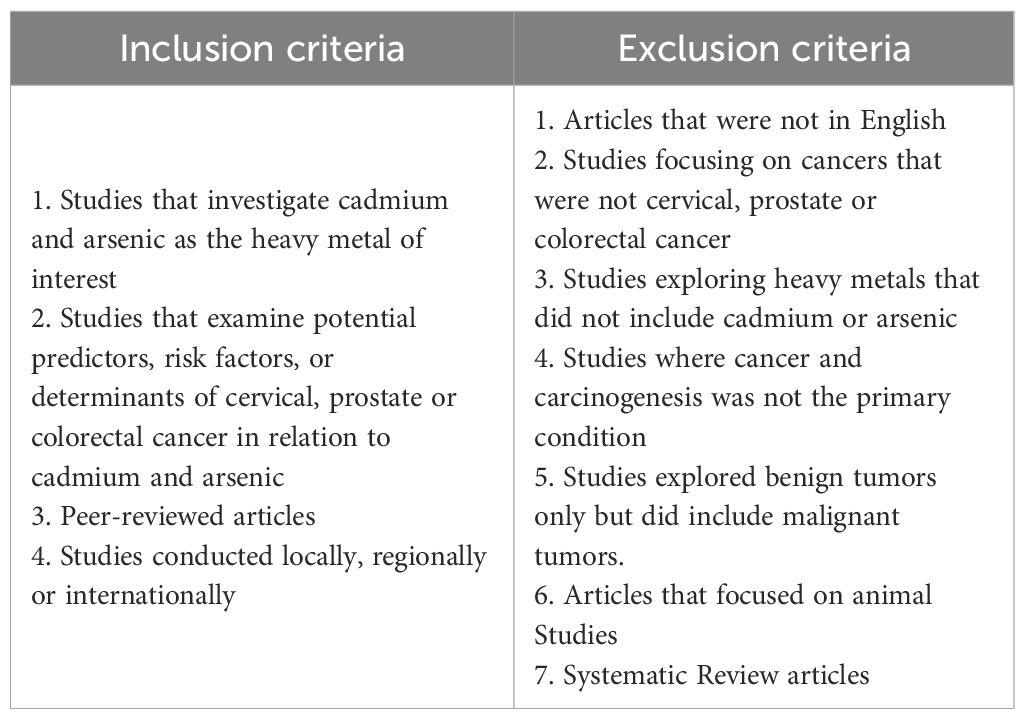
A search strategy was developed to search the PubMed and EBSCO host databases to retrieve articles on the cancer risk and cadmium & arsenic exposure. Articles retrieved were limited to those which had free full text available, human species studies, and English language for articles (Table 1).
There were no minimum or maximum date used in the search since the authors wanted to include as many studies as possible considering specificity of the study search. The primary keywords used in the search were “heavy metals,” or “arsenic” and “cadmium” with “carcinogenesis,” “cervical cancer,” “colorectal cancer,” and “prostate cancer.” The detailed search phrase can be found in Appendix 1 and 2.
The articles were selected to explore methodologies used to determine heavy metals exposure, their association with cervical, prostate and colorectal cancer risk, and subsequently, the potential impact of geographical location of the population. The inclusion and exclusion criteria are shown in Table 1.
The search terms used are listed in Appendix 1 and yielded 1397 results from PubMed and 25 from EBSCO (total 1422 articles which contained 53 duplicates). After the duplicates were removed, 1000 articles were automatically marked ineligible, and 9 articles were removed for other reasons (such articles that were outdated based on information included in more recent publications). The titles and abstracts of the remaining 360 articles were then screened by a single author according to the predetermined inclusion and exclusion criteria outlined in Table 1. After the title and abstract screening, 196 articles were eligible for full text review and ultimately 48 articles were included in this study for analysis. The management of citations throughout this process was facilitated using Mendeley reference manager. To address the research questions, a thematic approach/strategy was used to provide a comparative description of the studies. A summary of the search and inclusion & exclusion process is presented in the Preferred Reporting Items for Systematic Reviews and Meta-analyses extension for scoping review (PRISMA-ScR) flow diagram (Figure 1) (27).
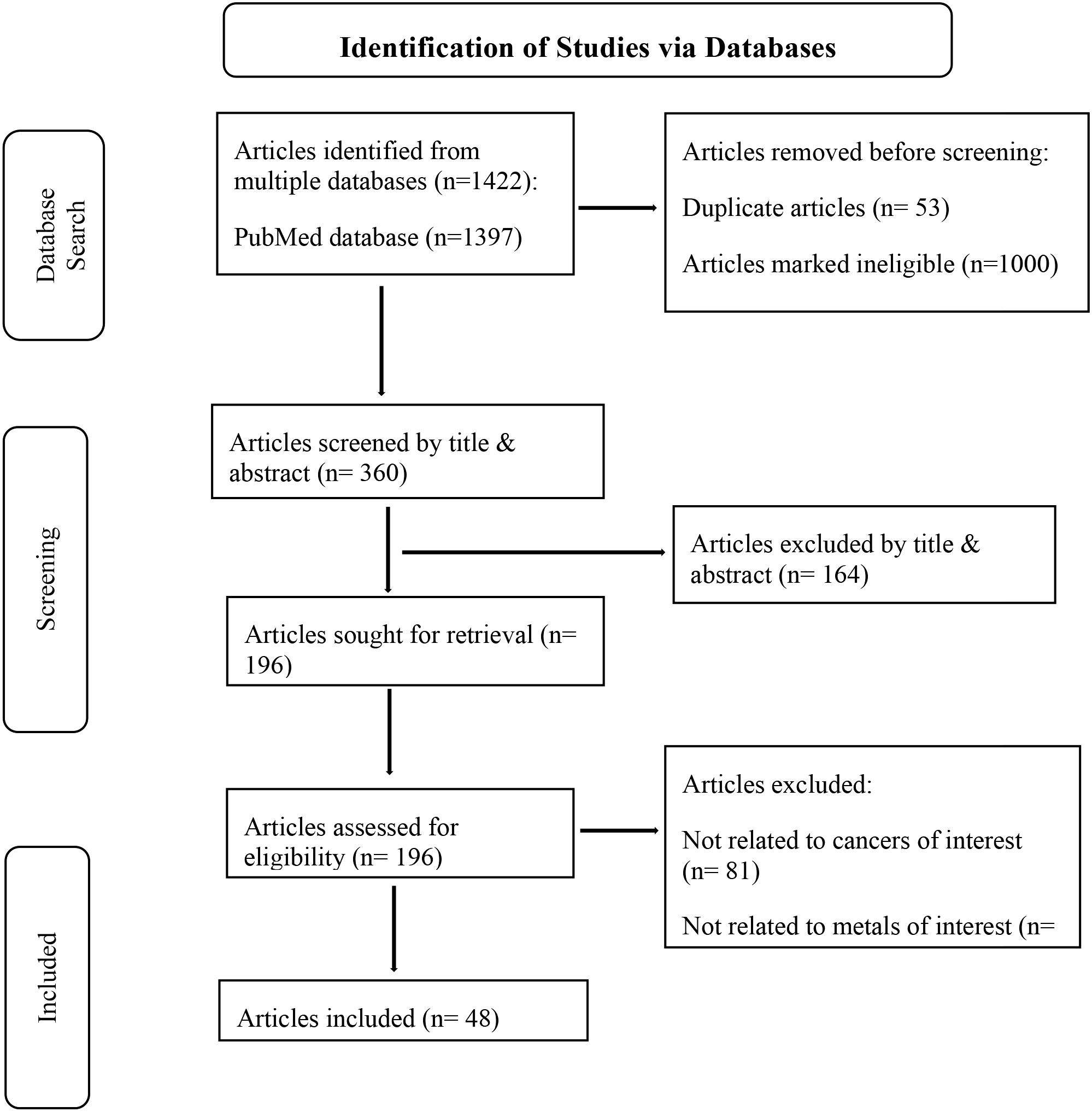
Figure 1. PRISMA-ScR diagram highlighting summary of the inclusion and exclusion process (27).
3 Results
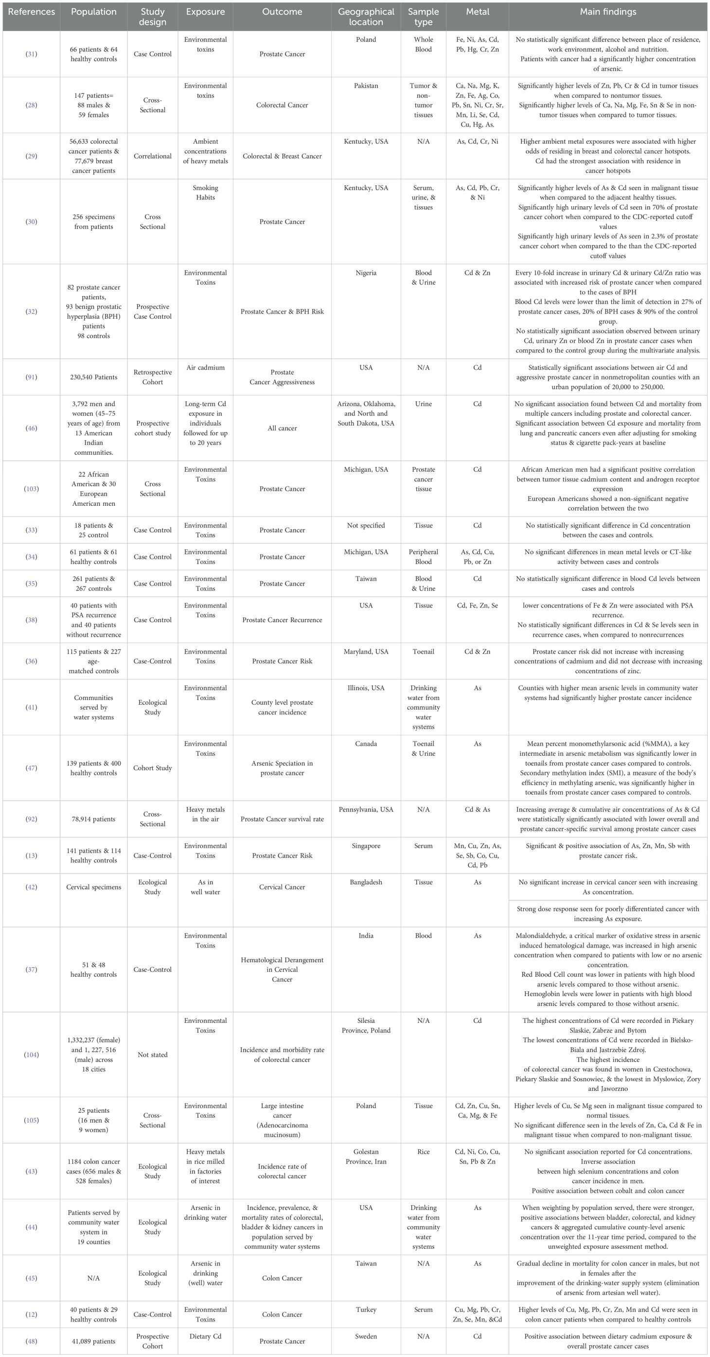
Following the search strategy, the included articles were sorted into two categories by the reviewers. The first category was observational studies (Table 2) and the second category was experimental studies (Table 3). The articles had publication dates ranging from 2000 to 2024.
3.1 Observational studies
In total there were 26 observational studies (Table 2), comprising approximately 3 million individuals from 12 countries identified and included in this review. There were various study designs used including cross sectional, case-control, cohort and ecological (Figure 2). In studies where cancer tissue was compared to non-cancer tissue in the same patient, there were significantly elevated levels of heavy metals in the cancer tissues (28–30). Case control studies which compared the concentration/levels of cadmium or arsenic in the biological samples of healthy controls and patients were ambivalent (12, 13, 31–37). While some studies showed significant differences between the concentration of the metals in the control group and patients (12, 13, 31), other studies reported no statistically significant difference (33–36). One case control study explored the risk of prostate cancer recurrence by comparing the levels of various metals in resected tissue samples of patients with PSA recurrence and those without recurrence (38). The study found no statistically significant differences when the levels were compared. Another study compared the level of arsenic, estimated malondialdehyde (MDA), hemoglobin, and red blood cell count in patients with cervical cancer and healthy controls to explore if there is a correlation between arsenic and MDA, hemoglobin and red blood cell count (37). The results of the study found that MDA was increased in high arsenic concentration when compared to patients with low or no arsenic concentration. Additionally, the red blood cell counts and hemoglobin levels of patients with high blood arsenic levels was found to be lower than patients without arsenic. Malondialdehyde is a reactive compound that is a physiological metabolite of lipid peroxidation of polyunsaturated fatty acids and is a critical marker of oxidative stress in arsenic induced hematological damage (37, 39, 40). A few ecological studies explored the potential relationship between the metals of interest in various environments and prevalence, incidence, & mortality of CPC cancers (41–45). One study that explored the arsenic levels in drinking water found that areas with higher mean arsenic levels in community water systems had significantly higher prostate cancer incidence (41). Another study that explored the possible relationship between the arsenic content in well water used for drinking and cervical cancer found a strong dose response seen for poorly differentiated cancer with increasing arsenic exposure, however, it did not find any significant increase in cervical cancer cases when the concentration of arsenic was increased (42). There were 4 cohort studies (46–48), 3 being prospective and one retrospective. The cohort studies made suggestions on the relationship between the metals of interest and the mortality, aggressiveness, incidence of the CPC cancers.
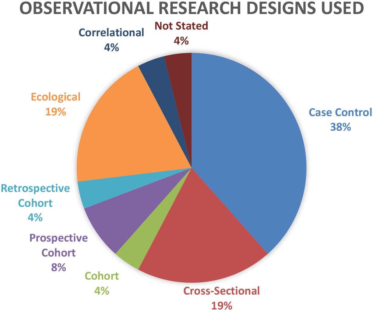
Figure 2. Case control studies accounted for 38% of the total observational studies used by researchers, while ecological and cross-sectional studies both accounted for 19% each.
3.2 Experimental studies
In total, there were 22 experimental studies (Table 3) included in this review. The method used was mostly cell assays. The relationship of arsenic and cadmium with various cellular processes, complexes and genes that work together to result in carcinogenesis were highlighted in the studies reviewed. This includes the induction of Erk (p44/42) and Mek 1/2 which represents the Erk/MAPK pathway activation (49, 50); Modulation of the phosphoinositide 3 kinase (PI3K/Akt) pathway by oncogene induction such as P110α, Akt, mTOR, NFKB1 and RAF (51); the increased expression of KRAS thus activating the RAS/ERK pathway (52); the modulation of epithelial-mesenchymal transition (EMT) related markers, N-cadherin, vimentin, E-cadherin and ZEB1 (53); and the activation of proto-oncogenes c-myc, p53 c-jun and c-fos (54, 55). Through these different cellular mechanisms cadmium and arsenic were found to promote the formation, migration, proliferation and invasion of cancer cells (Figure 3) (31, 51, 56–59).
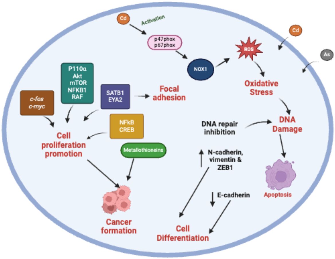
Figure 3. Chronic and acute metal exposure indirectly results in production of ROS which leads to oxidative stress in the cells. This activation leads to inhibition of DNA repair, DNA damage, activation of metallothioneins. Uncontrolled cell proliferation is the result of the effect of metal exposure on expression of genes such as c-fos, c-myc this activating the Ras/ERK (extracellular signal-regulated kinase) pathway. Metal exposure also results in disruption of epigenetic mechanisms such as DNA methylation by activating the PI3K/AKT/mTOR pathway result in promotion of cell proliferation.
One of the potential mechanisms for cadmium carcinogenesis explored in this review, is that chronic and acute exposure in cells results in oxidative stress and subsequent oxidative DNA damage which has been implicated in carcinogenesis (60–62). Oxidative stress is an imbalance between an antioxidant and pro-oxidant state in cells and tissues which results in excessive production of reactive oxygen species (ROS) (63, 64). It has been hypothesized that heavy metals such as cadmium and arsenic do not increase the production of ROS directly, but rather indirectly by disruption of essential metal homeostasis. This is because transporters in human cells that are specific for toxic metal transport (analogous to copper, zinc or other essential metal transporters) have yet to be identified (65, 66). Due to its biophysiochemical similarities to essential elements such as copper, zinc, and iron (67), it has been hypothesized that cadmium transport happens by mimicking these essential metals. Heavy metals have been shown to replace essential elements at the binding sites of their respective transporter proteins (67). For example, copper, zinc, and the metal ion binding protein metallothionein, have been implicated in the transport of cadmium (65, 66, 68, 69). Thus, the heavy metals can sometimes either inhibit enzymatic pathways or act as cofactors of the pathways (67, 70–72).
Another potential mechanism of cadmium as well as arsenic induced carcinogenesis explored in this review include the attenuation of apoptosis and alterations in gene expression (54, 55, 57, 58, 73). In one study cadmium and arsenic enhanced the expression of metallothioneins in prostate cancer cells, specifically MT1 MT2 & MT3 thus resulting in significant increase in cancer cell proliferation & invasion (58). Metallothioneins are stress-inducible cysteine-rich proteins with free radical scavenging capacity (74). Metallothioneins are associated with the maintenance of the homeostasis of the essential metal ions Zn and Cu (75). There are isoforms of metallothioneins; MT-1 and MT-2 (expressed in almost all tissues), MT-3 (brain specific), and MT-4 (expressed mainly in squamous epithelium) (76). The role of epithelial-mesenchymal transition (EMT) related markers was also explored where cadmium Cd exposure resulted in up-regulated the expressions of N-cadherin, vimentin, and ZEB1, while it down-regulated expression E-cadherin (53). Epithelial-mesenchymal transition (EMT) is a biologic process in which a polarized epithelial cell undergoes multiple biochemical changes and becomes mesenchymal cell phenotype, these changes include enhanced migratory & invasive properties, and elevated resistance to apoptosis (77). There are various markers for EMT, but those highlighted in the current review fall into three categories; they are epithelial markers (E-cadherin) and mesenchymal markers (vimentin & N-cadherin) and Zinc finger E-box binding homeobox 1 (ZEB1) which plays a critical role in the epithelial-to-mesenchymal transition (EMT) process (77, 78). Cadherins calcium dependent glycoproteins that make up what is referred to as a superfamily of adhesion molecules, which include other molecules such as integrins, selectins, and immunoglobulins. They are located on cell-surface membranes and play key roles in intercellular adhesions (79, 80).
When exploring the possible therapeutic options to combat metal-induced carcinogenesis, one experimental study explored the chemoprotective effects of psoralidin on cadmium induced prostate cancer cells (81). Psoralidin is a naturally occurring compound that has been hypothesized to have chemoprotective effects (82). Pal et al. (81) found that the addition of psoralidin to cadmium induced prostate cancer cells, resulted in an inhibition of cell growth, possibly by the inhibition of placenta specific 8 (PLAC8) expression. PLAC8 is a lysosomal protein that has been implicated in organ development and tumorigenesis (83). Another study found that resveratrol reversed the migratory and invasive ability of Cd-exposed colorectal cells (53). Resveratrol is a naturally occurring compound that can be found mainly in plants such as grapes and berries and has been found to have anti-cancer properties (84).
Finally, of the 22 experimental studies included in this review, only 4 studies did analysis on cervical cancer. There were no studies found on the effects of cadmium on cervical cancer. All four experimental studies that have been included explored the therapeutic effects of arsenic trioxide on cervical cancer cells. These studies differed from all the other studies included as they explored the anti-cancer effects of arsenic trioxide rather than a potential carcinogenic effect (85–88). These studies have been included in this review as they show a contrast to all the other studies covered in the review.
4 Discussion
At the start of this review, a preliminary search of MEDLINE, the Cochrane Database of Systematic Reviews and JBI Evidence Synthesis was conducted and similar reviews and meta-analysis on this topic have been referenced in this article but not included in the list of articles analyzed. One systematic review of note was published during the writing of the present review and explored a similar topic of heavy metals in biological samples of patients with breast, lung, prostate and gastric cancers (89). This systematic review explored cadmium and arsenic as risks for prostate cancer. The review, however, differs from the present one in that it did not go specifically into methodologies to determine exposure. Additionally, while the review briefly referenced the relationship that exists between soil contamination by heavy metals and lung cancer incidence, it did not explore the influence of geographical location. Another review of note explored the association of cadmium exposure and prostate cancer (90). This paper was a systematic review which included a meta-analysis and was considered by the authors of this review because it met the inclusion criteria.
During the implementation of the search strategy for this review, the reviewers realized that in attempting to answer the two research questions, there were quite a few methodologies that overlapped with each other when exploring cancer carcinogenesis. It is important to note that with the complex nature of carcinogenesis, oftentimes a mixed approach is prudent. Thus, it was important that both experimental and observational studies were highlighted in this review.
The varying results shown in the observational studies included in this review show the need for ongoing work. While a majority of studies found statistically significant relationships between cadmium and/or arsenic and CPC cancers (12, 13, 28–32, 37, 41, 47, 48, 91, 92), a small number did not find a statistically significant relationship (33–36). The ecological studies, though providing the lowest level of evidence were consistent, and showed trends in increasing cancer mortality and incidence with increasing metal concentration (41–45) which would warrant further in-depth studies to confirm.
4.1 Studies focused on geographical location as a risk factor for CPC cancers
Only 25% of the total number of studies reviewed focused specifically on geographical location as a risk factor for CPC. It is important to note that multiple studies have been done examining the pollution of heavy metals in the environment (7, 8, 93) and the resulting exposure to humans. More work is needed to explore the relationship between cancer (incidence, prevalence and mortality) in the population and the specific geographical location of the population affected. Additionally, it is of importance to note that some of the studies presented were ecological in nature which provided the lowest level of evidence in reviews, therefore with the use of aggregated data, establishing a direct relationship is not possible at the individual level (94). However, of importance, is that some studies (29, 41, 91, 92), reported a significant difference in incidence or survival of cancer in areas of high heavy metal concentrations even after adjusting for potential covariates.
4.2 Gaps in experimental research
As stated in the results, only 4 experimental studies explored the heavy metal of interest in cervical cancer, The studies covered explored the anti-cancer properties of arsenic trioxide. Arsenic trioxide (AS2O3) which is also referred to as white arsenic (95), is an old drug that was reintroduced into modern medicine due to its chemotherapeutic properties and is currently used to treat leukemia that is unresponsive to first-line treatment (96, 97). In medicine, the use of arsenic trioxide is tightly regulated, and the benefits have been found to outweigh the risks in cancer treatments (98).
Arsenic trioxide differs from environmental arsenic which has been shown to have negative health effects and has been explored in the observation and experimental studies in this review (96). The main difference between arsenic trioxide and the more toxic forms of inorganic arsenic has been hypothesized to be their metabolism in the body. Arsenic compounds are metabolized by oxidative methylation reactions in which inorganic arsenic is sequentially methylated to form mono-, di-, and trimethylated products (99, 100). Sometimes, arsenic metabolism results in arsenate (pentavalent) and arsenite (trivalent) which are the most toxic forms of arsenic implicated in carcinogenesis (44, 45).
The studies explored in this review, show that there are potential chemoprotective effects of arsenic trioxide on HPV cervical cancer cells (85–88). It would be interesting to explore the effects of inorganic arsenic on cervical cancer cells in future work.
5 Limitations
This review acknowledges the complexity of carcinogenesis as it relates to heavy metals and therefore, the authors acknowledge that some methodologies used to determine risks may be limited. Observational studies that collect retrospective data, rely heavily on accurate & legible record keeping. As such some records may not have details on the questions developed for the research and therefore with the possibility of large amounts of data being missing due to the information not recorded, ineligible or incomplete. Additionally, it is understood that studies exploring a specific factor may be confounded by factors not considered during the completion of analysis. We have highlighted these potential confounders in the studies where necessary.
The reviewers used a systematic search strategy to retrieve several articles to answer the research questions of this review. We acknowledge that there may have been relevant studies published in other languages that were omitted from this review. Additionally, this review did not include a search of grey literature (101). This omission of the grey literature further restricted the findings presented in the review to include only information that has been reported by scientific journals which may introduce bias in the review (102). Finally, we also acknowledge that the protocol for this review was not published and note this as limitation in the study.
6 Conclusions
As evidenced in this review, there has been several studies conducted that explore heavy metal exposure and the risks for carcinogenesis. The methodologies used to determine these risks are quite uniform across the various studies. The results of observational studies done, point to potential and often critical relationships between cadmium or arsenic and cancer development and in some cases, aggressiveness of the cancers of interest. The results of experimental studies done, point to multiple signaling pathways that result in carcinogenesis including the promotion of ROS production, inhibition of DNA repair, regulation of metallothioneins by miRNAs and transcription factors, promotion of cancer cell proliferation & invasion though p53 protein, E-cadherin, transcription factors, increased expression of proto-oncogenes as well as disruption of processes such DNA methylation by activation of the PI3K/AKT/mTOR pathway.
On the other hand, there is a paucity of studies dealing with the potential influence of geographical location in relation to the risks of carcinogenesis. Additionally, majority of the studies explored covered prostate and colorectal cancers, however there is a paucity of studies (both observational and experimental) exploring the relationship between cadmium or arsenic and cervical cancer.
This gap in knowledge shows that more insightful research is needed to improve on the current knowledge of heavy metals and carcinogenesis. It is understood that the relationship between heavy metals and carcinogenesis is indeed a complex one that cannot be linked to only one factor. It is the hope of the authors that as the relationship between the two are continually explored from different approaches that there will be meaningful contributions to public health policies in the approach to these cancers in terms of screening, as well as the planning of community education, environmental management plans and the sensitization about the risk factors for these cancers.
Data availability statement
The original contributions presented in the study are included in the article/Supplementary Material. Further inquiries can be directed to the corresponding author.
Author contributions
JB: Conceptualization, Methodology, Writing – original draft, Writing – review & editing. SM: Conceptualization, Methodology, Supervision, Writing – review & editing. IA: Supervision, Writing – review & editing.
Funding
The author(s) declare that no financial support was received for the research, and/or publication of this article.
Acknowledgments
This review is to contribute towards to the work done in partial fulfillment of a PhD degree program of the University of the West Indies, Mona, Jamaica for Ms. J. Bailey.
Conflict of interest
The authors declare that the research was conducted in the absence of any commercial or financial relationships that could be construed as a potential conflict of interest.
Generative AI statement
The author(s) declare that no Generative AI was used in the creation of this manuscript.
Publisher’s note
All claims expressed in this article are solely those of the authors and do not necessarily represent those of their affiliated organizations, or those of the publisher, the editors and the reviewers. Any product that may be evaluated in this article, or claim that may be made by its manufacturer, is not guaranteed or endorsed by the publisher.
Supplementary material
The Supplementary Material for this article can be found online at: https://www.frontiersin.org/articles/10.3389/fonc.2025.1569816/full#supplementary-material
References
1. Baudino TA. Send orders for reprints tocmVwcmludHNAYmVudGhhbXNjaWVuY2UuYWU=targeted cancer therapy: the next generation of cancer treatment. Current Drug Discovery Technologies (Bentham Science Publishers), UAE (2015).
2. De Marzo AM, DeWeese TL, Platz EA, Meeker AK, Nakayama M, Epstein JI, et al. Pathological and molecular mechanisms of prostate carcinogenesis: Implications for diagnosis, detection, prevention, and treatment. J Cell Biochem. (2004) 91:459–77. doi: 10.1002/jcb.10747
3. Porkka KP and Visakorpi T. Molecular mechanisms of prostate cancer. Eur Urol. (2004) 45:683–91. doi: 10.1016/s0302-2838(04)00017-x
4. Tariq K and Ghias K. Colorectal cancer carcinogenesis: a review of mechanisms. Cancer Biol Med Cancer Biol Med. (2016) 13:120–35. doi: 10.20892/j.issn.2095-3941.2015.0103
5. Yugawa T and Kiyono T. Molecular mechanisms of cervical carcinogenesis by high-risk human papillomaviruses: Novel functions of E6 and E7 oncoproteins. Rev Med Virol. (2009) 19:97–113. doi: 10.1002/rmv.605
6. Zhao L, Zhang Z, Lou H, Liang J, Yan X, Li W, et al. Exploration of the molecular mechanisms of cervical cancer based on mRNA expression profiles and predicted microRNA interactions. Oncol Lett. (2018) 15:8965–72. doi: 10.3892/ol.2018.8494
7. Ali H, Khan E, and Ilahi I. Environmental chemistry and ecotoxicology of hazardous heavy metals: environmental persistence, toxicity, and bioaccumulation. Journal of Chemistry (John Wiley & Sons Ltd), USA (2019). doi: 10.1155/2019/6730305.
8. Sall ML, Diaw AKD, Gningue-Sall D, Efremova Aaron S, and Aaron JJ. Toxic heavy metals: impact on the environment and human health, and treatment with conducting organic polymers, a review. Environ Sci pollut Res. (2020) 27:29927–42. doi: 10.1007/s11356-020-09354-3
9. Briffa J, Sinagra E, and Blundell R. Heavy metal pollution in the environment and their toxicological effects on humans. Heliyon. (2020) 6: e04691. doi: 10.1016/j.heliyon.2020.e04691
10. Tchounwou PB, Yedjou CG, Patlolla AK, and Sutton DJ. Heavy metals toxicity and the environment. Experientia supplementum (2012) 101:133–64. doi: 10.1007/978-3-7643-8340-4_6
11. Jaiswal AS, Armas ML, Connors SK, Panda H, and Narayan S. Exposure to environmental mutagens: APC and colorectal carcinogenesis. In: Environmental factors, genes, and the development of human cancers. Springer New York, New York, NY (2010). p. 303–29.
12. Emre O, Demir H, Dogan E, Esen R, Gur T, Demir C, et al. Plasma concentrations of some trace element and heavy metals in patients with metastatic colon cancer *. J Cancer Ther [Internet]. (2013) 4:1085–90. doi: 10.4236/jct.201346124PublishedOnlineAugust2013
13. Lim JT, Tan YQ, Valeri L, Lee J, Geok PP, Chia SE, et al. Association between serum heavy metals and prostate cancer risk – A multiple metal analysis. Environ Int. (2019) 1:132. doi: 10.1016/j.envint.2019.105109
14. Vella V, Malaguarnera R, Lappano R, Maggiolini M, and Belfiore A. Recent views of heavy metals as possible risk factors and potential preventive and therapeutic agents in prostate cancer. Mol Cell Endocrinol. (2017) 457:57–72. doi: 10.1016/j.mce.2016.10.020
15. Wu H, Wang M, Raman JD, and McDonald AC. Association between urinary arsenic, blood cadmium, blood lead, and blood mercury levels and serum prostate-specific antigen in a population-based cohort of men in the United States. PloS One. (2021) 16:e0250744. doi: 10.1371/journal.pone.0250744
16. Hsueh YM, Su CT, Shiue HS, Chen WJ, Pu YS, Lin YC, et al. Levels of plasma selenium and urinary total arsenic interact to affect the risk for prostate cancer. Food Chem Toxicology. (2017) 107::167–75. doi: 10.1016/j.fct.2017.06.031
17. Svartengren M, Elinder CG, Friberg L, and Lind B. Distribution and concentration of cadmium in human kidney. Environmental Research 39:1–7 (1986). doi: 10.1016/s0013-9351(86)80002-0
18. Lindqvist B, Nystrom K, Stegmayr B, Wirell’ M, and Eriksson A. Concentration in human kidney biopsies. Scand J Urol Nephrol. (1989) 23:213–17. doi: 10.3109/00365598909180844
19. Spickett JT and Lazner J. Cadmium concentrations in human kidney and liver tissues from western Australia. Bull Environm. Contam. Toxicol. (1979) 23:627–30. doi: 10.1007/bf01770016
20. lalor G, Rattray R, and Williams N. Wright Cadmium levels in kidney and liver of Jamaicans at autopsy. West Indian Med J. (2004) 53:76–80.
21. Johnson AHM, Lalor GC, Preston J, Robotham H, Thompson C, and Vutchkov MK. Heavy metals in Jamaican surface soils. Environ Geochemistry Health. (1996) 18:113–21. doi: 10.1007/bf01771287
22. Lalor GC. Review of cadmium transfers from soil to humans and its health effects and Jamaican environment. Sci Total Environment. (2008) 400:162–72. doi: 10.1016/j.scitotenv.2008.07.011
23. Bailey J, Bazuaye-Alonge P, and Mcfarlane S. The putative role of environmental chemical exposure in the development of cervical, prostate, and colorectal (CPC) cancers in Jamaica. World Cancer Res J. (2023) 10:1–11. doi: 10.32113/wcrj_20235_2542
24. Lalor G, Rattray R, Simpson P, and Vutchkov MK. Geochemistry of an arsenic anomaly in st. Elizabeth. Environmental Geochemistry and Health (Kluwer AcademicPublishers). Jamaica (1999).
25. Pollock D, Peters MDJ, Khalil H, McInerney P, Alexander L, Tricco AC, et al. Recommendations for the extraction, analysis, and presentation of results in scoping reviews. JBI Evid Synth. (2023) 21:520–32. doi: 10.11124/jbies-22-00123
26. Aromataris E, Lockwood C, Porritt K, Pilla B, and Jordan Z. editors. JBI Manual for Evidence Synthesis. JBI (2024). https://synthesismanual.jbi.global.
27. Tricco AC, Lillie E, Zarin W, O’Brien KK, Colquhoun H, Levac D, et al. PRISMA extension for scoping reviews (PRISMA-ScR): Checklist and explanation. Ann Internal Med. (2018) 169:467–73. doi: 10.7326/m18-0850
28. Qayyum MA, Farooq T, Baig A, Bokhari TH, Anjum MN, Mahmood MH ur R, et al. Assessment of essential and toxic elemental concentrations in tumor and non-tumor tissues with risk of colorectal carcinoma in Pakistan. J Trace Elements Med Biol. (2023) 79:79. doi: 10.1016/j.jtemb.2023.127234
29. Tomlinson MM, Pugh F, Nail AN, Newton JD, Udoh K, Abraham S, et al. Heavy-metal associated breast cancer and colorectal cancer hot spots and their demographic and socioeconomic characteristics. Cancer Causes Control. (2024). 35:1367–81. doi: 10.1007/s10552-024-01894-0
30. Tyagi B, Chandrasekaran B, Tyagi A, Shukla V, Saran U, Tyagi N, et al. Exposure of environmental trace elements in prostate cancer patients: A multiple metal analysis. Toxicol Appl Pharmacol. (2023) 479:479. doi: 10.1016/j.taap.2023.116728
31. Drozdz-Afelt JM and Koim-Puchowska B. Kaminski Concentration of trace elements in blood of Polish patients with prostate cancer. Environ Toxicol Pharmacol. (2024) 107:104425. doi: 10.1016/j.etap.2024.104425
32. Bede-Ojimadu O, Nnamah N, Onuegbu J, Grant-Weaver I, Barraza F, Orakwe J, et al. Cadmium exposure and the risk of prostate cancer among Nigerian men: Effect modification by zinc status. J Trace Elements Med Biol. (2023) 78:78. doi: 10.1016/j.jtemb.2023.127168
33. Lee JD, Wu SM, Lu LY, Yang YT, and Jeng SY. Cadmium concentration and metallothionein expression in prostate cancer and benign prostatic hyperplasia of humans. J Formosan Med Assoc. (2009) 108:554–9. doi: 10.1016/s0929-6646(09)60373-9
34. Neslund-Dudas C, Mitra B, Kandegedara A, Chen D, Schmitt S, Shen M, et al. Association of metals and proteasome activity in erythrocytes of prostate cancer patients and controls. Biol Trace Elem Res. (2012) 149:5–9. doi: 10.1007/s12011-012-9391-z
35. Chen YC, Pu YS, Wu HC, Wu TT, Lai MK, Yang CY, et al. Cadmium burden and the risk and phenotype of prostate cancer. BMC Cancer. (2009) 9:429. doi: 10.1186/1471-2407-9-429
36. Platz EA, Helzlsouer KJ, Hoffman SC, Morris JS, Baskett CK, and Comstock GW. Prediagnostic toenail cadmium and zinc and subsequent prostate cancer risk. Prostate. (2002) 52:288–96. doi: 10.1002/pros.10115
37. Kumar R, Trivedi V, Murti K, Dey A, Singh JK, Nath A, et al. Arsenic exposure and haematological derangement in cervical cancer cases in India. Asian Pacific J Cancer Prev. (2015) 16:6397–400. doi: 10.7314/apjcp.2015.16.15.6397
38. Sarafanov AG, Todorov TI, Centeno JA, MacIas V, Gao W, Liang WM, et al. Prostate cancer outcome and tissue levels of metal ions. Prostate. (2011) 71:1231–8. doi: 10.1002/pros.21339
39. Wang JP, Maddalena R, Zheng B, Zai C, Liu F, and Ng JC. Arsenicosis status and urinary malondialdehyde (MDA) in people exposed to arsenic contaminated-coal in China. Environ Int. (2009) 35:502–6. doi: 10.1016/j.envint.2008.07.016
40. Tan Q, Lv Y, Zhao F, Zhou J, Yang Y, Liu Y, et al. Association of low blood arsenic exposure with level of malondialdehyde among Chinese adults aged 65 and older. Sci Total Environment. (2021), 758:758. doi: 10.1016/j.scitotenv.2020.143638
41. Bulka CM, Jones RM, Turyk ME, Stayner LT, and Argos M. Arsenic in drinking water and prostate cancer in Illinois counties: An ecologic study. Environ Res. (2016) 148:450–6. doi: 10.1016/j.envres.2016.04.030
42. Mostafa MG, Queen ZJ, and Cherry N. Histopathology of cervical cancer and arsenic concentration in well water: An ecological analysis. Int J Environ Res Public Health. (2017) 14:1185. doi: 10.3390/ijerph14101185
43. Kiani B, Hashemi Amin F, Bagheri N, Bergquist R, Mohammadi AA, Yousefi M, et al. Association between heavy metals and colon cancer: an ecological study based on geographical information systems in North-Eastern Iran. BMC Cancer. (2021) 21:414. doi: 10.1186/s12885-021-08148-1
44. Krajewski AK, Jimenez MP, Rappazzo KM, Lobdell DT, and Jagai JS. Aggregated cumulative county arsenic in drinking water and associations with bladder, colorectal, and kidney cancers, accounting for population served. J Expo Sci Environ Epidemiol. (2021) 31:979–89. doi: 10.1038/s41370-021-00314-8
45. Yang CY, Chang CC, Ho SC, and Chiu HF. s colon cancer mortality related to arsenic exposure? J Toxicol Environ Health - Part A: Curr Issues. (2008) 71:533–8. doi: 10.1080/15287390801907509
46. García-Esquinas E, Pollan M, Tellez-Plaza M, Francesconi KA, Goessler W, Guallar E, et al. Cadmium exposure and cancer mortality in a prospective cohort: The strong heart study. Environ Health Perspect. (2014) 122:363–70. doi: 10.1289/ehp.1306587
47. Keltie E, Hood KM, Cui Y, Sweeney E, Ilie G, Adisesh A, et al. Arsenic speciation and metallomics profiling of human toenails as a biomarker to assess prostate cancer cases: atlantic PATH cohort study. Front Public Health. (2022) 10. doi: 10.3389/fpubh.2022.818069
48. Julin B, Wolk A, Johansson JE, Andersson SO, Andrén O, and Kesson A. Dietary cadmium exposure and prostate cancer incidence: A population-based prospective cohort study. Br J Cancer. (2012) 107:895–900. doi: 10.1038/bjc.2012.311
49. Dasgupta P, Kulkarni P, Bhat NS, Majid S, Shiina M, Shahryari V, et al. Activation of the Erk/MAPK signaling pathway is a driver for cadmium induced prostate cancer. Toxicol Appl Pharmacol. (2020) 401:401. doi: 10.1016/j.taap.2020.115102
50. Benbrahim-Tallaa L and Webber MM. Waalkes MMechanisms of acquired androgen independence during arsenic-induced Malignant transformation of human prostate epithelial cells. Environ Health Perspect. (2007) 115:243–7. doi: 10.1289/ehp.9630
51. Kulkarni P, Dasgupta P, Bhat NS, Hashimoto Y, Saini S, Shahryari V, et al. Role of the PI3K/Akt pathway in cadmium induced Malignant transformation of normal prostate epithelial cells. Toxicol Appl Pharmacol. (2020) 409:409. doi: 10.1016/j.taap.2020.115308
52. Ngalame NNO, Waalkes MP, and Tokar EJ. Silencing KRAS overexpression in cadmium-transformed prostate epithelial cells mitigates Malignant phenotype HHS public access. Chem Res Toxicol. (2016) 29:1458–67. doi: 10.1021/acs.chemrestox.6b00137
53. Qian Y, Wang R, Wei W, Wang M, and Wang S. Resveratrol reverses the cadmium-promoted migration, invasion, and epithelial–mesenchymal transition procession by regulating the expression of ZEB1. Hum Exp Toxicol. (2021) 40:S331–8. doi: 10.1177/09603271211041678
54. Achanzar WE, Achanzar KB, Lewis JG, and Webber MM. Waalkes MCadmium induces c-myc, p53, and c-jun expression in normal human prostate epithelial cells as a prelude to apoptosis. Toxicol Appl Pharmacol. (2000) 164:291–300. doi: 10.1006/taap.1999.8907
55. Misra UK, Gawdi G, and Pizzo SV. Induction of mitogenic signalling in the 1LN prostate cell line on exposure to submicromolar concentrations of cadmium. Cell Signal. (2003) 15:1059–70. doi: 10.1016/s0898-6568(03)00117-7
56. Kolluru V, Tyagi A, Chandrasekaran B, and Damodaran C. Profiling of differentially expressed genes in cadmium-induced prostate carcinogenesis. Toxicol Appl Pharmacol. (2019) 375:57–63. doi: 10.1016/j.taap.2019.05.008
57. Benbrahim-Tallaa L and Webber MM. Waalkes MAcquisition of androgen independence by human prostate epithelial cells during arsenic-induced Malignant transformation. Environ Health Perspect. (2005) 113:1134–9. doi: 10.1289/ehp.7832
58. Juang HH, Chung LC, Sung HC, Feng TH, Lee YH, Chang PL, et al. Metallothionein 3: An androgen-upregulated gene enhances cell invasion and tumorigenesis of prostate carcinoma cells. Prostate. (2013) 73:1495–506. doi: 10.1002/pros.22697
59. Hu W, Xia M, Zhang C, Song B, Xia Z, Guo C, et al. Chronic cadmium exposure induces epithelial mesenchymal transition in prostate cancer cells through a TGF-β-independent, endoplasmic reticulum stress induced pathway (2021). Available online at: https://www.elsevier.com/open-access/userlicense/1.0/. (Accessed October 23, 2024)
60. Tyagi A, Chandrasekaran B, Navin AK, Shukla V, Baby BV, Ankem MK, et al. Molecular interplay between NOX1 and autophagy in cadmium-induced prostate carcinogenesis. Free Radic Biol Med. (2023) 199:44–55. doi: 10.1016/j.freeradbiomed.2023.02.007
61. Kolluru V, Tyagi A, Chandrasekaran B, Ankem M, and Damodaran C. Induction of endoplasmic reticulum stress might be responsible for defective autophagy in cadmium-induced prostate carcinogenesis. Toxicol Appl Pharmacol. (2019) 373:62–8. doi: 10.1016/j.taap.2019.04.012
62. Naji S, Issa K, Eid A, Iratni R, and Eid AH. Cadmium induces migration of colon cancer cells: Roles of reactive oxygen species, p38 and cyclooxygenase-2. Cell Physiol Biochem. (2019) 52:1517–34. doi: 10.33594/000000106
63. Pizzino G, Irrera N, Cucinotta M, Pallio G, Mannino F, Arcoraci V, et al. Oxidative stress: harms and benefits for human health. Oxid Med Cell Longevity. (2017) 2017:8416763. doi: 10.1155/2017/8416763
64. Sies H. Oxidative stress: Concept and some practical aspects. Antioxidants. (2020) 9:1–6. doi: 10.3390/antiox9090852
65. Van Kerkhove E, Pennemans V, and Swennen Q. Cadmium and transport of ions and substances across cell membranes and epithelia. BioMetals. (2010) 23:823–55. doi: 10.1007/s10534-010-9357-6
66. Zalups RK and Ahmad S. Molecular handling of cadmium in transporting epithelia. Toxicol Appl Pharmacol. (2003) 186:163–88. doi: 10.1016/s0041-008x(02)00021-2
67. Witkowska D, Słowik J, and Chilicka K. Review heavy metals and human health: Possible exposure pathways and the competition for protein binding sites. Molecules. (2021) 26:6060. doi: 10.3390/molecules26196060
68. Himeno S, Sumi D, and Fujishiro H. Toxicometallomics of cadmium, manganese and arsenic with special reference to the roles of metal transporters. Toxicological Res Korean Soc Toxicol. (2019) 35:311–7. doi: 10.5487/tr.2019.35.4.311
69. Sabolić I, Breljak D, Škarica M, and Herak-Kramberger CM. Role of metallothionein in cadmium traffic and toxicity in kidneys and other mammalian organs. BioMetals. (2010) 23:897–926. doi: 10.1007/s10534-010-9351-z
70. Ballatori N. Metal Transport across Cell Membranes Occurs by Three General Mechanisms. Environ Health Perpectives. (2002) 110:689–94. Available online at: http://ehpnet1.niehs.nih.gov/docs/2002/suppl-5/689-694ballatori/abstract.html. (Accessed October 28, 2024).
71. Bridges CC and Zalups RK. Molecular and ionic mimicry and the transport of toxic metals. Toxicol Appl Pharmacol. (2005) 204:274–308. doi: 10.1016/j.taap.2004.09.007
72. Chmielowska-Bąk J, Izbiańska K, and Deckert J. The toxic Doppelganger: on the ionic and molecular mimicry of cadmium. Acta Biochim Polinica [Internet]. (2013) 60:369–74. Available online at: www.actabp.pl. (Accessed October 28, 2024).
73. Kolluru V, Pal D, Papu John AMS, Ankem MK, Freedman JH, and Damodaran C. Induction of Plac8 promotes pro-survival function of autophagy in cadmium-induced prostate carcinogenesis. Cancer Lett. (2017) 408:121–9. doi: 10.1016/j.canlet.2017.08.023
74. Si M and Lang J. The roles of metallothioneins in carcinogenesis. J Hematol Oncol. (2018) 11. doi: 10.1186/s13045-018-0645-x
75. Krężel A and Maret W. The functions of metamorphic metallothioneins in zinc and copper metabolism. Int J Mol Sci. (2017) 18. doi: 10.3390/ijms18061237
76. Coyle P, Philcox JC, Carey LC, and Rofe AM. Review Metallothionein: The multipurpose protein. CMLS Cell Mol Life Sci. (2002) 59:627–47. doi: 10.1007/s00018-002-8454-2
77. Kalluri R and Weinberg RA. The basics of epithelial-mesenchymal transition. J Clin Invest. (2009) 119:1420–8. doi: 10.1172/jci39104
78. Larsen JE, Nathan V, Osborne JK, Farrow RK, Deb D, Sullivan JP, et al. ZEB1 drives epithelial-to-mesenchymal transition in lung cancer. J Clin Invest. (2016) 126:3219–35. doi: 10.1172/jci76725
79. Lee NP, Poon RTP, Shek FH, Ng IOL, and Luk JM. Role of cadherin-17 in oncogenesis and potential therapeutic implications in hepatocellular carcinoma. Biochim Biophys Acta - Rev Cancer. (2010) 1806:138–45. doi: 10.1016/j.bbcan.2010.05.002
80. Paschos KA, Canovas D, and Bird NC. The role of cell adhesion molecules in the progression of colorectal cancer and the development of liver metastasis. Cell Signalling. (2009) 21:665–74. doi: 10.1016/j.cellsig.2009.01.006
81. Pal D, Suman S, Kolluru V, Sears S, Das TP, Alatassi H, et al. Inhibition of autophagy prevents cadmium-induced prostate carcinogenesis. Br J Cancer. (2017) 117:56–64. doi: 10.1038/bjc.2017.143
82. Sharifi-Rad J, Kamiloglu S, Yeskaliyeva B, Beyatli A, Alfred MA, Salehi B, et al. Pharmacological activities of psoralidin: A comprehensive review of the molecular mechanisms of action. Front Pharmacol. (2020) 11. doi: 10.3389/fphar.2020.571459
83. Mao M, Cheng Y, Yang J, Chen Y, Xu L, Zhang X, et al. Multifaced roles of PLAC8 in cancer. biomark Res. (2021) 9. doi: 10.1186/s40364-021-00329-1
84. Ren B, Kwah MXY, Liu C, Ma Z, Shanmugam MK, Ding L, et al. Resveratrol for cancer therapy: Challenges and future perspectives. Cancer Lett. (2021) 515:515:63–72. doi: 10.1016/j.canlet.2021.05.001
85. Wang X, Li D, Ghali L, Xia R, Munoz LP, Garelick H, et al. Therapeutic potential of delivering arsenic trioxide into HPV-infected cervical cancer cells using liposomal nanotechnology. Nanoscale Res Lett. (2016) 11:1–8. doi: 10.1186/s11671-016-1307-y
86. Wang H, Gao P, and Zheng J. Arsenic trioxide inhibits cell proliferation and human papillomavirus oncogene expression in cervical cancer cells. Biochem Biophys Res Commun. (2014) 451:556–61. doi: 10.1016/j.bbrc.2014.08.014
87. Yu J, Qian H, Li Y, Wang Y, Zhang X, Liang X, et al. Arsenic trioxide (As2O3) reduces the invasive and metastatic properties of cervical cancer cells in vitro and in vivo. Gynecol Oncol. (2007) 106:400–6. doi: 10.1016/j.ygyno.2007.04.016
88. Zhang L, Zhou Y, Kong J, Zhang L, Yuan M, Xian S, et al. Effect of arsenic trioxide on cervical cancer and its mechanisms. Exp Ther Med. (2020) 20:1–1. doi: 10.3892/etm.2020.9299
89. Coradduzza D, Congiargiu A, Azara E, Mammani IMA, De Miglio MR, Zinellu A, et al. Heavy metals in biological samples of cancer patients: a systematic literature review. BioMetals. (2024). 401:115102. doi: 10.1007/s10534-024-00583-4
90. Ju-Kun S, Yuan DB, Rao HF, Chen TF, Luan BS, Xu XM, et al. Association between cd exposure and risk of prostate cancer. Med (United States). (2016) 95. doi: 10.1097/md.0000000000002708
91. Vijayakumar V, Abern MR, Jagai JS, and Kajdacsy-balla A. Observational study of the association between air cadmium exposure and prostate cancer aggressiveness at diagnosis among a nationwide retrospective cohort of 230,540 patients in the United States. Int J Environ Res Public Health. (2021) 18. doi: 10.3390/ijerph18168333
92. McDonald AC, Gernand J, Geyer NR, Wu H, Yang Y, and Wang M. Ambient air exposures to arsenic and cadmium and overall and prostate cancer–specific survival among prostate cancer cases in Pennsylvania, 2004 to 2014. Cancer. (2022) 128:1832–9. doi: 10.1002/cncr.34128
93. Wright V, Jones S, and Omoruyi FO. Effect of bauxite mineralized soil on residual metal levels in some post harvest food crops in Jamaica. Bull Environ Contam Toxicol. (2012) 89:824–30. doi: 10.1007/s00128-012-0763-z
94. Cohen HW. American journal of hypertension, vol. 18. Oxford University Press (2005). p. 750. doi: 10.1016/j.amjhyper.2005.04.002
95. Paul NP, Galván AE, Yoshinaga-Sakurai K, Rosen BP, and Yoshinaga M. Arsenic in medicine: past, present and future. BioMetals. (2023) 36:283–301. doi: 10.1007/s10534-022-00371-y
96. Ghiuzeli CM, Stýblo M, Saunders J, Calabro A, Budman D, Allen S, et al. The pharmacokinetics of therapeutic arsenic trioxide in acute promyelocytic leukemia patients. Leuk Lymphoma. (2022) 63:653–63. doi: 10.1080/10428194.2021.1978084
97. Alimoghaddam K. A review of arsenic trioxide and acute promyelocytic leukemia. Int J Hematology-Oncology Stem Cell Res IJHOSCR. (2014) 8:44–54.
98. Yang MH, Wan WQ, Luo JS, Zheng MC, Huang K, Yang LH, et al. Multicenter randomized trial of arsenic trioxide and Realgar-Indigo naturalis formula in pediatric patients with acute promyelocytic leukemia: Interim results of the SCCLG-APL clinical study. Am J Hematol. (2018) 93:1467–73. doi: 10.1002/ajh.25271
99. Bertolero F, Pozzr G, Sabbioni E, and Saffiotti U. Cellular uptake and metabolic reduction of pentavalent to trivalent arsenic as determinants of cytotoxicity and morphological transformation. Carcinogenesis. (1987) 8. Available online at: http://carcin.oxfordjournals.org/. (Accessed January 3, 2025).
100. Khairul I and Wang Q. Metabolism, toxicity and anticancer activities of arsenic compounds. Oncotarget. (2017) 8. Available online at: www.impactjournals.com/oncotarget/. (Accessed January 3, 2025).
101. Mahood Q, Van Eerd D, and Irvin E. Searching for grey literature for systematic reviews: Challenges and benefits. Res Synth Methods. (2014) 5:221–34. doi: 10.1002/jrsm.1106
102. Pappas C and Williams I. Grey literature: Its emerging importance. J Hosp Librariansh. (2011) 11:228–34. doi: 10.1080/15323269.2011.587100
103. Neslund-Dudas CM, McBride RB, Kandegedara A, Rybicki BA, Kryvenko ON, Chitale D, et al. Association between cadmium and androgen receptor protein expression differs in prostate tumors of African American and European American men. J Trace Elements Med Biol. (2018) 48:233–8. doi: 10.1016/j.jtemb.2018.04.006
104. Rogala D, Marchwińska-Wyrwal E, Spychala A, and Hajok I. Incidence of colorectal cancer in urban population exposed to cadmium. Pol J Environ Stud. (2019) 28:3395–400. doi: 10.15244/pjoes/92707
105. Klimczak M, Dziki A, Kilanowicz A, Sapota A, Duda-Szymańska J, and Daragó A. Concentrations of cadmium and selected essential elements in Malignant large intestine tissue. Prz Gastroenterol. (2016) 11:24–9. doi: 10.5114/pg.2015.52563
106. Prajapati A, Chauhan G, Shah H, and Gupta S. Oncogenic transformation of human benign prostate hyperplasia with chronic cadmium exposure. J Trace Elements Med Biol. (2020) 62. doi: 10.1016/j.jtemb.2020.126633
107. Benbrahim-Tallaa L, Waterland RA, Styblo M, Achanzar WE, and Webber MM. Waalkes MMolecular events associated with arsenic-induced Malignant transformation of human prostatic epithelial cells: Aberrant genomic DNA methylation and K-ras oncogene activation. Toxicol Appl Pharmacol. (2005) 206:288–98. doi: 10.1016/j.taap.2004.11.017
Keywords: cadmium, arsenic, carcinogenesis, colorectal cancer, prostate cancer, cervical cancer, heavy metals, environmental toxins
Citation: Bailey J, McFarlane S and Amarakoon I (2025) Heavy metal carcinogenicity: a scoping review of observational & experimental evidence. Front. Oncol. 15:1569816. doi: 10.3389/fonc.2025.1569816
Received: 01 February 2025; Accepted: 16 July 2025;
Published: 11 August 2025.
Edited by:
David R. Wallace, Oklahoma State University Center for Health Sciences, United StatesReviewed by:
Felix M. Onyije, International Agency for Research on Cancer (IARC), FranceFrancisco Palencia-Sánchez, Pontifical Javeriana University, Colombia
Copyright © 2025 Bailey, McFarlane and Amarakoon. This is an open-access article distributed under the terms of the Creative Commons Attribution License (CC BY). The use, distribution or reproduction in other forums is permitted, provided the original author(s) and the copyright owner(s) are credited and that the original publication in this journal is cited, in accordance with accepted academic practice. No use, distribution or reproduction is permitted which does not comply with these terms.
*Correspondence: Julian Bailey, anVsaWFuLmJhaWxleUBteW1vbmEudXdpLmVkdQ==; anVsaWFuX2JhaWxleTE0QHlhaG9vLmNvbQ==; Shelly McFarlane, c2hlbGx5Lm1jZmFybGFuZTAyQHV3aW1vbmEuZWR1Lmpt
 Julian Bailey
 Shelly McFarlane
 Icolyn Amarakoon3