- Zhengzhou University People’s Hospital, Henan Provincial People’s Hospital, Zhengzhou, Henan, China
Phosphaturic mesenchymal tumor (PMT) is a rare tumor disease that often leads to tumor-induced osteomalacia (TIO). The typical manifestations of this disease include hypophosphatemia and osteomalacia. The main symptom in most PMT patients is diffuse pain throughout the body. However, we report a PMT patient with typical histological features but without hypophosphatemia and TIO. This patient only presented with pain in the right knee joint. At 6 months and 1 year after surgery, follow-up revealed that the patient’s swelling and pain in the right knee joint had disappeared. Moreover, the imaging and biochemical examinations conducted at the local hospital showed normal results.
1 Introduction
Phosphaturic mesenchymal tumor (PMT) is a rare tumor originating from bone and soft tissue, usually presenting as tumor-induced osteomalacia (TIO) (1). TIO is a paraneoplastic syndrome that can be caused by various other diseases in addition to PMT, such as odontogenic fibroma, and hemangiopericytoma (2). PMT can produce a variety of phosphatonins, including fibroblast growth factor 23 (FGF-23), secreted frizzled-related protein 4 (sFRP-4), and matrix extracellular phosphor-glycoprotein (MEPE) (3, 4). Among these phosphatonins, FGF-23 plays the most critical role. FGF-23 can act on the epithelial cells of the proximal renal tubules, reducing the reabsorption of phosphate while promoting its excretion (5). This process leads to the continuous consumption of phosphate in the body, ultimately resulting in hypophosphatemia in patients. In 1987, the concept of PMT was proposed by Weidner and Santa (6). PMT was included in the classification of tumors of soft tissue and bone in 2013 (7). The non-phosphaturic variant of PMT is uncommon (8–10). So far, only slightly more than 1,000 cases of TIO have been documented worldwide, including those caused by PMT (11). Studies have shown that PMT is the main type of tumor that causes TIO (12). The clinical manifestations of TIO are mainly related to tumor-induced hypophosphatemia, rather than the direct effect of the tumor itself (13). Since hypophosphatemia can be caused by a variety of different etiologies, this makes the clinical manifestations of TIO non-specific. Patients often present with a variety of symptoms such as fatigue, bone pain, fractures, and muscle weakness (14). Because of its rare occurrence, relatively small size, slow benign growth, and nonspecific systemic manifestations, it typically takes 5–7 years from tumor appearance to disease diagnosis (15). This case report discusses a rare case of PMT of the proximal right fibula, and the patient was free of hypophosphataemia and osteomalacia. It is hoped that this case will provide a valuable clinical reference for the early diagnosis and treatment of this condition.
2 Case presentation
A 65-year-old female patient presented with persistent pain and discomfort in the right knee for 3 months with no obvious trigger, and the pain worsened with activity. After taking anti-inflammatory and analgesic drugs at home, the patient’s knee pain symptoms were not significantly relieved, so she went to Henan Provincial People’s Hospital. The patient had no muscle or bone pain in other parts of the body except for the right knee. Physical examination of the patient revealed a slight swelling of the right knee joint, normal skin temperature around the knee joint, pronounced pressure pain at the posterolateral side of the knee joint, and normal muscle strength and tone. She had no past medical history other than a history of cerebral infarction and hypertension, and no family members were found to have a history of similar illnesses.
2.1 Diagnostic assessment
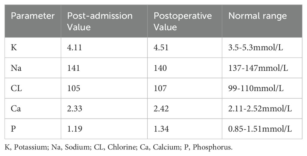
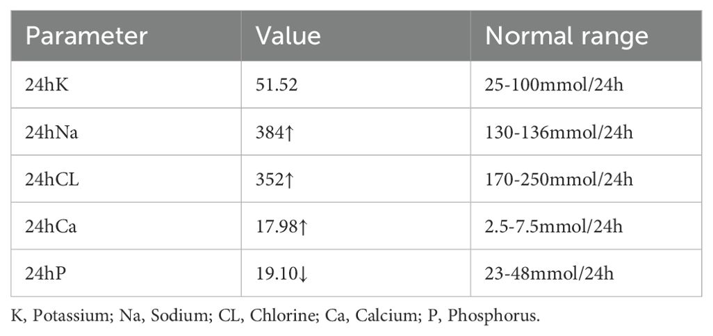
The above signs and symptoms are of limited value in the diagnosis of the disease. We then performed further laboratory and imaging studies on the patient. It is worth noting that the patient’s blood routine test results are normal, and so is the serum phosphorus level (Table 1). Additionally, the anteroposterior X-ray of the patient’s right knee joint showed a bone defect in the proximal end of the right fibula, with heterogeneous bone density. (Figure 1A). Integrating the aforementioned findings, we hypothesized that the patient might have a bone tumour. Therefore, the patient underwent a series of in-depth examinations, including tumour marker laboratory tests, whole-body bone imaging, computed tomography (CT), and magnetic resonance imaging (MRI). As shown in Supplementary Table S1, the tumor marker tests, including AFP, CEA, CA242, CA125, CA153, CA199, TPSA, FPSA, β-HCG, HGH, FE, and NSE, were all within normal range. 99mTc-MDP whole-body bone imaging revealed a focal area of increased radiotracer uptake at the proximal end of the right fibula, while the remainder of the skeleton exhibited no significant abnormalities in radiological distribution (Figure 2). The CT results of the right knee joint showed a soft-tissue mass at the proximal part of the right fibula (Figure 1C). Additionally, the results of plain and enhanced MRI of the right knee joint indicated a soft-tissue mass at the proximal part of the right fibula, with bone resorption of the proximal fibula, suggesting a neoplastic lesion (Figure 1B). To identify the patient’s tumor type and formulate the subsequent treatment plan, we conducted a needle biopsy on the patient’s tumor tissue. The biopsy results indicate that it is suspected to be PMT. In light of the characteristics of the PMT’s mechanism of action, we measured the 24-hour urinary electrolyte levels of the patients prior to surgery. The results indicated that the patients’ 24-hour urinary phosphorus levels decreased, while the 24-hour urinary calcium, chlorine and sodium levels increased (Table 2).
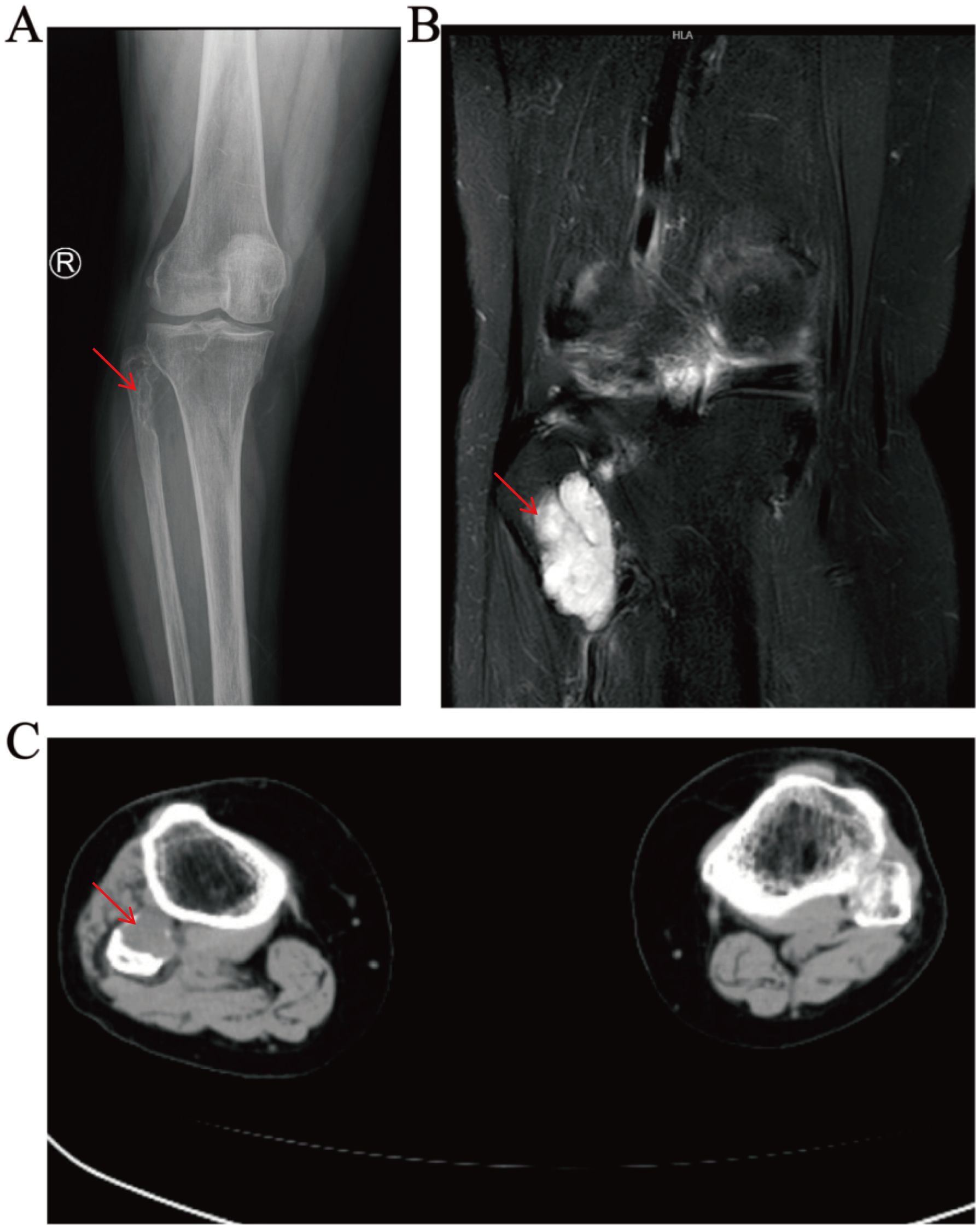
Figure 1. Preoperative imaging of the patient. (A) X-ray: bone defect in the proximal part of the fibula with uneven bone density, (B) enhanced MRI: tibiofibular interosseous soft-tissue mass, (C) CT: fibular head and tibiofibular interosseous soft-tissue mass.
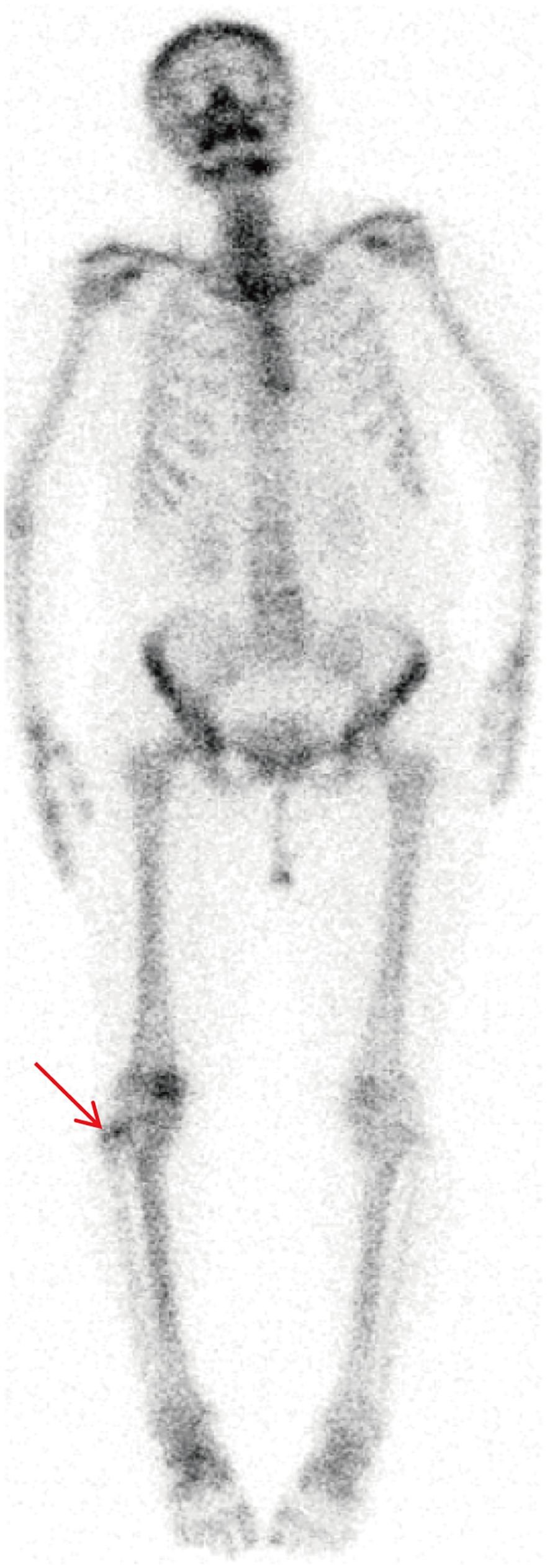
Figure 2. 99mTc-MDP scan showing a dense shadow of radioactivity distribution in the right proximal fibula.
2.2 Treatment
Previous research findings indicate that surgical resection of PMT represents the curative treatment approach (16). Therefore, we decided to surgically remove the diseased part of the proximal fibula and send the excised sample for pathological examination (Figure 3A). The tumor in this patient has distinct histological features: the tumor tissue is highly vascularized, and there are focal flocculent calcifications. (Figures 3B, C). The immunohistochemical results show: Vimentin (+), SMA (+), Bcl-2 (+), CD56 (+), CD34 (+), CD31 (+), Ki67 (1%+), ERG (+), CK (AE1/AE3) (-), INI-1 (+), SATB2 (+), SSTR2 (-) (Figures 3E, F). Based on the histological features and immunohistochemical results, the final diagnosis is PMT.
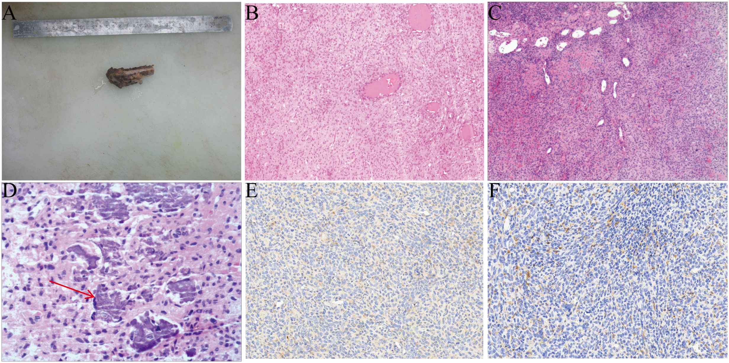
Figure 3. The pathology and immunohistochemistry of this case. (A) This is the tumor removed through surgery. (B-C) The tumor is rich in blood vessels. (D) Focal flocculent calcifications. (E, F) The expression of CD56 and SATB2 (×20).
2.3 Outcome and follow-up
After the surgery, the patient’s right knee joint pain was significantly alleviated. The results of blood routine tests, liver and kidney function tests, and serum electrolyte tests one week after the surgery were all normal (Table 1). At six months and one year after the surgery, we followed up with the patient separately. The patient’s pain and swelling in the right knee joint had disappeared, and the biochemical and imaging examination results at the local hospital were all normal, with no signs of recurrence.
3 Discussion
3.1 Clinical features
PMT is an exceptionally rare neoplasm, typically observed in middle-aged individuals and infrequently encountered in elderly patients (17–20). It has the potential to arise in any site within the skeletal system, albeit with a predilection for the appendicular skeleton, skull, and paranasal sinuses (21–24). PMTs are usually less than 5 cm in size and rarely cause localized pain related to the tumor site (4). The typical clinical manifestation of PMT is diffuse musculoskeletal pain, which is caused by secondary osteomalacia due to phosphate loss in patients (25, 26). This leads to an average time from symptom onset to final diagnosis that is typically as long as three years (27, 28). Notably, our patient exhibited distinct clinical characteristics. The patient in question was a 65-year-old female who initially complained of right knee pain lasting for 3 months, without any concurrent musculoskeletal pain upon presentation. The clinical presentation of this patient deviated from the conventional paradigm of PMT, highlighting the importance of considering individual variability in clinical diagnosis and expanding our diagnostic approach to facilitate earlier recognition of this uncommon neoplasm.
3.2 Pathogenesis
Most PMT-associated TIO is attributed to the overexpression of FGF-23 (29). FGF-23 diminishes the capacity of proximal renal tubules to reabsorb phosphate and augments phosphate excretion by the kidneys (30, 31). Furthermore, FGF-23 inhibits the activity of 1-α-hydroxylase, subsequently decreasing the synthesis of 1,25-dihydroxycholecalciferol (4). Other phosphatonins secreted by PMT, such as fibroblast growth factor 7 (FGF-7), secreted frizzled-related protein 4 (sFRP-4), and matrix extracellular phosphoglycoprotein (MEPE), can also lead to abnormal phosphate metabolism (32). Ultimately, these alterations result in hypophosphatemia and osteomalacia. Nonetheless, our patient exhibited neither hypophosphatemia nor any clinical manifestations of osteomalacia. Likewise, Folpe et al. identified cases lacking a known history of phosphaturia (17). They speculated that, in such instances, the tumor may secrete inactive or insufficient FGF-23, or possibly none at all. An alternative explanation posits that the patient possesses the capacity to compensate for increased FGF-23 secretion via alternative mechanisms. This may encompass compensatory regulation by other pathways or hormones involved in phosphate metabolism, thereby preventing the onset of hypophosphatemia and osteomalacia. Since we did not test for FGF-23, we are unable to speculate whether there is a compensatory mechanism in our patient. We speculate that the absence of hypophosphatemia and osteomalacia in our patient may be attributed to the early detection and resection of the tumor before the substantial production of humoral factors.
3.3 Imaging features
On CT examination, bone lesions in PMT patients typically present as osteolytic, and often contain internal matrix (33). On MRI examination, PMT usually shows isointensity on T1, hyperintensity on T2, with marked enhancement, and often accompanied by areas of low T2 signal (34). In addition, since PMT is a metabolically active tumor, radionuclide scans based on somatostatin receptor expression are of particular importance. In clinical practice, 99mTc-sestamibi scintigraphy, 111In-pentetreotide scintigraphy, 68Ga-DOTATATE PET/CT, and 18F-FDG PET/CT are commonly used to identify PMT (35). Existing research has shown that 68Ga-DOTATATE imaging is particularly sensitive in detecting occult tumors (36). CT examination of the patient revealed a cystic hypodense lesion with localized osteolytic alterations in the proximal region of the right fibula. MRI examination further elucidated that the proximal aspect of the right fibula exhibited isosignal intensity on T1-weighted images and high signal intensity on T2-weighted images. During contrast-enhanced scanning, the lesion area demonstrated significant heterogeneous enhancement. Furthermore, whole-body bone scintigraphy indicated the presence of a focal area of increased radiolabeled tracer uptake in the proximal portion of the right fibula. The imaging features of the patient are highly consistent with the typical imaging characteristics of PMT reported previously.
3.4 Pathologic and immunohistochemical features
PMT is a rare bone tumor with unique pathological features. There are four subtypes, namely osteoblastoma-like type, non-ossifying fibroma-like type, ossifying fibroma-like type, and mixed connective tissue type (6). Among these subtypes, the mixed connective tissue type is the relatively most common variant (17). The histological features of PMT include a small to moderate amount of bland spindle cells, small cell nuclei, inconspicuous nucleoli, a small number of mitotic figures, dirty calcified stroma, hemangiopericytoma-like vessels, osteoclast-like giant cells, fat, microcysts, hemorrhage, and other pathological changes (37, 38). Although immunohistochemical examination is not absolutely necessary for the diagnosis of PMT, it still has certain value in auxiliary diagnosis. Relevant studies have shown that in PMT cases without TIO, FGF-23 immunostaining is usually negative (39). In addition, PMT tumor cells can express a variety of markers, including vimentin, D2-40, SSTR2, ERG, FLI1, CD56, SMA, and SATB2 (10, 40–43). The research by Houang et al. pointed out that FGF-23 and SSTR2 are relatively sensitive for the diagnosis of PMT, but their specificity is relatively low (41). The FN1-FGFR1 fusion gene can be detected in most PMT cases, while the FN1-FGF1 fusion gene can be detected in a few cases (44, 45). Previous studies have shown that immunohistochemical detection of FGFR1 is helpful for the diagnosis of PMT, and meanwhile, RT-PCR detection of FGF-23 is a relatively sensitive method for diagnosing PMT (46). The tumor tissue in our patient exhibited typical histological features of PMT, including abundant calcified stroma and rich vasculature. Immunohistochemical results showed that the tumor cells expressed Vimentin, SMA, Bcl-2, CD56, CD34, CD31, Ki-67, ERG, RB, INI-1, and SATB2, but were negative for SSTR2 and CK. The detection of FGF-23 expression in tumor tissues is of significant value for differentiating PMT from other mesenchymal tumors with overlapping histological features (9). However, due to the retrospective nature of the case report, the expression of FGF-23 was not detected, which is a limitation of this study. These findings are highly consistent with the histological features and immunophenotype of PMT reported in previous studies. Ultimately, these characteristics enabled us to accurately diagnose the patient with PMT. Although the biopsy initially suggested that the lesion in this case was PMT, it is generally not recommended to perform biopsies on patients suspected of having PMT. This is because the biopsy procedure carries certain risks, such as the potential for tumor cell implantation, which may lead to local recurrence (47). This, in turn, can adversely affect disease control and subsequent treatment for the patient.
3.5 Treatments
In most cases, PMT is benign and can be completely cured by surgical excision. Negative surgical margins are a key factor in reducing the risk of recurrence (48). Radiotherapy is applicable for PMT that cannot be surgically resected (4). For cases where the tumor cannot be clinically localized, a pharmacological treatment approach, including the administration of phosphorus and calcitriol, is recommended (7, 37). Our patient resumed a normal diet after surgery and did not receive pharmacological treatment. The patient’s knee pain significantly improved after the surgery. We followed up with the patient at 6 months and 1 year postoperatively. Biochemical and imaging tests performed at the local hospital showed no signs of recurrence.
4 Conclusion
In this case, we present a PMT patient without hypophosphatemia and osteomalacia. The patient only presented with pain in the right knee joint. After a pathological examination of the tumor tissue, we found typical histological features of PMT. Therefore, when dealing with non-phosphaturic variant of PMT patients, histopathological examination of the tumor tissue is mandatory for accurate diagnosis and also for formulating subsequent treatment plans.
Data availability statement
The original contributions presented in the study are included in the article/supplementary material. Further inquiries can be directed to the corresponding author.
Ethics statement
The studies involving humans were approved by Ethics Committee of Henan Provincial People’s Hospital. The studies were conducted in accordance with the local legislation and institutional requirements. The participants provided their written informed consent to participate in this study. Written informed consent was obtained from the participant/patient(s) for the publication of this case report.
Author contributions
QC: Writing – original draft, Writing – review & editing, Conceptualization, Data curation, Formal Analysis, Investigation, Methodology, Project administration, Resources, Software, Supervision, Validation, Visualization. CC: Conceptualization, Data curation, Formal Analysis, Investigation, Methodology, Project administration, Resources, Software, Supervision, Validation, Visualization, Writing – review & editing. ZD: Conceptualization, Data curation, Formal Analysis, Funding acquisition, Investigation, Methodology, Project administration, Resources, Software, Supervision, Validation, Visualization, Writing – original draft, Writing – review & editing.
Funding
The author(s) declare that financial support was received for the research and/or publication of this article. The present study was supported by the National Natural Science Foundation of China (82002300); Henan Province Health Science and Technology innovation outstanding young talents training program (YXKC2021046).
Acknowledgments
We thank the patient and her guardians for giving their consent for the publication of this case report.
Conflict of interest
The authors declare that the research was conducted in the absence of any commercial or financial relationships that could be construed as a potential conflict of interest.
Generative AI statement
The author(s) declare that no Generative AI was used in the creation of this manuscript.
Publisher’s note
All claims expressed in this article are solely those of the authors and do not necessarily represent those of their affiliated organizations, or those of the publisher, the editors and the reviewers. Any product that may be evaluated in this article, or claim that may be made by its manufacturer, is not guaranteed or endorsed by the publisher.
Supplementary material
The Supplementary Material for this article can be found online at: https://www.frontiersin.org/articles/10.3389/fonc.2025.1597194/full#supplementary-material
References
1. Adnan Z, Nikomarov D, Weiler-Sagie M, Roguin Maor N. Phosphaturic mesenchymal tumors among elderly patients: A case report and review of literature. Endocrinol Diabetes Metab Case Rep. (2019) 2019:18-0139. doi: 10.1530/edm-18-01396
2. Jiang Y, Xia WB, Xing XP, Silva BC, Li M, Wang O, et al. Tumor-induced osteomalacia: An important cause of adult-onset hypophosphatemic osteomalacia in China: Report of 39 cases and review of the literature. J Bone Miner Res. (2012) 27:9::1967–75. doi: 10.1002/jbmr.1642
3. Berndt T, Craig TA, Bowe AE, Vassiliadis J, Reczek D, Finnegan R, et al. Secreted frizzled-related protein 4 is a potent tumor-derived phosphaturic agent. J Clin Invest (2003) 112:5:785–94J Clin Invest. doi: 10.1172/jci18563
4. Hudairy RE, Meliti A, Mokhtar G, Alanazi M. Phosphaturic mesenchymal tumor: A case report of a rare entity. Cureus. (2022) 14:2:e22712. doi: 10.7759/cureus.22712
5. Rad SG, Bahrami A, Houshyar J, Farrin N, Najafipour F, Amirazad H. A case report of tumor-induced osteomalacia (tio) caused by central giant cell granuloma. Bone Rep. (2024) 22:101804. doi: 10.1016/j.bonr.2024.101804
6. Weidner N, Santa Cruz D. Phosphaturic mesenchymal tumors. A polymorphous group causing osteomalacia or rickets. Cancer. (1987) 59:8:1442–54. doi: 10.1002/1097-0142(19870415)59:8<1442::aid-cncr2820590810>3.0.co;2-q
7. Beygi S, Denio A, Sharma TS. The foot that broke both hips: A case report and literature review of tumor-induced osteomalacia. Case Rep Rheumatol. (2017) 2017:3191673. doi: 10.1155/2017/3191673
8. Honda R, Kawabata Y, Ito S, Kikuchi F. Phosphaturic mesenchymal tumor, mixed connective tissue type, non-phosphaturic variant: Report of a case and review of 32 cases from the Japanese published work. J Dermatol. (2014) 41:9:845–9. doi: 10.1111/1346-8138.12602
9. Palmisano B, Pace A, Colangelo L, Polimeni R, Sonato C, Messineo D, et al. Non-phosphaturic” variant of phosphaturic mesenchymal tumor of the middle ear expressing multiple phosphatonins. Ear Nose Throat J. (2023) 1455613221149635. doi: 10.1177/01455613221149635
10. Shiba E, Matsuyama A, Shibuya R, Yabuki K, Harada H, Nakamoto M, et al. Immunohistochemical and molecular detection of the expression of fgf23 in phosphaturic mesenchymal tumors including the non-phosphaturic variant. Diagn Pathol. (2016) 11:26. doi: 10.1186/s13000-016-0477-3
11. Rendina D, Abate V, Cacace G, D’Elia L, De Filippo G, Del Vecchio S, et al. Tumor-induced osteomalacia: A systematic review and individual patient’s data analysis. J Clin Endocrinol Metab. (2022) 107:8:e3428–e36. doi: 10.1210/clinem/dgac253
12. Abate V, Vergatti A, De Filippo G, Damiano V, Menale C, D’Elia L, et al. Clinical characteristics of Malignant phosphaturic mesenchymal tumor causing tumor-induced osteomalacia. J Clin Endocrinol Metab. (2024) 109:3:e1006–e11. doi: 10.1210/clinem/dgad690
13. Wang Y, Liu S, Li C, Song W, Zhang Y, Wang J. Phosphaturic mesenchymal tumor of the popliteal fossa: A case report and literature review. Front Oncol. (2024) 14:1501499. doi: 10.3389/fonc.2024.1501499
14. Erben RG. Physiological actions of fibroblast growth factor-23. Front Endocrinol (Lausanne). (2018) 9:267. doi: 10.3389/fendo.2018.00267
15. Richardson AL, Richardson OK. Phosphaturic mesenchymal tumor: Case report. Radiol Case Rep. (2019) 14:12:1518–24. doi: 10.1016/j.radcr.2019.09.027
16. Benson JC, Trejo-Lopez JA, Nassiri AM, Eschbacher K, Link MJ, Driscoll CL, et al. Phosphaturic mesenchymal tumor. AJNR Am J Neuroradiol. (2022) 43:6:817–22. doi: 10.3174/ajnr.A7513
17. Folpe AL, Fanburg-Smith JC, Billings SD, Bisceglia M, Bertoni F, Cho JY, et al. Most osteomalacia-associated mesenchymal tumors are a single histopathologic entity: An analysis of 32 cases and a comprehensive review of the literature. Am J Surg Pathol. (2004) 28:1:1–30. doi: 10.1097/00000478-200401000-00001
18. Bahrami A, Weiss SW, Montgomery E, Horvai AE, Jin L, Inwards CY, et al. Rt-pcr analysis for fgf23 using paraffin sections in the diagnosis of phosphaturic mesenchymal tumors with and without known tumor induced osteomalacia. Am J Surg Pathol. (2009) 33:9:1348–54 https://doi. doi: 10.1097/PAS.0b013e3181aa2311
19. Agaimy A, Michal M, Chiosea S, Petersson F, Hadravsky L, Kristiansen G, et al. Phosphaturic mesenchymal tumors: Clinicopathologic, immunohistochemical and molecular analysis of 22 cases expanding their morphologic and immunophenotypic spectrum. . Am J Surg Pathol. (2017) 41:10:1371–80. doi: 10.1097/pas.0000000000000890
20. Jung GH, Kim JD, Cho Y, Chung SH, Lee JH, Sohn KR. A 9-month-old phosphaturic mesenchymal tumor mimicking the intractab le rickets. J Pediatr Orthop B. (2010) 19:1:127–32. doi: 10.1097/BPB.0b013e32832f59cb
21. Carter JM, Caron BL, Dogan A, Folpe AL. A novel chromogenic in situ hybridization assay for fgf23 mrna in phosphaturic mesenchymal tumors. Am J Surg Pathol. (2015) 39:1:75–83. doi: 10.1097/pas.0000000000000290
22. Kobayashi H, Akiyama T, Okuma T, Shinoda Y, Oka H, Ito N, et al. Three-dimensional fluoroscopic navigation-assisted surgery for tumors in patients with tumor-induced osteomalacia in the bones. Comput Assist Surg (Abingdon). (2017) 22:1:14–9. doi: 10.1080/24699322.2017.1282044
23. Kobayashi H, Ito N, Akiyama T, Okuma T, Kinoshita Y, Ikegami M, et al. Prevalence and clinical outcomes of hip fractures and subchondral insufficiency fractures of the femoral head in patients with tumour-induced osteomalacia. Int Orthop. (2017) 41:12:2597–603. doi: 10.1007/s00264-017-3610-3
24. Zuo QY, Wang H, Li W, Niu XH, Huang YH, Chen J, et al. Treatment and outcomes of tumor-induced osteomalacia associated with phosphaturic mesenchymal tumors: Retrospective review of 12 patients. BMC Musculoskelet Disord. (2017) 18:1:403. doi: 10.1186/s12891-017-1756-1
25. Hodgson SF, Clarke BL, Tebben PJ, Mullan BP, Cooney WP, Shives TC. Oncogenic osteomalacia: Localization of underlying peripheral mesenchymal tumors with use of tc 99m sestamibi scintigraphy. Endocr Pract. (2006) ;12:1:35–42. doi: 10.4158/ep.12.1.35
26. Montanari A, Pirini MG, Lotrecchiano L, Di Prinzio L, Zavatta G. Phosphaturic mesenchymal tumors with or without phosphate metabolism derangements. Curr Oncol. (2023) 30:8:7478–88. doi: 10.3390/curroncol30080541
27. Feng J, Jiang Y, Wang O, Li M, Xing X, Huo L, et al. The diagnostic dilemma of tumor induced osteomalacia: A retrospective analysis of 144 cases. Endocr J. (2017) 64:7:675–83. doi: 10.1507/endocrj.EJ16-0587
28. Wang H, Zhong D, Liu Y, Jiang Y, Qiu G, Weng X, et al. Surgical treatments of tumor-induced osteomalacia lesions in long bones: Seventeen cases with more than one year of follow-up. J Bone Joint Surg Am. (2015) 97:13:1084–94. doi: 10.2106/jbjs.N.01299
29. Riminucci M, Collins MT, Fedarko NS, Cherman N, Corsi A, White KE, et al. Fgf-23 in fibrous dysplasia of bone and its relationship to renal phosphate wasting. J Clin Invest. (2003) 112:5:683–92. doi: 10.1172/jci18399
30. Murer H, Hernando N, Forster I, Biber J. Proximal tubular phosphate reabsorption: Molecular mechanisms. Physiol Rev. (2000) 80:4:1373–409. doi: 10.1152/physrev.2000.80.4.1373
31. Shimada T, Mizutani S, Muto T, Yoneya T, Hino R, Takeda S, et al. Cloning and characterization of fgf23 as a causative factor of tumor-induced osteomalacia. Proc Natl Acad Sci U S A. (2001) 98:11:500–5. doi: 10.1073/pnas.101545198
32. Ghorbani-Aghbolaghi A, Darrow MA, Wang T. Phosphaturic mesenchymal tumor (pmt): Exceptionally rare disease, yet crucial not to miss. Autops Case Rep. (2017) 7:3:32–7. doi: 10.4322/acr.2017.031
33. Minisola S, Fukumoto S, Xia W, Corsi A, Colangelo L, Scillitani A, et al. Tumor-induced osteomalacia: A comprehensive review. Endocr Rev. (2023) ;44:2:323–53. doi: 10.1210/endrev/bnac026
34. Tungenwar S, Subith S, Mukadam M, Kokate S, Gunay S. Phosphaturic mesenchymal tumor in the proximal femur presenting as tumor-induced osteomalacia: A case report and literature review. J Orthop Case Rep. (2024) 14:2:23–8. doi: 10.13107/jocr.2024.v14.i02.4204
35. Broski SM, Folpe AL, Wenger DE. Imaging features of phosphaturic mesenchymal tumors. Skeletal Radiol. (2019) 48:1::119–27. doi: 10.1007/s00256-018-3014-5
36. El-Maouche D, Sadowski SM, Papadakis GZ, Guthrie L, Cottle-Delisle C, Merkel R, et al. (68)ga-dotatate for tumor localization in tumor-induced osteomalacia. J Clin Endocrinol Metab. (2016) 101:10:3575–81. doi: 10.1210/jc.2016-2052
37. Agarwal N, Kale SS, Kumari K. Tumor-induced osteomalacia due to a phosphaturic mesenchymal tumor in the cervical spine: A case report and literature review. Neurol India. (2019) 67:5:1334–40. doi: 10.4103/0028-3886.271274
38. Gardner KH, Shon W, Folpe AL, Wieland CN, Tebben PJ, Baum CL. Tumor-induced osteomalacia resulting from primary cutaneous phosphaturic mesenchymal tumor: A case and review of the medical literature. J Cutan Pathol. (2013) 40:9:780–4. doi: 10.1111/cup.12209
39. Wu H, Bui MM, Zhou L, Li D, Zhang H, Zhong D. Phosphaturic mesenchymal tumor with an admixture of epithelial and mesenchymal elements in the jaws: Clinicopathological and immunohistochemical analysis of 22 cases with literature review. Mod Pathol. (2019) 32:2:189–204. doi: 10.1038/s41379-018-0100-0
40. Tajima S, Fukayama M. Possibility of d2–40 as a diagnostic and tumor differentiation-suggestive marker for some of phosphaturic mesenchymal tumors. Int J Clin Exp Pathol. (2015) 8:8:9390–6.
41. Houang M, Clarkson A, Sioson L, Elston MS, Clifton-Bligh RJ, Dray M, et al. Phosphaturic mesenchymal tumors show positive staining for somatostatin receptor 2a (sstr2a). Hum Pathol. (2013) 44:12:2711–8. doi: 10.1016/j.humpath.2013.07.016
42. Tajima S, Takashi Y, Ito N, Fukumoto S, Fukuyama M. Erg and fli1 are useful immunohistochemical markers in phosphaturic mesenchymal tumors. Med Mol Morphol. (2016) 49:4:203–9. doi: 10.1007/s00795-015-0115-2
43. Li D, Zhu R, Zhou L, Zhong D. Clinical, histopathologic, subtype, and immunohistochemical analysis of jaw phosphaturic mesenchymal tumors. Med (Baltimore). (2020) 99:7:e19090. doi: 10.1097/md.0000000000019090
44. Lee JC, Su SY, Changou CA, Yang RS, Tsai KS, Collins MT, et al. Characterization of fn1-fgfr1 and novel fn1-fgf1 fusion genes in a large series of phosphaturic mesenchymal tumors. Mod Pathol. (2016) 29:11:1335–46. doi: 10.1038/modpathol.2016.137
45. Lee JC, Jeng YM, Su SY, Wu CT, Tsai KS, Lee CH, et al. Identification of a novel fn1-fgfr1 genetic fusion as a frequent event in phosphaturic mesenchymal tumour. J Pathol. (2015) 235:4:539–45. doi: 10.1002/path.4465
46. Tajima S, Fukayama M. Fibroblast growth factor receptor 1 (fgfr1) expression in phosphaturic mesenchymal tumors. Int J Clin Exp Pathol. (2015) 8:8:9422–7.
47. Crotti C, Bartoli F, Coletto LA, Manara M, Marini E, Daolio PA, et al. Tumor induced osteomalacia: A single center experience on 17 patients. Bone. (2021) 152:116077. doi: 10.1016/j.bone.2021.116077
Keywords: phosphaturic mesenchymal tumors, tumor-induced osteomalacia, fibroblast growth factor 23, pain, hypophosphatemia, osteomalacia
Citation: Cao Q, Cheng C and Dai Z (2025) Case Report: Phosphaturic mesenchymal tumor presenting solely as knee pain without hypophosphatemia. Front. Oncol. 15:1597194. doi: 10.3389/fonc.2025.1597194
Received: 25 March 2025; Accepted: 16 April 2025;
Published: 08 May 2025.
Edited by:
Luciano Colangelo, Sapienza University of Rome, ItalyReviewed by:
Alessandro Corsi, Sapienza University of Rome, ItalyGaetano Paride Arcidiacono, University of Padua, Italy
Copyright © 2025 Cao, Cheng and Dai. This is an open-access article distributed under the terms of the Creative Commons Attribution License (CC BY). The use, distribution or reproduction in other forums is permitted, provided the original author(s) and the copyright owner(s) are credited and that the original publication in this journal is cited, in accordance with accepted academic practice. No use, distribution or reproduction is permitted which does not comply with these terms.
*Correspondence: Zhipeng Dai, ZGFpenBAenp1LmVkdS5jbg==
 Qianqian Cao
 Cheng Cheng
 Zhipeng Dai