- 1Department of Family Medicine and Public Health, University of California, San Diego, San Diego, CA, United States
- 2Department of Population Health, University of Toledo, Toledo, OH, United States
- 3Evidence-Based Practice Research Program, Mayo Clinic, Rochester, MN, United States
Objectives: Among women of childbearing potential aged 15–39, cancer incidence is 52.3 per 100,000 annually. Women newly diagnosed with cancer often have just 2–6 weeks to decide whether to pursue fertility preservation (FP) before commencing treatment. The recommended waiting period to conceive post-treatment ranges from 6 months to 5 years, depending on cancer type, treatment, and age. With >18 million young adult cancer survivors worldwide, identifying factors affecting fertility preservation and live birth outcomes is more critical than ever. This is the first systematic review to explore whether AMH levels before, during, or after chemotherapy predict pregnancy outcomes resulting from re-utilization of stored oocytes/embryos or spontaneous conception in cancer patients undergoing FP. It also evaluates the optimal timing for post-treatment AMH recovery and how this may inform fertility success and decision-making for cancer patients pursuing FP.
Methods: A review of PubMed and Web of Science identified 458 studies until November 2024. After a full-text review of 38 studies, seven met the eligibility criteria: if they were peer-reviewed, in English, enrolled female cancer patients undergoing FP before chemotherapy, measured AMH, and reported pregnancy or live birth rates after chemotherapy. Study quality and relevance were categorized as high, moderate, or low. Of the seven studies, one was highly relevant, four were moderately relevant, and two were of lower relevance.
Results: The majority of studies focused on patients with breast cancer or lymphoma, comprising three prospective and four retrospective designs. Oocyte cryopreservation emerged as the most commonly used fertility preservation method. Among those who used stored specimens, baseline AMH levels ranging from ~2.1 to 2.8 ng/mL were related to live birth rates of 35–42%. Notably, spontaneous conception was more frequent than assisted reproduction. AMH recovery timelines varied widely, with follow-up periods spanning 1 to 36 months, yet no clear optimal timeframe for ovarian reserve restoration emerged.
Conclusion: In female cancer patients, pre-treatment Anti-Müllerian Hormone (AMH) levels may offer valuable insight to help inform fertility preservation decisions aimed at achieving future live births. This first-of-its-kind systematic review lays the groundwork for future research by identifying key knowledge gaps and emerging areas of clinical relevance.
1 Introduction
Preserving fertility and the constraints of a limited reproductive window are key concerns for many cancer survivors (1–3). For young survivors, achieving remission and remaining cancer-free are paramount, but for many, the ability to start a family represents a crucial milestone in reclaiming a sense of normalcy (4–6). However, delaying cancer treatment to pursue fertility preservation can have serious consequences for prognosis. A recent study found that postponing treatment by just one month, regardless of the treatment modalities (i.e., surgery, chemotherapy, and radiation), was associated with a 6-13% increase in mortality risk, with the danger compounding as delays lengthen (7).
Anti-Müllerian Hormone (AMH) is a widely recognized marker of ovarian reserve and has been proposed as a valuable tool in fertility counseling for cancer patients considering fertility preservation before undergoing gonadotoxic chemotherapy. However, the impact of AMH fluctuations on key clinical outcomes, such as pregnancy rates and live birth success resulting from re-utilized oocytes/embryos or spontaneous conception, remain largely unknown (1, 8–11). Addressing these gaps is essential for improving fertility counseling and optimizing reproductive decision-making for cancer survivors.
1.1 AMH and fertility preservation
For cancer survivors considering fertility preservation, pre-treatment AMH may provide an estimate of ovarian reserve, aiding in counselling and optimizing fertility preservation strategies.
For those undergoing fertility preservation, post-chemotherapy AMH recovery levels may provide valuable insights into the recovery of follicle growth in the functional ovarian reserve (12, 13). Sustained low AMH concentrations post-chemotherapy, especially with alkylating agents, may significantly decrease the conception window, increasing the risk of premature ovarian failure and decreasing the likelihood of pregnancy in a cancer patient population (13).
The decision to delay cancer treatment in order to pursue fertility preservation through oocyte or embryo cryopreservation emphasizes the need to better understand the potential utility of AMH as a biomarker for reproductive outcomes. While the current evidence base is limited, AMH may offer insight into chemotherapy-induced ovarian dysfunction (14–17) and could potentially inform the likelihood of future clinical pregnancy and live birth using stored oocytes/embryos from fertility preservation. Further research is needed to clarify AMH’s prognostic value and to determine its role in guiding fertility preservation counseling and decision-making in the oncofertility setting.
At present, no specific guidelines exist to help couples assess whether their chances of achieving a healthy live birth are better with fertility preservation techniques compared to relying solely on natural conception after cancer treatment.
1.2 Study objectives
AMH’s ability to predict spontaneous pregnancy has been assessed across various populations, including healthy but predominantly obese women aged 30-44, cancer patients treated with GnRH, and young breast cancer patients not using fertility preservation (18–21). Thus far, the literature has only extrapolated the role of AMH to cancer patients undergoing fertility preservation without accompanying evidence-based knowledge.
Our study aims to address a very specialized gap by conducting a systematic review of the existing literature to evaluate whether AMH levels prior to, during, or post-chemotherapy, could serve as a reliable predictor of success rates (including re-utilization of oocytes/embryos or unassisted spontaneous conception) for pregnancy and healthy live births among cancer patients undergoing fertility preservation The absence of research evaluating predictive biomarkers of fertility preservation success, particularly the likelihood of achieving a healthy pregnancy or live birth after chemotherapy, underscores the need for further investigation in this field.
The second aim of this systematic review is to identify an optimal time interval (i.e., a suitable period to get pregnant) after chemotherapy when AMH levels tend to recover most robustly among those women undergoing fertility preservation.
2 Methods
2.1 Search strategy
Two authors (NR and HKC) independently conducted a systematic search following PRISMA guidelines. The search included PubMed and Web of Science to identify relevant studies published up to November 2024. After removing duplicates, studies were initially screened based on their titles and abstracts, with full texts retrieved for the remaining studies. Two authors (HKC and MP) then evaluated the full texts against the predefined inclusion and exclusion criteria to finalize the studies for the systematic review.
2.2 Inclusion exclusion criteria
Studies were included if they: (i) were peer-reviewed, original research articles published in English, (ii) enrolled female cancer patients who underwent fertility preservation (e.g., oocyte or embryo cryopreservation) prior to cancer treatment, (iii) measured anti-Müllerian hormone (AMH) as one of the biomarkers, and (iv) reported pregnancy and/or live birth rates after chemotherapy as primary or secondary outcomes.
Studies were excluded if: (i) they did not involve fertility preservation prior to cancer treatment or did not evaluate subsequent IVF outcomes, (ii) they did not measure AMH, (iii) if the study sample consisted of childhood cancer patients who exclusively used tissue cryopreservation (iv) they were not in English or included only animal or laboratory-based research, male, transgender, or pediatric populations, or (v) they were reviews, systematic reviews, meta-analyses, case reports, conference abstracts, committee announcements, or protocol papers without original data.
2.3 Data extraction
The authors collectively extracted and compiled data from the selected studies, ensuring agreement, consistency, and accuracy. The extracted data included study details such as the first author, year of publication, and country, as well as study characteristics, including the hypothesis, study design, cancer type, sample size, and participant age. Additionally, data were collected on fertility preservation, AMH measurement techniques, the interpretation of AMH levels, and reported pregnancy outcomes. Specific details on AMH measurement techniques included the timing of measurements, AMH levels, units of measurement, and the type of values were reported.
2.4 Study quality and relevance assessment
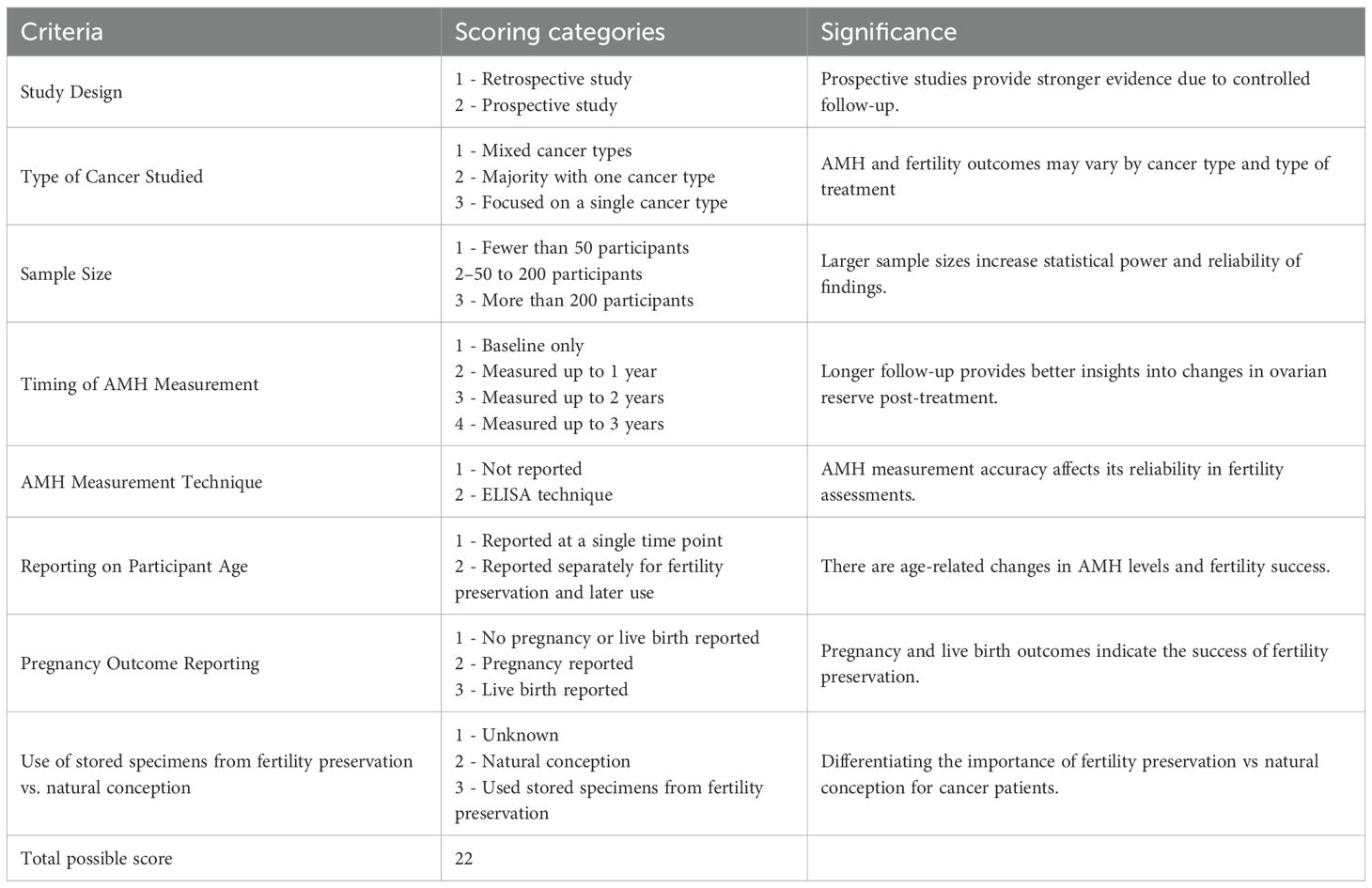
To evaluate the relevance and quality of included studies in addressing our research hypothesis, a scoring system was developed, reflecting the overall quality and rigor of the studies as well as their alignment with our specific research question (Table 1).
Two reviewers (HKC, MP) assigned a score for each study ranging from 7 to 20, based on factors including study design, type of cancer, sample size, technique and timing of AMH measurement, patient age at specimen use for fertility preservation, and presence of pregnancy outcomes (Table 1). Given the uniqueness of the hypothesis and paucity of availability studies, relevance was determined by the alignment with our study hypothesis. Studies scoring above 15 were classified as high-quality and highly relevant, those between 11 and 14 as moderate-quality and moderately relevant, and those between 7 and 10 as lower-quality and less relevant.
3 Results
3.1 Study selection
The process of study identification and selection, based on PRISMA guidelines, is illustrated in Figure 1. The initial search strategy identified 525 studies. After removing duplicates, 458 articles remained and were screened based on their titles and abstracts. From this, 38 articles were shortlisted for full-text review and assessed against the inclusion criteria.
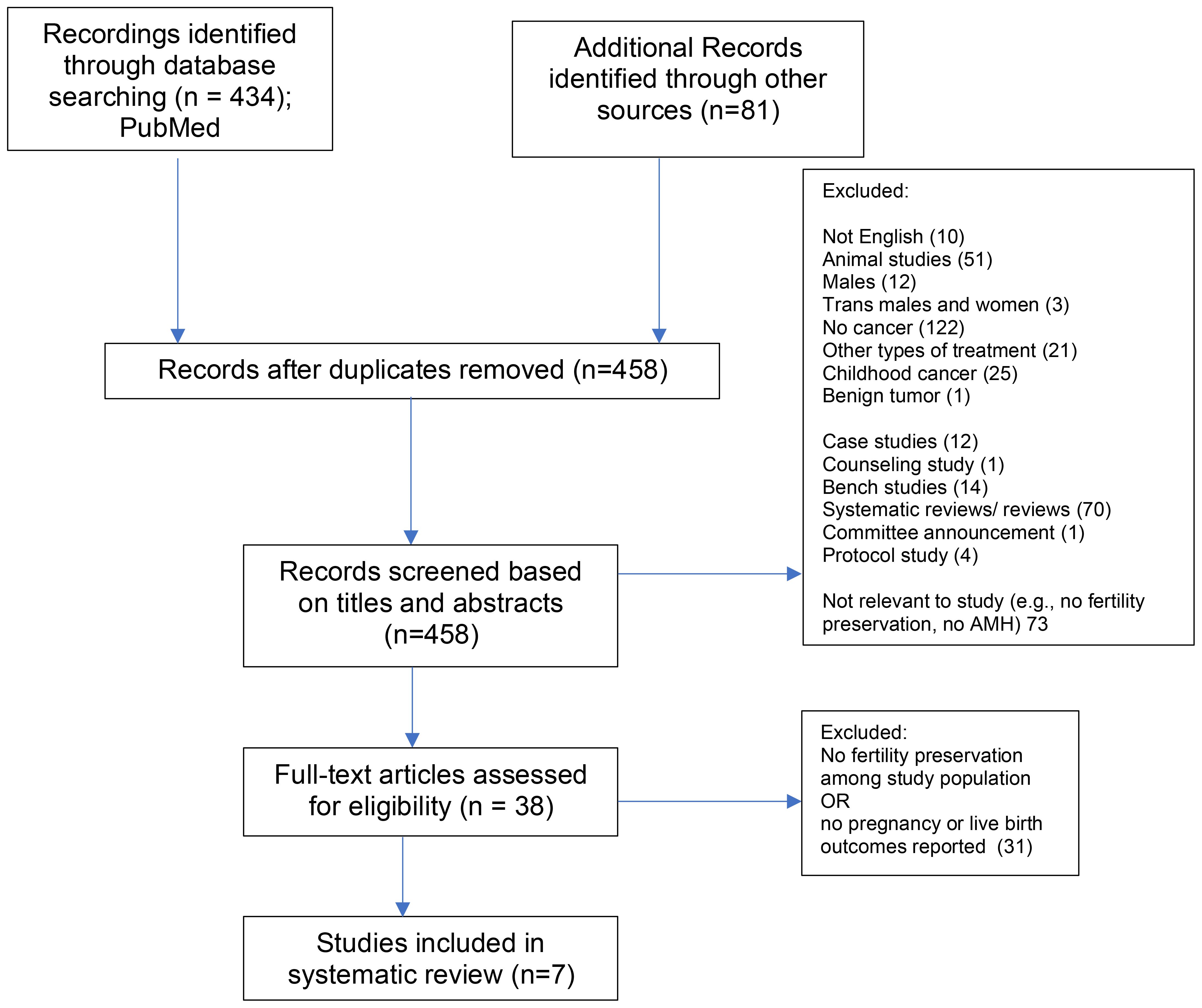
Figure 1. PRISMA Chart. From: Page MJ, McKenzie JE, Bossuyt PM, Boutron I, Hoffmann TC, Mulrow CD, et al. The PRISMA 2020 statement: an updated guideline for reporting systematic reviews. BMJ 2021;372:n71. doi: 10.1136/bmj.n71
The most frequent reasons for exclusion during the full-text review included a lack of cancer diagnosis, the absence of fertility preservation or Anti-Müllerian Hormone (AMH) assessments, and articles that were systematic or review papers. Other common grounds for exclusion involved non-English publications, animal studies, and populations outside the review scope (e.g., exclusively male, transgender-inclusive, or childhood cancer cohorts). Additionally, an attempt was made to identify relevant studies through reference lists, but no additional studies were found.
3.2 Study characteristics
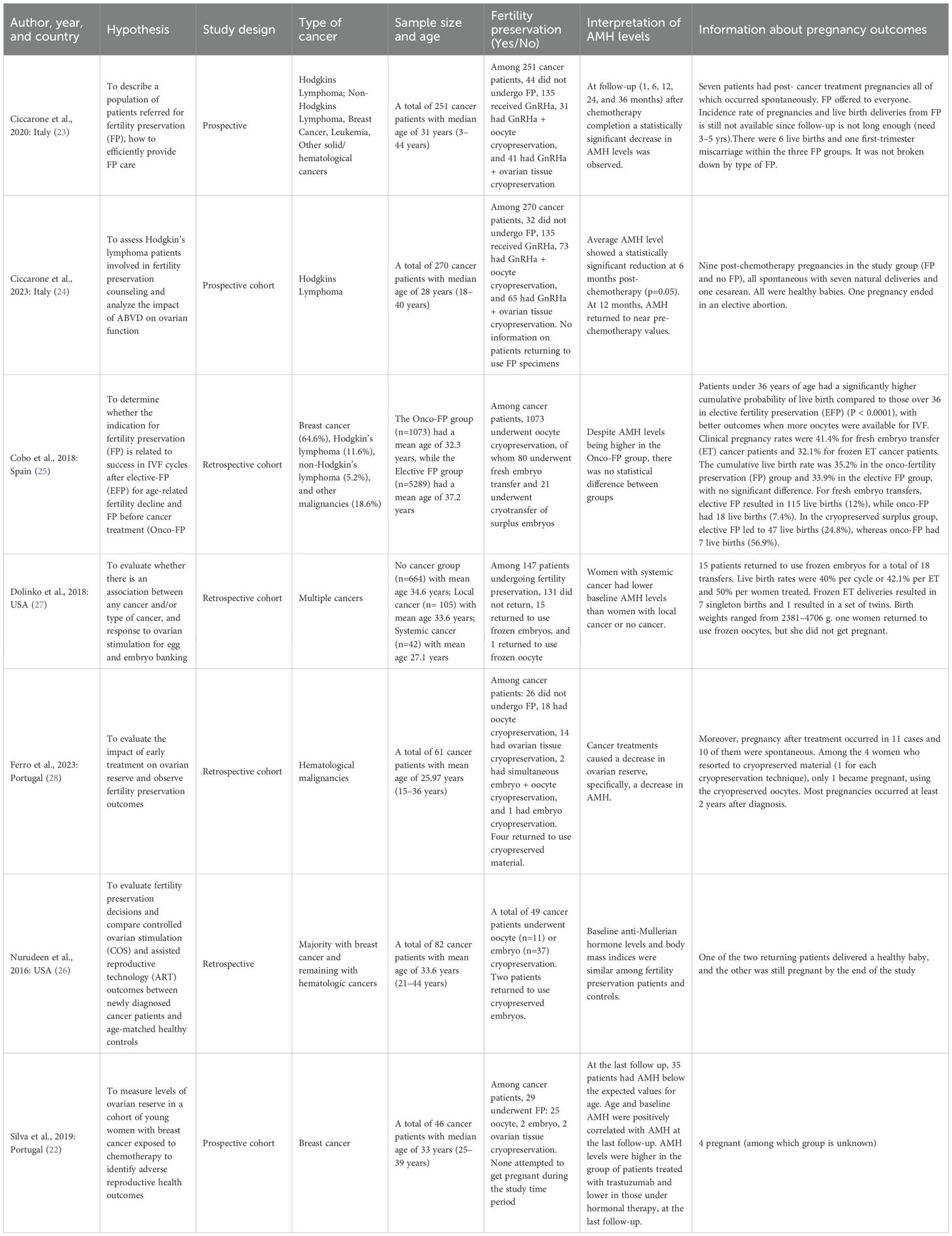
Ultimately, seven studies met the inclusion criteria and were included in the review (Table 2) (22–28). These studies, published between 2016 and 2023, examined fertility preservation among cancer patients and highlighted AMH as a key biomarker of ovarian reserve. Of these, three were prospective (22–24) and four were retrospective (25–28). These studies took place in Portugal (22), Spain (25), Italy (23, 24, 28), and the United States (26, 27). Sample sizes ranged widely from fewer than 50 participants in smaller cohorts (e.g., Silva et al. (22)) to larger-scale analyses exceeding 5000 (e.g., Cobo et al. (25)).
Across the included studies, the most frequent diagnoses were breast cancer (22, 25–27) and Hodgkin’s or non-Hodgkin’s lymphoma (23, 24, 28), with some also including patients with other hematologic malignancies. While several studies exclusively enrolled cancer patients, subdividing them according to the treatments received (22–24, 28), others incorporated comparison groups, such as age-matched healthy individuals (26), elective fertility preservation cohorts (25), or those with male-factor infertility (27).
AMH levels were measured at baseline before chemotherapy in all included studies, with several also reporting repeated assessments during or after treatment to evaluate changes in ovarian reserve (22–28).
3.3 Quality and relevance assessment of included studies
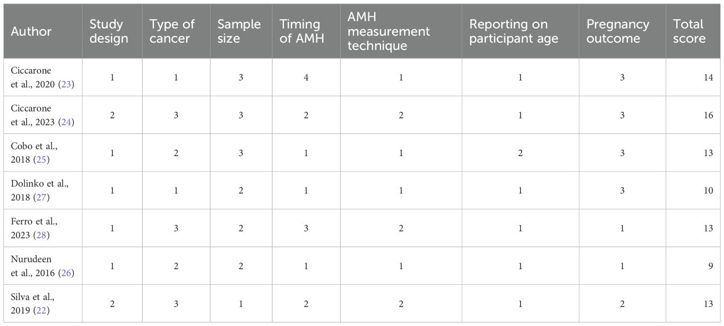
The quality and relevance assessment of the included studies are presented in Table 3. Among the seven studies evaluated, one study (24) scored 16, classifying it as highly relevant (score >15). Four studies (22, 23, 25, 28) scored 13 or 14, placing them in the moderate relevance category (scores between 11 and 14). Finally, Nurudeen et al., scored 9, and Dolinko et al., scored 10, classifying them as lower relevance (scores between 7 and 10) (26, 27).
3.4 Cancer patients undergoing fertility preservation
The studies by Dolinko et al. (27), Ciccarone et al. (2020) (23), Nurudeen et al. (26), and Cobo et al. (25) included only cancer patients who had undergone fertility preservation. In contrast, other studies included mixed populations: Ciccarone (2023) (24) reported that 88% of participants had used fertility preservation, Ferro reported 58% (28), and Silva reported 76% (22).
3.5 AMH levels
All extracted data on AMH levels, including timing of AMH measurements, AMH levels, and AMH measurement assay techniques, are summarized in Table 4.
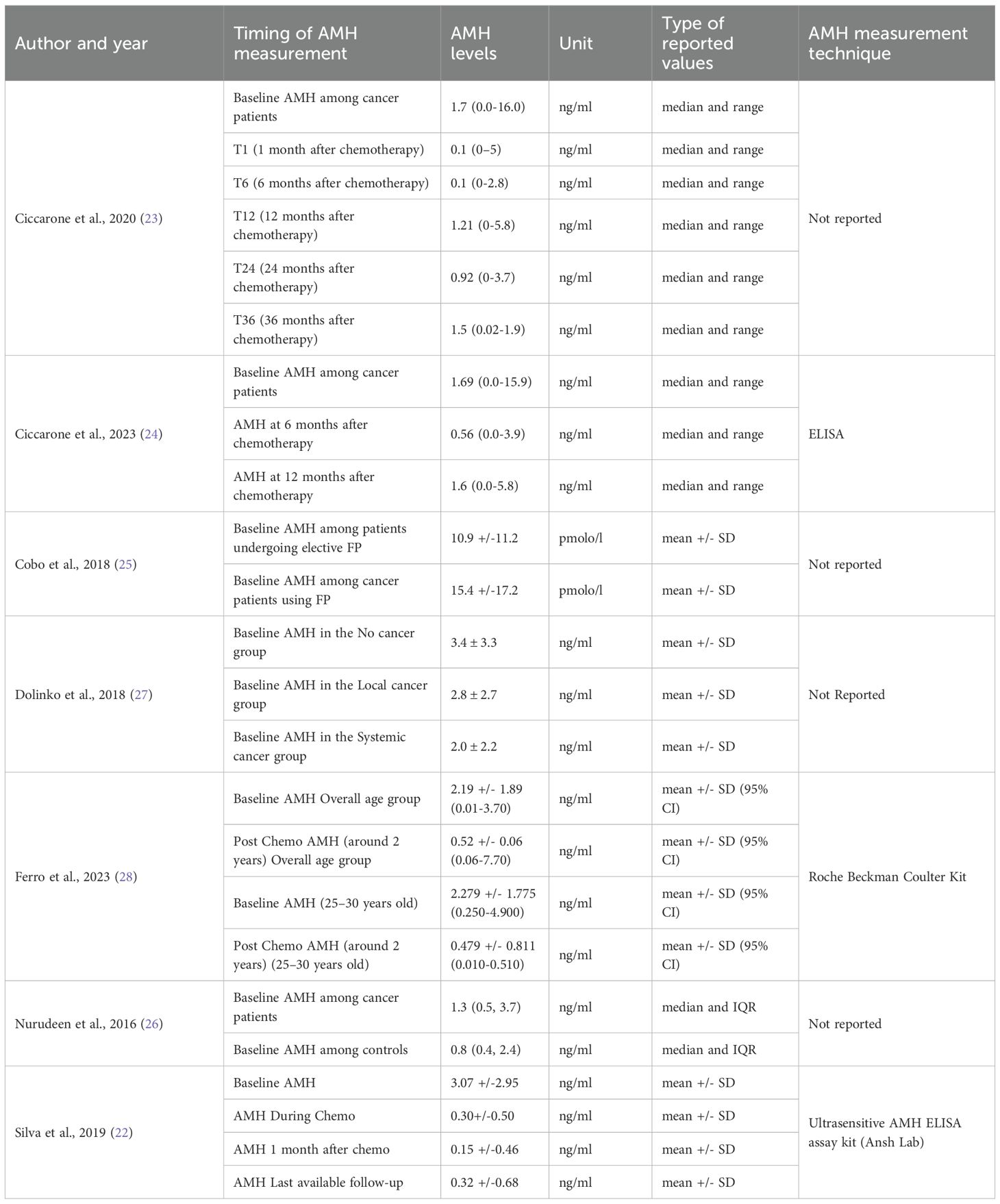
Table 4. Summary of AMH levels in cancer patients undergoing fertility preservation and control groups.
3.5.1 Baseline AMH
At baseline, AMH levels were highest in healthy controls (Dolinko et al., 2018: 3.4 ± 3.3 ng/ml) (27) and in breast cancer patients before chemotherapy (Silva et al., 2019: 3.07 ± 2.95 ng/ml) (22). Compared to breast cancer patients, those with hematologic malignancies had lower baseline AMH levels, as reported by Ferro et al., 2023: 2.19 ± 1.89 ng/ml (28); Ciccarone et al., 2020: 1.7 ng/ml (0.0-16.0 range) (23); and Ciccarone et al., 2023: 1.69 (0.0-16.0) (24).
Similarly, in a cohort of predominantly newly diagnosed breast and some hematologic cancer patients, Nurudeen et al. observed a median baseline AMH level of 1.3 ng/ml (IQR: 0.5 - 3.7) (26). Among cancer patients undergoing fertility preservation, Cobo et al. included primarily breast cancer patients, reporting baseline AMH levels of 15.4 ± 17.2 pmol/l (25), which converts to 2.16 ± 2.41 ng/ml using the standardized conversion factor (1 pmol/l ≈ 0.14 ng/ml). This value is comparable to other studies.
AMH patterns may, in part, reflect the younger ages of the study sample. For example, the mean or median ages at baseline were 33.6 years (range 21-44) in Nurudeen et al. (26), 25.97 years (range 15-36) in Ferro et al. (28), 28 years (range 18-40) in Ciccarone et al., 2023 (24), and 32.3 years at baseline Cobo et al. (25).
3.5.2 AMH during chemotherapy
Among the studies included in this review, Silva et al. was the only one to measure AMH levels during chemotherapy (22). In breast cancer patients, AMH levels dropped from 3.07 ng/ml at baseline to 0.30 ± 0.50 ng/ml during chemotherapy, highlighting the immediate gonadotoxic effects of cancer treatment (22).
3.5.3 AMH after chemotherapy
Across all studies, AMH levels remained low after chemotherapy, with varying degrees of recovery depending on cancer type, treatment regimen, and patient age.
In breast cancer patients, Silva et al. reported significantly reduced mean AMH levels post-treatment, dropping from 3.07 ng/ml at baseline to 0.15 ± 0.46 ng/ml one month after chemotherapy (22). At the last available follow-up, AMH levels showed only minimal improvement to 0.32 ± 0.68 ng/ml, indicating limited ovarian function recovery.
For patients with hematologic malignancies, long-term recovery of AMH was similarly limited among those undergoing fertility preservation or using natural conception. Ferro et al. observed a significant decline from 2.19 ± 1.89 ng/mL at baseline to 0.52 ± 0.06 ng/mL two years after chemotherapy, highlighting a sustained negative impact on ovarian reserve (28).
A longitudinal assessment by Ciccarone et al., 2020 (23), which included patients with Hodgkin’s lymphoma, non-Hodgkin’s lymphoma, breast cancer, and leukemia, found AMH levels fluctuated over time rather than a linear recovery trajectory. However, median AMH levels remained below baseline throughout the follow-up.
The degree of AMH reduction varied by treatment regimen. BEACOPP (bleomycin, etoposide, doxorubicin, cyclophosphamide, vincristine, procarbazine, prednisolone) resulted in the most substantial decline (28), whereas ABV-treated Hodgkins’ lymphoma patients exhibited partial AMH recovery (23, 28). Ciccarone et al., 2023 reported that in the entire ABVD cohort, median AMH initially declined but rebounded, approaching slightly below baseline levels (24). Similarly, Ferro et al. found that AMH levels in ABVD-treated patients were significantly higher than those receiving other regimens (1.573 ± 1.385 [0.060–3.700] vs. 0.342 ± 0.672 [0.010–3.00] ng/mL, p = 0.058), though still lower than baseline values (28).
The significant decline in AMH levels was particularly pronounced in younger patients (ages 25–30), whose AMH levels dropped markedly from 2.279 ± 1.775 ng/mL before treatment to 0.479 ± 0.811 ng/mL afterward (p = 0.033) (28); however, they did not compare this drop in older patients The pronounced drop in AMH was likely due to the BEACOPP chemotherapy regimen which is very gonadotoxic.
3.5.4 Timing of AMH measurements after chemotherapy among fertility preservation patients
The second aim of this systematic review was to identify an optimal time interval after chemotherapy when AMH levels would tend to recover most robustly.
Ciccarone et al., 2023 reported that in hematological patients, median AMH levels initially declined to 0.56 ng/ml (0.0-3.9) at six months post-chemotherapy but rebounded to 1.6 ng/ml (0.0-5.8) by 12 months, approaching but remaining slightly below the baseline of 1.69 ng/ml (0.0-15.9) (24).
Ciccarone et al., 2020 assessed AMH recovery at 1, 6, 12, 24, and 36 months post-chemotherapy (23). After an initial improvement within the first year, median AMH levels dropped at 24 months before slightly rebounding at 36 months. This pattern could potentially be attributed to the very small sample sizes during follow-up (T0: n=219; T1: n=64; T6: n=27; T12: n=21; T24: n=18; T36: n=9).
Silva et al. followed premenopausal breast cancer patients for 6–35 months post-chemotherapy, and AMH levels remained significantly reduced at the last available follow-up compared to baseline (22).
The reviewed studies did not establish a uniform timeframe for ovarian reserve restoration. Follow-up periods varied markedly across studies, with 2 years reported by Ferro et al. (28), 36 months by Ciccarone et al., 2020 (23), 12 months by Ciccarone et al., 2023 (24), and individualized follow-up schedules by Silva et al. (22), thereby preventing a clear consensus on the optimal timeframe for AMH recovery.
3.6 Fertility preservation
Among the seven studies, oocyte cryopreservation emerged as the most frequently utilized fertility preservation method (22, 24, 25). Cobo et al. reported the largest cohort, with 1,073 patients undergoing oocyte cryopreservation (25).
Embryo cryopreservation was less commonly used, with some patients opting exclusively for this method (26, 28).
Ovarian tissue cryopreservation was more frequently employed in patients receiving highly gonadotoxic chemotherapy (23, 24, 28), and was often combined with GnRHa therapy to further protect ovarian function (23, 24).
Additionally, two patients in Ferro et al. study underwent both oocyte and embryo cryopreservation simultaneously (28).
3.7 Pregnancy and reproductive outcomes
Although fertility preservation was pursued by cancer patients in all seven studies, most participants did not return to use their stored reproductive material (e.g., cryopreserved oocytes, embryos, or ovarian tissue) during the study follow-up period. In Silva et al., none of the breast cancer patients who cryopreserved oocytes attempted pregnancy using their stored gametes (22). Similarly, Cobo et al. reported that many patients who had undergone fertility preservation had not yet pursued embryo transfer (25).
Among those cancer patients who did attempt pregnancy using their cryopreserved specimens, success rates varied. Cobo et al. reported a baseline AMH level of 2.16 ng/ml and clinical pregnancy rates of 41.4% for fresh embryo transfers and 32.1% for frozen transfers among onco-fertility patients, yielding a cumulative live birth rate of 35.2% (25). Similarly, Dolinko et al. documented baseline AMH levels of 2.8 ng/ml, with a 40% live birth rate per cycle and a 42.1% success rate per embryo transfer among 15 of the 147 cancer patients who proceeded with frozen embryo transfers (27).
Notably, spontaneous pregnancies were more common than those achieved through assisted reproductive technologies (23, 24, 28). Ferro et al. reported 11 pregnancies, 10 of which occurred spontaneously (28), while Ciccarone et al., 2023 documented nine pregnancies, all conceived naturally, without the use of cryopreserved material (24).
4 Discussion
We conducted a systematic review of the existing evidence on AMH screening in female cancer survivors before, during, and after chemotherapy, focusing on those wanting to pursue fertility preservation. We sought to identify gaps in the literature regarding AMH as a prognostic indicator of reproductive outcomes, highlighting areas that require further investigation. By synthesizing the available data, we aspired to: i) inform future research on AMH screening and fertility preservation, and ii) contribute to the development of data-driven recommendations for clinical practice.
Our analysis revealed that AMH levels were highest in healthy controls compared to those with local and systemic cancers (26), and among breast cancer patients prior to chemotherapy compared to during and after chemotherapy (at 1 month and at last available follow-up) (22) (Table 4). In contrast, cohorts predominantly with breast cancer and hematologic cancer patients had higher AMH levels at baseline compared to controls (Table 4) (25, 26). Only one study (22) assessed AMH levels during chemotherapy, reporting a dramatic AMH decline in breast cancer patients.
Following chemotherapy, AMH levels remained consistently low across all four studies that reported post-treatment values. However, the degree of AMH recovery varied, influenced by cancer type, treatment regimen, and patient age.
4.1 In the future, could AMH potentially serve as a reliable and valid biomarker of pregnancy and live birth in women with cancer who undergo fertility preservation
Anti-Müllerian hormone (AMH) is widely recognized as a marker of ovarian reserve, but its predictive value for reproductive success, particularly pregnancy and live birth in cancer patients undergoing fertility preservation, remains uncertain. While AMH has been useful in assessing ovarian reserve, its role as a predictor of pregnancy and live births remains unclear in the context of cancer patients undergoing fertility preservation. High AMH levels prior to chemotherapy permit cancer patients to cryopreserve more oocytes/embryos, thereby increasing their chances of successful subsequent pregnancy outcomes using preserved gametes/embryos.
Only three studies (25–27) provided relevant data on cancer patients who returned to use their cryopreserved specimens post-chemotherapy, offering limited but valuable insights into pregnancy and live birth outcomes (Table 2). Cobo et al. (25) reported baseline AMH levels of 2.2 ng/ml, along with higher clinical pregnancy rates following fresh embryo transfers, yielding a cumulative live birth rate of 35%. Similarly, Dolinko et al. (27) documented baseline AMH levels of 2.8 ng/ml with a 40% live birth rate per cycle (Table 2) among 15 patients who utilized frozen embryos. Nurudeen et al. recorded baseline AMH levels of 1.3 ng/ml and two pregnancies from those who used preserved specimens, one of which resulted in a live birth, while the other was still pregnant at the study conclusion (25).
Hence, these findings suggest that baseline AMH levels may serve as a valuable reference for women considering fertility preservation. However, it is important to note that these three studies provided only baseline AMH data, with no information on post-treatment levels. More significantly, none of these studies examined nor linked baseline AMH levels with fertility preservation birth outcomes in individual cancer patients, which was a central focus of our investigation. In summary, high AMH levels prior to chemotherapy may permit cancer patients to cryopreserve more oocytes/embryos, thereby potentially increasing their chances of successful pregnancy outcomes using preserved gametes/embryos.
Interestingly, spontaneous pregnancies were more commonly reported in all studies than those achieved through assisted reproductive technologies, likely due to limited follow-up. Of note, the total number of women attempting to achieve pregnancy post-chemotherapy were not reported in these studies. Nevertheless, Ferro et al. (28) reported 10 natural pregnancies out of eleven, while Ciccarone et al., 2023 (24) documented all nine pregnancies as spontaneous. Additionally, Ciccarone et al., 2020 (23) recorded all seven pregnancies as spontaneous. However, it remains unclear from these three studies, as well as Silva et al. (22), whether these cancer patients/survivors returned to use their stored specimens or failed to conceive naturally during the study period.
Our findings encapsulate the current state of knowledge of AMH and fertility preservation outcomes in cancer survivors and will pave the way for future advancements in the field. The limited number of studies, coupled with the variability of follow-up periods, hindered the comprehensive assessment of post-treatment AMH recovery. As a result, definitive conclusions regarding the optimal time frame for utilizing stored reproductive specimens or the period when AMH levels tend to recover most robustly remain elusive.
These findings highlight the need for further research to establish whether AMH, beyond its role as an ovarian reserve marker, can serve as a clinically meaningful predictor of successful conception and live birth in cancer survivors using their specimens for fertility preservation. Future studies should focus on standardized AMH measurement methods, longitudinal studies tracking AMH recovery post-chemotherapy, and larger, more diverse patient cohorts to strengthen the evidence base.
4.2 Study details that prevented us from advancing knowledge in the field
4.2.1 AMH measures
Determining the role of AMH in predicting fertility preservation success is exceedingly challenging due to a host of study limitations. Variable study designs (e.g., retrospective, prospective, randomized clinical trials) performed across different countries complicate direct comparisons. This inconsistency makes it difficult to interpret whether AMH measurements prior to treatment can reliably reflect a woman’s reproductive potential or fertility preservation outcomes. Additionally, AMH levels may be influenced by individual factors such as body mass index (29, 30), BRCA gene status (31–35), as well as cancer treatment characteristics (36–39), complicating interpretation. Additional factors including AMH measurement techniques, patient’s age, and cancer types were summarized in this systematic review (Tables 2, 4).
The use of a single AMH assay during and after chemotherapy for cancer treatment provides limited insight into ovarian reserve. Since AMH levels fluctuate over time and are influenced by variations in measurement techniques, relying on a single measurement offers incomplete insight. Furthermore, studies that tracked women to conception measured AMH only once without monitoring its changes over time, which limits the ability to understand the full impact on fertility.
4.2.2 AMH measurement techniques
AMH measurement techniques have evolved, but earlier assays lacked sufficient sensitivity to detect very low AMH levels, further limiting their utility. More recently, automated assays from Beckman Coulter and Roche Diagnostics have improved precision and sensitivity. However, significant variations in laboratory techniques still exist. For example, assays like ELISA produce different results compared to the Gen2 Beckman manual method, resulting in inter- and intra-assay discrepancies. Some assays, such as those from Ansh Laboratories, have yielded even higher AMH values, further complicating interpretation (40).
Beyond assay variation, differences in sample storage and stability may further contribute to inconsistent findings. The absence of assay standardization raises concerns about reliability and reproducibility, highlighting the need for an international consensus to improve AMH measurement accuracy and interpretation (41).
4.2.3 Clinical factors affecting AMH
Further complexity of interpreting AMH as a prognostic marker arises from the diverse fertility preservation methods (e.g., cryopreserved embryos, oocytes, and tissues), cancer types, and chemotherapy regimens, all of which impact AMH levels and reproductive outcomes. Additionally, variability in patient demographics, including age, ethnicity, and cancer diagnosis, further complicate the consistent interpretation of AMH as a prognostic marker of fertility.
The administration of a GnRH analogs (GnRHa) during chemotherapy to reduce ovarian failure and increase pregnancy rate is well documented. Nevertheless, the available data on AMH behavior during concurrent administration of chemotherapy and GnRHa administration are inconsistent, adding another layer of complexity (42).
A critical limitation in many studies is the lack of clarity regarding the utilization of stored specimens during fertility preservation. Many studies fail to specify whether women attempted pregnancy using their preserved specimens or conceived naturally. Additionally, the number of patients returning to use their fertility preservation specimens was exceedingly low, making it challenging to draw robust conclusions about the effectiveness of fertility preservation strategies in cancer survivors.
4.3 Systematic review limitations
Many studies in this systematic review had small sample sizes (e.g., n=46), with cancer survivor groups ranging from 46 to 1073.
Cancer diagnoses spanned multiple types and stages, yet these details were often not reported, limiting the ability to assess their impact. Additionally, treatment specifics, including type, duration, cumulative drug dose, radiation dose, and targeted location, were rarely disclosed.
Control groups included both healthy women and cancer patients who did not undergo the same treatments, introducing potential bias. Furthermore, the follow-up durations for cancer survivors varied considerably, making it difficult to assess long-term reproductive outcomes. The timeframes ranged from 1 month to 36 months following chemotherapy (Table 4), mirroring the clinical uncertainty about the optimal follow-up period needed to observe successful healthy live births.
Most studies primarily examined ovarian reserve rather than clinically meaningful endpoints such as pregnancy or live birth. This likely reflects the difficulty in recruiting a sufficient number of cancer patients who return to use their cryopreserved specimens and complete fertility preservation. Statistical measures (medians vs. means) further hindered AMH comparisons across studies.
Potential bias could exist in the interpretation of pregnancy rates due to associated co-morbidities and male infertility. Additionally, confounding factors were inconsistently accounted for, and lifestyle influences such as smoking, alcohol use, recreational drugs, caffeine intake, psychological stress, physical activity, vitamin D levels, and obesity were largely overlooked. Many studies also lacked details on medical and reproductive histories, as well as prior hormone use. These gaps further limited the ability to draw definitive conclusions about AMH’s predictive value for fertility preservation outcomes.
The limited number of eligible studies (n=7) reflects the nascence of this research rather than shortcomings in our process. The novelty of our hypothesis of measuring AMH levels before, during, and after chemotherapy in women who elected fertility preservation meant working with a small and heterogeneous evidence base. Each of the studies contributed important but partial insights, often focusing on one or two discrete time points (e.g., before or after chemotherapy).
As a result of these deficiencies, the current body of evidence remains insufficient to draw definitive conclusions about whether AMH can reliably predict fertility outcomes in cancer survivors choosing fertility preservation. The limitations are not a reflection of the quality of our review, but rather a reflection of the early stage of development (infancy) of this important and complex field.
4.4 Systematic review advantages
To date, this systematic review is the first to comprehensively identify, synthesize, and evaluate all available evidence on AMH and fertility preservation outcomes in cancer survivors. Beyond consolidating existing knowledge, our analysis also highlighted critical gaps in the literature, underscoring areas that require further investigation. By employing a precise and transparent methodology, we minimized bias and strengthened the reliability of our findings.
The clinical relevance of our research question further strengthens the significance and credibility of this review. Understanding which women are most likely to achieve successful fertility preservation outcomes whether through the use of cryopreserved oocytes/embryos or through natural conception after completing cancer treatment, is essential for the growing population of reproductive-aged cancer patients seeking to preserve their future fertility. Our systematic review provides the backbone for future research by identifying key exposures, outcomes, and potential confounders that should be considered to answer this important question.
4.5 Conclusions and future recommendations
Further research is needed to define optimal AMH levels before, during, and after cancer treatment as well as to understand the timeline for AMH restoration and its implications for fertility preservation outcomes. Future studies should identify optimal AMH levels in conjunction with such factors as age, type of cancer, and treatment regimens, along with the ideal post-chemotherapy AMH timeframe (e.g., 24 or 36 months) for achieving a healthy live birth following fertility preservation.
To improve the quality of future studies, it is essential to standardize AMH measurement techniques, coordinate the timing of assessments, and focus on similar cancer types. Separating natural and assisted reproductive outcomes will also enhance the reliability of findings. These improvements will offer valuable insights to inform critical decisions for female cancer patients, clinicians, and policymakers, ultimately helping to optimize strategies and outcomes of fertility preservation–a life-transforming option for many cancer survivors.
Author contributions
HK: Funding acquisition, Project administration, Data curation, Supervision, Writing – review & editing, Methodology, Conceptualization, Writing – original draft, Investigation. MP: Writing – original draft, Writing – review & editing, Investigation, Formal Analysis, Methodology, Data curation, Supervision. NR: Methodology, Writing – review & editing, Writing – original draft, Investigation, Project administration, Data curation.
Funding
The author(s) declare financial support was received for the research and/or publication of this article. This research was supported by faculty retention funds provided to Dr. Hillary Klonoff-Cohen by the College of Applied Health Sciences at the University of Illinois Urbana-Champaign.
Conflict of interest
The authors declare that the research was conducted in the absence of any commercial or financial relationships that could be construed as a potential conflict of interest.
The author(s) declared that they were an editorial board member of Frontiers, at the time of submission. This had no impact on the peer review process and the final decision.
Generative AI statement
The author(s) declare that no Generative AI was used in the creation of this manuscript.
Any alternative text (alt text) provided alongside figures in this article has been generated by Frontiers with the support of artificial intelligence and reasonable efforts have been made to ensure accuracy, including review by the authors wherever possible. If you identify any issues, please contact us.
Publisher’s note
All claims expressed in this article are solely those of the authors and do not necessarily represent those of their affiliated organizations, or those of the publisher, the editors and the reviewers. Any product that may be evaluated in this article, or claim that may be made by its manufacturer, is not guaranteed or endorsed by the publisher.
Supplementary material
The Supplementary Material for this article can be found online at: https://www.frontiersin.org/articles/10.3389/fonc.2025.1683794/full#supplementary-material
References
1. Cameron K, Sammel MD, Prewitt M, and Gracia C. Differential rates of change in measures of ovarian reserve in young cancer survivors across the reproductive lifespan. J Clin Endocrinol Metab. (2019) 104:1813–22. doi: 10.1210/jc.2018-02257
2. Kinahan KE, Didwania A, and Nieman CL. Childhood cancer: fertility and psychosocial implications. Cancer Treat Res. (2007) 138:191–200. doi: 10.1007/978-0-387-72293-1_14
3. Schover LR. Psychosocial aspects of infertility and decisions about reproduction in young cancer survivors: a review. Med Pediatr Oncol. (1999) 33:53–9. doi: 10.1002/(SICI)1096-911X(199907)33:1<53::AID-MPO10>3.0.CO;2-K
4. Benedict C, Shuk E, and Ford JS. Fertility issues in adolescent and young adult cancer survivors. J Adolesc Young Adult Oncol. (2016) 5:48–57. doi: 10.1089/jayao.2015.0024
5. Gorman JR, Usita PM, Madlensky L, and Pierce JP. Young breast cancer survivors: their perspectives on treatment decisions and fertility concerns. Cancer Nurs. (2011) 34:32–40. doi: 10.1097/NCC.0b013e3181e4528d
6. Schover LR. Motivation for parenthood after cancer: a review. J Natl Cancer Inst Monogr. (2005) 34):2–5. doi: 10.1093/jncimonographs/lgi010
7. Hanna TP, King WD, Thibodeau S, Jalink M, Paulin GA, Harvey-Jones E, et al. Mortality due to cancer treatment delay: systematic review and meta-analysis. BMJ. (2020) 371:m4087. doi: 10.1136/bmj.m4087
8. Lee JH and Choi YS. The role of gonadotropin-releasing hormone agonists in female fertility preservation. Clin Exp Reprod Med. (2021) 48:11–26. doi: 10.5653/cerm.2020.04049
9. Çelebi F, Ordu Ç, Ilgün S, Oztürk A, Erdoğan Iyigün Z, Alço G, et al. The effect of systemic chemotherapy on ovarian function: A prospective clinical trial. Eur J Breast Health. (2020) 16:177–82. doi: 10.5152/ejbh.2020.5114
10. Dillon KE, Sammel MD, Ginsberg JP, Lechtenberg L, Prewitt M, and Gracia CR. Pregnancy after cancer: results from a prospective cohort study of cancer survivors. Pediatr Blood Cancer. (2013) 60:2001–6. doi: 10.1002/pbc.24701
11. Dillon KE, Sammel MD, Prewitt M, Ginsberg JP, Walker D, Mersereau JE, et al. Pretreatment antimüllerian hormone levels determine rate of posttherapy ovarian reserve recovery: acute changes in ovarian reserve during and after chemotherapy. Fertil Steril. (2013) 99:477–83. doi: 10.1016/j.fertnstert.2012.09.039
12. Anderson RA and Su HI. The clinical value and interpretation of anti-Müllerian hormone in women with cancer. Front Endocrinol. (2020) 11:574263. doi: 10.3389/fendo.2020.574263
13. Decanter C, Elefant E, Poirot C, and Courbiere B. What reproductive follow-up for adolescent and young women after cancer? A review. Reprod BioMed Online. (2024) 49:103891. doi: 10.1016/j.rbmo.2024.103891
14. Kapoor M, Swamy AM, Sundriyal D, Khanna M, Sinha N, Karthik J, et al. Effects of chemotherapy on fertility and fertility preservation strategies for the women of childbearing potential undergoing chemotherapy: A comprehensive review. Indian J Surg Oncol. (2025) 16:401–. doi: 10.1007/s13193-024-02103-9
15. Grisendi V, Mastellari E, and La Marca A. Ovarian reserve markers to identify poor responders in the context of Poseidon classification. Front Endocrinol. (2019) 10:281. doi: 10.3389/fendo.2019.00281
16. Tal R and Seifer DB. Ovarian reserve testing: a user’s guide. Am J Obstet Gynecol. (2017) 217:129–40. doi: 10.1016/j.ajog.2017.02.027
17. Anderson RA, Rosendahl M, Kelsey TW, and Cameron DA. Pretreatment anti-Müllerian hormone predicts for loss of ovarian function after chemotherapy for early breast cancer. Eur J Cancer Oxf Engl 1990. (2013) 49:3404–11. doi: 10.1016/j.ejca.2013.07.014
18. Steiner AZ, Pritchard D, Stanczyk FZ, Kesner JS, Meadows JW, Herring AH, et al. Association between biomarkers of ovarian reserve and infertility among older women of reproductive age. JAMA. (2017) 318:1367–76. doi: 10.1001/jama.2017.14588
19. Hamy AS, Porcher R, Eskenazi S, Cuvier C, Giacchetti S, Coussy F, et al. Anti-Müllerian hormone in breast cancer patients treated with chemotherapy: a retrospective evaluation of subsequent pregnancies. Reprod BioMed Online. (2016) 32:299–307. doi: 10.1016/j.rbmo.2015.12.008
20. Janse F, Donnez J, Anckaert E, de Jong FH, Fauser BCJM, and Dolmans MM. Limited value of ovarian function markers following orthotopic transplantation of ovarian tissue after gonadotoxic treatment. J Clin Endocrinol Metab. (2011) 96:1136–44. doi: 10.1210/jc.2010-2188
21. Demeestere I, Brice P, Peccatori FA, Kentos A, Dupuis J, Zachee P, et al. No evidence for the benefit of gonadotropin-releasing hormone agonist in preserving ovarian function and fertility in lymphoma survivors treated with chemotherapy: final long-term report of a prospective randomized trial. J Clin Oncol Off J Am Soc Clin Oncol. (2016) 34:2568–74. doi: 10.1200/JCO.2015.65.8864
22. Silva C, Ribeiro Rama AC, Reis Soares S, Moura-Ramos M, and Almeida-Santos T. Adverse reproductive health outcomes in a cohort of young women with breast cancer exposed to systemic treatments. J Ovarian Res. (2019) 12:102. doi: 10.1186/s13048-019-0581-6
23. Ciccarone M, Hohaus S, Pulsoni A, Cavaceppi P, Franzò S, Fabbri R, et al. Preliminary results of a counselling programme for fertility preservation in female cancer patients: The experience of the GEMME DORMIENTI network. Eur J Cancer Care (Engl). (2020) 29:e13174. doi: 10.1111/ecc.13174
24. Ciccarone M, Cavaceppi P, Tesei C, Brunetti S, Pulsoni A, Annibali O, et al. Effects of ABVD chemotherapy on ovarian function: epidemiology, hormonal dosages and ultrasound morphologic analyses in 270 patients with Hodgkin’s disease. Front Oncol. (2023) 13:1059393. doi: 10.3389/fonc.2023.1059393
25. Cobo A, García-Velasco J, Domingo J, Pellicer A, and Remohí J. Elective and Onco-fertility preservation: factors related to IVF outcomes. Hum Reprod Oxf Engl. (2018) 33:2222–31. doi: 10.1093/humrep/dey321
26. Nurudeen SK, Douglas NC, Mahany EL, Sauer MV, and Choi JM. Fertility preservation decisions among newly diagnosed oncology patients: A single-center experience. Am J Clin Oncol. (2016) 39:154–9. doi: 10.1097/COC.0000000000000031
27. Dolinko AV, Farland LV, Missmer SA, Srouji SS, Racowsky C, and Ginsburg ES. Responses to fertility treatment among patients with cancer: a retrospective cohort study. Fertil Res Pract. (2018) 4:3. doi: 10.1186/s40738-018-0048-2
28. Ferro R, Pais AS, Sousa AP, and Almeida Santos T. Fertility preservation in women with haematological Malignancies. Hum Fertil Camb Engl. (2023) 26:998–1007. doi: 10.1080/14647273.2022.2042605
29. Song Y and Liu H. A review on the relationship between anti-mullerian hormone and fertility in treating young breast cancer patients. BMC Womens Health. (2021) 21:295. doi: 10.1186/s12905-021-01420-3
30. Buyuk E, Seifer DB, Illions E, Grazi RV, and Lieman H. Elevated body mass index is associated with lower serum anti-mullerian hormone levels in infertile women with diminished ovarian reserve but not with normal ovarian reserve. Fertil Steril. (2011) 95:2364–8. doi: 10.1016/j.fertnstert.2011.03.081
31. Lambertini M, Goldrat O, Ferreira AR, Dechene J, Azim HA Jr., Desir J, et al. Reproductive potential and performance of fertility preservation strategies in BRCA-mutated breast cancer patients. Ann Oncol. (2018) 29:237–43. doi: 10.1093/annonc/mdx639
32. Grynberg M, Dagher Hayeck B, Papanikolaou EG, Sifer C, Sermondade N, and Sonigo C. BRCA1/2 gene mutations do not affect the capacity of oocytes from breast cancer candidates for fertility preservation to mature in vitro. Hum Reprod. (2019) 34:374–9. doi: 10.1093/humrep/dey358
33. Raimondo D, Raffone A, Neola D, Genovese F, Travaglino A, Aguzzi A, et al. Molecular factors predicting ovarian chemotoxicity in fertile women: A systematic review. Cancers. (2024) 16:2793. doi: 10.3390/cancers16162793
34. Oktay KH, Bedoschi G, Goldfarb SB, Taylan E, Titus S, Palomaki GE, et al. Increased chemotherapy-induced ovarian reserve loss in women with germline BRCA mutations due to oocyte deoxyribonucleic acid double strand break repair deficiency. Fertil Steril. (2020) 113:1251–60.e1. doi: 10.1016/j.fertnstert.2020.01.033
35. Van Der Perk M, Broer L, Yasui Y, Robison L, Hudson M, Laven J, et al. Effect of genetic variation in CYP450 on gonadal impairment in a European cohort of female childhood cancer survivors, based on a candidate gene approach: results from the panCareLIFE study. Cancers. (2021) 13:4598. doi: 10.3390/cancers13184598
36. Weidlinger S, Weidlinger M, Schramm RM, Vidal A, Pape J, Karrer T, et al. High impact of chemotherapy on ovarian reserve in breast cancer survivors of reproductive age: A systematic review and meta-analysis. Breast Edinb Scotl. (2025) 82:104514. doi: 10.1016/j.breast.2025.104514
37. von Wolff M, Germeyer A, Böttcher B, Magaton IM, Marcu I, Pape J, et al. Evaluation of the gonadotoxicity of cancer therapies to improve counseling of patients about fertility and fertility preservation measures: protocol for a retrospective systematic data analysis and a prospective cohort study. JMIR Res Protoc. (2024) 13:e51145. doi: 10.2196/51145
38. Singh MP, Kalra R, Agresta F, Leos A, Alwis SM, Polyakov A, et al. Age and serum anti-Müllerian hormone levels as predictors of time to return of menses after chemotherapy. Reprod Fertil. (2025) 6:e240046. doi: 10.1530/RAF-24-0046
39. Goldfarb SB, Turan V, Bedoschi G, Taylan E, Abdo N, Cigler T, et al. Impact of adjuvant chemotherapy or tamoxifen-alone on the ovarian reserve of young women with breast cancer. Breast Cancer Res Treat. (2021) 185:165–73. doi: 10.1007/s10549-020-05933-7
40. Su HI, Sammel MD, Homer MV, Bui K, Haunschild C, and Stanczyk FZ. Comparability of antimüllerian hormone levels among commercially available immunoassays. Fertil Steril. (2014) 101:1766–72.e1. doi: 10.1016/j.fertnstert.2014.02.046
41. Victoria M, Labrosse J, Krief F, Cédrin-Durnerin I, Comtet M, and Grynberg M. Anti Müllerian Hormone: More than a biomarker of female reproductive function. J Gynecol Obstet Hum Reprod. (2019) 48:19–24. doi: 10.1016/j.jogoh.2018.10.015
Keywords: Anti-Mullerian Hormone, neoplasm, cancer, fertility preservation, pregnancy
Citation: Klonoff-Cohen H, Polavarapu M and Ramachandran N (2025) Anti-Mullerian Hormone and conception timing as predictors of live births in cancer patients using fertility preservation: a systematic review. Front. Oncol. 15:1683794. doi: 10.3389/fonc.2025.1683794
Received: 11 August 2025; Accepted: 29 September 2025;
Published: 09 October 2025.
Edited by:
Ruifang Wu, Peking University, ChinaReviewed by:
Gedis Grudzinskas, Royal College of Obstetricians and Gynecologists, United KingdomMayank Kapoor, All India Institute of Medical Sciences, Rishikesh, India
Copyright © 2025 Klonoff-Cohen, Polavarapu and Ramachandran. This is an open-access article distributed under the terms of the Creative Commons Attribution License (CC BY). The use, distribution or reproduction in other forums is permitted, provided the original author(s) and the copyright owner(s) are credited and that the original publication in this journal is cited, in accordance with accepted academic practice. No use, distribution or reproduction is permitted which does not comply with these terms.
*Correspondence: Hillary Klonoff-Cohen, aGtsb25vZmZjb2hlbkBoZWFsdGgudWNzZC5lZHU=; a2xvbm9mZmNAaWxsaW5vaXMuZWR1
 Hillary Klonoff-Cohen
 Mounika Polavarapu
 Neha Ramachandran3