- 1Department of Pediatrics, Osaka Medical and Pharmaceutical University Hospital, Takatsuki, Japan
- 2Tanaka OD Hypotension Clinic, Osaka, Japan
- 3Department of Pediatrics, Saiseikai Ibaraki Hospital, Ibaraki, Japan
- 4Department of Pediatrics, Saiseikai Suita Hospital, Suita, Japan
- 5Department of Pediatrics, Hokusetsu General Hospital, Suita, Japan
Background: Abdominal compression is an effective nonpharmacological treatment for orthostatic intolerance. Although the efficacy of abdominal compression bands has been studied in adults, studies involving children are limited. This study investigates the efficacy of abdominal compression bands in children diagnosed with postural tachycardia syndrome (POTS), using a larger cohort and incorporating autonomic nervous function assessment via frequency analysis.
Methods: A crossover study was conducted with 23 patients with POTS (mean age 13.4 ± 0.8 years), with and without abdominal compression bands (20 mmHg). Standing symptoms, hemodynamics (blood pressure, heart rate [HR], cardiac index [CI], stroke volume [SV], and total peripheral resistance [TPR]), and cardiovascular autonomic function were assessed by spectrum analysis during standing tests. These variables were compared with and without bands.
Results: When standing with the abdominal compression band, subjective symptoms improved, increases in heart rate (HR) and decreases in stroke volume (SV) were suppressed, blood pressure was maintained, and cardiac parasympathetic function was improved. By contrast, in the supine position, patients showed an increased HR, decreased CI, increased TPR, lower cardiac parasympathetic function, and higher cardiac sympathetic function.
Conclusion: Abdominal compression bands may relieve symptoms while standing and improve hemodynamic and autonomic nervous functions in children with POTS.
1 Introduction
Orthostatic intolerance refers to a spectrum of disorders involving autonomic dysfunction, including postural tachycardia syndrome (POTS) and orthostatic hypotension (OH), which are associated with severe autonomic failure (1).
In Japan, orthostatic dysregulation (OD) is often used synonymously with orthostatic intolerance. Approximately 10% of junior high and high school students are estimated to suffer from OD, a leading cause of school absenteeism (2).
The pathophysiology of OD arises from impaired compensatory regulatory mechanisms that manage the dynamic circulatory changes during orthostasis. These mechanisms include circulating blood volume, cardiac output, peripheral vascular characteristics, cerebral circulatory regulatory characteristics, and autonomic nervous system integration. OD is a functional physical disorder caused primarily by autonomic nervous system dysfunction, leading to abnormal regulation of circulation (3). The diagnosis and treatment of OD in Japanese children are guided by the recommendations of the Japanese Society of Psychosomatic Pediatrics. These guidelines prioritize guidance and education for parents and children, non-pharmacological treatments, contact with school personnel, medication, strategies of psychosocial intervention, and psychotherapy, with nonpharmacological therapy being as important as disease education (4).
Many OD practice guidelines recommend therapeutic orthotics, such as abdominal compression bands and compression socks, as nonpharmacological therapies (5, 6). Studies have reported an improved orthostatic tolerance to abdominal compression in adult patients (7, 8). Patients with POTS, a subtype of OD, have been shown to have increased venous pooling in the lower extremities and intra-abdominal organs during orthostasis, particularly in the intra-abdominal organs rather than in the lower extremities (8, 9). Therefore, abdominal compression bands, which aim to increase venous return by externally applying pressure to blood pooling in the intra-abdominal organs, are considered effective. However, their effectiveness in children has only been reported in nine cases conducted by Tanaka (10).
In this study, we aimed to perform a crossover analysis with and without abdominal compression bands, involving a larger pediatric population compared to previous studies. Additionally, we assessed autonomic nervous system function using frequency analysis to further examine the efficacy of abdominal compression bands in children with POTS.
2 Materials and methods
2.1 Participants
Twenty-three patients (13 boys and 10 girls, mean age: 13.4 ± 0.8 years, mean BMI-SD: −0.9 ± 0.2) aged 12–15 years were included in this study. They visited the Department of Pediatrics at Osaka Medical and Pharmaceutical University Hospital between October 2023 and August 2024 and were diagnosed with POTS based on the diagnostic criteria of the Japanese Society of Psychosomatic Pediatrics at the initial standing test before starting pharmacotherapy. Common underlying diseases, including iron deficiency anemia and thyroid dysfunction, were excluded from the blood tests.
2.2 Study design
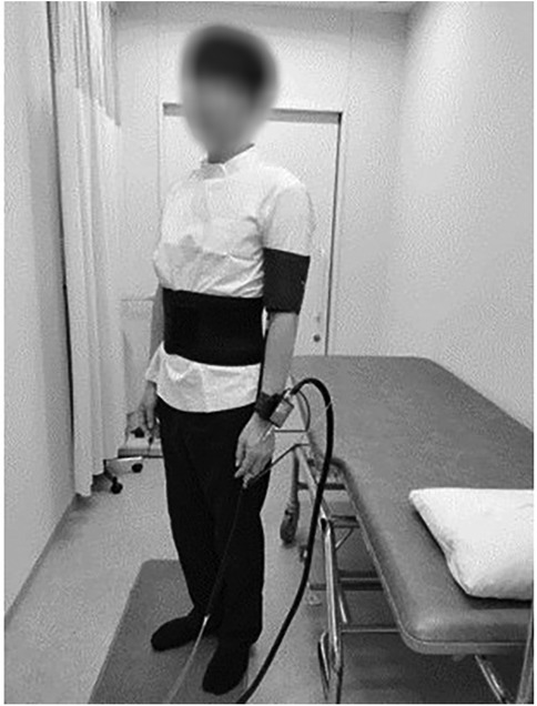
A two-group alternating crossover AB/BA design (Figure 1) was used to evaluate the effect of the abdominal compression band. Each patient underwent two consecutive standing tests in the morning. Participants were alternately assigned to an AB or BA sequence. The group assigned to AB underwent a standing test in the supine and standing positions for 5 min each with the abdominal compression band (Figure 2), followed by a 5 min interval. After the first standing test and an interval, the second standing test was performed in the supine and standing positions for 5 min each without the band. Similarly, the participants assigned to BA performed the first standing test without the band, took an interval and performed the second standing test with the band.
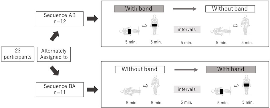
Figure 1. A crossover study; 5 min each in the supine and standing positions. 23 participants were alternately assigned to sequence AB/BA. Each standing test was performed at a 5 min interval, with and without the abdominal compression band.
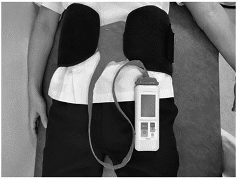
The abdominal compression band (made by ATERCEL, China) measured 19.5 cm in width. A portable contact pressure measuring device (PalmQ, CAPE, Kanagawa, Japan) was placed under the abdominal band at a pressure to 20 mmHg (Figure 3).
The evaluation variables were standing-related symptoms, hemodynamics (blood pressure (BP), heart rate (HR), cardiac index (CI), stroke volume (SV), and total peripheral resistance (TPR)), and cardiovascular autonomic function in the standing test and were compared with and without wearing the abdominal compression band.
Each evaluation parameter is described as follows:
2.2.1 Standing symptoms
Five symptoms—dizziness, blurred vision, headache, palpitations, and fatigue—were evaluated through participant interviews conducted after the test. Symptom intensity was rated on an 11-point visual analog scale ranging from 0 (no symptoms) to 10 (severe symptoms).
2.2.2 Hemodynamics
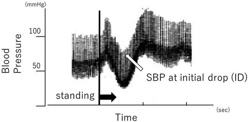
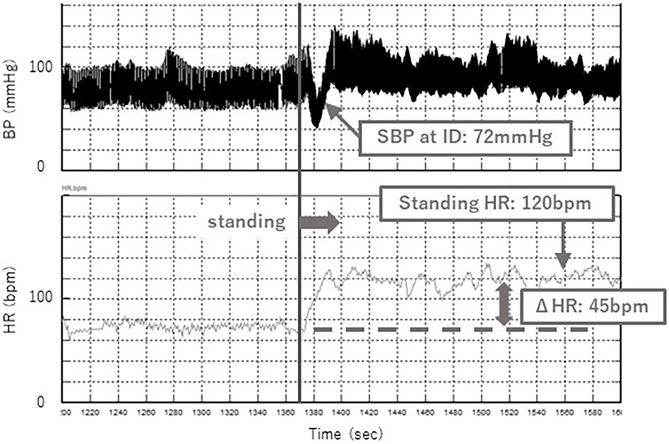
Systolic blood pressure (SBP) in the supine position, at initial drop (ID) and during standing were measured using continuous noninvasive finger arterial pressure measurement (Finapres, FMS, Amsterdam, The Netherlands). ID is defined as the initial drop in SBP immediately after standing (Figure 4). Muscle contractions of the lower extremities and abdomen while standing cause a temporary increase in venous return and sudden increases in right atrial pressure. The baroreceptor reflex then decreases systemic vascular resistance, resulting in ID. ID in SBP occurs in the first 5–15 s on standing, and a transient fall of up to 40 mmHg SBP is considered normal (11). ID is related to dizziness and blurred vision.
HR, CI, SV, and TPR were also measured as well as SBP. The SBP, HR, CI, SV, and TPR were averaged from 3–5 min after standing.
The amount of change (standing value minus supine value) was calculated for SBP and HR, which were defined as ΔSBP and ΔHR each. The rate of change (standing value divided by supine value) was calculated for CI, SV, and TPR.
In addition, since it is possible that body size may affect the change in venous return with the abdominal compression band, we evaluated the correlation between the BMI-SD and the rate of change in SV, which is one of the indices reflecting venous return. The rate of change in SV was calculated dividing the change in SV with the band by the change in SV without the band.
2.2.3 Autonomic nervous function
Heart rate and blood pressure variability during the standing test were evaluated by frequency analysis. Two frequency components were evaluated: the high-frequency (HF; 0.15–0.4 Hz) and low-frequency (LF; 0.04–0.15 Hz) components.
It is generally accepted that the HF component of RR interval variability (RR-HF) is mediated by cardiac parasympathetic tone generated by respiration, whereas the LF component of RR interval variability (RR-LF) is mediated by both cardiac sympathetic and parasympathetic tone. The ratio of the LF to HF components of RR interval variability (RR-LF/HF) is considered an index of cardiac sympathetic tone (12, 13). In contrast, the LF component of BP variability has been reported to parallel sympathetic vasomotor activity (12, 14). Therefore, we used the LF component of DBP variability (DBP-LF) to evaluate sympathetic vasomotor activity.
A Finometer cuff (model 1; FMS, Amsterdam, The Netherlands) was placed on the middle phalanx of the third finger on the right hand. To insure optimal Finometer BP measurements, we used appropriate cuff sizes (S or M) according to the manufacturer's instructions. Spectral analysis was conducted using the maximum entropy method (MemCalc for Windows version 1.2; Suwa Trust, Tokyo, Japan) on the time-series data from 3–5 min for each variable.
2.3 Statistical analysis
Corresponding paired tests were conducted on measurements before and after the intervention. The Wilcoxon signed-rank test was used owing to the lack of normality in the aggregated data. Statistical significance was set at p < 0.05. Data were summarized using medians and interquartile ranges. All statistical analyses were conducted using JMP version 17 (SAS Institute, Cary, NC, USA).
3 Result
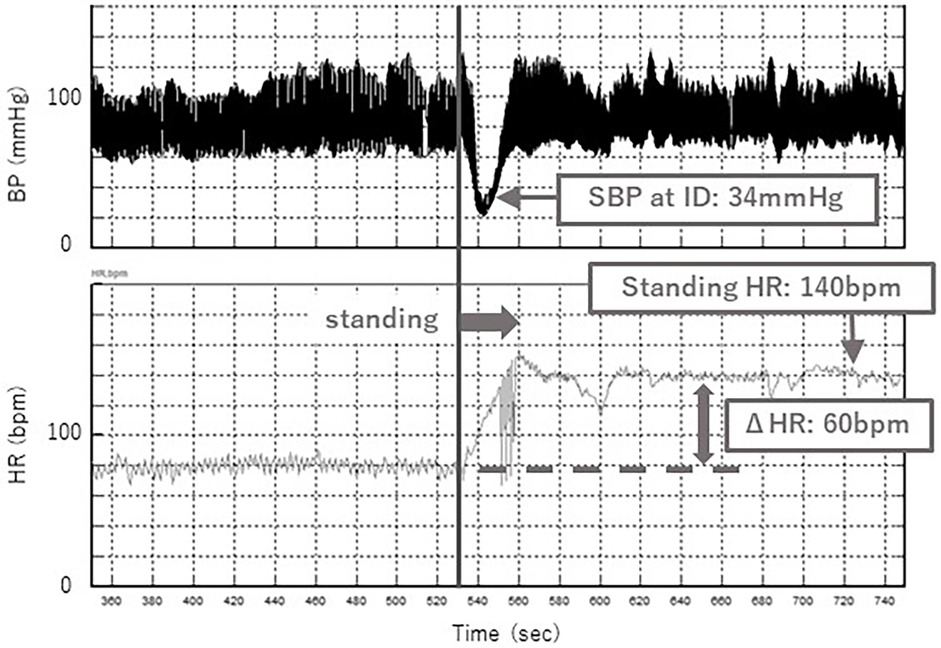
A typical case of the standing test with and without the abdominal compression band is shown in Figures 5, 6. Without the band, a decrease in SBP at ID and an increase in HR were observed after standing up (Figure 5), while these changes were significantly suppressed with the band (Figure 6). Orthostatic symptoms were also markedly improved, and the visual analog scale of headache in this patient improved from 7/10 without the band to 1/10 with the band.
3.1 Standing symptoms
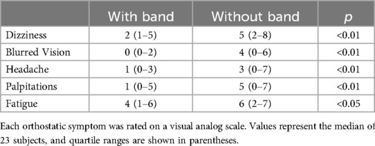
When wearing the abdominal compression band, standing-related symptoms (dizziness, blurred vision, headache, palpitations, and fatigue) improved significantly compared with when not wearing it (Table 1).
3.2 Hemodynamics
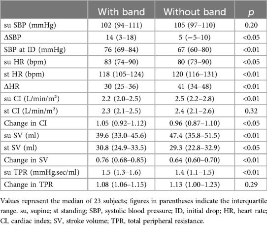
SBP in the supine position was not significantly different with and without the band. Change in SBP (ΔSBP) was significantly greater with the band (Table 2).
ID was significantly higher with the band.
Supine HR was significantly higher with the band and HR while standing was significantly higher without the band. The change in HR (ΔHR) was significantly smaller with the band (Figure 7). The CI in the supine position was significantly smaller with the band, but the CI in standing position is no significantly different with and without the band. For SV, the values with the band were significantly lower than those without the band in the supine position, however, the values with the band were significantly greater than those without the band in the standing position (Figure 8). There was no correlation between BMI-SD and the rate of change in stroke volume (p = 0.91). Supine TPR was significantly higher with the band, and the rate of change in TPR was not significantly different between the two groups.
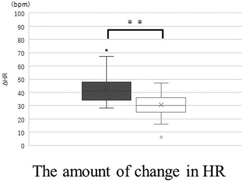
Figure 7. The amount of change (standing value minus supine value) in HR without band (▪) and with band (□). Box plot, median (IQR), mean values (×), with outliers. **p < 0.01. HR, heart rate.

Figure 8. The values in standing SV without band (▪) and with band (□). Box plot, median (IQR), mean values (×). *p < 0.05. SV, stroke volume.
3.3 Frequency analysis
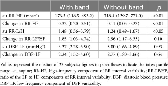
For RR-HF, RR-HF in the supine position showed a significant decrease with the band. RR-HF change in the standing position was significantly higher with the band (Figure 9) (Table 3).
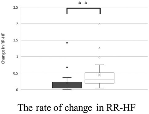
Figure 9. The rate of change (standing value divided by supine value) in RR-HF without band (▪) and with band (□). Box plot, median (IQR), mean values (×), with outliers. **p < 0.01.
For RR-LF/HF, RR-LF/HF in the supine position was significantly higher with the band. However, there was no significant difference between the two groups in terms of RR-LF/HF changes during standing.
Regarding LF-DBP, there were no significant differences in the rate of change of LF-DBP in the supine position and LF-DBP in the standing position with or without the band.
4 Discussion
This study evaluated the efficacy of the abdominal compression band in pediatric patients with POTS and examined its effects on standing, hemodynamics, and autonomic nervous function.
When a person stands, approximately 500–800 ml of blood moves to the abdomen and lower extremities. As a normal response, the autonomic nervous system (sympathetic and parasympathetic) is activated to maintain blood pressure through increased HR and peripheral vasoconstriction. Simultaneously, the abdominal and lower extremity muscles rhythmically contract to compress the capillary vessels and maintain venous return (15). By contrast, the pathophysiology of POTS involves the inability to maintain adequate venous return in an standing position, leading to compensatory tachycardia (16). The pathogenesis of POTS varies widely and is classified into the following subtypes: hyperadrenergic, neuropathic, hypovolemic, and autoimmune POTS. These mechanisms represent a complex pathogenesis involving circulatory abnormalities, such as impaired blood volume regulation, cardiovascular deconditioning, sympathetic nervous system hyperreactivity, and autoantibody involvement. Key pathophysiological features of POTS include abnormal peripheral vascular function, partial denervation of the lower extremities, and increased venous pooling, which are associated with decreased circulating blood volume (6).
Patients with POTS experienced a reduction in dizziness while standing, blurred vision, headache, palpitations, and fatigue when wearing an abdominal compression band. Diedrich et al. reported that venous retention in the abdominal vessels during standing is two to three times greater than that in the lower extremities (17). It is speculated that the abdominal compression band reduces blood retention in the abdominal veins and improves hemodynamics by maintaining the circulating blood volume, which contributes to symptom relief while standing. The mechanism of headache in patients with POTS is unclear, although cerebral hypoperfusion may be a potential cause of orthostatic headache (18). We hypothesized that improved hemodynamics contributed to headache relief.
Significant improvements were also observed in hemodynamic parameters, with the abdominal compression band increasing changes in CI during standing, suppressing decreases in SV and increases in HR, and reducing decreases in ID. In the standing position, the abdominal compression band stabilized blood circulation, potentially alleviating the significant HR increase characteristic of POTS.
Conversely, in the supine position, decreased CI and SV, and increased TPR and HR were observed, possibly due to partial inhibition of venous return.
The band pressure was set at 20 mmHg, which is significantly higher than the venous pressure in the abdomen (approximately 5.5 mmHg) (19). The band pressure was also set at 20 mmHg in this study, based on reports that the compression garment should be worn at 20–30 mmHg to increase venous return (20, 21) and a previous study by Tanaka (10), where 20 mmHg was used. This indicates the need to adjust the band pressure to suit the hemodynamics of each patient.
The CI and SV in the supine was lower with the band. There was no significant difference in CI when standing with or without the band, but SV was larger when standing with the band. These results show that venous return while standing was maintained by wearing the band. CI is affected by HR, but the HR was significantly higher without the band, so we consider that there was no significant difference in CI while standing with or without the band.
The frequency analysis of autonomic nervous system function showed that wearing the abdominal compression band tended to decrease cardiac parasympathetic function and increase cardiac sympathetic function in the supine position. In the supine position, wearing the abdominal compression band decreased venous return, which decreased parasympathetic activity via the vagus nerve by decreasing the intraventricular pressure in the right atrium. However, the decrease in cardiac output due to reduced venous return potentially caused a decrease in blood pressure, triggering sympathetic activation in response to high-pressure system receptors.
Notably, the decrease in the cardiac parasympathetic function was alleviated while standing. Although a decrease in cardiac parasympathetic function caused an increase in HR, the increased venous return and hemodynamic stabilization due to the abdominal compression band may have prevented a significant decrease in cardiac parasympathetic function, preventing an uncontrolled increase in HR.
These findings support the efficacy of the abdominal compression band as an adjunctive therapy, highlighting its advantages in improving hemodynamics and enhancing autonomic nervous function in children with POTS. Because some participants reported discomfort with the abdominal compression band, we recommend that it be worn while symptoms occur, rather than throughout the day. The study suggested that effective use of the abdominal compression band is likely to contribute to an improvement in patients' quality of life (QOL).
5 Limitation
This study has some limitations. First, further studies are needed to determine whether the band pressure setting is appropriate for achieving an effective result for each patient. Second, this study focused on the short-term effects and not on the long-term effects on patient health. Third, previous studies have reported greater symptomatic improvement with beta-blockers compared with the abdominal compression band alone (22) and additional benefits when combined with physical measures, such as leg crossing (23), which may be a topic for future research. Fourth, assuming that the abdominal compression band increases venous return, cerebral blood flow may be changed. The correlation between the abdominal compression band and cerebral blood flow is another interesting research topic. Fifth, the sample size of this study was small and was not compared to healthy participants. Future studies should increase the sample size and make comparisons with healthy participants. Sixth, there are racial differences in autonomic nervous function (24). This study used standardized diagnostic criteria for pediatric POTS in Japanese patients (The diagnostic criterion for pediatric POTS in Europe and the United States is an increase in heart rate of 40 bpm or more, while in Japan it is 35 bpm or more.). Similar studies in other countries are awaited.
6 Conclusion
The abdominal compression band has shown the potential to relieve symptoms while standing and improve hemodynamics and autonomic nervous function in children with POTS.
Data availability statement
The datasets presented in this study can be found in online repositories. The names of the repository/repositories and accession number(s) can be found in the article/Supplementary Material.
Ethics statement
The studies involving humans were approved by the Ethics Committee of Osaka Medical and Pharmaceutical University. The studies were conducted in accordance with the local legislation and institutional requirements. Written informed consent for participation in this study was provided by the participants' legal guardians/next of kin. Written informed consent was obtained from the individual(s) for the publication of any potentially identifiable images or data included in this article.
Author contributions
GY: Conceptualization, Data curation, Formal analysis, Investigation, Methodology, Resources, Visualization, Writing – original draft, Writing – review & editing. SY: Conceptualization, Data curation, Formal analysis, Funding acquisition, Investigation, Methodology, Resources, Visualization, Writing – original draft, Writing – review & editing. HT: Conceptualization, Formal analysis, Supervision, Writing – review & editing. YK: Data curation, Resources, Writing – review & editing. AK: Data curation, Resources, Writing – review & editing. YO: Data curation, Resources, Writing – review & editing. MM: Data curation, Resources, Writing – review & editing. AA: Supervision, Writing – review & editing.
Funding
The author(s) declare that financial support was received for the research and/or publication of this article. This research was supported by a research grant program from the Japanese Society of Psychosomatic Pediatrics in 2022.
Acknowledgments
The authors would like to express our gratitude to all those who contributed to this research.
Conflict of interest
The authors declare that this study was conducted in the absence of any commercial or financial relationships that could be construed as potential conflicts of interest.
Generative AI statement
The author(s) declare that no Generative AI was used in the creation of this manuscript.
Publisher's note
All claims expressed in this article are solely those of the authors and do not necessarily represent those of their affiliated organizations, or those of the publisher, the editors and the reviewers. Any product that may be evaluated in this article, or claim that may be made by its manufacturer, is not guaranteed or endorsed by the publisher.
References
1. Pena C, Moustafa A, Mohamed AR, Grubb B. Autoimmunity in syndromes of orthostatic intolerance: an updated review. J Pers Med. (2024) 14:435. doi: 10.3390/jpm14040435
2. Shinno K, Nagamitsu S. Toward the goal of leaving no one behind: orthostatic dysregulation. JMA J. (2023) 6:334–6. doi: 10.31662/jmaj.2022-0225
3. Japanese Society of Psychosomatic Pediatrics. Japanese Clinical Guidelines for Psychosomatic Pediatrics: Five Guidelines for Daily Clinical Practice. Japan: Nankodo (2015). p. 26–85.
4. Tanaka H, Fujita Y, Takenaka Y, Kajiwara S, Masutani S, Ishizaki Y, et al. Japanese clinical guidelines for juvenile orthostatic dysregulation version 1. Pediatr Int. (2009) 51:169–79. doi: 10.1111/j.1442-200X.2008.02783.x
5. Raj SR, Guzman JC, Harvey P, Richer L, Schondorf R, Seifer C, et al. Canadian cardiovascular society position statement on postural orthostatic tachycardia syndrome (POTS) and related disorders of chronic orthostatic intolerance. Can J Cardiol. (2020) 36:357–72. doi: 10.1016/j.cjca.2019.12.024
6. Vernino S, Bourne KM, Stiles LE, Grubb BP, Fedorowski A, Stewart JM, et al. Postural orthostatic tachycardia syndrome (POTS): state of the science and clinical care from a 2019 national institutes of health expert consensus meeting—part 1. Auton Neurosci. (2021) 235:102828. doi: 10.1016/j.autneu.2021.102828
7. Oyake K, Katai M, Yoneyama A, Ikegawa H, Kani S, Momose K. Comparisons of heart rate variability responses to head-up tilt with and without abdominal and lower-extremity compression in healthy young individuals: a randomized crossover study. Front Physiol. (2023) 14:1269079. doi: 10.3389/fphys.2023.1269079
8. Bourne KM, Sheldon RS, Hall J, Lloyd M, Kogut K, Sheikh N, et al. Compression garment reduces orthostatic tachycardia and symptoms in patients with postural orthostatic tachycardia syndrome. J Am Coll Cardiol. (2021) 77:285–96. doi: 10.1016/j.jacc.2020.11.040
9. Tani H, Singer W, McPhee BR, Opfer-Gehrking TL, Haruma K, Kajiyama G, et al. Splanchnic-mesenteric capacitance bed in the postural tachycardia syndrome (POTS). Auton Neurosci. (2000) 86:107–13. doi: 10.1016/S1566-0702(00)00205-8
10. Tanaka H, Yamaguchi H, Tamai H. Treatment of orthostatic intolerance with inflatable abdominal band. Lancet. (1997) 349:175. doi: 10.1016/S0140-6736(97)24003-1
11. Wieling W, Krediet CTP, Van Dijk N, Linzer M, Tschakovsky ME. Initial orthostatic hypotension: review of a forgotten condition. Clin Sci (Lond). (2007) 112(3):157–65. doi: 10.1042/CS20060091
12. Task Force of the European Society of Cardiology, and the North American Society of Pacing and Electrophysiology. Heart rate variability: standards of measurement, physiological interpretation and clinical use. Circulation. (1996) 93:1043–65.8598068
13. Pagani M, Lombardi F, Guzzetti S, Rimoldi O, Furlan RA, Pizzinelli P, et al. Power spectral analysis of heart rate and arterial pressure variabilities as a marker of sympatho-vagal interaction in man and conscious dog. Circ. Res. (1986) 59:178–93. doi: 10.1161/01.res.59.2.178
14. Barnett SR, Morin RJ, Kiely DK, Gagnon M, Azhar G, Knight EL, et al. Effects of age and gender on autonomic control of blood pressure dynamics. Hypertension. (1999) 33:1195–200. doi: 10.1161/01.hyp.33.5.1195
15. Mallick D, Goyal L, Chourasia P, Zapata MR, Yashi K, Surani S. COVID-19 induced postural orthostatic tachycardia syndrome (POTS): a review. Cureus. (2023) 15:e36955. doi: 10.7759/cureus.36955
16. Sebastian SA, Co EL, Panthangi V, Jain E, Ishak A, Shah Y, et al. Postural orthostatic tachycardia syndrome (POTS): an update for clinical practice. Curr Probl Cardiol. (2022) 47:101384. doi: 10.1016/j.cpcardiol.2022.101384
17. Diedrich A, Biaggioni I. Segmental orthostatic fluid shifts. Clin Auton Res. (2004) 14:146–7. doi: 10.1007/s10286-004-0188-9
18. Iser C, Arca K. Headache and autonomic dysfunction: a review. Curr Neurol Neurosci Rep. (2022) 22:625–34. doi: 10.1007/s11910-022-01225-3
19. Barrett KE, Barman SM, Brooks HL, Yuan JX-J. Ganong’s Review of Medical Physiology. 26th ed New York: McGraw-Hill Education (2019). p. 570.
20. Podoleanu C, Maggi R, Brignole M, Croci F, Incze A, Solano A, et al. Lower limb and abdominal compression bandages prevent progressive orthostatic hypotension in elderly persons: a randomized single-blind controlled study. J Am Coll Cardiol. (2006) 48:1425–32. doi: 10.1016/j.jacc.2006.06.052
21. Chelimsky G, Chelimsky T. Non-pharmacologic management of orthostatic hypotension. Auton Neurosci. (2020) 229:102732. doi: 10.1016/j.autneu.2020.102732
22. Smith EC, Diedrich A, Raj SR, Gamboa A, Shibao CA, Black BK, et al. Splanchnic venous compression enhances the effects of β-blockade in the treatment of postural tachycardia syndrome. J Am Heart Assoc. (2020) 9:e016196. doi: 10.1161/JAHA.120.016196
23. Smit AAJ, Wieling W, Fujimura J, Denq JC, Opfer-Gehrking TL, Akarriou M, et al. Use of lower abdominal compression to combat orthostatic hypotension in patients with autonomic dysfunction. Clin Auton Res. (2004) 14:167–75. doi: 10.1007/s10286-004-0187-x
Keywords: postural tachycardia syndrome, abdominal compression band, children, cardiovascular autonomic function, hemodynamic
Citation: Yamawake G, Yoshida S, Tanaka H, Kurooka Y, Kubo A, Ota Y, Mizutani M and Ashida A (2025) Evaluation of abdominal compression band for pediatric postural tachycardia syndrome: a crossover study. Front. Pediatr. 13:1573091. doi: 10.3389/fped.2025.1573091
Received: 8 February 2025; Accepted: 29 April 2025;
Published: 22 May 2025.
Edited by:
Jiro Takeuchi, Hyogo Medical University, JapanReviewed by:
He Jiang, Children's Hospital of Capital Institute of Pediatrics, ChinaYusaku Nagatomo, Kyushu University Hospital, Japan
Copyright: © 2025 Yamawake, Yoshida, Tanaka, Kurooka, Kubo, Ota, Mizutani and Ashida. This is an open-access article distributed under the terms of the Creative Commons Attribution License (CC BY). The use, distribution or reproduction in other forums is permitted, provided the original author(s) and the copyright owner(s) are credited and that the original publication in this journal is cited, in accordance with accepted academic practice. No use, distribution or reproduction is permitted which does not comply with these terms.
*Correspondence: Seiji Yoshida, c2VpamkueW9zaGlkYUBvbXB1LmFjLmpw
 Ginroku Yamawake
 Seiji Yoshida
 Hidetaka Tanaka
 Yusuke Kurooka1