- 1Department of Rehabilitation Medicine, Ganzhou People’s Hospital, Ganzhou, China
- 2Medical Department, Ganzhou Rongjiang New District People's Hospital, Ganzhou, China
Adolescent scoliosis (AS) is a complex spinal deformity characterized by a curvature exceeding 10 degrees, affecting 1%–3% of adolescents globally. Despite extensive research, its etiology remains multifactorial, involving genetic, biomechanical, neuromuscular, and environmental factors. This review synthesizes recent advances in understanding the pathogenesis of AS and explores the latest developments in non-surgical rehabilitation strategies, including physical therapy, bracing, exercise therapy, and psychological interventions. Emerging technologies, such as artificial intelligence, wearable devices, and virtual reality, are revolutionizing diagnostic accuracy and treatment personalization. The review also highlights the critical role of multidisciplinary collaboration and psychological support in improving patient outcomes. By identifying key research gaps and proposing innovative future directions—such as the integration of epigenetics, advanced biomechanical modeling, and AI-driven precision rehabilitation—this article aims to provide clinicians and researchers with a comprehensive framework for managing AS. Ultimately, this review underscores the importance of early detection, personalized treatment, and long-term follow-up in enhancing the quality of life for adolescents with scoliosis.
1 Introduction
Adolescent scoliosis (AS), characterized by a spinal curvature of more than 10 degrees, is a prevalent condition affecting approximately 1%–3% of adolescents worldwide (1). This spinal deformity not only poses significant physical health challenges but also impacts mental health and quality of life. Despite extensive research, the exact etiology of adolescent scoliosis remains elusive, with multiple theories explaining its development (2). Understanding the underlying causes is essential for developing preventive strategies, enabling early diagnosis, and implementing effective rehabilitation to improve patients’ quality of life. Over the past decade, significant progress has been made in understanding the etiology, diagnosis, and treatment of AS, leading to the development of more sophisticated and individualized rehabilitation strategies (3–5).
The importance of researching the causes of scoliosis in adolescents and providing effective rehabilitation cannot be overstated. Early detection and targeted rehabilitation can halt the progression of scoliosis, alleviate symptoms, reduce the need for invasive treatments such as surgery, and improve the overall physical and mental health of patients (6). Additionally, understanding the underlying causes of the disease can lead to the development of targeted therapies that address specific disease mechanisms (7–9). This is particularly important given the heterogeneity of scoliosis presentations and the varying responses to current treatment modalities (10).
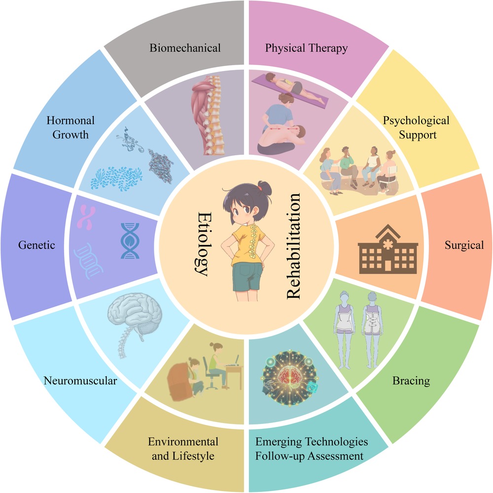
The purpose of this review is to provide a comprehensive overview of the latest research advances in the etiology and rehabilitation of adolescent scoliosis, as shown in Figure 1. By systematically reviewing and analyzing recent literature, we delve into the underlying etiology of scoliosis and elaborate on the principles, applications, and effects of various rehabilitation treatments. This article not only provides clinicians with a comprehensive reference for management strategies but also offers valuable insights into future research directions.

Figure 1. An overview of the etiology of scoliosis and rehabilitation programs. Major etiologic factors include genetic, hormonal and growth factors, biomechanical, neuromuscular, and environmental and lifestyle factors. In terms of rehabilitation, there are currently physical therapy, bracing, surgery, psychological support, emerging technologies, and follow-up assessments.
2 Etiology of adolescent scoliosis
2.1 Genetic factors
Genetic factors play a crucial role in the etiology of Adolescent scoliosis. Recent advances in molecular biology and genetics have revealed the complexity and diversity of genetic factors in the pathogenesis of AS (11). Family and twin studies have provided initial evidence of the importance of genetic factors. Family studies indicate that first-degree relatives of AS patients have a significantly higher risk of developing the condition compared to the general population, suggesting a strong genetic component (12). Twin studies further support this, with monozygotic twins showing a significantly higher concordance rate for AS than dizygotic twins, indicating that genetic factors play a significant role in the onset of AS (13).
Genome-wide association studies (GWAS) and candidate gene studies are the primary methods for uncovering the genetic basis of AS. GWAS scans the genomes of large numbers of AS patients and healthy controls to identify genetic loci associated with AS. For example, studies have identified multiple single nucleotide polymorphisms (SNPs) in regions such as 6p24.1, 10q24.31, and 19p13.3 that are significantly associated with AS (14, 15). These SNPs may affect genes related to spinal development and bone metabolism, thereby increasing the risk of AS. Candidate gene studies focus on genes known to be involved in skeletal development and connective tissue metabolism, such as MATN1, GPR126, and LBX1 (16–18). Mutations or polymorphisms in these genes may affect spinal growth and stability, leading to the development of AS.
The specific mechanisms by which genetic factors contribute to AS are not fully understood, but existing research suggests that genetic factors may influence spinal growth rate, bone mechanical properties, and neuromuscular function, collectively contributing to the onset of AS (19). For example, certain gene mutations may cause asymmetric growth of the spinal growth plates, leading to scoliosis (20). Additionally, genetic factors may affect the mechanical properties of bones, making the spine more susceptible to deformation under external forces. Neuromuscular system dysfunction, potentially linked to genetic factors, may also contribute to muscle imbalance around the spine and the development of scoliosis (21).
2.2 Hormonal and growth factors
Adolescence, the peak period for AS onset, is characterized by significant hormonal changes and rapid growth. Hormonal and growth factors may interact to influence spinal development and stability.
Growth hormone (GH) and insulin-like growth factor-1 (IGF-1) are key hormones regulating bone growth and metabolism (22, 23). Studies suggest that AS patients may have abnormal levels of GH and IGF-1, leading to asymmetric growth of the spinal growth plates. For example, one study found that serum IGF-1 levels were significantly higher in AS patients compared to healthy controls, indicating that IGF-1 may play an important role in the pathogenesis of AS (24). Additionally, abnormalities in GH and IGF-1 may affect the mechanical properties of bones, making the spine more susceptible to deformation under external forces (25).
Sex hormones, particularly estrogen and testosterone, also play significant roles in the pathogenesis of AS. Estrogen influences bone growth and remodeling, potentially playing a key role in the onset and progression of AS. Studies suggest that AS patients may have abnormal estrogen levels, leading to asymmetric growth of the spinal growth plates and the development of scoliosis (26). For example, one study found that serum estrogen levels were significantly higher in AS patients compared to healthy controls, suggesting that estrogen may play an important role in the pathogenesis of AS (27). Additionally, estrogen may affect the mechanical properties of bones, making the spine more susceptible to deformation under external forces.
The role of testosterone in the pathogenesis of AS is not fully understood, but existing research suggests that testosterone may influence bone growth and remodeling, affecting spinal development and stability (28). For example, one study found that serum testosterone levels were significantly lower in AS patients compared to healthy controls, suggesting that testosterone may have a protective role in the pathogenesis of AS (29). Additionally, testosterone may affect the mechanical properties of bones, making the spine more susceptible to deformation under external forces.
2.3 Biomechanical factors
Biomechanical factors play a significant role in the pathogenesis of adolescent scoliosis (30). Abnormal mechanical properties and load distribution of the spine are considered key factors in the development and progression of AS. The mechanical properties of the spine, including stiffness, flexibility, and stability, undergo significant changes during adolescence, potentially affecting normal spinal development (31).
Abnormal load distribution is an important biomechanical factor in AS (32). Normally, the spine distributes loads evenly through its complex structure (e.g., vertebrae, intervertebral discs, and ligaments), maintaining spinal stability and balance. However, in AS patients, load distribution may become abnormal, leading to asymmetric loading and the development of scoliosis. For example, studies have shown that in AS patients, the spine experiences uneven load distribution under external forces, causing certain areas of the spine to bear excessive pressure, leading to scoliosis (33).
Posture and movement habits are also important biomechanical factors influencing the development of AS. Poor posture and movement habits may lead to asymmetric loading of the spine, increasing the risk of AS (34). For example, maintaining poor sitting or standing postures for extended periods may cause certain areas of the spine to bear excessive pressure, leading to scoliosis (35). Additionally, certain movement habits, such as overusing one side of the body or engaging in asymmetrical movements, may also lead to asymmetric loading of the spine, increasing the risk of AS (36).
2.4 Neuromuscular factors
Dysfunction of the neuromuscular system may lead to muscle imbalance around the spine, affecting spinal stability and normal development (37).
Muscle imbalance is an important neuromuscular factor in AS (38). Normal spinal development and stability depend on the balance and coordination of surrounding muscles. However, in AS patients, the balance of muscles around the spine may be disrupted, leading to asymmetric loading and the development of scoliosis (39). For example, studies have shown that AS patients may have asymmetric tension and relaxation of muscles around the spine, causing certain areas of the spine to bear excessive pressure, leading to scoliosis. Additionally, muscle imbalance may affect the spinal growth plates, leading to asymmetric growth and the development of scoliosis (40).
Neurological abnormalities are also an important neuromuscular factor in AS. Dysfunction of the nervous system may lead to impaired control and coordination of muscles around the spine, affecting spinal stability and normal development (41). For example, studies have shown that AS patients may have neurological dysfunction, leading to impaired control and coordination of muscles around the spine, resulting in scoliosis (42). Additionally, neurological abnormalities may affect the spinal growth plates, leading to asymmetric growth and the development of scoliosis.
2.5 Environmental and lifestyle factors
Although genetic and biomechanical factors are the primary causes of AS, environmental and lifestyle factors should not be overlooked (43). These factors may influence spinal development and load distribution, increasing the risk of AS (44).
Nutritional factors are important environmental factors influencing the development of AS (45). Malnutrition or nutritional imbalances may affect bone growth and development, increasing the risk of AS. For example, calcium and vitamin D deficiencies may reduce the mechanical properties of bones, making the spine more susceptible to deformation under external forces (46). Studies have shown that AS patients have significantly lower serum calcium and vitamin D levels compared to healthy controls, suggesting that nutritional factors may play an important role in the pathogenesis of AS (47). Additionally, malnutrition may affect the spinal growth plates, leading to asymmetric growth and the development of scoliosis.
Physical activity is another important lifestyle factor (48). Moderate physical activity helps maintain spinal health and stability. Studies have shown that AS patients have significantly different physical activity habits compared to healthy controls, suggesting that physical activity may play an important role in the pathogenesis of AS (49).
Other environmental and lifestyle factors, such as posture habits, backpack weight, and sleeping posture, may also influence the development of AS (50). For example, maintaining poor sitting or standing postures for extended periods may lead to asymmetric loading of the spine, increasing the risk of AS (51). Studies have shown that AS patients have significantly different posture habits compared to healthy controls, suggesting that posture habits may play an important role in the pathogenesis of AS (52). Additionally, backpack weight and sleeping posture may affect spinal load distribution, increasing the risk of AS (53).
3 Advances in the diagnosis of adolescent scoliosis
In recent years, significant progress has been made in the diagnosis of adolescent scoliosis, particularly in imaging and early screening. Traditional x-ray remains the gold standard for diagnosing scoliosis, accurately measuring the Cobb angle and assessing the severity of the curvature (54). However, x-ray carries the risk of radiation exposure, especially when repeated examinations are required (55). Therefore, low-dose x-ray techniques and digital x-ray imaging systems have been gradually introduced into clinical practice to reduce radiation dose and improve image quality (56). The EOS slot-scanning 2D/3D system has 50%–80% lower radiation compared to conventional radiographs (57).
In addition to x-ray, magnetic resonance imaging (MRI) and computed tomography (CT) also play important roles in the diagnosis of scoliosis. MRI provides detailed three-dimensional images of the spine and surrounding soft tissues, aiding in the assessment of the spinal cord and nerve roots, particularly in complex cases and preoperative evaluations (58). CT scans provide high-resolution images of bony structures, helping to assess vertebral rotation and deformity (59). In recent years, the application of three-dimensional reconstruction techniques and computer-aided diagnosis systems has further improved the accuracy and efficiency of imaging diagnosis (60).
Early screening is key to preventing and controlling the progression of scoliosis (61–63). The most commonly used screening method is the Adam's Forward bend test (FBT), which can be performed with or without scoliometer measurement (64). Moiré Topography is another screening method that has been used (62). This involves using a luminescent imaging device to project a series of lines onto the patient's back. These lines are then distorted by the contours of the patient's back to form a three-dimensional map. This map is then photographed and can be evaluated for asymmetric markings called “Moiré fringes”. If more than 2 Moiré fringes are present, a referral to a specialist is required (65).
In recent years, artificial intelligence (AI)-based screening systems have emerged, analyzing patients’ posture and spinal morphology to automatically detect early signs of scoliosis (66, 67). These technologies not only improve the efficiency and accuracy of screening but also provide powerful tools for large-scale epidemiological surveys (68).
4 Rehabilitation methods for adolescent scoliosis
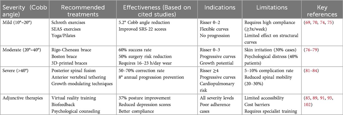
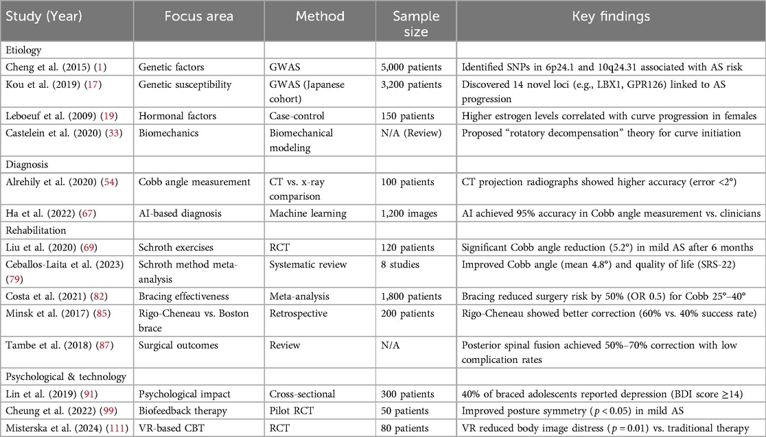
Rehabilitation methods for adolescent scoliosis are diverse, primarily including physical therapy, bracing, and surgical treatment. Each method has its unique advantages and scope of application, and an individualized treatment plan is usually required based on the patient's specific condition. Additionally, psychological support is crucial in the rehabilitation process of scoliosis. Table 1 shows the treatments for adolescent scoliosis by severity. Table 2 shows the included studies and summary of key findings.
4.1 Physical therapy
Physical therapy is the foundation of scoliosis rehabilitation, aiming to improve spinal symmetry and function through specific exercises and posture training (69). It mainly includes manual therapy, traction therapy, and electrical stimulation. Manual therapy uses specific techniques to improve spinal mobility, relieve muscle tension, and reduce pain (70). Traction therapy uses mechanical force to stretch the spine, helping to improve spinal alignment and alleviate nerve compression symptoms (71). Electrical stimulation therapy stimulates paraspinal muscles to enhance muscle strength and improve posture control (72).
In recent years, kinematic-based physical therapy methods have gained attention. By analyzing patients’ movement patterns, individualized training plans can be developed to further improve treatment outcomes. Studies have shown that physical therapy not only improves spinal morphology but also enhances muscle strength and endurance, improving patients’ quality of life (73–75). The Schroth method is a three-dimensional exercise therapy specifically designed for scoliosis, using specific breathing patterns and posture correction exercises to improve spinal alignment and strengthen core muscles. Both the Schrott Method and core stabilization exercises have a positive impact on patients with idiopathic scoliosis (76–78). The recent meta-analysis showed that Schroth therapy reduced Cobb's angle by an average of 4.8° (79, 80). SEAS (Scientific Exercises Approach to Scoliosis) is another evidence-based exercise therapy emphasizing neuromuscular control and posture re-education (81). Other exercise therapies such as yoga, Pilates, and swimming can also serve as adjunct treatments, improving flexibility, muscle strength, and cardiopulmonary function (70).
4.2 Bracing
Bracing is an important treatment for moderate to severe scoliosis, particularly in patients with immature skeletons (82). Braces apply external pressure to effectively halt the progression of scoliosis. In recent years, brace design and materials have continuously improved, with new braces such as the Rigo-Cheneau brace and SpineCor brace offering better comfort and corrective effects (83). The choice of brace should be individualized based on the patient's curve type, Cobb angle, and skeletal maturity. The effectiveness of bracing is closely related to wearing time and compliance, with a recommended daily wearing time of 16–23 hours (84). Studies have shown that for adolescent idiopathic scoliosis patients with Cobb angles between 25°–40°, bracing can significantly reduce the need for surgery (85).
Additionally, with advancements in technology and medical science, computer-aided design and 3D printing have become more sophisticated, allowing braces to be customized based on the patient's specific anatomical structure, further improving treatment outcomes and patient compliance (86).
4.3 Surgical treatment
Surgery is necessary for severe cases of scoliosis or in cases where braces do not work. Traditional surgical methods include posterior spinal fusion and anterior spinal fusion using metal rods and screws to correct the spinal deformity (87, 88). In recent years, minimally invasive surgical techniques and navigation systems have dramatically reduced surgical trauma and complications, and improved the accuracy and safety of surgery (89). In addition, newer surgical approaches such as growth rods and vertebral tethering have provided more treatment options for patients with incomplete skeletal growth. Studies have shown that surgical treatment can significantly improve spinal morphology and function, but postoperative rehabilitation and long-term follow-up are equally important to ensure a favorable outcome for patients (90).
4.4 Psychological support
Scoliosis not only affects patients’ physical health but also has a profound impact on their mental health. Many patients feel self-conscious due to spinal deformity and abnormal posture, leading to anxiety, depression, and other psychological issues (91). Therefore, psychological support is crucial in the rehabilitation process of scoliosis.
Studies have shown that psychological interventions can significantly improve patients’ mental state, enhance their confidence in rehabilitation, and improve treatment compliance (92). Psychological support methods are diverse, including psychological counseling, cognitive-behavioral therapy, and group support (93). Psychological counseling helps patients understand and accept their condition through in-depth communication, alleviating psychological stress (94). Cognitive-behavioral therapy changes patients’ negative thought patterns, enhancing their ability to cope with the disease and improving quality of life (95). Group support provides a platform for patients to share experiences and encourage each other, fostering a sense of belonging and support, and enhancing confidence in rehabilitation. Additionally, family support plays an important role in the psychological rehabilitation of scoliosis patients. Family members’ understanding and support provide emotional comfort and practical help, enhancing patients’ motivation for rehabilitation (96). Therefore, rehabilitation teams should encourage family members to actively participate in the rehabilitation process, providing necessary psychological support and emotional care.
4.5 Emerging technologies and follow-up assessment
Emerging treatment technologies offer new hope for scoliosis rehabilitation. Virtual reality technology provides real-time feedback through immersive environments, enhancing the fun and effectiveness of exercise therapy (97). Robot-assisted rehabilitation training offers precise force control and movement trajectories, helping to improve posture control and muscle coordination (98). Biofeedback therapy uses sensors to monitor muscle activity and posture changes, helping patients better master correct movement patterns (99). Additionally, the evaluation of rehabilitation outcomes and long-term follow-up are crucial for optimizing treatment plans. Common evaluation indicators include changes in Cobb angle, trunk rotation angle, quality of life scores, and pulmonary function tests. Long-term follow-up studies show that early, standardized rehabilitation can significantly improve prognosis, reduce the need for surgery, and enhance patients’ quality of life (100). However, more high-quality randomized controlled trials are needed to compare the advantages and disadvantages of different rehabilitation methods and explore individualized treatment strategies.
4.6 Critical appraisal of therapeutic conflicts
The synthesis of current evidence reveals fundamental tensions between rehabilitation efficacy and practical implementation that demand reconciliation. While Schroth method demonstrates significant Cobb angle reduction [5.2° in RCTs (69)], SEAS exercises exhibit superior neuromuscular control in EMG studies (81), suggesting an unresolved dichotomy between structural correction and functional adaptation that may require phenotype-specific treatment selection. This conflict is compounded by the bracing paradox, where despite proven 50% surgery reduction (82), 40% of adolescents develop clinically significant depression during treatment (91), exposing critical gaps in our risk-benefit calculus that must weigh radiographic outcomes against psychosocial morbidity. Particularly problematic are the inconsistent adherence rates across modalities—Schroth maintains 65%–80% compliance in controlled trials (79) but drops to 52% in real-world bracing applications (84), while combined approaches show superior efficacy [60% surgery risk reduction (85)] yet demand impractical resource investment. These conflicts underscore the necessity of stratified protocols: Schroth for flexible curves >15° where structural correction dominates, SEAS for early postural dysfunction, and restricted bracing (25–40° progressive curves) with embedded mental health monitoring. The field urgently requires pragmatic trials comparing long-term outcomes of these approaches, particularly for the 20–25° “gray zone” where current guidance remains equivocal.
5 Discussion and future perspectives
Adolescent scoliosis is a multifactorial spinal deformity influenced by genetic, biomechanical, neuromuscular, and environmental factors. While significant progress has been made in understanding its etiology and developing rehabilitation strategies, several critical research gaps remain. Addressing these gaps is essential for advancing the field and improving patient outcomes.
5.1 Unresolved etiological mechanisms
Despite advances in genetic research, the precise mechanisms by which genetic variants contribute to AS remain poorly understood. While genome-wide association studies have identified several susceptibility loci, the functional roles of these genetic variants in spinal development and disease progression are yet to be fully elucidated (101). For example, the interaction between genetic factors and environmental triggers (e.g., mechanical loading, hormonal changes) during critical periods of spinal growth warrants further investigation. Future studies should employ functional genomics and single-cell sequencing technologies to uncover the molecular pathways underlying AS pathogenesis.
Additionally, the role of epigenetic modifications in AS has been largely unexplored. Epigenetic changes, such as DNA methylation and histone modifications, may mediate the effects of environmental factors on gene expression, potentially contributing to disease heterogeneity (102). Longitudinal studies tracking epigenetic changes in AS patients could provide insights into disease progression and identify novel therapeutic targets.
5.2 Biomechanical and neuromuscular interactions
The biomechanical and neuromuscular mechanisms driving spinal deformity in AS are complex and not fully understood. While abnormal spinal loading and muscle imbalance are recognized as key factors, their interplay with genetic and hormonal influences remains unclear (103). Advanced biomechanical modeling, coupled with real-time motion analysis, could help elucidate how these factors interact to initiate and perpetuate spinal curvature. Furthermore, the development of patient-specific biomechanical models using 3D imaging and computational simulations may enable personalized risk assessment and treatment planning.
Neuromuscular control deficits in AS patients also require further investigation. Emerging evidence suggests that proprioceptive dysfunction and central nervous system abnormalities may contribute to postural instability and curve progression (104). Neuroimaging studies, such as functional MRI and diffusion tensor imaging, could provide valuable insights into the neural correlates of AS and inform the development of targeted neuromodulation therapies (105).
5.3 Optimization of rehabilitation strategies
While non-surgical interventions, such as physical therapy and bracing, have shown promise in managing AS, their efficacy varies widely among patients. This heterogeneity highlights the need for personalized rehabilitation protocols based on individual patient characteristics, including curve type, skeletal maturity, and genetic profile.
The selection of rehabilitation strategies for scoliosis should be based on the patient's age, Cobb angle, skeletal maturity (Risser sign), and risk of progression. According to the International Scientific Society on Scoliosis Orthopaedic and Rehabilitation Treatment guidelines, Schroth therapy is recommended for mild idiopathic scoliosis (Cobb angle 10°–25°), particularly in adolescents, as it focuses on three-dimensional breathing exercises and postural correction to improve muscular symmetry and spinal alignment. For moderate scoliosis (Cobb angle 25°–45°) in skeletally immature patients (Risser 0–2), a combination of custom orthotic bracing (e.g., Boston or Chêneau brace) and Schroth therapy is advised to reduce curve progression and avoid surgical intervention. Patients with severe curves (>45°) or rapid progression should be referred for surgical evaluation. Evidence supports that conservative management integrating Schroth exercises and bracing significantly reduces surgical rates (106–108). Evidence also suggests that the combined Schroeder + brace group has a 60% lower surgical rate than brace alone (10).
In addition, machine learning algorithms could be employed to analyze large datasets and identify predictors of treatment response, enabling the development of precision rehabilitation strategies (109). Emerging technologies, such as wearable sensors and virtual reality (VR), offer new opportunities for enhancing rehabilitation outcomes. Wearable devices can provide real-time feedback on posture and movement, facilitating adherence to exercise programs (110). VR-based rehabilitation platforms could create immersive environments for motor learning and postural training, potentially improving patient engagement and outcomes (111). However, the long-term efficacy and cost-effectiveness of these technologies require rigorous evaluation through randomized controlled trials.
5.4 Psychological and social dimensions
The psychological impact of AS on adolescents is profound, yet often underaddressed in clinical practice. While psychological interventions, such as cognitive-behavioral therapy and group support, have shown promise, their integration into standard care remains limited. Future research should explore the effectiveness of digital mental health interventions, such as mobile apps and online support groups, in addressing the psychosocial needs of AS patients. Additionally, the role of family support in rehabilitation outcomes warrants further investigation, as family dynamics may significantly influence treatment adherence and patient well-being.
5.5 Long-term outcomes and transition to adulthood
The long-term outcomes of AS patients, particularly those transitioning from adolescence to adulthood, are poorly understood. While early intervention can halt curve progression, the impact of AS on adult spinal health, quality of life, and socioeconomic outcomes remains unclear. Longitudinal studies tracking AS patients into adulthood are needed to evaluate the durability of treatment effects and identify risk factors for late-onset complications, such as degenerative spinal disorders. Furthermore, the development of transition programs to support AS patients as they move from pediatric to adult care could improve continuity of care and long-term outcomes.
5.6 Integration of multidisciplinary approaches
The management of AS requires a multidisciplinary approach, yet the integration of diverse specialties (e.g., orthopedics, physical therapy, psychology) into cohesive care teams remains challenging. Future research should focus on developing standardized protocols for multidisciplinary collaboration, as well as evaluating the impact of team-based care on patient outcomes. Telemedicine platforms could facilitate communication among care providers and enable remote monitoring of patients, particularly in underserved areas.
5.7 Emerging technologies and big data
The integration of artificial intelligence and big data analytics into AS research holds immense potential. AI algorithms could be used to analyze large-scale datasets, such as electronic health records and imaging studies, to identify novel risk factors and predict disease progression (112). Additionally, the development of AI-driven diagnostic tools could enhance early detection and screening efforts, particularly in resource-limited settings. However, the ethical and regulatory challenges associated with AI in healthcare must be carefully addressed to ensure patient safety and data privacy.
6 Limitations and advantages
This review integrates research on adolescent idiopathic scoliosis, including the fields of genetics, biomechanics, and rehabilitation, and proposes clinical treatment guidelines based on the Cobb angle and the Risser sign. It explores the controversy over the Schroth method vs. SEAS exercises and the balance between the effectiveness of brace therapy (50% reduction in surgery rates) and the psychological impact (40% depression rate), and suggests optimizing protocols through phenotypic typing and mental health screening.
Although the research recognizes the role of new technologies (e.g., VR to improve body image, AI to measure Cobb angle), it also points out limitations, such as the wide variation in rehabilitation study designs, the lack of long-term comparative data on surgical vs. non-surgical treatments, and the predominantly white and Asian study populations.
The review informs clinical practice while pointing to future research directions, including multicenter rehabilitation trials, ethnically-specific guidelines, and standardized assessment methods, with an emphasis on combining genetics, biomechanics, and technological advances to achieve individualized treatment.
7 Conclusion
In conclusion, while significant progress has been made in understanding and managing adolescent scoliosis, several critical research gaps remain. Addressing these gaps will require a multidisciplinary approach, leveraging advances in genetics, biomechanics, neuroscience, and digital health technologies. Based on this review, early detection combined with AI-assisted personalized rehabilitation is the most promising direction. By focusing on these innovative research directions, we can develop more effective, personalized, and accessible strategies for preventing and treating AS, ultimately improving the quality of life for patients worldwide.
Author contributions
HK: Software, Conceptualization, Visualization, Writing – original draft. LC: Writing – original draft. MH: Writing – review & editing, Supervision. JC: Project administration, Supervision, Writing – review & editing.
Funding
The author(s) declare that no financial support was received for the research and/or publication of this article.
Conflict of interest
The authors declare that the research was conducted in the absence of any commercial or financial relationships that could be construed as a potential conflict of interest.
Generative AI statement
The author(s) declare that no Generative AI was used in the creation of this manuscript.
Publisher's note
All claims expressed in this article are solely those of the authors and do not necessarily represent those of their affiliated organizations, or those of the publisher, the editors and the reviewers. Any product that may be evaluated in this article, or claim that may be made by its manufacturer, is not guaranteed or endorsed by the publisher.
References
1. Cheng JC, Castelein RM, Chu WC, Danielsson AJ, Dobbs MB, Grivas TB, et al. Adolescent idiopathic scoliosis. Nat Rev Dis Primers. (2015) 1:15030. doi: 10.1038/nrdp.2015.30
2. Almahmoud OH, Baniodeh B, Musleh R, Asmar S, Zyada M, Qattousah H. Overview of adolescent idiopathic scoliosis and associated factors: a scoping review. Int J Adolesc Med Health. (2023) 35(6):437–41. doi: 10.1515/ijamh-2023-0166
3. Zhang X, Dai X, Chen Y, Yang H. Adolescent idiopathic scoliosis: a case report and review of experiences. Asian J Surg. (2024) 47(10):4548–9. doi: 10.1016/j.asjsur.2024.07.256
4. Angelliaume A, Pfirrmann C, Alhada T, Sales de Gauzy J. Non-operative treatment of adolescent idiopathic scoliosis. Orthop Traumatol Surg Res. (2025) 111(1S):104078. doi: 10.1016/j.otsr.2024.104078
5. Foley Davelaar CM, Weber Goff E, Granger JE, Gill DE, Dela Cruz NMR, Sugimoto D. Conservative treatments of adolescent idiopathic scoliosis: physical Therapists’ perspectives. Clin Pediatr (Phila). (2024) 63(8):1132–8. doi: 10.1177/00099228231208609
6. Jinnah AH, Lynch KA, Wood TR, Hughes MS. Adolescent idiopathic scoliosis: advances in diagnosis and management. Curr Rev Musculoskelet Med. (2025) 18(2):54–60. doi: 10.1007/s12178-024-09939-2
7. Khatami N, Caraus I, Rahaman M, Nepotchatykh E, Elbakry M, Elremaly W, et al. Genome-wide profiling of circulating micrornas in adolescent idiopathic scoliosis and their relation to spinal deformity severity, and disease pathophysiology. Sci Rep. (2025) 15(1):5305. doi: 10.1038/s41598-025-88985-3
8. Wang X, Yue M, Cheung JPY, Cheung PWH, Fan Y, Wu M, et al. Impaired glycine neurotransmission causes adolescent idiopathic scoliosis. J Clin Invest. (2024) 134(2):e168783. doi: 10.1172/JCI168783
9. Shao Z, Zhang Z, Tu Y, Huang C, Chen L, Sun A, et al. A targeted antibody-based array reveals a Serum protein signature as biomarker for adolescent idiopathic scoliosis patients. BMC Genomics. (2023) 24(1):522. doi: 10.1186/s12864-023-09624-7
10. Gamiz-Bermudez F, Obrero-Gaitan E, Zagalaz-Anula N, Lomas-Vega R. Corrective exercise-based therapy for adolescent idiopathic scoliosis: systematic review and meta-analysis. Clin Rehabil. (2022) 36(5):597–608. doi: 10.1177/02692155211070452
11. Jiang X, Liu F, Zhang M, Hu W, Zhao Y, Xia B, et al. Advances in genetic factors of adolescent idiopathic scoliosis: a bibliometric analysis. Front Pediatr. (2023) 11:1301137. doi: 10.3389/fped.2023.1301137
12. Faldini C, Manzetti M, Neri S, Barile F, Viroli G, Geraci G, et al. Epigenetic and genetic factors related to curve progression in adolescent idiopathic scoliosis: a systematic scoping review of the current literature. Int J Mol Sci. (2022) 23(11):5914. doi: 10.3390/ijms23115914
13. Peng Y, Wang SR, Qiu GX, Zhang JG, Zhuang QY. Research progress on the etiology and pathogenesis of adolescent idiopathic scoliosis. Chin Med J (Engl). (2020) 133(4):483–93. doi: 10.1097/CM9.0000000000000652
14. Ogura Y, Takahashi Y, Kou I, Nakajima M, Kono K, Kawakami N, et al. A replication study for association of 53 single nucleotide polymorphisms in a scoliosis prognostic test with progression of adolescent idiopathic scoliosis in Japanese. Spine (Phila Pa 1976). (2013) 38(16):1375–9. doi: 10.1097/BRS.0b013e3182947d21
15. Ghanbari F, Otomo N, Gamache I, Iwami T, Koike Y, Khanshour AM, et al. Interrogating causal effects of body composition and puberty-related risk factors on adolescent idiopathic scoliosis: a two-sample Mendelian randomization study. JBMR Plus. (2023) 7(12):e10830. doi: 10.1002/jbm4.10830
16. Soto ME, Fuentevilla-Alvarez G, Koretzky SG, Vargas-Alarcon G, Torres-Paz YE, Meza-Toledo SE, et al. Analysis of Gpr126 polymorphisms and their relationship with scoliosis in Marfan syndrome and Marfan-like syndrome in Mexican patients. Biomol Biomed. (2023) 23(6):976–83. doi: 10.17305/bb.2023.9268
17. Kou I, Otomo N, Takeda K, Momozawa Y, Lu HF, Kubo M, et al. Genome-wide association study identifies 14 previously unreported susceptibility loci for adolescent idiopathic scoliosis in Japanese. Nat Commun. (2019) 10(1):3685. doi: 10.1038/s41467-019-11596-w
18. Moon ES, Kim HS, Sharma V, Park JO, Lee HM, Moon SH, et al. Analysis of single nucleotide polymorphism in adolescent idiopathic scoliosis in Korea: for personalized treatment. Yonsei Med J. (2013) 54(2):500–9. doi: 10.3349/ymj.2013.54.2.500
19. Leboeuf D, Letellier K, Alos N, Edery P, Moldovan F. Do estrogens impact adolescent idiopathic scoliosis? Trends Endocrinol Metab. (2009) 20(4):147–52. doi: 10.1016/j.tem.2008.12.004
20. Wang Y, Pessin JE. Mechanisms for fiber-type specificity of skeletal muscle atrophy. Curr Opin Clin Nutr Metab Care. (2013) 16(3):243–50. doi: 10.1097/MCO.0b013e328360272d
21. Will RE, Stokes IA, Qiu X, Walker MR, Sanders JO. Cobb angle progression in adolescent scoliosis begins at the intervertebral disc. Spine (Phila Pa 1976). (2009) 34(25):2782–6. doi: 10.1097/BRS.0b013e3181c11853
22. Ziv-Baran T, Modan-Moses D, Zacay G, Ackshota N, Levy-Shraga Y. Growth hormone treatment and the risk of adolescent scoliosis: a large matched cohort study. Acta Paediatr. (2023) 112(6):1240–8. doi: 10.1111/apa.16749
23. Guan M, Wang H, Fang H, Zhang C, Gao S, Zou Y. Association between Igf1 gene single nucleotide polymorphism (Rs5742612) and adolescent idiopathic scoliosis: a meta-analysis. Eur Spine J. (2017) 26(6):1624–30. doi: 10.1007/s00586-016-4742-7
24. Zhang HQ, Lu SJ, Tang MX, Chen LQ, Liu SH, Guo CF, et al. Association of estrogen receptor beta gene polymorphisms with susceptibility to adolescent idiopathic scoliosis. Spine (Phila Pa 1976). (2009) 34(8):760–4. doi: 10.1097/BRS.0b013e31818ad5ac
25. Andrews A, Maharaj A, Cottrell E, Chatterjee S, Shah P, Denvir L, et al. Genetic characterization of short stature patients with overlapping features of growth hormone insensitivity syndromes. J Clin Endocrinol Metab. (2021) 106(11):e4716–e33. doi: 10.1210/clinem/dgab437
26. Rao J, Qian S, Li X, Xu Y. Single nucleotide polymorphisms of estrogen receptors are risk factors for the progression of adolescent idiopathic scoliosis: a systematic review and meta-analyses. J Orthop Surg Res. (2024) 19(1):605. doi: 10.1186/s13018-024-05102-2
27. Zhao L, Roffey DM, Chen S. Association between the estrogen receptor beta (Esr2) Rs1256120 single nucleotide polymorphism and adolescent idiopathic scoliosis: a systematic review and meta-analysis. Spine (Phila Pa 1976). (2017) 42(11):871–8. doi: 10.1097/BRS.0000000000001932
28. Szulc P, Uusi-Rasi K, Claustrat B, Marchand F, Beck TJ, Delmas PD. Role of sex steroids in the regulation of bone morphology in men. The MINOS study. Osteoporos Int. (2004) 15(11):909–17. doi: 10.1007/s00198-004-1635-0
29. Peper JS, Brouwer RM, Schnack HG, van Baal GC, van Leeuwen M, van den Berg SM, et al. Sex steroids and brain structure in pubertal boys and girls. Psychoneuroendocrinology. (2009) 34(3):332–42. doi: 10.1016/j.psyneuen.2008.09.012
30. Sarwark JF, Castelein RM, Maqsood A, Aubin CE. The biomechanics of induction in adolescent idiopathic scoliosis: theoretical factors. J Bone Joint Surg Am. (2019) 101(6):e22. doi: 10.2106/JBJS.18.00846
31. Wu L, Zheng A, Guan T, Lei L. Biomechanical analysis of scoliosis correction under the influence of muscular and external forces. J Clin Neurosci. (2025) 132:110991. doi: 10.1016/j.jocn.2024.110991
32. Chen H, Schlosser TPC, Brink RC, Colo D, van Stralen M, Shi L, et al. The height-width-depth ratios of the intervertebral discs and vertebral bodies in adolescent idiopathic scoliosis vs controls in a Chinese population. Sci Rep. (2017) 7:46448. doi: 10.1038/srep46448
33. Castelein RM, Pasha S, Cheng JC, Dubousset J. Idiopathic scoliosis as a rotatory decompensation of the spine. J Bone Miner Res. (2020) 35(10):1850–7. doi: 10.1002/jbmr.4137
34. Chen X, Ye Y, Zhu Z, Zhang R, Wang W, Wu M, et al. Association between incorrect postures and curve magnitude of adolescent idiopathic scoliosis in China. J Orthop Surg Res. (2024) 19(1):300. doi: 10.1186/s13018-024-04767-z
35. Levi D, Springer S, Parmet Y, Ovadia D, Ben-Sira D. Acute muscle stretching and the ability to maintain posture in females with adolescent idiopathic scoliosis. J Back Musculoskelet Rehabil. (2019) 32(4):655–62. doi: 10.3233/BMR-181175
36. Jamison M, Glover M, Peterson K, DeGregorio M, King K, Danelson K, et al. Lumbopelvic postural differences in adolescent idiopathic scoliosis: a pilot study. Gait Posture. (2022) 93:73–7. doi: 10.1016/j.gaitpost.2022.01.002
37. Mayer OH. Scoliosis and the impact in neuromuscular disease. Paediatr Respir Rev. (2015) 16(1):35–42. doi: 10.1016/j.prrv.2014.10.013
38. Wishart BD, Kivlehan E. Neuromuscular scoliosis: when, who, why and outcomes. Phys Med Rehabil Clin N Am. (2021) 32(3):547–56. doi: 10.1016/j.pmr.2021.02.007
39. Loughenbury PR, Tsirikos AI. Current concepts in the treatment of neuromuscular scoliosis: clinical assessment, treatment options, and surgical outcomes. Bone Jt Open. (2022) 3(1):85–92. doi: 10.1302/2633-1462.31.BJO-2021-0178.R1
40. von der Hoh NH, Schleifenbaum S, Schumann E, Heilmann R, Volker A, Heyde CE. Etiology, epidemiology, prognosis and biomechanical principles of neuromuscular scoliosis. Orthopade. (2021) 50(8):608–13. doi: 10.1007/s00132-021-04126-4
41. Dinh HTP, Yamato Y, Hasegawa T, Yoshida G, Banno T, Arima H, et al. Adolescent thoracic scoliosis due to giant ganglioneuroma: a two-case report and literature review. Nagoya J Med Sci. (2024) 86(4):711–9. doi: 10.18999/nagjms.86.4.711
42. Hu ZS, Zhao ZH, Tseng CC, Li J, Man GC, Lam TP, et al. Abnormal activity of sympathetic nervous system in girls with adolescent idiopathic scoliosis: a cross-sectional study. Biomed Environ Sci. (2018) 31(9):700–4. doi: 10.3967/bes2018.094
43. Yang J, Huang S, Cheng M, Tan W, Yang J. Postural habits and lifestyle factors associated with adolescent idiopathic scoliosis (ais) in China: results from a big case-control study. J Orthop Surg Res. (2022) 17(1):472. doi: 10.1186/s13018-022-03366-0
44. Watanabe K, Michikawa T, Yonezawa I, Takaso M, Minami S, Soshi S, et al. Physical activities and lifestyle factors related to adolescent idiopathic scoliosis. J Bone Joint Surg Am. (2017) 99(4):284–94. doi: 10.2106/JBJS.16.00459
45. Ng PTT, Tucker K, Zahir SF, Izatt MT, Straker L, Claus A. Comparison of physiological and behavioral nutrition-related factors in people with and without adolescent idiopathic scoliosis, from cohort data at 8 to 20 years. JBMR Plus. (2024) 8(3):ziad013. doi: 10.1093/jbmrpl/ziad013
46. Llopis-Ibor CI, Mariscal G, de la Rubia Orti JE, Barrios C. Incidence of vitamin D deficiency in adolescent idiopathic scoliosis: a meta-analysis. Front Endocrinol (Lausanne). (2023) 14:1250118. doi: 10.3389/fendo.2023.1250118
47. Alsiddiky A, Alfadhil R, Al-Aqel M, Ababtain N, Almajed N, Bakarman K, et al. Assessment of serum vitamin D levels in surgical adolescent idiopathic scoliosis patients. BMC Pediatr. (2020) 20(1):202. doi: 10.1186/s12887-020-02114-9
48. Glavas J, Rumboldt M, Karin Z, Matkovic R, Bilic-Kirin V, Buljan V, et al. The impact of physical activity on adolescent idiopathic scoliosis. Life (Basel). (2023) 13(5):1180. doi: 10.3390/life13051180
49. Qi X, Peng C, Fu P, Zhu A, Jiao W. Correlation between physical activity and adolescent idiopathic scoliosis: a systematic review. BMC Musculoskelet Disord. (2023) 24(1):978. doi: 10.1186/s12891-023-07114-1
50. Khadour FA, Khadour YA, Albarroush D. Association between postural habits and lifestyle factors of adolescent idiopathic scoliosis in Syria. Sci Rep. (2024) 14(1):26784. doi: 10.1038/s41598-024-77712-z
51. Zhu L, Ru S, Wang W, Dou Q, Li Y, Guo L, et al. Associations of physical activity and screen time with adolescent idiopathic scoliosis. Environ Health Prev Med. (2023) 28:55. doi: 10.1265/ehpm.23-00004
52. de Assis SJC, Sanchis GJB, de Souza CG, Roncalli AG. Influence of physical activity and postural habits in schoolchildren with scoliosis. Arch Public Health. (2021) 79(1):63. doi: 10.1186/s13690-021-00584-6
53. Baroni MP, Sanchis GJ, de Assis SJ, dos Santos RG, Pereira SA, Sousa KG, et al. Factors associated with scoliosis in schoolchildren: a cross-sectional population-based study. J Epidemiol. (2015) 25(3):212–20. doi: 10.2188/jea.JE20140061
54. Alrehily F, Hogg P, Twiste M, Johansen S, Tootell A. The accuracy of Cobb angle measurement on ct scan projection radiograph images. Radiography (Lond). (2020) 26(2):e73–e7. doi: 10.1016/j.radi.2019.11.001
55. Hamilton M, Kendall E. Radiation exposure from the patient perspective: an argument for the inclusion of dose history. Health Phys. (2023) 125(3):198–201. doi: 10.1097/HP.0000000000001705
56. Kolck J, Ziegeler K, Walter-Rittel T, Hermann KGA, Hamm B, Beck A. Clinical utility of postprocessed low-dose radiographs in skeletal imaging. Br J Radiol. (2022) 95(1130):20210881. doi: 10.1259/bjr.20210881
57. Boissonnat G, Morichau-Beauchant P, Reshef A, Villa C, Desaute P, Simon AC. Performance of automatic exposure control on dose and image quality: comparison between slot-scanning and flat-panel digital radiography systems. Med Phys. (2023) 50(2):1162–84. doi: 10.1002/mp.15954
58. Duncombe P, Izatt MT, Pivonka P, Claus A, Little JP, Tucker K. Quantifying muscle size asymmetry in adolescent idiopathic scoliosis using three-dimensional magnetic resonance imaging. Spine (Phila Pa 1976). (2023) 48(24):1717–25. doi: 10.1097/BRS.0000000000004715
59. Li J, Gao J, Zhao Z, Canavese F, Cai Q, Li Y, et al. Quantitative computed tomography assessment of bone mineral density in adolescent idiopathic scoliosis: correlations with Cobb angle, vertebral rotation, and risser sign. Transl Pediatr. (2024) 13(4):610–23. doi: 10.21037/tp-24-74
60. Zhang M, Chen W, Wang S, Lei S, Liu Y, Zhang J, et al. Clinical validation of the differences between two-dimensional radiography and three-dimensional computed tomography image measurements of the spine in adolescent idiopathic scoliosis. World Neurosurg. (2022) 165:e689–e96. doi: 10.1016/j.wneu.2022.06.128
61. Scaturro D, de Sire A, Terrana P, Costantino C, Lauricella L, Sannasardo CE, et al. Adolescent idiopathic scoliosis screening: could a school-based assessment protocol be useful for an early diagnosis? J Back Musculoskelet Rehabil. (2021) 34(2):301–6. doi: 10.3233/BMR-200215
62. Oetgen ME, Heyer JH, Kelly SM. Scoliosis screening. J Am Acad Orthop Surg. (2021) 29(9):370–9. doi: 10.5435/JAAOS-D-20-00356
63. Zou Y, Lin Y, Meng J, Li J, Gu F, Zhang R. The prevalence of scoliosis screening positive and its influencing factors: a school-based cross-sectional study in Zhejiang province, China. Front Public Health. (2022) 10:773594. doi: 10.3389/fpubh.2022.773594
64. Dunn J, Henrikson NB, Morrison CC, Blasi PR, Nguyen M, Lin JS. Screening for adolescent idiopathic scoliosis: evidence report and systematic review for the US preventive services task force. JAMA. (2018) 319(2):173–87. doi: 10.1001/jama.2017.11669
65. Kuroki H, Nagai T, Chosa E, Tajima N. School scoliosis screening by moire topography - overview for 33 years in Miyazaki Japan. J Orthop Sci. (2018) 23(4):609–13. doi: 10.1016/j.jos.2018.03.005
66. Oquendo Y, Hollyer I, Maschhoff C, Calderon C, DeBaun M, Langner J, et al. Mobile device-based 3d scanning is superior to scoliometer in assessment of adolescent idiopathic scoliosis. Spine Deform. (2025) 13(2):529–37. doi: 10.1007/s43390-024-01007-6
67. Ha AY, Do BH, Bartret AL, Fang CX, Hsiao A, Lutz AM, et al. Automating scoliosis measurements in radiographic studies with machine learning: comparing artificial intelligence and clinical reports. J Digit Imaging. (2022) 35(3):524–33. doi: 10.1007/s10278-022-00595-x
68. Tingsheng L, Chunshan L, Shudan Y, Xingwei P, Qiling C, Minglu Y, et al. Validation of artificial intelligence in the classification of adolescent idiopathic scoliosis and the compairment to clinical manual handling. Orthop Surg. (2024) 16(8):2040–51. doi: 10.1111/os.14144
69. Liu D, Yang Y, Yu X, Yang J, Xuan X, Yang J, et al. Effects of specific exercise therapy on adolescent patients with idiopathic scoliosis: a prospective controlled cohort study. Spine (Phila Pa 1976). (2020) 45(15):1039–46. doi: 10.1097/BRS.0000000000003451
70. Chen Y, Zhang Z, Zhu Q. The effect of an exercise intervention on adolescent idiopathic scoliosis: a network meta-analysis. J Orthop Surg Res. (2023) 18(1):655. doi: 10.1186/s13018-023-04137-1
71. Liang Y, Zhu Z, Zhao C, Xu S, Guo C, Zhao D, et al. The impact of halo-pelvic traction on sagittal kyphosis in the treatment of severe scoliosis and kyphoscoliosis. J Orthop Surg Res. (2024) 19(1):652. doi: 10.1186/s13018-024-04985-5
72. Hatzilazaridis I, Hatzitaki V, Antoniadou N, Samoladas E. Postural and muscle responses to galvanic vestibular stimulation reveal a vestibular deficit in adolescents with idiopathic scoliosis. Eur J Neurosci. (2019) 50(10):3614–26. doi: 10.1111/ejn.14525
73. Ma K, Wang C, Huang Y, Wang Y, Li D, He G. The effects of physiotherapeutic scoliosis-specific exercise on idiopathic scoliosis in children and adolescents: a systematic review and meta-analysis. Physiotherapy. (2023) 121:46–57. doi: 10.1016/j.physio.2023.07.005
74. Li X, Shen J, Liang J, Zhou X, Yang Y, Wang D, et al. Effect of core-based exercise in people with scoliosis: a systematic review and meta-analysis. Clin Rehabil. (2021) 35(5):669–80. doi: 10.1177/0269215520975105
75. Dimitrijevic V, Raskovic B, Popovic M, Viduka D, Nikolic S, Drid P, et al. Treatment of idiopathic scoliosis with conservative methods based on exercises: a systematic review and meta-analysis. Front Sports Act Living. (2024) 6:1492241. doi: 10.3389/fspor.2024.1492241
76. Dimitrijevic V, Viduka D, Scepanovic T, Maksimovic N, Giustino V, Bianco A, et al. Effects of schroth method and core stabilization exercises on idiopathic scoliosis: a systematic review and meta-analysis. Eur Spine J. (2022) 31(12):3500–11. doi: 10.1007/s00586-022-07407-4
77. Dimitrijevic V, Scepanovic T, Jevtic N, Raskovic B, Milankov V, Milosevic Z, et al. Application of the Schroth method in the treatment of idiopathic scoliosis: a systematic review and meta-analysis. Int J Environ Res Public Health. (2022) 19(24):16730. doi: 10.3390/ijerph192416730
78. Park JH, Jeon HS, Park HW. Effects of the Schroth exercise on idiopathic scoliosis: a meta-analysis. Eur J Phys Rehabil Med. (2018) 54(3):440–9. doi: 10.23736/S1973-9087.17.04461-6
79. Ceballos-Laita L, Carrasco-Uribarren A, Cabanillas-Barea S, Perez-Guillen S, Pardos-Aguilella P, Jimenez Del Barrio S. The effectiveness of Schroth method in Cobb angle, quality of life and trunk rotation angle in adolescent idiopathic scoliosis: a systematic review and meta-analysis. Eur J Phys Rehabil Med. (2023) 59(2):228–36. doi: 10.23736/S1973-9087.23.07654-2
80. You MJ, Lu ZY, Xu QY, Chen PB, Li B, Jiang SD, et al. Effectiveness of physiotherapeutic scoliosis-specific exercises on 3-dimensional spinal deformities in patients with adolescent idiopathic scoliosis: a systematic review and meta-analysis. Arch Phys Med Rehabil. (2024) 105(12):2375–89. doi: 10.1016/j.apmr.2024.04.011
81. Romano M, Negrini A, Parzini S, Tavernaro M, Zaina F, Donzelli S, et al. Seas (scientific exercises approach to scoliosis): a modern and effective evidence based approach to physiotherapic specific scoliosis exercises. Scoliosis. (2015) 10:3. doi: 10.1186/s13013-014-0027-2
82. Costa L, Schlosser TPC, Jimale H, Homans JF, Kruyt MC, Castelein RM. The effectiveness of different concepts of bracing in adolescent idiopathic scoliosis (ais): a systematic review and meta-analysis. J Clin Med. (2021) 10(10):2145. doi: 10.3390/jcm10102145
83. Landauer F, Trieb KP. Scoliosis: brace treatment - from the past 50 years to the future. Medicine (Baltimore). (2022) 101(37):e30556. doi: 10.1097/MD.0000000000030556
84. Rahimi S, Kiaghadi A, Fallahian N. Effective factors on brace compliance in idiopathic scoliosis: a literature review. Disabil Rehabil Assist Technol. (2020) 15(8):917–23. doi: 10.1080/17483107.2019.1629117
85. Minsk MK, Venuti KD, Daumit GL, Sponseller PD. Effectiveness of the rigo cheneau versus Boston-style orthoses for adolescent idiopathic scoliosis: a retrospective study. Scoliosis Spinal Disord. (2017) 12:7. doi: 10.1186/s13013-017-0117-z
86. Nathan P, Chou SM, Liu G. A review on different methods of scoliosis brace fabrication. Prosthet Orthot Int. (2023) 47(4):424–33. doi: 10.1097/PXR.0000000000000195
87. Tambe AD, Panikkar SJ, Millner PA, Tsirikos AI. Current concepts in the surgical management of adolescent idiopathic scoliosis. Bone Joint J. (2018) 100-B(4):415–24. doi: 10.1302/0301-620X.100B4.BJJ-2017-0846.R2
88. Lin Y, Chen W, Chen A, Li F, Xiong W. Anterior versus posterior selective fusion in treating adolescent idiopathic scoliosis: a systematic review and meta-analysis of radiologic parameters. World Neurosurg. (2018) 111:e830–e44. doi: 10.1016/j.wneu.2017.12.161
89. Virk S, Qureshi S. Navigation in minimally invasive spine surgery. J Spine Surg. (2019) 5(Suppl 1):S25–30. doi: 10.21037/jss.2019.04.23
90. Senkoylu A, Riise RB, Acaroglu E, Helenius I. Diverse approaches to scoliosis in young children. EFORT Open Rev. (2020) 5(10):753–62. doi: 10.1302/2058-5241.5.190087
91. Lin T, Meng Y, Ji Z, Jiang H, Shao W, Gao R, et al. Extent of depression in juvenile and adolescent patients with idiopathic scoliosis during treatment with braces. World Neurosurg. (2019) 126:e27–32. doi: 10.1016/j.wneu.2019.01.095
92. van Agteren J, Iasiello M, Lo L, Bartholomaeus J, Kopsaftis Z, Carey M, et al. A systematic review and meta-analysis of psychological interventions to improve mental wellbeing. Nat Hum Behav. (2021) 5(5):631–52. doi: 10.1038/s41562-021-01093-w
93. Masi G, Carucci S, Muratori P, Balia C, Sesso G, Milone A. Contemporary diagnosis and treatment of conduct disorder in youth. Expert Rev Neurother. (2023) 23(12):1277–96. doi: 10.1080/14737175.2023.2271169
94. de Kleine RA, Smits JAJ, Hofmann SG. Advancements in cognitive behavioral therapy. Psychiatr Clin North Am. (2024) 47(2):xiii–xv. doi: 10.1016/j.psc.2024.03.001
95. Clark E, MacCrosain A, Ward NS, Jones F. The key features and role of peer support within group self-management interventions for stroke? A systematic review. Disabil Rehabil. (2020) 42(3):307–16. doi: 10.1080/09638288.2018.1498544
96. Wang J, Hu S, Wang L. Multilevel analysis of personality, family, and classroom influences on emotional and behavioral problems among Chinese adolescent students. PLoS One. (2018) 13(8):e0201442. doi: 10.1371/journal.pone.0201442
97. Feng H, Li C, Liu J, Wang L, Ma J, Li G, et al. Virtual reality rehabilitation versus conventional physical therapy for improving balance and gait in Parkinson’s disease patients: a randomized controlled trial. Med Sci Monit. (2019) 25:4186–92. doi: 10.12659/MSM.916455
98. Rodgers H, Bosomworth H, Krebs HI, van Wijck F, Howel D, Wilson N, et al. Robot assisted training for the upper limb after stroke (ratuls): a multicentre randomised controlled trial. Lancet. (2019) 394(10192):51–62. doi: 10.1016/S0140-6736(19)31055-4
99. Cheung MC, Yip J, Lai JSK. Biofeedback posture training for adolescents with mild scoliosis. Biomed Res Int. (2022) 2022:5918698. doi: 10.1155/2022/5918698
100. Birch NC, Tsirikos AI. Long-term follow-up of patients with idiopathic scoliosis: providing appropriate continuing care. Bone Joint J. (2023) 105-B(2):99–100. doi: 10.1302/0301-620X.105B2.BJJ-2022-1298
101. Montemurro N, Ricciardi L, Scerrati A, Ippolito G, Lofrese G, Trungu S, et al. The potential role of dysregulated mirnas in adolescent idiopathic scoliosis and 22q11.2 deletion syndrome. J Pers Med. (2022) 12(11):1925. doi: 10.3390/jpm12111925
102. Sun D, Ding Z, Hai Y, Cheng Y. Advances in epigenetic research of adolescent idiopathic scoliosis and congenital scoliosis. Front Genet. (2023) 14:1211376. doi: 10.3389/fgene.2023.1211376
103. Pizones J, Chang DG, Suk SI, Izquierdo E. Current biomechanical theories on the etiopathogenesis of idiopathic scoliosis. Spine Deform. (2024) 12(2):247–55. doi: 10.1007/s43390-023-00787-7
104. Zhang Y, Chai T, Weng H, Liu Y. Pelvic rotation correction combined with Schroth exercises for pelvic and spinal deformities in mild adolescent idiopathic scoliosis: a randomized controlled trial. PLoS One. (2024) 19(7):e0307955. doi: 10.1371/journal.pone.0307955
105. Soh RCC, Chen BZ, Hartono S, Lee MS, Lee W, Lim SL, et al. The hindbrain and cortico-reticular pathway in adolescent idiopathic scoliosis. Clin Radiol. (2024) 79(5):e759–e66. doi: 10.1016/j.crad.2024.01.027
106. Chen J, Xu T, Zhou J, Han B, Wu Q, Jin W, et al. The superiority of Schroth exercise combined brace treatment for mild-to-moderate adolescent idiopathic scoliosis: a systematic review and network meta-analysis. World Neurosurg. (2024) 186:184–96.e9. doi: 10.1016/j.wneu.2024.03.103
107. Negrini S, Donzelli S, Aulisa AG, Czaprowski D, Schreiber S, de Mauroy JC, et al. 2016 Sosort guidelines: orthopaedic and rehabilitation treatment of idiopathic scoliosis during growth. Scoliosis Spinal Disord. (2018) 13:3. doi: 10.1186/s13013-017-0145-8
108. Weinstein SL, Dolan LA, Wright JG, Dobbs MB. Effects of bracing in adolescents with idiopathic scoliosis. N Engl J Med. (2013) 369(16):1512–21. doi: 10.1056/NEJMoa1307337
109. Ohyama S, Maki S, Kotani T, Ogata Y, Sakuma T, Iijima Y, et al. Machine learning algorithms for predicting future curve using first and second visit data in female adolescent idiopathic scoliosis patients. Eur Spine J. (2025). doi: 10.1007/s00586-025-08680-9
110. Xuan L, Lei L, Shao M, Han Q. Design and development of an intelligent wearing system for adolescent spinal orthotics. Med Biol Eng Comput. (2024) 62(9):2653–67. doi: 10.1007/s11517-024-03082-3
111. Misterska E, Tomaszewski M, Gorski F, Gapsa J, Slysz A, Glowacki M. Assessing the efficacy of cognitive-behavioral therapy on body image in adolescent scoliosis patients using virtual reality. J Clin Med. (2024) 13(21):6422. doi: 10.3390/jcm13216422
Keywords: adolescent scoliosis, etiology, rehabilitation, physical therapy, bracing
Citation: Kuang H, Chen L, Huang M and Chen J (2025) Management of adolescent scoliosis: a comprehensive review of etiology and rehabilitation. Front. Pediatr. 13:1596400. doi: 10.3389/fped.2025.1596400
Received: 19 March 2025; Accepted: 30 June 2025;
Published: 16 July 2025.
Edited by:
Davide Bizzoca, University of Bari Aldo Moro, ItalyReviewed by:
Jean Claude De Mauroy, Independent Researcher, Lyon, FranceVanja Dimitrijević, University of Novi Sad, Serbia
Copyright: © 2025 Kuang, Chen, Huang and Chen. This is an open-access article distributed under the terms of the Creative Commons Attribution License (CC BY). The use, distribution or reproduction in other forums is permitted, provided the original author(s) and the copyright owner(s) are credited and that the original publication in this journal is cited, in accordance with accepted academic practice. No use, distribution or reproduction is permitted which does not comply with these terms.
*Correspondence: Miao Huang, MTc5MTIwMzc0MUBxcS5jb20=; Jianbin Chen, emhvdXlleXVob3VAMTYzLmNvbQ==
 Hongwei Kuang1
 Miao Huang
