- 1Bioinnovation Center, Buddhist Tzu Chi Medical Foundation, Hualien, Taiwan
- 2Department of Neurosurgery, Hualien Tzu Chi Hospital, Buddhist Tzu Chi Medical Foundation, Hualien, Taiwan
- 3Department of Biological Science and Technology, College of Life Sciences, China Medical University, Taichung, Taiwan
- 4Ph.D. Program for Biotechnology Industry, China Medical University, Taichung, Taiwan
- 5School of Pharmacy, China Medical University, Taichung, Taiwan
- 6Cardiovascular and Mitochondrial Related Disease Research Center, Hualien Tzu Chi Hospital, Buddhist Tzu Chi Medical Foundation, Hualien, Taiwan
- 7Laboratory of Exercise Biochemistry, University of Taipei, Taipei, Taiwan
- 8School of Physical Education and Sports Science, Soochow University, Suzhou, China
- 9Department of Medical Laboratory and Biotechnology, Chung Shan Medical University, Taichung, Taiwan
- 10Clinical Laboratory, Chung Shan Medical University Hospital, Taichung, Taiwan
- 11Department of Orthopedic Surgery, Chung Shan Medical University Hospital, Taichung, Taiwan
- 12School of Medicine, Chung Shan Medical University, Taichung, Taiwan
- 13School of Medicine, College of Medicine, China Medical University, Taichung, Taiwan
- 14Division of Cardiovascular Medicine, Department of Internal Medicine, China Medical University Hospital, Taichung, Taiwan
- 15Home Run Biotechnology Co., Ltd., Tainan, Taiwan
- 16Graduate Institute of Biomedical Sciences, China Medical University, Taichung, Taiwan
- 17Department of Medical Research, China Medical University Hospital, China Medical University, Taichung, Taiwan
- 18Department of Medical Laboratory Science and Biotechnology, Asia University, Taichung, Taiwan
- 19Center of General Education, Tzu Chi University, Hualien, Taiwan
- 20Department of Emergency Medicine, China Medical University Hospital, Taichung, Taiwan
Background: Diabetic nephropathy (DN), a leading cause of chronic kidney disease and end-stage renal disease, is a serious complication of type 2 diabetes mellitus (T2DM). Current therapies primarily slow disease progression but are unable to reverse kidney damage, highlighting the need for novel therapy to treat DN.
Objective: This study evaluated the therapeutic potential of Cordyceps militaris (C. militaris) cultivated on Ginkgo biloba (G. biloba) seeds in ameliorating T2DM and its complications, especially DN. A T2DM mouse model was established using ApoE knockout mice fed a Western diet (WD).
Results: Treatment with the specially cultivated C. militaris ameliorated hyperglycemia, dyslipidemia and hepatic dysfunction, while mitigating T2DM-induced renal damage. Key biochemical markers, including blood glucose, triglycerides, cholesterol, blood urea nitrogen (BUN), and creatinine, were significantly improved after treatment. Histopathologic analysis revealed restored renal morphology, reduced fibrosis and decreased amyloid deposition. Mechanistic studies showed downregulation of fibrosis-related proteins such as α-SMA, COL1, TIMP-1, CTGF, TGFβ1 and fibronectin, and upregulation of E-cadherin, Smad7 and Klotho, proteins with anti-fibrotic and renoprotective properties.
Conclusion: These results suggest that the specially cultivated C. militaris enhances metabolic regulation and renal repair mechanisms, effectively attenuating T2DM-induced renal damage. This unique cultivation approach enriches the bioactive properties of C. militaris and offers a promising natural therapeutic strategy for T2DM and DN. Further studies are needed to validate these results in clinical settings and to explore long-term efficacy and safety.
1 Introduction
Diabetes mellitus (DM) is a common and serious metabolic disorder that poses a significant challenge to modern healthcare systems. The global prevalence of DM has increased significantly in recent decades. According to the classification system established by the American Diabetes Association, there are two popular types of DM. Type 1 diabetes mellitus (T1DM) is an autoimmune disease characterized by the destruction of insulin-producing β-cells in the islets of Langerhans, accounting for approximately 5%–10% of cases. In contrast, type 2 diabetes mellitus (T2DM) is the more common DM, accounting for up to 90% of cases, and is attributed to a lack of insulin secretion, impaired insulin action, or both. The global rise in obesity is closely associated with a significant increase in the incidence of T2DM and is increasingly observed in younger obese individuals (Sagoo and Gnudi, 2020). One of the causes of T2DM is changes in the dietary habits of modern people, such as the Western Diet (WD) (Clemente-Suárez et al., 2023). Prolonged DM is associated with multiple organ failure and microvascular complications, including neuropathy, retinopathy, and nephropathy (Beckman and Creager, 2016). Diabetic nephropathy (DN), also known as diabetic kidney disease, is one of the major complications of DM and a leading cause of chronic kidney disease and end-stage renal disease. T2DM often progresses to DN, which is the leading cause of end-stage renal disease worldwide and is associated with significant morbidity and mortality (Maggiore et al., 2017). However, standard treatments for DN primarily slow its progression, but are unable to halt or reverse the disease. Therefore, the development of novel therapeutic strategies for the effective management of DN has become imperative (Samsu, 2021).
Traditional Chinese medicines and their bioactive metabolites possess protective properties against various stresses and diseases. These properties make them promising candidates for the development of innovative therapeutic approaches to effectively treat DN (Chang et al., 2013; Liu et al., 2020; Chang et al., 2021; Ho et al., 2024). Cordyceps militaris (C. militaris), a fungus belonging to the Ascomycota, is highly regarded for its medicinal and nutritional benefits. Known in Asia for centuries, C. militaris has been used both as a dietary supplement and in traditional medicine. C. militaris is a rich source of various bioactive metabolites such as cordycepin, adenosine, carotenoids, pentostatin, polysaccharides, proteins, ergosterol, and myriocin, among others (Zhang et al., 2019; Zeng et al., 2024). Pharmacological research has shown that C. militaris offers remarkable therapeutic benefits for a wide range of health problems. These include diseases of the neurological, respiratory, cardiovascular, hepatic, and renal systems (Zhang et al., 2019). For the treatment of DM and DN, C. militaris has been shown to modulate the gut microbiota by enhancing beneficial bacteria and regulating metabolites and metabolic pathways, thereby ameliorating T2DM in mice (Liu et al., 2023). Previous research shows that C. militaris extract has potent anti-diabetic and renal protective effects, making it a promising candidate for the development of new treatments for diabetes (Dong et al., 2014). In addition, cordycepin from C. militaris has ability against DN by reducing oxidative stress, inflammation, and apoptosis in HK-2 cells through the miR-193b-5p/MCL-1 pathway. In a mouse model, cordycepin improved renal function and pathology (Zheng et al., 2023). C. militaris polysaccharides are observed to improve renal function and mitigate inflammation and podocyte injury while restoring autophagy in a mouse model of streptozotocin-induced DN (Chen et al., 2019). In addition, C. militaris mitigated DN in a mouse model by reducing blood glucose, markers of renal dysfunction, pathological renal changes, and fibrosis-related proteins while improving lipid metabolism (Yu et al., 2016). These studies indicate the potential of C. militaris and its metabolites as a natural therapy for DM and diabetic kidney disease.
Ginkgo biloba (G. biloba), often referred to as the “living fossil,” is a deciduous tree belonging to the genus Ginkgo in the family Ginkgoaceae. Native to East Asia, this species has a long history of use in traditional Chinese medicine. It has been used in the treatment of several conditions, including cognitive impairment, respiratory diseases, and gastrointestinal disorders (Liu et al., 2022; Biernacka et al., 2023). Previous studies have highlighted the diverse therapeutic properties of G. biloba, including its anti-inflammatory and anticancer activities. In addition, G. biloba has been shown to exert protective effects on various physiological systems, such as the nervous and cardiovascular systems. It also plays a role in preventing damage associated with diabetes, particularly in mitigating the development of diabetes-induced cataracts (Belwal et al., 2019). G. biloba seeds, which are rich in starch, protein, and ginkgo oil, serve a dual purpose as both a food and a medicinal agent. They show significant potential in combating inflammation, oxidative stress, bacterial infections, tumor progression, and nerve damage (Liu et al., 2022). A study shows that G. biloba seeds alleviate hyperglycemia-induced oxidative stress, reduce insulin resistance, enhance antioxidant defenses, and reduce inflammation in pancreatic β-cells, liver, and kidneys in a T2DM mouse model (Jing et al., 2021). However, G. biloba seeds contain toxic metabolites that pose potential health risks and require the development and implementation of specific detoxification strategies, especially when used as a food or medicinal agent (Boateng and Yang, 2021; Biernacka et al., 2023).
Although G. biloba seeds have the potential to alleviate complications associated with DM, their toxicity limits their widespread applicability and safe use. C. militaris shows significant health benefits in the management of DM. The bioactive metabolites in C. militaris contribute to improved metabolic regulation, antioxidant protection, and anti-inflammatory effects, making them valuable in alleviating complications associated with DM, particularly DN. However, improving the efficacy of C. militaris remains an area of research that warrants further investigation. Therefore, in this study, G. biloba seeds were used as a culture medium for C. militaris to explore its potential therapeutic effects in the context of DM and DN. The research aimed to evaluate the functional properties of C. militaris cultivated on G. biloba seeds and its efficacy in alleviating the complications associated with DM. Using a T2DM mouse model, the study demonstrated that this specially cultivated C. militaris mitigated hyperglycemia, dyslipidemia, liver dysfunction and renal damage, especially DN. The results highlighted the bioactive properties of the specially cultivated C. militaris in improving metabolic health and reversing kidney injury, underscoring its potential as a novel treatment strategy for DM and DN.
2 Materials and methods
2.1 Materials
All chemicals used in this study were analytical grade. They were purchased primarily from Sigma-Aldrich (St. Louis, MO, United States) and Merck (Darmstadt, Germany). Any exceptions were explicitly mentioned. The original C. militaris (Cordycipitaceae; C. militaris (L.) Link) was obtained from the Bioresource Collection and Research Center (BCRC34380; Food Industry Research and Development Institute, Hsinchu, Taiwan; Hsinchu, Taiwan). Prof. Tsung-Jung Ho of the Department of Chinese Medicine, Hualien Tzu Chi Hospital, Buddhist Tzu Chi Medical Foundation (Hualien, Taiwan) and the technical staff of HOME RUN Biotechnology Co., Ltd. (Tainan, Taiwan) performed the identification of the specially cultivated C. militaris. The specially cultivated C. militaris was examined morphologically and its identity was then confirmed using the 2018 (Version III, Chinese Edition) of the Taiwan Herbal Pharmacopoeia. The specially cultivated C. militaris was also analyzed using HPLC-MS (Protech Technology Enterprise Co., Ltd., Taipei, Taiwan) to identify its primary metabolites (Supplementary Material). The C. militaris cultivated on G. biloba seeds (Patent No. I687170 in Taiwan and CN112913572B in China) was processed into pills and manufactured by HOME RUN Biotechnology Co., Ltd. (Tainan, Taiwan). The WD diet (D12079B, carbohydrate: 43% kcal, fat: 40% kcal, and protein: 17% kcal) was purchased from Research Diets, Inc. (New Brunswick, NJ, Unites States). A variety of primary antibodies were used for analysis in the current study. These primary antibodies included mouse monoclonal anti-alpha smooth muscle actin (α-SMA; ab7817, Abcam, Cambridge, United Kingdom), rabbit polyclonal anti-collagen I (COL1; GTX20292, GeneTex, Irvine, CA, Unites States), mouse monoclonal anti-fibronectin (sc-8422, Santa Cruz Biotechnology, Santa Cruz, CA, Unites States), mouse monoclonal anti-glyceraldehyde-3-phosphate dehydrogenase (GAPDH; sc-32233, Santa Cruz Biotechnology), anti-connective tissue growth factor (CTGF; sc-365970, Santa Cruz Biotechnology), anti-tissue inhibitor of metalloproteinase-1 (TIMP-1; sc-365905, Santa Cruz Biotechnology), anti-E-cadherin (sc-8426, Santa Cruz Biotechnology), goat polyclonal anti-transforming growth factor β1 (TGFβ1; sc-31609, Santa Cruz Biotechnology), rabbit polyclonal anti-Smad7 (sc-11392, Santa Cruz Biotechnology), rabbit polyclonal anti-Klotho (A12028, Abclonal, Woburn, MA, Unites States), and mouse monoclonal anti-beta-actin (β-actin, sc-47778, Santa Cruz Biotechnology). The corresponding secondary antibodies (sc-2357, sc-2354, and sc-516102) were purchased from Santa Cruz Biotechnology.
2.2 Animal experimental design
The Institutional Animal Care and Use Committee (IACUC) of Hualien Tzu Chi Hospital, Hualien, Taiwan, approved the animal experiment (approval number: 113–43). Four-week-old male C57BL/6JNarl mice (n = 9, RMRC11005) and male Apoeem1Narl/Narl mice (ApoE KO mice, n = 27, RMRC13302) were obtained from the National Institutes of Applied Research National Center for Biomodels in Taipei, Taiwan (http://www.nlac.org.tw/RMRC/webc/html/data/show.aspx?ix=1&page=1&kw=13302). The ApoE KO mice were generated using CRISPR/Cas9 technology. All animals were housed under controlled conditions, maintained at 22°C ± 2°C with a 12-h light-dark cycle and 55% ± 5% relative humidity, and provided with standard rodent chow and water ad libitum. Mice were allowed a 2-week acclimation period before the start of experimental procedures. The C57BL/6JNarl mice (n = 9) served as the control group, while the ApoE KO mice were randomly divided into three experimental groups (n = 9 per group): ApoE KO mice fed a standard diet (ApoE KO mice), ApoE KO mice fed a WD (ApoE KO mice with WD), and ApoE KO mice fed a WD and this specially cultivated C. militaris (ApoE KO mice with WD + C. militaris). The experimental period was 16 weeks. Starting at week 8, two C. militaris pills (30 mg each, containing 111.3 μg cordycepin and 9.4 μg adenosine) were administered orally every 2 days. Fasting serum glucose was monitored weekly using Accu-Chek® guide test strips and Accu-Chek® guide meter (Fritz Hoffmann-La Roche AG, Basel, Switzerland), and a serum glucose level higher than 200 mg/dL was considered indicative of T2DM (Chiang et al., 2021; Ohno et al., 2022). At the end of 16 weeks, all mice were sacrificed and the organs were harvested for further analysis. The different biochemical indicators in serum were measured by Arkray Automated analyzer for clinical chemistry (SPOTCHEM EZ SP-4 430, Arkray Inc., Kyoto, Japan).
2.3 Preparation of paraffin-embedded tissue sections
Kidney tissues from each experimental group were fixed in 10% formalin for 2 weeks. The samples were then dehydrated through a graded series of ethanol solutions and embedded in paraffin. The paraffin-embedded tissue blocks were then sectioned at 4 μm thickness for further analysis. Kidney sections were deparaffinized with xylene and rehydrated through a graded series of ethanol solutions for further staining (Tsai et al., 2020a; Tsai et al., 2020b).
2.4 Hematoxylin and eosin staining
The rehydrated kidney sections were sequentially stained with hematoxylin and eosin followed by rinsing in water. The slides were then dehydrated through a graded ethanol series and immersed twice in xylene. Images of the stained sections were captured using an OLYMPUS BX53 microscope (Olympus® Corporation, Shinjuku-ku, Tokyo, Japan) (Lin et al., 2021).
2.5 Masson’s trichrome staining
The rehydrated kidney sections were stained with Masson’s trichrome dye for 5 min and then rinsed with water. After staining, the slides were dehydrated through a graded ethanol series and immersed twice in xylene. Images of stained tissues were captured using an OLYMPUS BX53 microscope (Olympus® Corporation) (Lai et al., 2023).
2.6 Picrosirius red staining
Rehydrated kidney tissue sections were stained for collagen type I and type III fibers using the Picrosirius Red Stain Kit (ab150681, Abcam) according to the manufacturer’s protocol. After staining, the sections were thoroughly rinsed to remove excess dye. High-resolution images of the stained tissues were captured using an OLYMPUS BX53 microscope (Olympus® Corporation) (Lattouf et al., 2014).
2.7 Immunohistochemical staining
Rehydrated kidney tissue sections were treated with a permeabilization solution and blocking buffer to reduce nonspecific binding, followed by thorough washing with PBS. The sections were incubated with the primary antibody, anti-α-SMA (diluted in 1% horse serum), for 1 h to target specific protein expression. After further washing with PBS, the slides were processed using a horseradish peroxidase-conjugated avidin-biotin complex (ABC) from the Vectastain Elite ABC Kit (Vector Laboratories, Burlingame, CA). The chromogenic detection was performed using NovaRed substrate (Vector Laboratories, Burlingame, CA), and slides were counterstained with hematoxylin for nuclei visualization. Finally, photomicrographs of the stained sections were captured using an OLYMPUS BX53 microscope (Olympus® Corporation) (Tsai et al., 2020a; Tsai et al., 2020b).
2.8 Toluidine blue staining
Rehydrated kidney tissue sections were stained with toluidine blue solution (ScyTek Laboratories, West Logan, UT, Unites States) according to the manufacturer’s protocol. Upon completion of the staining process, the sections were thoroughly rinsed to remove excess dye. High-resolution images of the stained tissues were captured using an OLYMPUS BX53 microscope (Olympus® Corporation) (Niculae et al., 2017).
2.9 Tissue protein extraction and western blotting
Kidney tissues from each experimental group were homogenized in lysis buffer containing 50 mM Tris-HCl pH 7.4, 150 mM sodium chloride, 1 mM ethylenediaminetetraacetic acid, 1% nonylphenoxypolyethoxylethanol, 0.25% deoxycholic acid, and supplemented with phosphatase inhibitor cocktail 2 (Cat# P5726, Sigma-Aldrich) and protease inhibitor cocktail (Cat# S8830, Sigma-Aldrich) at a ratio of 100 mg tissue per 1 mL lysis buffer. Homogenized samples were stored at −80°C overnight and then centrifuged at 10,000 g for 30 min at 4°C. The supernatant was collected and protein concentrations were determined using the Lowry protein assay. Calculations were performed using Microsoft Excel (Microsoft Corporation, Redmond, WA, Unites States). Protein aliquots were prepared by mixing with 5× loading dye and then heated at 95°C for 5 min. Equal amounts of protein from each sample were separated by 8%, 10%, or 12% sodium dodecyl sulfate-polyacrylamide gel electrophoresis and transferred to 0.45 µm pore size polyvinylidene difluoride (PVDF) membranes (GE Healthcare UK, Ltd., Amersham, United Kingdom). Membranes were blocked in a buffer containing 5% skim milk in TBST buffer (20 mM Tris-HCl, pH 7.6, 150 mM sodium chloride, and 0.1% polysorbate 20) for 1 h at 25°C to prevent non-specific binding. After blocking, the membranes were incubated overnight at 4°C with primary antibodies diluted in blocking buffer. The next day, the membranes were incubated with horseradish peroxidase-conjugated secondary antibodies, and protein bands were visualized using an Image Bright 1,500 imaging system (Thermo Fisher Scientific, Waltham, MA, Unites States) with Immobilon Western chemiluminescent horseradish peroxidase substrate (WBKLS0500, Merck Millipore, Burlington, MA, Unites States) (Lin et al., 2023; Lin et al., 2024). GAPDH or β-actin were used as internal control proteins.
2.10 Statistical analysis
Statistical analyses were performed using GraphPad Prism software (version 6.0, CA, Unites States). Data are presented as the mean ± standard deviation (SD) of independent experiments. One-way analysis of variance (ANOVA) followed by Tukey’s post hoc test was used to assess the statistical significance of differences between group means. A p-value of less than 0.05 was considered statistically significant, whereas p-values of less than 0.01 and 0.001 were considered highly significant (p < 0.01 and p < 0.001, respectively).
3 Results
3.1 Beneficial effects of the specially cultivated C. militaris on mice with T2DM
First, to evaluate the therapeutic potential of this specially cultivated C. militaris in the treatment of T2DM, experiments were conducted using an established T2DM mouse model to evaluate the effect of C. militaris on body weight and serum glucose levels in ApoE KO mice fed a WD over a 16-week period. The results showed significant differences in body weight between the experimental groups. In the group of ApoE KO mice receiving a WD without any treatment, a significant increase in body weight was observed during the early weeks of the study. However, this trend was reversed and body weight began to decrease after 14 weeks in the WD group (Figure 1). Conversely, the group of ApoE KO mice that received WD and C. militaris treatment starting at week 8 showed stable body weight throughout the experimental period (Figure 1). In addition to body weight, the study also monitored serum glucose levels, a critical biomarker in T2DM. Mice exposed to WD alone showed a significant increase in serum glucose levels by week 8 of the study (Figure 2). Treatment with this specially cultivated C. militaris began at this week, and a significant reduction in serum glucose levels was observed after 4 weeks of supplementation. This downward trend in serum glucose levels continued consistently through week 16 in the group treated with both WD and this specially cultivated C. militaris (Figure 2). These results indicate the potential of this specially cultivated C. militaris as a therapeutic agent in the management of DM. Its ability to attenuate hyperglycemia in a T2DM mouse model highlighted its feasibility for further investigation in the treatment of DM.

Figure 1. Changes in body weight of ApoE KO mice under different experimental conditions. The group of ApoE KO mice fed a Western diet (WD) without treatment showed an initial increase in body weight followed by a decrease after 14 weeks. In contrast, ApoE KO mice fed a WD and treated with this specially cultivated Cordyceps militaris from week 8 maintained a stable body weight throughout the experimental period. Data are expressed as mean ± SD. *** = p < 0.001 compared to ApoE KO mice fed a standard diet group; ## = p < 0.01 compared to ApoE KO mice fed with WD without any treatment.

Figure 2. Serum glucose levels in ApoE KO mice under different experimental conditions. The ApoE KO mice fed a Western diet (WD) alone showed a significant increase in serum glucose levels by week 8. After initiation of this specially cultivated Cordyceps militaris treatment at week 8, a progressive decrease in serum glucose levels was observed, which continued consistently until week 16. Data were expressed as mean ± SD. *** = p < 0.001 compared to ApoE KO mice fed a standard diet group; ### = p < 0.001, # = p < 0.05 compared to ApoE KO mice fed with WD without any treatment.
3.2 The specially cultivated C. militaris alleviated dyslipidemia and hepatic dysfunction in a T2DM mouse model
Next, to evaluate the potential therapeutic effects of this specially cultivated C. militaris on metabolic health, a series of biochemical analyses were performed in a T2DM mouse model. The present study used ApoE KO mice fed WD to mimic the metabolic disturbances of T2DM. The results showed that WD significantly elevated blood lipid levels, as evidenced by marked increases in triglyceride and total cholesterol levels (Figure 3A). In addition, WD resulted in significant liver dysfunction as evidenced by elevated serum concentrations of aspartate aminotransferase (AST) and alanine aminotransferase (ALT) (Figure 3B), two well-established markers of liver injury. Interestingly, when the ApoE KO mice with WD received this specially cultivated C. militaris treatment, a marked improvement in these pathological markers was observed. Specifically, triglyceride and total cholesterol levels were significantly reduced in the C. militaris-treated group compared to the WD-only group (Figure 3A). Similarly, the elevated levels of AST and ALT, which are indicative of liver injury, were also significantly reduced after C. militaris administration (Figure 3B). These findings suggest that the metabolic abnormalities induced by T2DM, including dyslipidemia and liver dysfunction, were ameliorated. It suggested that this specially cultivated C. militaris appeared to exert a protective effect by ameliorating lipid abnormalities and reducing liver damage induced by T2DM.
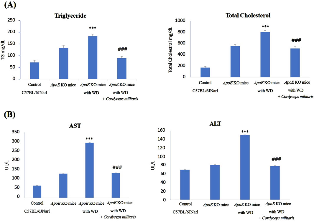
Figure 3. Biochemical analysis of blood lipid levels and liver function in ApoE KO mice under different experimental conditions. (A) The ApoE KO mice fed a Western diet (WD) showed significantly elevated levels of triglycerides and total cholesterol, which were reduced after this specially cultivated Cordyceps militaris treatment. (B) Elevated serum concentrations of aspartate aminotransferase (AST) and alanine aminotransferase (ALT), markers of liver dysfunction induced by WD, were significantly reduced in the Cordyceps militaris-treated group. Data were expressed as mean ± SD. *** = p < 0.001 compared to ApoE KO mice fed a standard diet group; ### = p < 0.001 compared to ApoE KO mice fed with WD without any treatment.
3.3 C. militaris reduced renal dysfunction in a mouse model of T2DM
DN is recognized as one of the primary and most serious complications of T2DM, often leading to progressive renal dysfunction. To evaluate the beneficial effects of this specially cultivated C. militaris on renal dysfunction in T2DM, various indicators of renal health were evaluated in this study. The values such as kidney weight, BUN and serum creatinine levels were measured to determine the extent of renal damage and the therapeutic potential of this specially cultivated C. militaris. The results showed that the fibrosis morphologies, including dark brown and shrinking morphology, were observed in ApoE KO mice fed with WD. Similarly, a significant reduction in kidney weight was observed in ApoE KO mice fed with WD, accompanied by markedly elevated levels of BUN and serum creatinine. (Figures 4A,B). Because glomerular abnormalities are the hallmark of DN and lead to structural destruction of the kidney (Kaya et al., 2021), histopathologic analysis of renal glomeruli was performed using Hematoxylin and eosin staining. The stained tissue sections revealed pronounced morphological abnormalities in renal glomeruli, in the WD-fed ApoE KO mice (Figure 5). Remarkably, treatment with this specially cultivated C. militaris was found to alleviate these pathological changes. In T2DM mice receiving C. militaris, kidney weight was improved. Similarly, elevated BUN and serum creatinine levels were significantly reduced (Figure 4). Histological examination further confirmed these results, as the structural integrity of renal glomeruli was visibly restored in C. militaris-treated mice, with morphology comparable to that of healthy controls (Figure 5). The data suggest that this specially cultivated C. militaris not only improved biochemical markers of renal health, but also facilitated the recovery of renal morphology associated with DN in a T2DM mouse model.
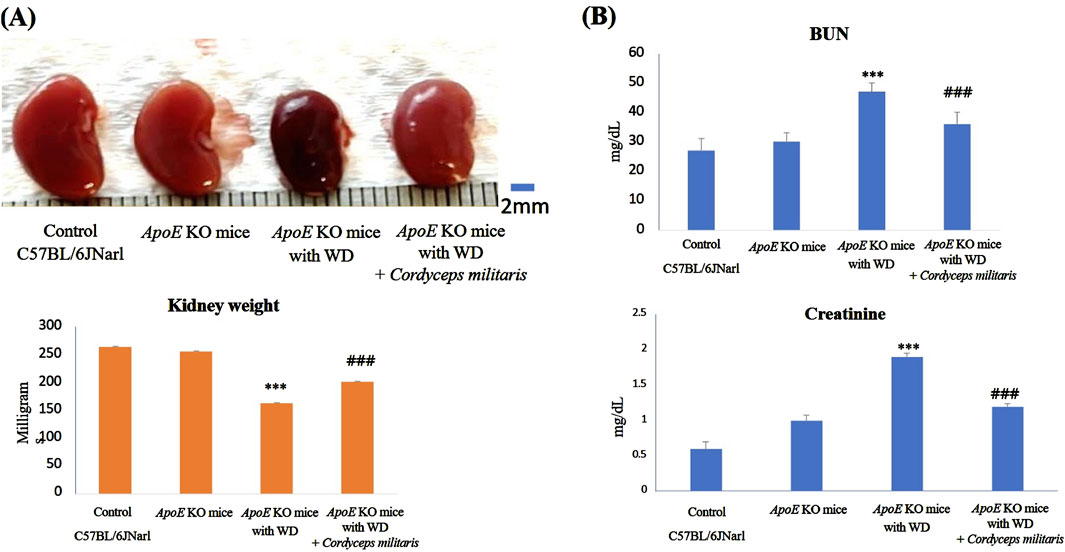
Figure 4. Evaluation of renal dysfunction and histopathologic changes in ApoE KO mice under different experimental conditions. (A) The ApoE KO mice fed a Western diet (WD) showed changes in renal morphology and a decrease in renal weight. (B) The ApoE KO mice showed significantly elevated blood urea nitrogen (BUN) and serum creatinine levels, indicating renal dysfunction. However, this specially cultivated Cordyceps militaris treatment improved the kidney and significantly decreased the BUN and serum creatinine levels. Data were expressed as mean ± SD. *** = P < 0.001 compared to ApoE KO mice fed a standard diet group; ### = P < 0.001 compared to ApoE KO mice with WD without any treatment.
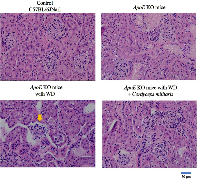
Figure 5. Restoration of renal glomerular morphology by Cordyceps militaris treatment in ApoE KO mice. Histopathological analysis using Hematoxylin and eosin staining showed severe morphological abnormalities in renal glomeruli, including shrinkage and atrophy, in WD-fed mice. These pathological changes were ameliorated in the Cordyceps militaris-treated group and the glomerular morphology resembled that of healthy controls.
3.4 Therapeutic effects of C. militaris on diabetes-induced renal fibrosis
Renal fibrosis is recognized as a hallmark pathological feature of DN and contributes significantly to its progression and severity (Xue et al., 2023). In this study, the potential therapeutic effects of this specially cultivated C. militaris on DM-induced renal fibrosis were thoroughly investigated by applying various tissue staining techniques and Western blot analysis. Masson’s trichrome staining (Figure 6A, blue area) and picrosirius red staining (Figure 6B, red area) were used to evaluate collagen deposition within the renal tissue. The staining results showed a significant increase in collagen accumulation in the kidneys of WD-fed ApoE KO mice. Remarkably, C. militaris treatment attenuated this collagen deposition as shown in the staining images (Figure 6). Furthermore, the expression of α-smooth muscle actin (α-SMA), a widely recognized marker of fibrosis, was significantly increased in the kidneys of ApoE KO mice fed with WD. However, after C. militaris administration, α-SMA levels were significantly reduced, highlighting the potential antifibrotic effects of this specially cultivated C. militaris in diabetic kidneys (Figure 7A). To further evaluate the extent of amyloid deposition, toluidine blue staining was performed. Toluidine blue staining revealed pronounced amyloid deposits in the renal glomeruli of WD-fed ApoE KO mice. Notably, C. militaris treatment attenuated these deposits, suggesting its protective role against amyloid accumulation in the kidneys (Figure 7B). The results of these histologic stains were further supported by Western blot analysis, which detects the expression of key fibrosis-related proteins. In the kidneys of WD-fed ApoE KO mice, proteins such as collagen type I (COL1), fibronectin, connective tissue growth factor (CTGF), tissue inhibitor of metalloproteinases (TIMPs), and transforming growth factor β1 (TGFβ1) were upregulated. Conversely, E-cadherin (Chang et al., 2022) and Smad7 (Li et al., 2002), key proteins known for their anti-fibrotic properties, was remarkably downregulated. Importantly, C. militaris treatment reversed these pathological trends, demonstrating a reduction in the levels of pro-fibrotic proteins and an upregulation of Smad7 expression (Figure 8). In addition, the study observed that C. militaris treatment increased the expression of Klotho, an anti-aging protein with renoprotective properties, which was decreased in the kidneys of WD-fed ApoE KO mice (Figure 8). Taken together, the reversal of fibrotic markers, the reduction of amyloid deposition, and the enhancement of protective protein expression highlighted the therapeutic potential of this specially cultivated C. militaris in mitigating DM-induced renal fibrosis and kidney damage in DN.
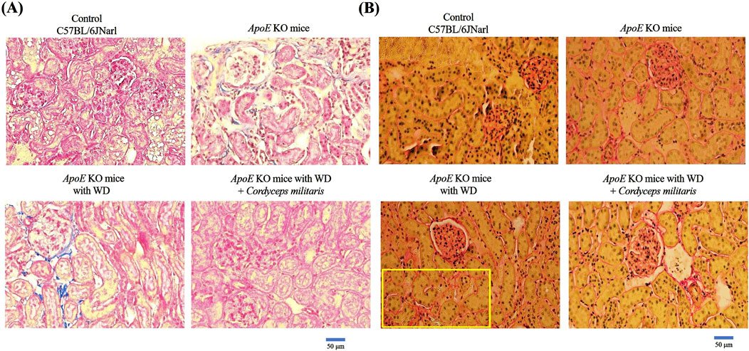
Figure 6. Evaluation of collagen deposition in renal tissue using histological staining. (A) Masson’s trichrome staining and (B) Picrosirius red staining revealed significant collagen accumulation in the kidneys of ApoE KO mice fed a Western diet (WD). This specially cultivated Cordyceps militaris treatment remarkably reduced collagen deposition as evidenced by reduced staining intensity and improved tissue morphology.
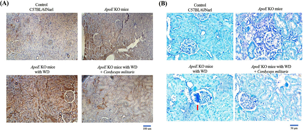
Figure 7. Analysis of fibrosis and amyloid deposition in the kidneys of ApoE KO mice. (A) Immunohistochemical staining revealed significantly increased expression of α-smooth muscle actin (α-SMA), a marker of fibrosis, in the kidneys of mice fed a Western diet (WD). This specially cultivated Cordyceps militaris treatment significantly reduced α-SMA expression, demonstrating its antifibrotic effects. (B) Toluidine blue staining showed extensive amyloid deposits in the renal glomeruli of WD-fed mice, which were attenuated after this specially cultivated Cordyceps militaris administration, indicating its protective role against amyloid accumulation.
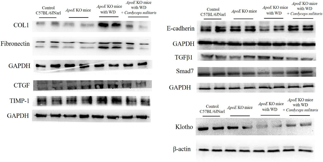
Figure 8. Western blot analysis of fibrosis-related proteins and Klotho expression in the kidneys of ApoE KO mice. Western diet (WD)-fed mice showed upregulation of fibrosis-associated proteins, including collagen type I (COL1), fibronectin, connective tissue growth factor (CTGF), tissue inhibitor of metalloproteinase 1 (TIMP-1), and transforming growth factor β1 (TGFβ1), and downregulation of anti-fibrotic proteins E-cadherin and Smad7. This specially cultivated Cordyceps militaris treatment reversed these trends by reducing pro-fibrotic proteins, restoring E-cadherin and Smad7 expression, and increasing Klotho level.
4 Discussion
A previous study mentions that C. militaris extracts significantly reduce blood glucose, cholesterol, and triglycerides, improve antioxidant capacity, and combat DN by reducing uric acid, creatinine, BUN, and urinary protein levels (Dong et al., 2014). The present study also demonstrates that C. militaris cultivated on G. biloba seeds has remarkable therapeutic potential by effectively addressing critical complications associated with DM. These included reducing hyperglycemia, alleviating dyslipidemia, and improving liver dysfunction. In addition, it specifically alleviated DN, a serious complication of DM, by reducing renal fibrosis while supporting renal function and metabolic balance. T2DM is a chronic metabolic disorder characterized by insulin resistance and relative insulin deficiency, resulting in elevated blood glucose levels (Galicia-Garcia et al., 2020). The present study demonstrates that this specially cultivated C. militaris effectively reduces serum glucose levels in a T2DM mouse model. This finding is consistent with previous research showing the hypoglycemic effects of C. militaris (Cheng et al., 2012). The diabetic serum lipid abnormalities commonly observed in T2DM are characterized by an atherogenic lipid profile with high levels of triglycerides, elevated low-density lipoprotein cholesterol, and decreased high-density lipoprotein cholesterol. This lipid imbalance significantly increases the risk of cardiovascular disease in people with DM (Kalra and Raizada, 2024). Moreover, an increase in total cholesterol is associated with a higher risk of DM-related cardiovascular diseases in diabetic patients, whereas a decrease in total cholesterol is associated with a lower risk of cardiovascular diseases (Khil et al., 2023). Non-alcoholic fatty liver disease has a significant and well-documented association with T2DM. It is estimated that fatty liver affects approximately 70%–80% of individuals diagnosed with T2DM, highlighting its high prevalence in this population. Diabetic dyslipidemia contributes significantly to the pathogenesis of fatty liver. The dysregulated lipid metabolism associated with diabetic dyslipidemia leads to excessive lipid accumulation in the liver, thereby promoting the development of hepatic steatosis (Han et al., 2019). Furthermore, associated liver enzymes such as AST and ALT are often significantly elevated in patients with diabetes, reflecting liver dysfunction (Al-Jameil et al., 2014). The current study confirms that WD significantly increases blood lipid levels (triglycerides and cholesterol) and causes liver dysfunction as indicated by elevated serum AST and ALT levels. However, treatment with this specially cultivated C. militaris improves these markers by reducing lipid abnormalities and mitigating liver damage. These results highlight the protective effects of C. militaris against T2DM-induced metabolic disorders, including dyslipidemia and liver injury. One study shows that patients with diabetic nephropathy have significantly higher levels of total cholesterol and triglycerides than those without nephropathy (Palazhy and Viswanathan, 2017). These findings underscore the link between lipid abnormalities and progression of diabetes-related kidney damage.
DN, the most common complication of DM, is the leading cause of end-stage renal disease in DM patients. Renal fibrosis mediated by TGF-β1 upregulation plays a key role in the onset and progression of diabetic nephropathy by promoting type I collagen synthesis and suppressing its degradation (Cheng et al., 2013; Zhang et al., 2021). Smad proteins are essential components of the downstream signaling pathway involved in TGF-β1-mediated renal fibrosis. While Smad2-4 are activated by TGF-β1 to drive renal fibrosis, Smad7, an inhibitory Smad, counteracts the process of renal fibrosis by suppressing the expression of fibrotic cytokines (Zhang et al., 2021). Additionally, the TGF-β signaling pathway promotes the downregulation of E-cadherin and the expression of mesenchymal markers such as α-SMA and fibronectin, leading to the transformation of injured renal epithelial cells into a mesenchymal-like state. This transition may contribute to increased extracellular matrix production and accumulation (Nagae et al., 2008; Gewin and Zent, 2012). CTGF is a known profibrotic mediator. It is also upregulated in human kidney proximal tubular cells (HKC-8) after TGF-β1 stimulation (Phanish et al., 2006). Additionally, TIMP-1, which inhibits interstitial collagenases and promotes fibrosis progression, is also induced by TGF-β1 stimulation (Zhou et al., 2007; Kim et al., 2018). Together, these studies demonstrate that TGF-β1 plays a central role in the initiation and progression of renal fibrosis. Moreover, Klotho, a protein known for its anti-aging properties, has been shown to be downregulated in T2DM and its associated complication, DN (Tang et al., 2023). In the current study, this specially cultivated C. militaris attenuates TGF-β1 expression to reduce fibrotic markers such as α-SMA, COL1, CTGF, TIMP-1 and fibronectin, and increases the expression of anti-fibrotic proteins such as Smad7 and E-cadherin to limit DM-induced renal fibrosis in a T2DM mouse model. Treatment with this specially cultivated C. militaris also increases Klotho expression. And then, this specially cultivated C. militaris presents ability to restore renal morphology and weight as well as normalize BUN and serum creatinine levels through changes in molecular mechanisms. Furthermore, amyloid plaques are observed in over 70% of patients with T2DM, and these deposits readily develop into mature fibrils (Smith et al., 2022). The kidney is one of the most common organs affected by amyloid deposition (Dember, 2006; Feitosa et al., 2022). The present study shows amyloid deposition in the renal glomeruli of WD-fed ApoE KO mice, which was reduced by the specially cultivated C. militaris treatment, indicating its potential protective effect in the kidney of T2DM. These findings highlight the therapeutic potential of this specially cultivated C. militaris in ameliorating renal injury and fibrosis in DN.
Although this study highlights the therapeutic potential of C. militaris cultivated on G. biloba seeds in mitigating T2DM and its related complications, it still has some research limitations. In terms of detection indicators, the focus is primarily on a few metabolic indicators, renal function indicators, and fibrosis-related indicators, with a lack of data on other physiological processes such as inflammatory factors and oxidative stress indicators. This limits a comprehensive understanding of the mechanism of action of C. militaris in the body. Protein detection is also limited to a few fibrosis-related and anti-fibrosis proteins and does not cover all key proteins involved in renal fibrosis. Regarding the experimental design, the lack of different dosage groups for C. militaris hinders the identification of an optimal therapeutic dose, reducing the clinical applicability of the results. Furthermore, the lack of comparative trials with clinical medications makes it difficult to evaluate the relative advantages of this specially cultivated C. militaris in clinical application. The study also relies on an animal model that may not fully capture the complexity of DN in humans, thus caution should be exercised in translating these results to clinical practice. In addition, the short duration of the study does not address the long-term safety and efficacy of C. militaris. Future research should include clinical trials to validate these findings in humans, explore potential side effects, and optimize dosing regimens. Nevertheless, the results underscore the promising potential of this specially cultivated C. militaris as a natural and effective therapeutic strategy for the management of T2DM and its associated complications.
5 Conclusion
In conclusion, this study highlighted the therapeutic potential of C. militaris cultivated on G. biloba seeds in the treatment of T2DM and its complications, especially DN. The results showed that this unique cultivation approach enhanced the bioactive properties of C. militaris, enabling it to effectively alleviate hyperglycemia, dyslipidemia, liver dysfunction, and renal damage. In addition, the treatment reduced renal fibrosis while promoting renal repair and metabolic balance. These results highlight the promise of this specially cultivated C. militaris as a natural and effective therapeutic option for T2DM and its associated complications, which warrants further investigation and clinical validation.
Data availability statement
The original contributions presented in the study are included in the article/Supplementary Material, further inquiries can be directed to the corresponding authors.
Ethics statement
The animal study was approved by the Institutional Animal Care and Use Committee of Hualien Tzu Chi Hospital (approval number: 113-43). The study was conducted in accordance with the local legislation and institutional requirements.
Author contributions
S-ZL: Conceptualization, Methodology, Project administration, Writing – review and editing. W-WK: Conceptualization, Methodology, Project administration, Writing – review and editing. BC-KT: Investigation, Methodology, Validation, Visualization, Writing – original draft. CP: Formal Analysis, Investigation, Methodology, Validation, Visualization, Writing – review and editing. C-HK: Formal Analysis, Investigation, Validation, Writing – review and editing. DJ-YH: Formal Analysis, Validation, Writing – review and editing. S-WK: Validation, Data curation, Writing – review and editing. P-YP: Data curation, Methodology, Writing – review and editing. S-JC: Funding acquisition, Resources, Visualization, Writing – review and editing. C-YH: Conceptualization, Funding acquisition, Project administration, Supervision, Writing – review and editing. K-HL: Conceptualization, Funding acquisition, Project administration, Supervision, Writing – review and editing.
Funding
The author(s) declare that financial support was received for the research and/or publication of this article. The authors acknowledge the China Medical University (CMU103-TC-03), China Medical University Hospital (DMR-108-057), Ministry of Science and Technology (Taiwan; MOST 110-2320-B-303-001-MY2 and MOST 111-2314-B-303-008-MY3), and National Science and Technology Council (Taiwan; NSTC 113-2811-B-303-002; NSTC 113-2314-B-303-024 and NSTC-113-2314-B-303-024) to support this study. The authors declare that this study received funding from Home Run Biotechnology Co., Ltd. (TCJ112-002). The funder was not involved in the study design, collection, analysis, interpretation of data, the writing of this article, or the decision to submit it for publication.
Acknowledgments
The authors gratefully acknowledge Prof. Tsung-Jung Ho of the Department of Traditional Chinese Medicine, Hualien Tzu Chi Hospital, Buddhist Tzu Chi Medical Foundation (Hualien, Taiwan), for his expertise in the morphological and related identification of this specially cultivated Cordyceps militaris. The authors also thank the Laboratory Animal Center at Tzu Chi University, Hualien, Taiwan, for their support in animal husbandry and care and the core facilities provided by Advanced Instrumentation Center of Department of Medicine Research, Hualien Tzu Chi Hospital, Buddhist Tzu Chi Medical Foundation, Hualien, Taiwan.
Conflict of interest
Author S-JC was employed by Home Run Biotechnology Co., Ltd.
The remaining authors declare that the research was conducted in the absence of any commercial or financial relationships that could be construed as a potential conflict of interest.
Generative AI statement
The author(s) declare that Generative AI was used in the creation of this manuscript. DeepL Writing was only used to check the English grammar error.
Publisher’s note
All claims expressed in this article are solely those of the authors and do not necessarily represent those of their affiliated organizations, or those of the publisher, the editors and the reviewers. Any product that may be evaluated in this article, or claim that may be made by its manufacturer, is not guaranteed or endorsed by the publisher.
Supplementary material
The Supplementary Material for this article can be found online at: https://www.frontiersin.org/articles/10.3389/fphar.2025.1562116/full#supplementary-material
Abbreviations
ALT, alanine aminotransferase; AST, aspartate aminotransferase; BNU, blood urea nitrogen; C. militaris, Cordyceps militaris; DM, Diabetes mellitus; DN, Diabetic nephropathy; G. biloba, Ginkgo biloba; T2DM, type 2 diabetes mellitus; WD, western diet.
References
Al-Jameil, N., Khan, F. A., Arjumand, S., Khan, M. F., and Tabassum, H. (2014). Associated liver enzymes with hyperlipidemic profile in type 2 diabetes patients. Int. J. Clin. Exp. Pathol. 7, 4345–4349.
Beckman, J. A., and Creager, M. A. (2016). Vascular complications of diabetes. Circ. Res. 118, 1771–1785. doi:10.1161/CIRCRESAHA.115.306884
Belwal, T., Giri, L., Bahukhandi, A., Tariq, M., Kewlani, P., Bhatt, I. D., et al. (2019). “Ginkgo biloba,” in Nonvitamin and nonmineral nutritional supplements. New York, NY, United States, Elsevier, 241–250.
Biernacka, P., Adamska, I., and Felisiak, K. (2023). The potential of ginkgo biloba as a source of biologically active compounds-A review of the recent literature and patents. Molecules 28, 3993. doi:10.3390/molecules28103993
Boateng, I. D., and Yang, X. M. (2021). Effect of different drying methods on product quality, bioactive and toxic components of Ginkgo biloba L. seed. J. Sci. Food Agric. 101, 3290–3297. doi:10.1002/jsfa.10958
Chang, W. S., Tsai, C. W., Yang, J. S., Hsu, Y. M., Shih, L. C., Chiu, H. Y., et al. (2021). Resveratrol inhibited the metastatic behaviors of cisplatin-resistant human oral cancer cells via phosphorylation of ERK/p-38 and suppression of MMP-2/9. J. Food Biochem. 45, e13666. doi:10.1111/jfbc.13666
Chang, Y.-M., Velmurugan, B. K., Kuo, W.-W., Chen, Y.-S., Ho, T.-J., Tsai, C.-T., et al. (2013). Inhibitory effect of alpinate Oxyphyllae fructus extracts on Ang II-induced cardiac pathological remodeling-related pathways in H9c2 cardiomyoblast cells. BioMedicine 3, 148–152. doi:10.1016/j.biomed.2013.05.001
Chang, Y. T., Chung, M. C., Chang, C. H., Chiu, K. H., Shieh, J. J., and Wu, M. J. (2022). Anti-EMT and anti-fibrosis effects of protocatechuic aldehyde in renal proximal tubular cells and the unilateral ureteral obstruction animal model. Pharm. Biol. 60, 1198–1206. doi:10.1080/13880209.2022.2088809
Chen, D. D., Xu, R., Zhou, J. Y., Chen, J. Q., Wang, L., Liu, X. S., et al. (2019). Cordyceps militaris polysaccharides exerted protective effects on diabetic nephropathy in mice via regulation of autophagy. Food Funct. 10, 5102–5114. doi:10.1039/c9fo00957d
Cheng, X., Gao, W., Dang, Y., Liu, X., Li, Y., Peng, X., et al. (2013). Both ERK/MAPK and TGF-Beta/Smad signaling pathways play a role in the kidney fibrosis of diabetic mice accelerated by blood glucose fluctuation. J. Diabetes Res. 2013, 463740. doi:10.1155/2013/463740
Cheng, Y. W., Chen, Y. I., Tzeng, C. Y., Chen, H. C., Tsai, C. C., Lee, Y. C., et al. (2012). Extracts of Cordyceps militaris lower blood glucose via the stimulation of cholinergic activation and insulin secretion in normal rats. Phytother. Res. 26, 1173–1177. doi:10.1002/ptr.3709
Chiang, C. J., Tsai, B. C., Lu, T. L., Chao, Y. P., Day, C. H., Ho, T. J., et al. (2021). Diabetes-induced cardiomyopathy is ameliorated by heat-killed Lactobacillus reuteri GMNL-263 in diabetic rats via the repression of the toll-like receptor 4 pathway. Eur. J. Nutr. 60, 3211–3223. doi:10.1007/s00394-020-02474-z
Clemente-Suárez, V. J., Beltrán-Velasco, A. I., Redondo-Flórez, L., Martín-Rodríguez, A., and Tornero-Aguilera, J. F. (2023). Global impacts of western diet and its effects on metabolism and health: a narrative review. Nutrients 15, 2749. doi:10.3390/nu15122749
Dember, L. M. (2006). Amyloidosis-associated kidney disease. J. Am. Soc. Nephrol. 17, 3458–3471. doi:10.1681/ASN.2006050460
Dong, Y., Jing, T., Meng, Q., Liu, C., Hu, S., Ma, Y., et al. (2014). Studies on the antidiabetic activities of Cordyceps militaris extract in diet-streptozotocin-induced diabetic Sprague-Dawley rats. Biomed. Res. Int. 2014, 160980. doi:10.1155/2014/160980
Feitosa, V. A., Neves, P., Jorge, L. B., Noronha, I. L., and Onuchic, L. F. (2022). Renal amyloidosis: a new time for a complete diagnosis. Braz J. Med. Biol. Res. 55, e12284. doi:10.1590/1414-431X2022e12284
Galicia-Garcia, U., Benito-Vicente, A., Jebari, S., Larrea-Sebal, A., Siddiqi, H., Uribe, K. B., et al. (2020). Pathophysiology of type 2 diabetes mellitus. Int. J. Mol. Sci. 21, 6275. doi:10.3390/ijms21176275
Gewin, L., and Zent, R. (2012). How does TGF-β mediate tubulointerstitial fibrosis? Semin. Nephrol. 32, 228–235. doi:10.1016/j.semnephrol.2012.04.001
Han, J. M., Kim, H. I., Lee, Y. J., Lee, J. W., Kim, K. M., and Bae, J. C. (2019). Differing associations between fatty liver and dyslipidemia according to the degree of hepatic steatosis in korea. J. Lipid Atheroscler. 8, 258–266. doi:10.12997/jla.2019.8.2.258
Ho, T. J., Ahmed, T., Shibu, M. A., Lin, Y. J., Shih, C. Y., Lin, P. Y., et al. (2024). A prospective review of the health-promoting potential of Jing Si Herbal Tea. Tzu Chi Med. J. 36, 1–22. doi:10.4103/tcmj.tcmj_194_23
Jing, F.-Y., Zhou, Y.-Z., Wang, H.-Y., Yin, X.-L., and Zhang, Y.-Q. (2021). Enhancing antioxidant and anti-hyperglycaemic functions of gingko biloba L. seeds using thermal detoxification. J. Funct. Foods 87, 104819. doi:10.1016/j.jff.2021.104819
Kalra, S., and Raizada, N. (2024). Dyslipidemia in diabetes. Indian Heart J. 76 (Suppl. 1), S80–s82. doi:10.1016/j.ihj.2023.11.002
Kaya, B., Paydas, S., Kuzu, T., Basak Tanburoglu, D., Balal, M., Eren Erdogan, K., et al. (2021). Primary glomerulonephritis in diabetic patients. Int. J. Clin. Pract. 75, e13713. doi:10.1111/ijcp.13713
Khil, J., Kim, S. M., Chang, J., Choi, S., Lee, G., Son, J. S., et al. (2023). Changes in total cholesterol level and cardiovascular disease risk among type 2 diabetes patients. Sci. Rep. 13, 8342. doi:10.1038/s41598-023-33743-6
Kim, K. K., Sheppard, D., and Chapman, H. A. (2018). TGF-β1 signaling and tissue fibrosis. Cold Spring Harb. Perspect. Biol. 10, a022293. doi:10.1101/cshperspect.a022293
Lai, C. H., Van Thao, D., Tsai, B. C., Hsieh, D. J., Chen, M. Y., Kuo, W. W., et al. (2023). Insulin-like growth factor II receptor alpha overexpression in heart aggravates hyperglycemia-induced cardiac inflammation and myocardial necrosis. Environ. Toxicol. 38, 676–684. doi:10.1002/tox.23717
Lattouf, R., Younes, R., Lutomski, D., Naaman, N., Godeau, G., Senni, K., et al. (2014). Picrosirius red staining: a useful tool to appraise collagen networks in normal and pathological tissues. J. Histochem Cytochem 62, 751–758. doi:10.1369/0022155414545787
Li, J. H., Zhu, H. J., Huang, X. R., Lai, K. N., Johnson, R. J., and Lan, H. Y. (2002). Smad7 inhibits fibrotic effect of TGF-Beta on renal tubular epithelial cells by blocking Smad2 activation. J. Am. Soc. Nephrol. 13, 1464–1472. doi:10.1097/01.asn.0000014252.37680.e4
Lin, J. Y., Tsai, B. C., Kao, H. C., Chiang, C. Y., Chen, Y. A., Chen, W. S., et al. (2023). Neuroprotective effects of probiotic lactobacillus reuteri GMNL-263 in the Hippocampus of streptozotocin-induced diabetic rats. Probiotics Antimicrob. Proteins 15, 1287–1297. doi:10.1007/s12602-022-09982-w
Lin, T. K., Tsai, C. L., Tsai, B. C., Kuo, C. H., Ho, T. J., Hsieh, D. J., et al. (2024). Low-concentration imiquimod treatment promotes enhanced skin barrier functions through epidermal melanization reaction regulation. Environ. Toxicol. 39, 4360–4371. doi:10.1002/tox.24332
Lin, W. Y., Tsai, B. C. K., Day, C. H., Chiu, P. L., Chen, R. J., Chen, M. Y. C., et al. (2021). Arecoline induces heart injure via Fas/Fas ligand apoptotic pathway in heart of Sprague–Dawley rat. Environ. Toxicol. 36, 1567–1575. doi:10.1002/tox.23153
Liu, S. P., Shibu, M. A., Tsai, F. J., Hsu, Y. M., Tsai, C. H., Chung, J. G., et al. (2020). Tetramethylpyrazine reverses high-glucose induced hypoxic effects by negatively regulating HIF-1α induced BNIP3 expression to ameliorate H9c2 cardiomyoblast apoptosis. Nutr. Metab. (Lond) 17, 12. doi:10.1186/s12986-020-0432-x
Liu, X., Dun, M., Jian, T., Sun, Y., Wang, M., Zhang, G., et al. (2023). Cordyceps militaris extracts and cordycepin ameliorate type 2 diabetes mellitus by modulating the gut microbiota and metabolites. Front. Pharmacol. 14, 1134429. doi:10.3389/fphar.2023.1134429
Liu, Y., Xin, H., Zhang, Y., Che, F., Shen, N., and Cui, Y. (2022). Leaves, seeds and exocarp of Ginkgo biloba L. (Ginkgoaceae): a Comprehensive Review of Traditional Uses, phytochemistry, pharmacology, resource utilization and toxicity. J. Ethnopharmacol. 298, 115645. doi:10.1016/j.jep.2022.115645
Maggiore, U., Budde, K., Heemann, U., Hilbrands, L., Oberbauer, R., Oniscu, G. C., et al. (2017). Long-term risks of kidney living donation: review and position paper by the ERA-EDTA DESCARTES working group. Nephrol. Dial. Transpl. 32, 216–223. doi:10.1093/ndt/gfw429
Nagae, T., Mori, K., Mukoyama, M., Kasahara, M., Yokoi, H., Suganami, T., et al. (2008). Adrenomedullin inhibits connective tissue growth factor expression, extracellular signal-regulated kinase activation and renal fibrosis. Kidney Int. 74, 70–80. doi:10.1038/ki.2008.98
Niculae, A., Peride, I., Vinereanu, V., Rădulescu, D., Bratu, O. G., Geavlete, B. F., et al. (2017). Nephrotic syndrome secondary to amyloidosis in a patient with monoclonal gammopathy with renal significance (MGRS). Rom. J. Morphol. Embryol. 58, 1065–1068.
Ohno, T., Miyasaka, Y., Yoshida, K., Kobayashi, M., Horio, F., Yokoi, N., et al. (2022). A novel model mouse for type 2 diabetes mellitus with early onset and persistent hyperglycemia. Exp. Anim. 71, 510–518. doi:10.1538/expanim.22-0061
Palazhy, S., and Viswanathan, V. (2017). Lipid abnormalities in type 2 diabetes mellitus patients with overt nephropathy. Diabetes Metab. J. 41, 128–134. doi:10.4093/dmj.2017.41.2.128
Phanish, M. K., Wahab, N. A., Colville-Nash, P., Hendry, B. M., and Dockrell, M. E. (2006). The differential role of Smad2 and Smad3 in the regulation of pro-fibrotic TGFbeta1 responses in human proximal-tubule epithelial cells. Biochem. J. 393, 601–607. doi:10.1042/BJ20051106
Sagoo, M. K., and Gnudi, L. (2020). “Diabetic nephropathy: an overview,” in Diabetic nephropathy: methods and protocols. New York, NY, United States, Humana Press Inc, 3–7.
Samsu, N. (2021). Diabetic nephropathy: challenges in pathogenesis, diagnosis, and treatment. Biomed. Res. Int. 2021, 1497449. doi:10.1155/2021/1497449
Smith, A. A., Moore, K. B. E., Ambs, P. M., Saraswati, A. P., and Fortin, J. S. (2022). Recent advances in the discovery of therapeutics to curtail islet amyloid polypeptide aggregation for type 2 diabetes treatment. Adv. Biol. (Weinh) 6, e2101301. doi:10.1002/adbi.202101301
Tang, A., Zhang, Y., Wu, L., Lin, Y., Lv, L., Zhao, L., et al. (2023). Klotho's impact on diabetic nephropathy and its emerging connection to diabetic retinopathy. Front. Endocrinol. (Lausanne) 14, 1180169. doi:10.3389/fendo.2023.1180169
Tsai, B.C.-K., Hsieh, D.J.-Y., Lin, W.-T., Tamilselvi, S., Day, C. H., Ho, T.-J., et al. (2020a). Functional potato bioactive peptide intensifies Nrf2-dependent antioxidant defense against renal damage in hypertensive rats. Food Res. Int. 129, 108862. doi:10.1016/j.foodres.2019.108862
Tsai, B.C.-K., Kuo, W.-W., Day, C. H., Hsieh, D.J.-Y., Kuo, C.-H., Daddam, J., et al. (2020b). The soybean bioactive peptide VHVV alleviates hypertension-induced renal damage in hypertensive rats via the SIRT1-PGC1α/Nrf2 pathway. J. Funct. Foods 75, 104255. doi:10.1016/j.jff.2020.104255
Xue, S., Li, Y. X., Lu, X. X., and Tang, W. (2023). Dapagliflozin can alleviate renal fibrosis in rats with streptozotocin-induced type 2 diabetes mellitus. Exp. Ther. Med. 26, 572. doi:10.3892/etm.2023.12271
Yu, S. H., Dubey, N. K., Li, W. S., Liu, M. C., Chiang, H. S., Leu, S. J., et al. (2016). Cordyceps militaris treatment preserves renal function in type 2 diabetic nephropathy mice. PLoS One 11, e0166342. doi:10.1371/journal.pone.0166342
Zeng, J., Zhou, Y., Lyu, M., Huang, X., Xie, M., Huang, M., et al. (2024). Cordyceps militaris: a novel mushroom platform for metabolic engineering. Biotechnol. Adv. 74, 108396. doi:10.1016/j.biotechadv.2024.108396
Zhang, J., Wen, C., Duan, Y., Zhang, H., and Ma, H. (2019). Advance in Cordyceps militaris (Linn) Link polysaccharides: isolation, structure, and bioactivities: a review. Int. J. Biol. Macromol. 132, 906–914. doi:10.1016/j.ijbiomac.2019.04.020
Zhang, Y., Jin, D., Kang, X., Zhou, R., Sun, Y., Lian, F., et al. (2021). Signaling pathways involved in diabetic renal fibrosis. Front. Cell Dev. Biol. 9, 696542. doi:10.3389/fcell.2021.696542
Zheng, R., Zhang, W., Song, J., Zhong, Y., and Zhu, R. (2023). Cordycepin from Cordyceps militaris ameliorates diabetic nephropathy via the miR-193b-5p/MCL-1 axis. Chin. Med. 18, 134. doi:10.1186/s13020-023-00842-5
Keywords: diabetes mellitus, diabetic nephropathy, Cordyceps militaris, Ginkgo biloba seeds, kidney
Citation: Lin S-Z, Kuo W-W, Tsai BC-K, Paul CR, Kuo C-H, Hsieh DJ-Y, Kao S-W, Pai P-Y, Chen S-J, Huang C-Y and Lin K-H (2025) Therapeutic potential of Cordyceps militaris cultivated with Ginkgo biloba seeds for alleviating western diet-induced type 2 diabetes and diabetic nephropathy. Front. Pharmacol. 16:1562116. doi: 10.3389/fphar.2025.1562116
Received: 04 February 2025; Accepted: 28 April 2025;
Published: 29 May 2025.
Edited by:
Ruiwen Zhang, University of Houston, United StatesReviewed by:
Xueqi Liu, First Affiliated Hospital of Anhui Medical University, ChinaJiahong Wang, Shenyang Pharmaceutical University, China
Copyright © 2025 Lin, Kuo, Tsai, Paul, Kuo, Hsieh, Kao, Pai, Chen, Huang and Lin. This is an open-access article distributed under the terms of the Creative Commons Attribution License (CC BY). The use, distribution or reproduction in other forums is permitted, provided the original author(s) and the copyright owner(s) are credited and that the original publication in this journal is cited, in accordance with accepted academic practice. No use, distribution or reproduction is permitted which does not comply with these terms.
*Correspondence: Chih-Yang Huang, Y3lodWFuZ0BtYWlsLmNtdS5lZHUudHc=; Kuan-Ho Lin, bGluODM5QG1haWwuY211LmVkdS50dw==
†These authors have contributed equally to this work
†ORCID: Chih-Yang Huang, https://orcid.org/0000-0003-2347-0411
 Shinn-Zong Lin
 Wei-Wen Kuo3,4,5
 Chia-Hua Kuo
 Chih-Yang Huang