- 1Guangxi Subtropical Crops Research Institute, Guangxi Academy of Agricultural Sciences, Nanning, China
- 2Guangxi Key Laboratory of Quality and Safety Control for Subtropical Fruits, Guangxi Subtropical Crops Research Institute, Nanning, China
- 3Institute of Molecular Plant Sciences, School of Biological Sciences, University of Edinburgh, Edinburgh, United Kingdom
Chronic inflammation and oxidative stress are pivotal drivers of pathological conditions, necessitating safer plant-derived therapeutic alternatives. This study elucidates the anti-inflammatory and antioxidant mechanisms of Nymphaea “Eldorado” flower water extract (NEWE) in a zebrafish (Danio rerio) model of copper sulfate (CuSO4)-induced inflammation. NEWE (25–100 μg/mL) attenuates CuSO4-triggered neutrophil migration and oxidative stress by downregulating proinflammatory genes (il1b, ptgs2a/b) and reducing reactive oxygen species (ROS) production. Transcriptomic profiling identified 339 differentially expressed genes (DEGs), enriched in cytokine signaling and redox regulation, with serpine1, stat3, and mmp9 emerging as key regulatory hubs. Widely targeted metabolomics revealed 891 bioactive compounds, including flavonoids and phenylpropanoids, with network pharmacology predicting multi-target interactions involving inflammatory and oxidative stress pathways. Molecular docking confirmed binding affinities between protocatechuic acid, L-pyroglutamic acid, and Serpine1’s active site, indicating direct interference with inflammation modulation. Collectively, these results establish NEWE as a polypharmacological agent that disrupts inflammation-oxidative stress crosstalk primarily through Serpine1-mediated pathways, offering a molecular foundation for plant-derived interventions against chronic inflammatory diseases.
1 Introduction
Inflammation and oxidative stress are hallmarks of chronic diseases. Chronic inflammation can lead to tissue damage and pathological conditions such as cardiovascular disease, diabetes, and cancer (Dhabhar, 2014; Edlow et al., 2022; Garbarino et al., 2021; Porter et al., 2020; Prata et al., 2024). Oxidative stress is caused by the disruption of redox homeostasis, causing excessive reactive oxygen species (ROS) production, cellular damage, and systemic inflammation (Jin and Kang, 2024; Vo et al., 2011), potentially leading to chronic diseases, including neurodegenerative disorders and metabolic syndrome (Lee et al., 2024). Environmental factors, poor diet, and chronic stress exacerbate these processes, heightening the risk of disease development (Lee et al., 2024; Prata et al., 2024). Nonsteroidal anti-inflammatory drugs and corticosteroids are commonly used to reduce inflammation (Ju et al., 2022; Sapieha et al., 2011; Zhou et al., 2014). However, the long-term use of these medications may cause adverse effects, including gastrointestinal bleeding, acute kidney injury, and increased cardiovascular risk (Atiquzzaman et al., 2019; Laine, 2001; Misurac et al., 2013). These detrimental effects underscore the urgent need to develop alternative treatments with plant-based anti-inflammatory and antioxidant compounds, aiming to minimize toxicity and the incidence of adverse effects (Haj et al., 2015; Leu et al., 2019).
Water lilies (Nymphaea L. Nymphaeaceae) are a rich source of bioactive compounds, including flavonoids, alkaloids, and phenols, exhibiting diverse therapeutic effects, including antimicrobial and antioxidant activities (Hsu et al., 2013; Ren et al., 2019; Sheichenko et al., 2019). Thus, determining the pharmacological activities and mechanisms of action of Nymphaea extracts remains to be investigated to explore their potential as anti-inflammation and anti-oxidative stress agents.
Zebrafish (Danio rerio) is a valuable model organism for natural product research to its genetic and physiological conservation with humans, rapid development, and transparent embryos that allow the visualization of biological processes and the trafficking of fluorescent cells in vivo (Howe et al., 2013; Postlethwait et al., 1998). Zebrafish have unique advantages over rodent models, including higher fecundity, cost-effectiveness, and suitability for high-throughput screening (Ali et al., 2011; Choi et al., 2021). Thus, zebrafish models are ideal for evaluating the pharmacological properties of plant compounds. Specifically, these models are used to assess the ability of plant compounds to modulate the copper sulfate (CuSO4)-induced activation and recruitment of fluorescently labeled neutrophils, thereby accelerating the discovery and development of new therapeutics (Singh et al., 2022; Xie et al., 2020; Zhang et al., 2023a).
This study investigated the anti-inflammatory and antioxidant effects of water lily (Nymphaea “Eldorado”) flower water extract (NEWE) using a zebrafish model of copper sulfate (CuSO4)-induced inflammation. Genes and metabolic pathways regulated by extract components were identified by transcriptomic and metabolomic analyses. Potential molecular targets of bioactive compounds were identified by network pharmacology and molecular docking. The results offer valuable insights into the mechanisms of action of NEWE, supporting its potential therapeutic use in reducing inflammation and oxidative stress to improve human health and wellbeing.
2 Materials and methods
2.1 Sample extraction
Fresh flowers of Nymphaea “Eldorado” were sourced from the Liuzhou Shixian Planting and Breeding Cooperative (Liuzhou, China). Fresh flowers on the first day of flowering were collected and washed with water (Supplementary Figure S1A). The flowers were placed in a container filled with water and exposed to daylight for 24 h. The flowers were opened, and stalks and sepals were removed. The ovary was cut by 1/3, and the flowers were dried at 70°C–80°C until the moisture content was less than 10% (Supplementary Figure S1B). The dried flowers were crushed in a grinder, and the powder was sieved through a 20-mesh sieve and stored at −20°C for later use.
The extract (5,120 mg) was placed in a 1 L flask and mixed with 400 mL of water (yielding a solid-to-solvent ratio of 1:78, w/v) at 100°C. After standing for 30 min at room temperature, the mixture was centrifuged. The supernatant was collected, and the pellet was resuspended in 400 mL of water at 100 C. After standing for 30 min, the sample was centrifuged as described above. The supernatant was collected and combined with the first supernatant. The stock solution (6,400 mg/L) was stored at −20°C.
2.2 Toxicity assessment
The stock solution was diluted to obtain working concentrations (100, 200, 400, 800, 1600, 3200, and 6,400 mg/L) (Supplementary Figure S1). At 72 h post-fertilization (hpf), AB strain wild-type zebrafish embryos were placed in 24-well plates with 10 embryos per well and three replicates per concentration (Supplementary Figure S1C). The culture medium was removed, and 2 mL of NEWE was added to each well. Based on the results of these assays, NEWE concentrations that were not toxic to zebrafish embryos were used in subsequent experiments (Supplementary Figure S1). All procedures involving animals were approved by the Animal Care and Use Committee of the Guangxi Academy of Agricultural Sciences and complied with Chinese government regulations.
2.3 Analysis of neutrophil migration
This study used the transgenic zebrafish line Tg(mpx:GFP), which expresses GFP under the neutrophil-specific myeloperoxidase promoter, as described previously (Renshaw et al., 2006). Zebrafish embryos were placed in 90 mm dishes containing 1×E3 medium (pH was adjusted to 7.0–7.2 using 5 mM NaCl, 0.33 mM CaCl2, 0.17 mM KCl, and 0.33 mM MgSO4) with 100 embryos per dish. At 24 hpf, dead embryos were removed, and healthy embryos were added. At 72 hpf, normal embryos were transferred to 24-well culture plates with 10 embryos per well and incubated with 20 μM CuSO4 + NEWE (0, 25, 50, 75, 100 μg/mL) for 2 h at 28°C ± 1°C to induce neutrophil migration. GFP-expressing neutrophils in the anterior yolk sac were observed under a fluorescence microscope (Axio Imager M2, Zeiss) at 2 h after CuSO4 + NEWE treament to evaluate the degree of inflammation.
2.4 Measurement of antioxidant activity
WT zebrafish embryos were incubated with NEWE (0, 25, 50, 75, 100, 1255 μg/mL) or 100 μM quercetin (positive control) for 1 h at 28°C ± 1°C, followed by treatment with 10 μM CuSO4 for 20 min as described in Section 2.3. After washing with culture medium, embryos were incubated with 20 μg/mL of the fluorescent indicator 2′,7′-dichlorodihydrofluorescin diacetate (DCFH-DA) for 1 h at 28°C ± 1°C. CuSO4-induced ROS in embryos was detected by the formation of the oxidized compound dichlorofluorescein by fluorescence microscopy (Axio Imager M2, Zeiss) (Andoh et al., 2006; Girard-Lalancette et al., 2009; Zhang et al., 2023a; Zhang et al., 2023b).
2.5 RNA extraction and quantitative real-time polymerase chain reaction (qRT-PCR)
Tg(mpx:GFP) zebrafish embryos were incubated with 20 μM 20 μM CuSO4+NEWE 2 h at 28°C ± 1°C as described in Section 2.3. RNA from each sample (50 μg/mL) was extracted using TRIzol and reverse transcribed using the PrimeScript RT reagent kit. qRT-PCR was performed using the TB Green Premix Ex Taq II reagent kit (TaKaRa, Dalian, China) on the LightCycler 480 II System (Roche, Indianapolis, IN, United States). The cycling conditions were as follows: initial denaturation at 95°C for 30 s, followed by 40 cycles of 95°C for 5 s and 60°C for 20 s. Melting curves were generated by holding at 95°C for 5 s and 65°C for 1 min and increasing to 95°C at a rate of 0.1°C per second. The primers used in this study are listed in Supplementary Table S1. Relative gene expression levels were quantified using the 2−ΔΔCT method (Livak and Schmittgen, 2001). The data were visualized using Origin 2021 (OriginLab, Northampton, MA, United States).
2.6 Library construction and RNA-seq analysis
Tg(mpx:GFP) zebrafish embryos were cultured as described in Section 2.3. Three treatment groups were established: control (untreated), model (treated with CuSO4), and treatment (treated with 20 µM CuSO4 + 50 μg/mL NEWE).
The construction of sequencing libraries and RNA-seq were performed using the Illumina HiSeq platform (Biomarker Biotechnology Corporation Co., Beijing, China). High-quality reads were assembled using StringTie. Genes with log2 fold-change >2.0 or < −2.0 and false discovery rate (FDR) < 0.01 were considered differentially expressed genes (DEGs). DEGs were identified using edgeR (https://bioconductor.org/packages/release/bioc/html/edgeR.html). Relative transcript abundance was measured as fragments per kilobase of exon per million fragments mapped using Cuffdiff, with an FPKM threshold set at a minimum of 1.0 to ensure the reliability of expression levels. Histograms, scatter plots, volcano plots, Venn diagrams, and heatmaps were generated on the Biomarker Cloud Platform (Biomarker Biotechnology Corporation Co., Beijing, China) or https://hiplot.com.cn/cloud-tool/drawing-tool/list.
2.7 Widely targeted metabolomic analysis
The stock solution was diluted to 50 mg/L in culture medium. Metabolomics analyses were performed by MetWare Biotechnology (Wuhan, China). Metabolite extraction, identification, and quantification were performed as described previously (Yuan et al., 2018). Samples were freeze-dried, ground into powder, and dissolved in 70% methanol as internal standard (1:30 v/v). The samples were centrifuged at 12,000 rpm for 3 min at 4 C, and the supernatant was filtered through a 0.22 μm membrane for analysis.
Metabolites were analyzed by ultra-performance liquid chromatography (Exion LC AD System, AB Sciex, Darmstadt, Germany) coupled with electrospray tandem mass spectrometry (ESI-Q TRAP-MS/MS (AB SCIEX Q TRAP 4000, Applied Biosystems, Foster City, CA, United States). Chromatographic separation was performed on an SB-C18 column (1.8 µm, 2.1 mm × 100 mm, Agilent). Elution was carried out using a system consisting of solvent A (0.1% formic acid in water) and solvent B (0.1% formic acid in acetonitrile) and a linear gradient from 95% A to 95% B in 9 min, 95% B for 1 min, 95% B to 95% A in 1.1 min, and 95% A in 2.9 min. The flow rate was 0.35 mL/min at 40°C, and the injection volume was 2 μL. The ESI conditions were as follows: source temperature, 550 C; ion spray voltage (IS), 5500 V (positive mode)/-4500 V (negative mode); gas pressure of 50, 60, and 25 psi for GS1, GS2, and CUR gases, respectively. The collision-activated dissociation was set to high. Multiple reaction monitoring mode was utilized for metabolite detection, with declustering potential and collision energy individually optimized for each transition.
Metabolites abundance was categorized into two groups (classes I and II) based on peak areas.
2.8 Prediction of target genes and network construction
Genes potentially targeted by metabolites were identified using the Traditional Chinese Medicine Systems Pharmacology Database and Analysis Platform (https://old.tcmsp-e.com/tcmsp.php) and SwissTargetsPrediction (http://www.swisstargetprediction.ch/). Drug-target networks integrating transcriptomic and metabolomics data were constructed using Cytoscape version 3.9.1 (Li et al., 2022).
2.9 Molecular docking
The molecular structure of L-pyroglutamic acid, monomyristin, and protocatechuic acid were obtained from the PubChem database (https://pubchem.ncbi.nlm.nih.gov/). The 3D structure of the target protein, Serpine1, also known as plasminogen activator inhibitor-1, was obtained from the Protein Data Bank (https://www.rcsb.org/structure/4DTE). Protein-metabolite interactions were predicted using AutoDock Tools version 1.5.7, which applies a semi-flexible docking approach to predict binding affinities by exploring ligand and receptor flexibility. The docking grid was centered on the protein’s active site, with grid spacing and box size parameters optimized to encapsulate potential binding pockets. For each ligand, binding modes were generated based on the lowest binding energy utilizing the Lamarckian Genetic Algorithm as the primary search strategy, which has been validated in several studies for its efficacy in docking simulations. The AutoDock results were visualized and analyzed using PyMOL version 4.6.0 (Morris et al., 2009; Seeliger and de Groot, 2010), allowing detailed observation of ligand binding conformations, hydrogen bonding, and hydrophobic interactions, providing insights into the stability and specificity of ligand binding to Serpine1.
2.10 Statistical analysis
Data normality was assessed using the Kolmogorov–Smirnov test. Normally distributed continuous variables were expressed as mean ± standard deviations, and the significance of differences between means was determined using paired Student’s t-test or one-way analysis of variance followed by Tukey’s post hoc test. Differences between groups were compared using the Kruskal–Wallis test and Dunn–Sidak multiple comparison test. Box plots were drawn using Origin 2021 (OriginLab, Northampton, MA, United States). P-values of less than 0.05 were considered statistically significant.
3 Results
3.1 Effect of NEWE on CuSO4-induced inflammation in zebrafish larvae
Acute toxicity assessment revealed that the 24-h LC50 of NEWE for 72 h post-fertilization (hpf) zebrafish embryos was 3200 μg/mL (Supplementary Figure S1D). Based on this safety profile, we evaluated the anti-inflammatory potential of safety concentrations of NEWE using a copper sulfate (CuSO4) induced inflammation model in 3 days post-fertilization (dpf) larvae, this model utilizes the chemotactic properties of CuSO4 to disrupt neutrophil homeostasis, enabling real-time tracking of GFP-labeled neutrophil migration toward lateral line neuromasts - a validated method for quantifying inflammatory responses in zebrafish (Renshaw et al., 2006). As show in Figure 1A, neutrophils primarily localized to the caudal hematopoietic tissue in the control group, consistent with normal distribution patterns (Zhang et al., 2023b), but CuSO4 treatment prompted neutrophil migration towards the horizontal midline, where they formed clusters near the lateral line neuromasts (Figure 1B), indicating a pronounced inflammatory response. However, treatment with varying concentrations of NEWE effectively inhibited CuSO4-induced neutrophil migration in a does dependent manner, suggesting a potential anti-inflammatory activity (Figure 1B). Further, we investigated the effect of NEWE on expression of inflammatory genes using qRT-PCR, and found CuSO4 treatment significantly upregulated the expression of inflammatory genes, interleukin 1 beta (il1b), nterleukin 11a (il11a), C-X-C motif chemokine ligand 8a (cxcl8a), and nuclear factor kappa B inhibitor alpha a (nfkbiaa). Specifically, il1b expression increased by 37.22-fold, il11a by 9.67-fold, cxcl8a by 4.59-fold, and nfkbiaa by 3.91-fold compared to controls, respectively. However, treatment with NEWE could restore the CuSO4-indecued inflammatory gene expression. For example, in the presence of 100 μg/mL NEWE CuSO4 induced il1b expression was reduced to 8.78-fold from 37.22-fold (Figures 1C–F). Moreover, NEWE could restore the CuSO4-induced expression of il11a, cxcl8a and nfkbiaa to control level (Figures 1C–F). Collectively, our findings demonstrate that NEWE could effectively mitigate CuSO4-induced inflammation by reducing neutrophil recruitment to inflammatory site and downregulating inflammatory gene expression, highlighting its potential anti-inflammatory agent.
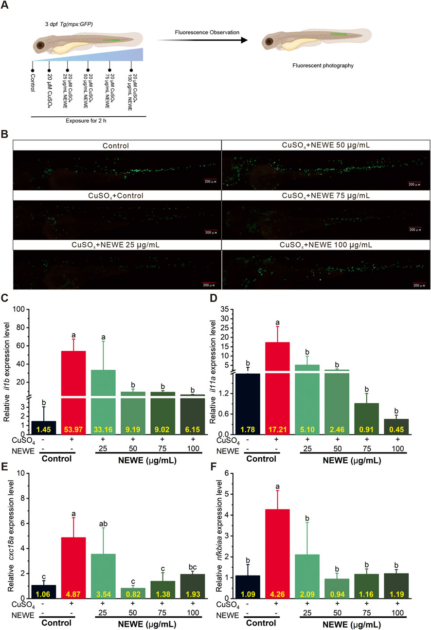
Figure 1. Effects of Nymphaea “Eldorado” flower water extract (NEWE) on CuSO4-induced inflammation in zebrafish larvae. (A) Flowchart of the experiments. (B) Fluorescence analysis of neutrophil migration in zebrafish larvae treated with CuSO4 in the presence or absence of NEWE. In the control group, neutrophils were localized predominantly to the caudal hematopoietic tissue in the ventral trunk and tail region. Upon CuSO4 treatment, neutrophils migrated towards the horizontal midline and accumulated near lateral line neuromasts, indicating an inflammatory response. Treatment with various concentrations of NEWE significantly inhibited CuSO4-induced neutrophil migration to inflammatory sites, suggesting an anti-inflammatory effect. GFP-expressing neutrophils are shown in green. Scale bar: 200 µm. (C–F) qRT-PCR analysis of the relative expression of inflammatory genes in zebrafish larvae. Gene expression was normalized to β-actin. Data are means ± SDs of three independent experiments. Different letters indicate significant differences (p < 0.05, Student’s t-test).
3.2 Antioxidant activity of NEWE
The antioxidant activity of NEWE was assessed by fluorescence microscopy and qRT-PCR analysis. The DCFH-DA fluorescence probe operates through a redox-sensitive mechanism: non-fluorescent DCFH-DA passively diffuses across cell membranes and is deacetylated by intracellular esterases to form DCFH, which becomes fluorescent DCF upon oxidation by reactive oxygen species (ROS) (Zhang et al., 2023b). This oxidation process generates fluorescence intensity proportional to ROS levels, allowing semi-quantitative assessment of oxidative stress in vivo (Nguyen et al., 2020). Fluorescence imaging showed that CuSO4 remarkably increased oxidative stress in zebrafish larvae, whereas 100 μM quercetin (positive control) and different concentrations of NEWE reduced fluorescence intensity (Figures 2A,B), suggesting NEWE possesses antioxidant properties by reducing CuSO4-induced oxidative stress levels. The qRT-PCR results showed that CuSO4 exposure led to an upregulation of the oxidative stress-related genes, ptgs2a and ptgs2b, which encode prostaglandin synthase 2a and 2b, respectively (Ishikawa et al., 2007). Specifically, CuSO4 induced expression of ptgs2a to 4.1-fold, and ptgs2b to 37.42, relative to control, respectively. In contrast, NEWE treatment at 25–125 μg/mL effectively reversed these effects, reducing ptgs2a and ptgs2b expression (p < 0.05, Figures 2C,D). These findings demonstrate that NEWE mitigates CuSO4-induced oxidative stress in zebrafish larvae.
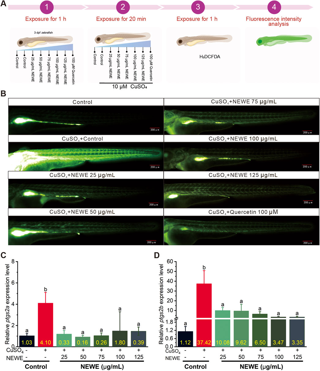
Figure 2. Fluorescence microscopy and qRT-PCR analysis of the antioxidant activity of Nymphaea “Eldorado” flower water extract (NEWE) in zebrafish larvae treated with CuSO4 in the presence and absence of NEWE or quercetin (positive control). (A) Flowchart of the experiments. (B) Fluorescence analysis of the antioxidant activity of NEWE in zebrafish larvae. The control (untreated) group shows baseline fluorescence. (C, D). qRT-PCR analysis of the relative expression of oxidative stress-related genes ptgs2a and ptgs2b. Error bars represent standard deviations, and different letters indicate significant differences (p < 0.05).
3.3 Transcriptomic analysis of NEWE’s anti-inflammatory mechanisms
In order to examine the molecular mechanism of NEWE’s anti-inflammatory, we performed RNA-seq to profile global gene expression changes. Volcano plots showed that distinct gene expression patterns across the control, model group (treated with CuSO4), and treatment groups (CuSO4 + 50 μg/mL NEWE) (Figures 3A–C). A total of 339 differentially expressed genes (DEGs) was shared across the three groups and were selected for further functional annotation (Figure 3D), genes previously validated by qRT-PCR - including il1b (interleukin 1 beta), ptgs2a/b (prostaglandin-endoperoxide synthase 2), and cxcl8a (C-X-C motif chemokine ligand 8a) - emerged as core regulatory nodes (Figure 3H). Gene Ontology (GO) analysis showed that the selected DEGs were enriched in several biological processes, cellular components, and molecular functions (Figures 3E,F). Notably, genes associated with inflammatory response were significantly enriched, indicating their potential role in inflammatory pathways. Additionally, genes related to antioxidant activity were differentially expressed (Figures 2C,D), suggesting that antioxidant mechanisms may help mitigate inflammation by NEWE. Thus, future research should screen for DEGs related to both inflammatory and antioxidant activities to elucidate molecular mechanisms underlying NEWE’s anti-inflammatory effects. A heatmap showed that the expression profiles of these DEGs varied between different treatments (Figure 3G). A protein-protein interaction (PPI) network analysis indetified 30 hub DEGs as potential targets of NEWE, of particular significance, stat3, serpine1, and mmp9 formed a tightly interconnected subnetwork that bridges inflammatory signaling and extracellular matrix reorganization (Figure 3H). These hub genes exhibited strong co-expression patterns with qRT-PCR validated targets, confirming their central role in NEWE-mediated anti-inflammatory responses.
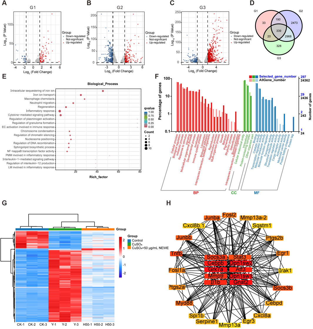
Figure 3. Differentially expressed genes (DEGs) and functional enrichment analyses of DEGs in zebrafish larvae treated with CuSO4 and Nymphaea “Eldorado” flower water extract. (A-C) DEGs in the control (untreated) group, model group (treated with CuSO4) vs control, and treatment group (treated with CuSO4 + 50 μg/mL NEWE) vs model group. Upregulated and downregulated genes are shown in red and blue, respectively. A total of 582, 5,970, and 3655 DEGs were identified in these groups. (D) DEGs shared between the groups. A total of 339 DEGs were shared across the three groups and were selected for further analysis. (E) Biological processes enriched in the selected DEGs. The size of each circle indicates the number of DEGs, and the color intensity represents the q-value. (F) Gene Ontology enrichment analysis of 339 DEGs. BP, biological process; CC, cellular component; MF, molecular function. (G) Expression profile of DEGs in each group. The color gradient from blue to red represents increasing expression levels. (H) Protein-protein interaction network of 30 hub DEGs, highlighting core interaction hubs with high connectivity. Nodes with low and high centrality are shown in yellow and red, respectively.
3.4 Chemical composition of NEWE and network pharmacology analysis
To further understand the molecular mechanism of NEWE in anti-inflammatory pathway, widely targeted metabolomic analysis was employed to identify potential compounds involved in NEWE-mediated anti-inflammatory (Supplementary Figure S2). A total of 891 compounds were identified and quantified, revealing a high degree of chemical diversity in the extract (Figures 4A,B). Flavonoids (172 compounds), alkaloids (85 compounds), lipids (97 compounds), and phenylpropanoids (142 compounds) were predominant. Subsequent network pharmacology analysis predicted potential biochemical interactions between these chemical compounds and target genes (Figures 4C–F; Supplementary Tables S2-S5). These interactions indicate the potential pharmacological relevance of these compounds and the importance of understanding these interactions to elucidate NEWE’s pharmacological effects.
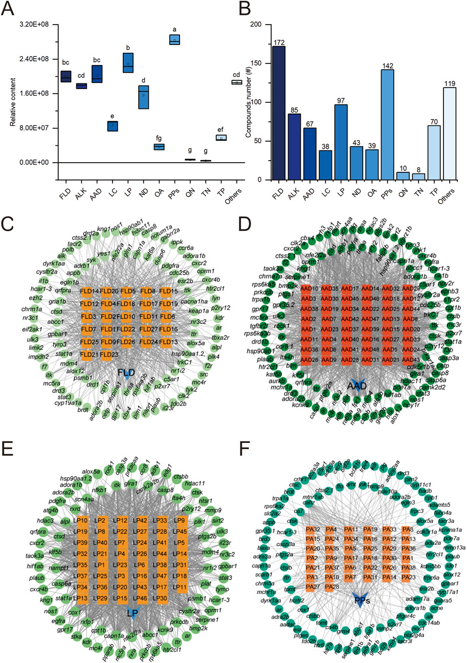
Figure 4. Network pharmacology analysis of major chemical constituents found in Nymphaea “Eldorado” flower water extract (NEWE) and their interactions with zebrafish target genes. (A) Relative abundance of major chemical constituents in NEWE. Different letters indicate significant differences (p < 0.05). (B) Number of unique chemical compounds in each category. (C–F) Interactions between chemical compounds (orange nodes) and target genes (green nodes). Abbreviations: FLD, flavonoids; ALK, alkaloids; AAD, amino acid derivatives; LC, lactones; LP, lipids; ND, nucleosides and nucleotides; OA, organic acids; PPs, phenylpropanoids; QN, quinones; TN, tannins; TP, terpenoids.
3.5 Integrated multi-omics and network pharmacology reveal a polypharmacological hub gene network mediating NEWE’s anti-inflammatory activity
To investigate the potential mechanisms by which NEWE’s diverse chemical components exert their anti-inflammatory effects at the molecular level, we conducted an integrated analysis of our metabolomics and transcriptomics data, focusing on the identified hub DEGs and their predicted interactions with the various classes of compounds present in NEWE. Venn diagrams (Figures 5A–D) showed that among the 30 hub DEGs associated with NEWE treatment, several genes were consistently targeted by different classes of compounds within NEWE, including flavonoids, amino acid derivatives, lipids, and phenylpropanoids. Specifically, 6 key genes, including il1b, stat3, serpine1, mmp9, ptgs2a, and ptgs2b showed overlap, and the recurring presence across multiple compound-targeted groups suggests that these genes play pivotal roles in the response to NEWE treatment. Figure 5E depicts a network pharmacology map linking these six genes to various chemical constituents, underscoring their potential importance in mediating NEWE’s therapeutic effects. Additionally, RNA-seq expression profiling (Figure 5F) shows that the expression patterns of these core genes were distinct across the control, model, and treatment groups and that treatment normalized the gene expression disrupted by CuSO4 exposure. These findings collectively suggest that the six hub genes (il1b, stat3, serpine1, mmp9, ptgs2a, and ptgs2b) form a coordinated regulatory network through which multiple bioactive compounds in NEWE exert polypharmacological effects, simultaneously targeting inflammatory signaling, oxidative stress responses, and extracellular matrix remodeling. This multi-target engagement mechanism provides a molecular rationale for the observed anti-inflammatory efficacy of NEWE and highlights these genes as potential biomarkers for evaluating plant-derived therapeutics.
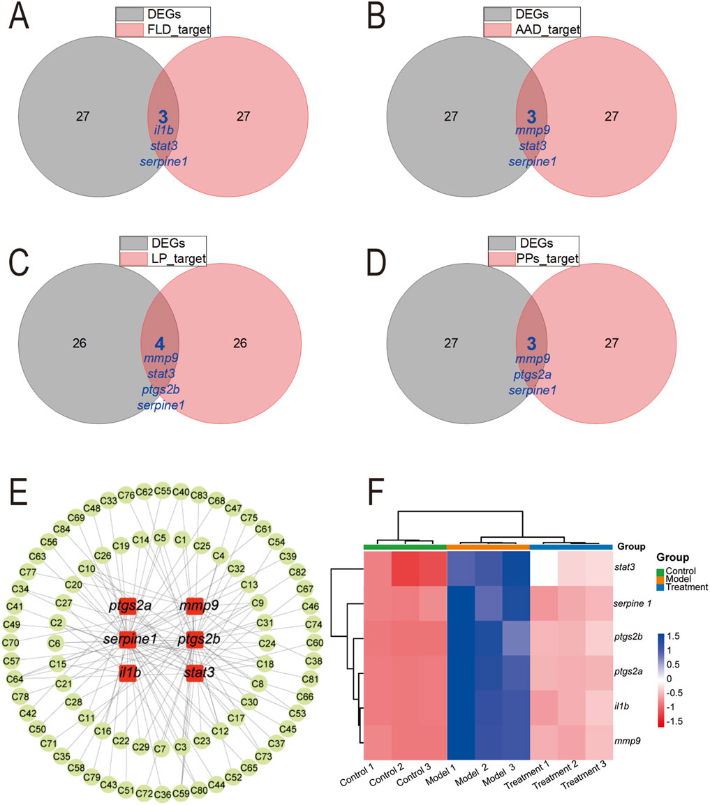
Figure 5. Analysis of hub differentially expressed genes (DEGs) associated with treatment with Nymphaea “Eldorado” flower water extract (NEWE) and genes targeted by four chemical compounds found in NEWE. (A–D) Overlap between DEGs and core target genes. (E) Interactions between six target genes (il1b, stat3, serpine1, mmp9, ptgs2a, and ptgs2b) (red nodes) and chemical compounds (green nodes). (F) Expression profile of target genes in different groups based on RNA-seq analysis. The color gradient from blue to red indicates increasing expression levels. Abbreviations: FLD, flavonoids; AAD, amino acid derivatives; LP, lipids; PPs, phenylpropanoids.
3.6 Expression of core target genes
To validate the transcriptional regulation of key inflammatory mediators identified through multi-omics integration, we quantified the expression dynamics of six core target genes using FPKM (Fragments Per Kilobase per Million mapped reads) values derived from RNA-seq data. This normalized metric accounts for both transcript length and sequencing depth, providing a reliable measure of relative gene expression levels across experimental groups. Upon treatment with CuSO4, six inflammatory genes were significantly upregulated: ptgs2a increased from 9.16 to 72.62 FPKM (approximately 7.9-fold), serpine 1 from 6.81 to 43.64 FPKM (around 6.4-fold), il1b from 0.42 to 50.39 FPKM (about 120-fold), mmp9 from 11.31 to 245.92 FPKM (approximately 21.7-fold), ptgs2b from 2.35 to 76.95 FPKM (around 32.8-fold), and stat3 from 10.85 to 26.90 FPKM (approximately 2.5-fold). Notably, il1b exhibited the most dramatic induction, consistent with its role as a primary cytokine driver in copper-induced inflammation. Treatment with NEWE reversed these effects, significantly reducing gene expression in comparison to the CuSO4 group. These results indicate that NEWE possesses a potential anti-inflammatory effect, mitigating the overexpression induced by CuSO4.
3.7 Molecular docking of Serpine1-targeting compounds and functional validation of transcriptional regulation
To validate the network pharmacology prediction that Serpine1 (plasminogen activator inhibitor-1) acts as a critical mediator of NEWE’s bioactivity, we conducted molecular docking analyses on three representative compounds identified in the metabolomic profile: L-pyroglutamic acid, monomyristin, and protocatechuic acid (Supplementary Table S2). As illustrated in Figure 7, docking revealed specific interactions within the Serpine1 substrate-binding cleft: L-pyroglutamic acid formed hydrogen bonds with Thr-183, Arg-348, and Lys-235 (binding energy = −2.67 kcal/mol); monomyristin bound Asp-209 and Lys-311, positioning its acyl chain in the hydrophobic S4 pocket (binding energe = −0.67 kcal/mol); protocatechuic acid engaged Asp-209 and Asn-243 via hydrogen bonds (binding energy = −1.26 kcal/mol). These residues are critical for the structural integrity of the reactive center loop.
The functional relevance of these predicted interactions was assessed by measuring the compounds’ effects on serpine1 gene expression in the zebrafish inflammation model. Treatment with L-pyroglutamic acid (0.20 mg/mL) significantly downregulated serpine1 expression (Figure 7G). Similarly, protocatechuic acid induced a dose-dependent and significant downregulation at concentrations of 0.01, 0.06, and 0.10 mg/mL (Figure 7I). In contrast, monomyristin treatment did not significantly alter serpine1 expression levels (Figure 7H).
The significant downregulation of serpine 1 by L-pyroglutamic acid and protocatechuic acid provides functional biological evidence supporting their predicted interaction with Serpine1 through molecular docking, aligning with the 6.4-fold serpine1 downregulation observed with whole NEWE extract (Figure 6B). This suggests these compounds contribute to NEWE’s anti-inflammatory effect, potentially through direct or indirect interference with Serpine1 function or its regulation.
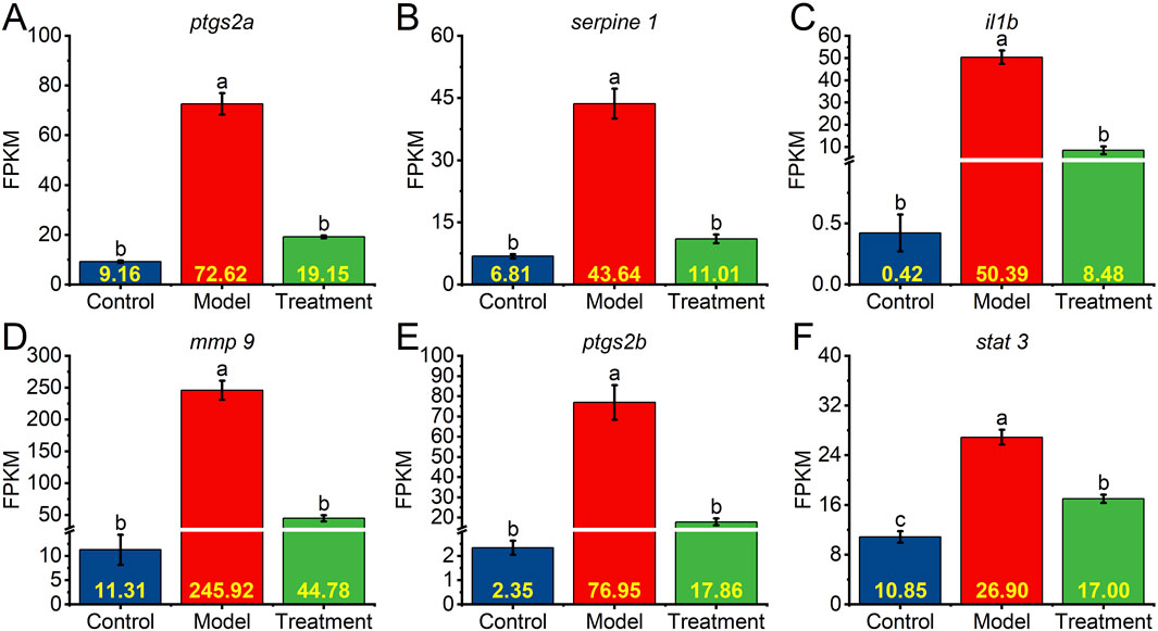
Figure 6. Expression levels of core target genes in different groups. Relative transcript levels were calculated as fragments per kilobase of exon per million fragments mapped (FPKM). (A–F) Relative expression of ptgs2a, serpine1, il1b, mmp9, ptgs2b, and stat3 in the control (untreated) group, model group (treated with CuSO4), and treatment group (treated with CuSO4 and NEWE). Different letters indicate significant differences (p < 0.05). Data are means and standard errors of the mean.
4 Discussion
This study demonstrated that Nymphaea “Eldorado” flower water extract (NEWE) exerts significant anti-inflammatory and antioxidant in a zebrafish model of copper sulfate-induced inflammation. The results provide a basis for using these natural bioactive compounds as inflammatory agents. Chronic inflammation is highly prevalent and impairs health-related quality of life. Thus, these compounds can help reduce the burden of inflammatory diseases (Hatch-McChesney and Smith, 2023; Pasiakos et al., 2016).
NEWE significantly reduced CuSO4-induced inflammation in zebrafish larvae, evidenced by a decrease in neutrophil migration and downregulation of the inflammatory genes il1b, il11a, cxcl8a, and nfkbiaa. Notably, these genes exhibited characteristic expression patterns in response to CuSO4 exposure, with il1b showing a remarkable 37.22-fold, consistent with their established roles as canonical inflammatory markers in zebrafish models (Barbalho et al., 2016; Bucher et al., 2018). These findings suggest that NEWE components attenuate acute inflammatory responses and thus can be used as anti-inflammatory agents.
Oxidative stress promotes inflammation and tissue damage, contributing to the progression of inflammatory diseases (Agnihotri et al., 2008; Alam et al., 2021; Bondia-Pons et al., 2012; Vo et al., 2011). Oxidative/ROS production assay and qRT-PCR analyses showed that NEWE treatment significantly reduced oxidative stress and inflammation in CuSO4-treated zebrafish larvae. The dual modulation of ptgs2a/b genes, which encode cyclooxygenase-2 isoforms critical for prostaglandin synthesis, further establishes their utility as oxidative stress biomarkers in this model. Specifically, NEWE attenuated neutrophil recruitment to inflammatory sites and downregulated key inflammatory genes, suggesting the inhibition of the oxidative stress-inflammation cascade triggered by CuSO4 exposure. These findings highlight the therapeutic potential of NEWE, as its antioxidative and anti-inflammatory effects may protect against inflammatory damage.
Transcriptomic analysis showed that NEWE treatment downregulated pro-inflammatory pathways and genes, including il1b, stat3, serpine1, mmp9, ptgs2a, and ptgs2b. mmp9 was mainly expressed in neutrophil (Duan et al., 2024), the identification of mmp9 as a differentially expressed gene (21.7-fold induction by CuSO4) is particularly noteworthy. Oxidative stress and inflammatory factors can upregulate the expression of serpine1 (PAI-1) (Simone et al., 2014), the 6.4-fold upregulation of serpine1 suggesting these genes may serve as secondary markers for chronic inflammatory progression. These results were corroborated by PPI network analyses and demonstrate NEWE’s potential to modulate inflammatory pathways, including those related to cytokine signaling and immune response regulation.
Widely targeted metabolomic analysis identified a diverse range of bioactive compounds, including flavonoids, alkaloids, amino acid derivatives, and phenylpropanoids. Flavonoids and phenylpropanoids have anti-inflammatory and antioxidant properties (Al-Khayri et al., 2022; Hasnat et al., 2024; Mykhailenko et al., 2021; Wang et al., 2022). Network pharmacology analysis showed that these compounds interacted with inflammatory genes, providing a molecular basis for the anti-inflammatory effects. The multi-target engagement observed - particularly the concurrent modulation of il1b, stat3, and ptgs2a/b - suggests these genes form a core biomarker network for evaluating anti-inflammatory efficacy. This polypharmacological approach mirrors recent strategies in complex disease biomarker discovery (Antolin et al., 2016; McDermott et al., 2013; Sun et al., 2019), where combined gene expression profiles outperform single-marker analyses. Understanding these interactions is crucial since multi-target therapeutics can have synergistic activity and reduce the side effects of single-target therapies.
The transcriptomic analysis revealed that NEWE treatment significantly attenuated CuSO4-induced inflammatory responses through coordinated downregulation of key pro-inflammatory mediators, including ptgs2a, serpine1, il1b, mmp9, ptgs2b, and stat3. This gene cluster demonstrates high diagnostic specificity for metal-induced inflammation, as evidenced by their minimal baseline expression (e.g., il1b FPKM 0.42 in controls vs. 50.39 in CuSO4 group) and strong response attenuation by NEWE. The hierarchical regulation observed - from early cytokine response genes (il1b) to downstream effectors (mmp9, serpine1) - provides a temporal framework for monitoring inflammatory progression. This comprehensive suppression of inflammatory pathway components underscores the extract’s potential as a therapeutic agent for inflammation-related pathologies.
Molecular docking revealed that key NEWE constituents -L-pyroglutamic acid, protocatechuic acid, monomyristin-exhibit binding affinities towards the active site of Serpine1, suggesting potential direct modulation of its function. This finding aligns with established bioactivities of these compounds: L-pyroglutamic acid demonstrates anti-inflammatory properties by suppressing LPS-induced NO production and exhibits neuroprotective effects in NGF-induced PC-12 cell differentiation (Gang et al., 2018). Protocatechuic acid possesses multi-faceted biological activities, including antioxidant, anti-inflammatory, and anti-hyperglycemic effects, mediated through interactions with diverse molecular targets (Lin et al., 2009). Monomyristin is characterized by significant antimicrobial and antifungal activities (Jumina et al., 2018). To functionally validate the predicted interactions with Serpine1 within our inflammatory context, we assessed the effects of individual compounds on serpine1 expression in our zebrafish inflammation model. Notably, L-pyroglutamic acid (0.20 mg/mL) significantly downregulated serpine1 transcription (Figure 7G), while protocatechuic acid induced dose-dependent suppression at 0.01, 0.06, and 0.10 mg/mL (Figure 7I). In contrast, monomyristin showed no significant effect (Figure 7H), highlighting compound-specific regulatory roles despite shared docking potential. These results align with transcriptomic data showing 6.4-fold serpine1 downregulation by NEWE and reinforce its role as a key pharmacological target.
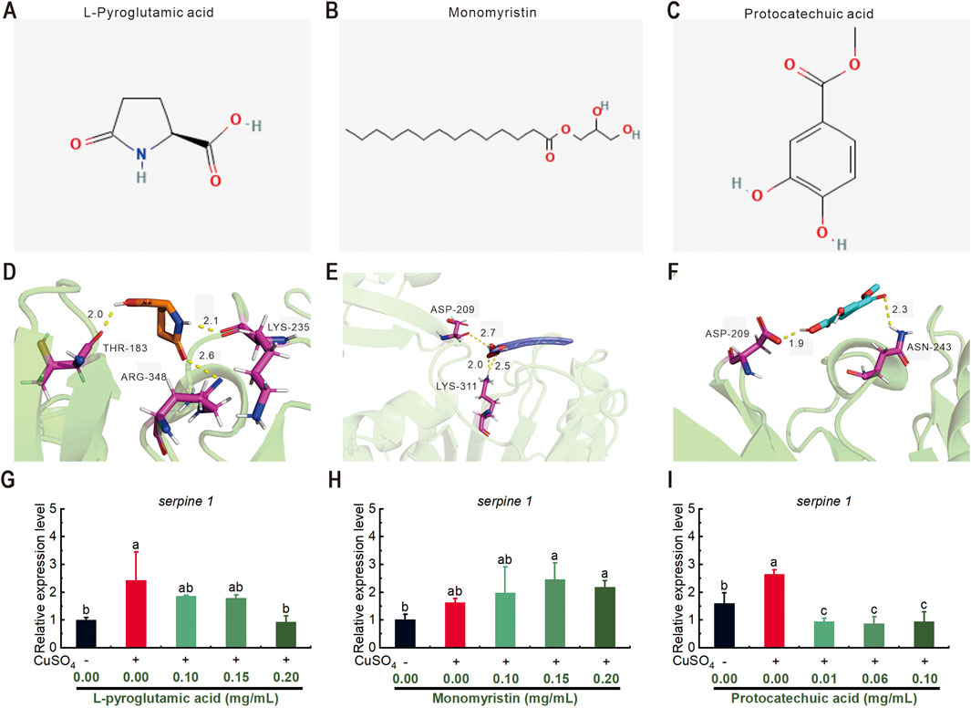
Figure 7. Molecular structure of three chemical compounds found in Nymphaea “Eldorado” flower water extract (NEWE) and their interactions with Serpine1 protein. (A–C) Molecular structure of L-pyroglutamic acid, monomyristin, and protocatechuic acid. (D–F) Interaction of these compounds with Serpine1. (D) L-pyroglutamic acid forms hydrogen bonds with Thr-183, Arg-348, and Lys-235. (E) Monomyristin establishes hydrogen bonds with Asp-209 and Lys-311. (F) Protocatechuic acid forms hydrogen bonds with Asp-209 and Asn-243. Molecular distances are shown in angstroms. Interacting residues in Serpine1 are shown in purple. (G–I) qRT-PCR analysis showing the relative expression of serpine1 following treatment with the three compounds. Error bars represent standard deviations, and different letters indicate significant differences (p < 0.05).
The central position of Serpine1(PAI-1) within our multi-omics network is mechanistically well supported. Serpine1 functions as a critical node linking inflammation, fibrosis, and oxidative stress (Liu, 2008). Its overexpression drives pathological processes such as collagen accumulation following inflammatory lung injury (Eitzman et al., 1996). Conversely, Serpine1 modulates key pathways: it controls abdominal aortic aneurysm formation via TGF-β/Smad2/3 signaling (Zhao et al., 2025), influences macrophage polarization to reduce cardiac fibrosis in inflammatory cardiomyopathy, and suppresses inflammation in experimental autoimmune encephalomyelitis (EAE) by inhibiting Th1 cell secretion of IFNγ and TNFα (Akbar et al., 2023). Our data extend this established paradigm to copper-induced inflammation, suggesting an evolutionarily conserved pro-inflammatory role for Serpine1.
However, while transcriptional and docking data strongly implicate Serpine1, functional validation of its necessity remains essential. Future studies should employ CRISPR/Cas9-mediated serpine1 knockout in zebrafish or siRNA silencing in mammalian macrophages (e.g., RAW264.7) to definitively establish whether Serpine1 inhibition is sufficient to recapitulate NEWE’s anti-inflammatory effects. Complementary protein-level assays (e.g., ELISA for Serpine 1, MMP9, IL-1β) are equally critical to confirm translational relevance of observed mRNA changes.
5 Conclusion
This comprehensive study establishes Nymphaea “Eldorado” flower water extract (NEWE) as a multifaceted therapeutic agent targeting inflammation-oxidative stress crosstalk through integrated molecular mechanisms. Key findings reveal that NEWE (25–100 μg/mL) anti-inflammatory effects by suppressing CuSO4-induced neutrophil migration and downregulating critical proinflammatory genes, notably reducing il1b expression by 8.78-fold and ptgs2a/b expression by 25.63- and 11.17-fold, respectively.
Transcriptomic profiling identified 339 differentially expressed genes, with hierarchical suppression of il1b (120-fold reduction), mmp9 (21.7-fold), and serpine1 (6.4-fold) forming a core anti-inflammatory signature. Network pharmacology revealed polypharmacological interactions between 891 identified metabolites and inflammation-related targets, particularly flavonoids and phenylpropanoids targeting the il1b-stat3-mmp9 axis. Molecular docking validated high-affinity binding of protocatechuic acid and L-pyroglutamic acid to Serpine1’s active site, providing structural evidence for pathway-specific modulation.
These findings systematically demonstrate that NEWE operates through a Serpine1-centric mechanism, coordinating transcriptional regulation of cytokine signaling (stat3, il1b), extracellular matrix remodeling (mmp9), and prostaglandin biosynthesis (ptgs2a/b). The multi-omics convergence of transcriptomic, metabolomic, and pharmacodynamic data positions NEWE as a promising candidate for developing plant-derived therapeutics against chronic inflammatory pathologies, offering advantages over conventional NSAIDs through its multi-target efficacy and reduced toxicity profile. Future studies should prioritize clinical validation of these zebrafish-derived mechanisms in mammalian systems and isolation of specific bioactive fractions for therapeutic optimization.
Data availability statement
The data presented in the study are deposited in the NCBI repository, accession number PRJNA1282926.
Ethics statement
The animal studies were approved by The animal study protocol was approved by Animal Experimental Ethical Inspection Form of Guangxi Academy of Agriculture Sciences (Approval No. GXNKYDWSY2023002; Approval Date: 12 July 2023). The studies were conducted in accordance with the local legislation and institutional requirements. Written informed consent was obtained from the owners for the participation of their animals in this study.
Author contributions
LM: Funding acquisition, Visualization, Methodology, Writing – original draft, Validation, Investigation, Conceptualization. LL: Validation, Conceptualization, Formal Analysis, Visualization, Writing – original draft. QQ: Investigation, Writing – original draft, Validation, Methodology. QwH: Visualization, Writing – original draft, Investigation, Methodology. QlH: Writing – original draft, Methodology, Investigation. YY: Writing – original draft, Validation, Methodology. LD: Writing – original draft, Formal Analysis, Methodology. GL: Validation, Investigation, Writing – original draft. JZ: Writing – original draft, Investigation, Methodology. BC: Formal Analysis, Validation, Writing – review and editing. XT: Writing – review and editing, Methodology, Investigation, Conceptualization, Formal Analysis.
Funding
The author(s) declare that financial support was received for the research and/or publication of this article. This research was funded by grants from the Guangxi Natural Science Foundation (Grant number 2024GXNSFAA010389), and the Special Project for Basic Scientific Research of Guangxi Academy of Agricultural Sciences (Grant number Guinongke 2021YT152, Guinongke 2025YP133).
Acknowledgments
We are grateful to Jinchun Yang and Xiaohong Deng for their assistance in zebrafish husbandry.
Conflict of interest
The authors declare that the research was conducted in the absence of any commercial or financial relationships that could be construed as a potential conflict of interest.
Generative AI statement
The author(s) declare that no Generative AI was used in the creation of this manuscript.
Publisher’s note
All claims expressed in this article are solely those of the authors and do not necessarily represent those of their affiliated organizations, or those of the publisher, the editors and the reviewers. Any product that may be evaluated in this article, or claim that may be made by its manufacturer, is not guaranteed or endorsed by the publisher.
Supplementary material
The Supplementary Material for this article can be found online at: https://www.frontiersin.org/articles/10.3389/fphar.2025.1612233/full#supplementary-material
References
Agnihotri, V. K., Elsohly, H. N., Khan, S. I., Smillie, T. J., Khan, I. A., and Walker, L. A. (2008). Antioxidant constituents of Nymphaea caerulea flowers. Phytochemistry 69, 2061–2066. doi:10.1016/j.phytochem.2008.04.009
Akbar, I., Tang, R., Baillargeon, J., Roy, A. P., Doss, P., Zhu, C., et al. (2023). Cutting edge: serpine1 negatively regulates Th1 cell responses in experimental autoimmune encephalomyelitis. J. Immunol. 211, 1762–1766. doi:10.4049/jimmunol.2300526
Alam, M. B., Naznin, M., Islam, S., Alshammari, F. H., Choi, H. J., Song, B. R., et al. (2021). High resolution mass spectroscopy-based secondary metabolite profiling of Nymphaea nouchali (burm. F) stem attenuates oxidative stress via regulation of MAPK/Nrf2/HO-1/ROS pathway stem attenuates oxidative stress via regulation of MAPK/Nrf2/HO-1/ROS pathway. Antioxidants (Basel) 10, 719. doi:10.3390/antiox10050719
Ali, S., Champagne, D. L., Spaink, H. P., and Richardson, M. K. (2011). Zebrafish embryos and larvae: a new generation of disease models and drug screens. Birth Defects Res. C Embryo Today 93, 115–133. doi:10.1002/bdrc.20206
Al-Khayri, J. M., Sahana, G. R., Nagella, P., Joseph, B. V., Alessa, F. M., and Al-Mssallem, M. Q. (2022). Flavonoids as potential anti-inflammatory molecules: a review. Molecules 27, 2901. doi:10.3390/molecules27092901
Andoh, Y., Mizutani, A., Ohashi, T., Kojo, S., Ishii, T., Adachi, Y., et al. (2006). The antioxidant role of a reagent, 2',7'-dichlorodihydrofluorescin diacetate, detecting reactive-oxygen species and blocking the induction of heme oxygenase-1 and preventing cytotoxicity. J. Biochem. 140, 483–489. doi:10.1093/jb/mvj187
Antolin, A. A., Workman, P., Mestres, J., and Al-Lazikani, B. (2016). Polypharmacology in precision oncology: current applications and future prospects. Curr. Pharm. Des. 22, 6935–6945. doi:10.2174/1381612822666160923115828
Atiquzzaman, M., Karim, M. E., Kopec, J., Wong, H., and Anis, A. H. (2019). Role of nonsteroidal antiinflammatory drugs in the association between osteoarthritis and cardiovascular diseases: a longitudinal study. Arthritis Rheumatol. 71, 1835–1843. doi:10.1002/art.41027
Barbalho, P. G., Lopes-Cendes, I., and Maurer-Morelli, C. V. (2016). Indomethacin treatment prior to pentylenetetrazole-induced seizures downregulates the expression of il1b and cox2 and decreases seizure-like behavior in zebrafish larvae. BMC Neurosci. 17, 12. doi:10.1186/s12868-016-0246-y
Bondia-Pons, I., Ryan, L., and Martinez, J. A. (2012). Oxidative stress and inflammation interactions in human obesity. J. Physiol. Biochem. 68, 701–711. doi:10.1007/s13105-012-0154-2
Bucher, S., Tete, A., Podechard, N., Liamin, M., Le Guillou, D., Chevanne, M., et al. (2018). Co-exposure to benzo[a]pyrene and ethanol induces a pathological progression of liver steatosis in vitro and in vivo. Sci. Rep. 8, 5963. doi:10.1038/s41598-018-24403-1
Choi, T.-Y., Choi, T.-I., Lee, Y.-R., Choe, S.-K., and Kim, C.-H. (2021). Zebrafish as an animal model for biomedical research. Exp. Mol. Med. 53, 310–317. doi:10.1038/s12276-021-00571-5
Dhabhar, F. S. (2014). Effects of stress on immune function: the good, the bad, and the beautiful. Immunol. Res. 58, 193–210. doi:10.1007/s12026-014-8517-0
Duan, C., Yu, X., Feng, X., Shi, L., and Wang, D. (2024). Expression profiles of matrix metalloproteinases and their inhibitors in nasal polyps. J. Inflamm. Res. 17, 29–39. doi:10.2147/jir.S438581
Edlow, B. L., Bodien, Y. G., Baxter, T., Belanger, H. G., Cali, R. J., Deary, K. B., et al. (2022). Long-term effects of repeated blast exposure in United States special operations forces personnel: a pilot study protocol. J. Neurotrauma 39, 1391–1407. doi:10.1089/neu.2022.0030
Eitzman, D. T., McCoy, R. D., Zheng, X., Fay, W. P., Shen, T., Ginsburg, D., et al. (1996). Bleomycin-induced pulmonary fibrosis in transgenic mice that either lack or overexpress the murine plasminogen activator inhibitor-1 gene. J. Clin. Invest 97, 232–237. doi:10.1172/jci118396
Gang, F. L., Zhu, F., Li, X. T., Wei, J. L., Wu, W. J., and Zhang, J. W. (2018). Synthesis and bioactivities evaluation of l-pyroglutamic acid analogues from natural product lead. Bioorg Med. Chem. 26, 4644–4649. doi:10.1016/j.bmc.2018.07.041
Garbarino, S., Lanteri, P., Bragazzi, N. L., Magnavita, N., and Scoditti, E. (2021). Role of sleep deprivation in immune-related disease risk and outcomes. Commun. Biol. 4, 1304. doi:10.1038/s42003-021-02825-4
Girard-Lalancette, K., Pichette, A., and Legault, J. (2009). Sensitive cell-based assay using DCFH oxidation for the determination of pro- and antioxidant properties of compounds and mixtures: analysis of fruit and vegetable juices. Food Chem. 115, 720–726. doi:10.1016/j.foodchem.2008.12.002
Haj, C. G., Sumariwalla, P. F., Hanus, L., Kogan, N. M., Yektin, Z., Mechoulam, R., et al. (2015). HU-444, a novel, potent anti-inflammatory, nonpsychotropic cannabinoid. J. Pharmacol. Exp. Ther. 355, 66–75. doi:10.1124/jpet.115.226100
Hasnat, H., Shompa, S. A., Islam, M. M., Alam, S., Richi, F. T., Emon, N. U., et al. (2024). Flavonoids: a treasure house of prospective pharmacological potentials. Heliyon 10, e27533. doi:10.1016/j.heliyon.2024.e27533
Hatch-McChesney, A., and Smith, T. J. (2023). Nutrition, immune function, and infectious disease in military personnel: a narrative review. Nutrients 15, 4999. doi:10.3390/nu15234999
Howe, K., Clark, M. D., Torroja, C. F., Torrance, J., Berthelot, C., Muffato, M., et al. (2013). The zebrafish reference genome sequence and its relationship to the human genome. Nature 496, 498–503. doi:10.1038/nature12111
Hsu, C. L., Fang, S. C., and Yen, G. C. (2013). Anti-inflammatory effects of phenolic compounds isolated from the flowers of Nymphaea mexicana Zucc. Food Funct. 4, 1216–1222. doi:10.1039/c3fo60041f
Ishikawa, T. O., Griffin, K. J., Banerjee, U., and Herschman, H. R. (2007). The zebrafish genome contains two inducible, functional cyclooxygenase-2 genes. Biochem. Biophys. Res. Commun. 352, 181–187. doi:10.1016/j.bbrc.2006.11.007
Jin, S., and Kang, P. M. (2024). A systematic review on advances in management of oxidative stress-associated cardiovascular diseases. Antioxidants (Basel) 13, 923. doi:10.3390/antiox13080923
Ju, Z., Li, M., Xu, J., Howell, D. C., Li, Z., and Chen, F.-E. (2022). Recent development on COX-2 inhibitors as promising anti-inflammatory agents: the past 10 years. Acta Pharm. Sin. B 12, 2790–2807. doi:10.1016/j.apsb.2022.01.002
Jumina, A., Fitria, A., Pranowo, D., Sholikhah, E. N., Kurniawan, Y. S., Kuswandi, B., et al. (2018). Monomyristin and monopalmitin derivatives: synthesis and evaluation as potential antibacterial and antifungal agents. Molecules 23, 3141. doi:10.3390/molecules23123141
Laine, L. (2001). Approaches to nonsteroidal anti-inflammatory drug use in the high-risk patient. Gastroenterology 120, 594–606. doi:10.1053/gast.2001.21907
Lee, O. Y. A., Wong, A. N. N., Ho, C. Y., Tse, K. W., Chan, A. Z., Leung, G. P., et al. (2024). Potentials of natural antioxidants in reducing inflammation and oxidative stress in chronic kidney disease. Antioxidants (Basel) 13, 751. doi:10.3390/antiox13060751
Li, X., Wei, S., Niu, S., Ma, X., Li, H., Jing, M., et al. (2022). Network pharmacology prediction and molecular docking-based strategy to explore the potential mechanism of Huanglian Jiedu Decoction against sepsis. Comput. Biol. Med. 144, 105389. doi:10.1016/j.compbiomed.2022.105389
Lin, C. Y., Huang, C. S., Huang, C. Y., and Yin, M. C. (2009). Anticoagulatory, antiinflammatory, and antioxidative effects of protocatechuic acid in diabetic mice. J. Agric. Food Chem. 57, 6661–6667. doi:10.1021/jf9015202
Liu, R. M. (2008). Oxidative stress, plasminogen activator inhibitor 1, and lung fibrosis. Antioxid. Redox Signal 10, 303–319. doi:10.1089/ars.2007.1903
Livak, K. J., and Schmittgen, T. D. (2001). Analysis of relative gene expression data using real-time quantitative PCR and the 2(-Delta Delta C(T)) method. Methods 25, 402–408. doi:10.1006/meth.2001.1262
McDermott, J. E., Wang, J., Mitchell, H., Webb-Robertson, B. J., Hafen, R., Ramey, J., et al. (2013). Challenges in biomarker discovery: combining expert insights with statistical analysis of complex omics data. Expert Opin. Med. Diagn 7, 37–51. doi:10.1517/17530059.2012.718329
Misurac, J. M., Knoderer, C. A., Leiser, J. D., Nailescu, C., Wilson, A. C., and Andreoli, S. P. (2013). Nonsteroidal anti-inflammatory drugs are an important cause of acute kidney injury in children. J. Pediatr. 162, 1153–1159. doi:10.1016/j.jpeds.2012.11.069
Morris, G. M., Huey, R., Lindstrom, W., Sanner, M. F., Belew, R. K., Goodsell, D. S., et al. (2009). AutoDock4 and AutoDockTools4: automated docking with selective receptor flexibility. J. Comput. Chem. 30, 2785–2791. doi:10.1002/jcc.21256
Mykhailenko, O., Korinek, M., Chen, B.-H., and Georgiyants, V. (2021). Phenolic compounds from iris hungarica as potential anti-inflammatory agents. Free Radic. Biol. Med. 165, 44–45. doi:10.1016/j.freeradbiomed.2020.12.385
Nguyen, T. H., Le, H. D., Kim, T. N. T., The, H. P., Nguyen, T. M., Cornet, V., et al. (2020). Anti-inflammatory and antioxidant properties of the ethanol extract of Clerodendrum cyrtophyllum turcz in copper sulfate-induced inflammation in zebrafish. Antioxidants (Basel) 9, 192. doi:10.3390/antiox9030192
Pasiakos, S. M., Margolis, L. M., Murphy, N. E., McClung, H. L., Martini, S., Gundersen, Y., et al. (2016). Effects of exercise mode, energy, and macronutrient interventions on inflammation during military training. Physiol. Rep. 4, e12820. doi:10.14814/phy2.12820
Porter, C. K., Riddle, M. S., Laird, R. M., Loza, M., Cole, S., Gariepy, C., et al. (2020). Cohort profile of a US military population for evaluating pre-disease and disease serological biomarkers in rheumatoid and reactive arthritis: Rationale, organization, design, and baseline characteristics. Contemp. Clin. Trials Commun. 17, 100522. doi:10.1016/j.conctc.2020.100522
Postlethwait, J. H., Yan, Y. L., Gates, M. A., Horne, S., Amores, A., Brownlie, A., et al. (1998). Vertebrate genome evolution and the zebrafish gene map. Nat. Genet. 18, 345–349. doi:10.1038/ng0498-345
Prata, C., Angeloni, C., and Maraldi, T. (2024). Strategies to counteract oxidative stress and inflammation in chronic-degenerative diseases 2.0. Int. J. Mol. Sci. 25, 5026. doi:10.3390/ijms25095026
Ren, Y., Yao, X., Xiao, J., Zheng, X., Ouyang, N.-n., Ouyang, Y., et al. (2019). Alkaloids components and pharmacological activities of lotus (Nelumbo nucifera gaertn) leaves. Nat. Prod. J. 9, 26–31. doi:10.2174/2210315508666180614123802
Renshaw, S. A., Loynes, C. A., Trushell, D. M., Elworthy, S., Ingham, P. W., and Whyte, M. K. (2006). A transgenic zebrafish model of neutrophilic inflammation. Blood 108, 3976–3978. doi:10.1182/blood-2006-05-024075
Sapieha, P., Stahl, A., Chen, J., Seaward, M. R., Willett, K. L., Krah, N. M., et al. (2011). 5-Lipoxygenase metabolite 4-HDHA is a mediator of the antiangiogenic effect of ω-3 polyunsaturated fatty acids. Sci. Transl. Med. 3, 69ra12. doi:10.1126/scitranslmed.3001571
Seeliger, D., and de Groot, B. L. (2010). Ligand docking and binding site analysis with PyMOL and autodock/vina. J. Comput. Aided Mol. Des. 24, 417–422. doi:10.1007/s10822-010-9352-6
Sheichenko, V. I., Tolkachev, O. N., Osipov, V. I., Sheichenko, O. P., Anufrieva, V. V., Timoshina, V. A., et al. (2019). Composition and biological activity of furanoquinolizidine alkaloids from yellow water-lily (Nuphar lutea L. smith) rhizomes. Pharm. Chem. J. 53, 553–558. doi:10.1007/s11094-019-02042-8
Simone, T. M., Higgins, S. P., Higgins, C. E., Lennartz, M. R., and Higgins, P. J. (2014). Chemical antagonists of plasminogen activator Inhibitor-1: mechanisms of action and therapeutic potential in vascular disease. J. Mol. Genet. Med. 8, 125. doi:10.4172/1747-0862.1000125
Singh, M., Guru, A., Sudhakaran, G., Pachaiappan, R., Mahboob, S., Al-Ghanim, K. A., et al. (2022). Copper sulfate induced toxicological impact on in-vivo zebrafish larval model protected due to acacetin via anti-inflammatory and glutathione redox mechanism. Comp. Biochem. Physiol. C Toxicol. Pharmacol. 262, 109463. doi:10.1016/j.cbpc.2022.109463
Sun, D., Ren, X., Ari, E., Korcsmaros, T., Csermely, P., and Wu, L. Y. (2019). Discovering cooperative biomarkers for heterogeneous complex disease diagnoses. Brief. Bioinform 20, 89–101. doi:10.1093/bib/bbx090
Vo, T. S., Ngo, D. H., Kim, J. A., Ryu, B., and Kim, S. K. (2011). An antihypertensive peptide from tilapia gelatin diminishes free radical formation in murine microglial cells. J. Agric. Food Chem. 59, 12193–12197. doi:10.1021/jf202837g
Wang, Y., Liu, X.-J., Chen, J.-B., Cao, J.-P., Li, X., and Sun, C.-D. (2022). Citrus flavonoids and their antioxidant evaluation. Crit. Rev. Food Sci. Nutr. 62, 3833–3854. doi:10.1080/10408398.2020.1870035
Xie, Y., Meijer, A. H., and Schaaf, M. J. M. (2020). Modeling inflammation in zebrafish for the development of anti-inflammatory drugs. Front. Cell Dev. Biol. 8, 620984. doi:10.3389/fcell.2020.620984
Yuan, H., Zeng, X., Shi, J., Xu, Q., Wang, Y., Jabu, D., et al. (2018). Time-course comparative metabolite profiling under osmotic stress in tolerant and sensitive Tibetan hulless barley. Biomed. Res. Int. 2018, 9415409. doi:10.1155/2018/9415409
Zhang, P., Liu, N., Xue, M., Zhang, M., Liu, W., Xu, C., et al. (2023a). Anti-inflammatory and antioxidant properties of beta-Sitosterol in copper sulfate-induced inflammation in zebrafish (Danio rerio). Antioxidants (Basel) 12, 391. doi:10.3390/antiox12020391
Zhang, P., Liu, N., Xue, M., Zhang, M., Xiao, Z., Xu, C., et al. (2023b). Anti-inflammatory and antioxidant properties of squalene in copper sulfate-induced inflammation in zebrafish (Danio rerio). Int. J. Mol. Sci. 24, 8518. doi:10.3390/ijms24108518
Zhao, M., Hu, L., Lin, Z., Yue, X., Zheng, X., Piao, M., et al. (2025). Plasminogen activator inhibitor 1 controls abdominal aortic aneurism formation via the modulation of TGF-β/Smad2/3 signaling in mice. FASEB J. 39, e70562. doi:10.1096/fj.202403133RR
Keywords: Nymphaea “Eldorado”, anti-inflammatory, antioxidant, zebrafish, transcriptomics, molecular docking
Citation: Mao L, Long L, Qin Q, Huang Q, Huang Q, Yu Y, Ding L, Liu G, Zhang J, Cui B and Tan X (2025) Nymphaea “Eldorado” flower extract targets serpine 1 to attenuate inflammatory and antioxidant crosstalk in zebrafish. Front. Pharmacol. 16:1612233. doi: 10.3389/fphar.2025.1612233
Received: 17 April 2025; Accepted: 23 June 2025;
Published: 11 July 2025.
Edited by:
Javier Echeverria, University of Santiago, ChileReviewed by:
Tayo Alex Adekiya, Howard University, United StatesHao Wu, Changsha University of Science and Technology, China
Yulin Dai, Changchun University of Chinese Medicine, China
Copyright © 2025 Mao, Long, Qin, Huang, Huang, Yu, Ding, Liu, Zhang, Cui and Tan. This is an open-access article distributed under the terms of the Creative Commons Attribution License (CC BY). The use, distribution or reproduction in other forums is permitted, provided the original author(s) and the copyright owner(s) are credited and that the original publication in this journal is cited, in accordance with accepted academic practice. No use, distribution or reproduction is permitted which does not comply with these terms.
*Correspondence: Beimi Cui, YmVpbWkuY3VpQGdtYWlsLmNvbQ==; Xiaohui Tan, eGlhb2h1aXRhbkBneGFhcy5uZXQ=
†These authors have contributed equally to this work
 Liyan Mao1,2†
 Lingyun Long
 Beimi Cui
 Xiaohui Tan