- 1Hospital of Stomatology, Guanghua School of Stomatology, Sun Yat-sen University, Guangzhou, China
- 2Guangdong Provincial Key Laboratory of Stomatology, Sun Yat-sen University, Guangzhou, China
- 3College of Stomatology, Chongqing Medical University, Chongqing, China
- 4Chongqing Key Laboratory of Oral Diseases, Chongqing, China
- 5Chongqing Municipal Key Laboratory of Oral Biomedical Engineering of Higher Education, Chongqing, China
Background/purpose: Maxillomandibular advancement (MMA) is widely used for treating obstructive sleep apnea (OSA) patients. However, conventional MMA may not be suitable for Eastern Asian patients with moderate or severe OSA, as it can lead to unsatisfactory postoperative facial appearance. Hence, modified MMA was reported. Our study aims to evaluate the therapeutic effects of modified MMA on OSA and patient satisfaction with facial appearance. In addition, anatomic and aerodynamic changes in the upper airway were explored.
Materials and methods: This retrospective study included 13 patients with moderate or severe OSA. Overnight polysomnography and the Epworth Sleepiness Scale (ESS) scores were recorded before operation and 6 months after operation to evaluate therapeutic outcomes. Spiral CT scans were performed for all patients to reconstruct 3D configurations of the bony structures and the upper airway. Computational fluid dynamics was performed to analyze aerodynamic characteristics. In addition, correlations between bone segment movements and improvement in airway parameters were examined.
Results: Modified MMA achieved successful therapeutic and esthetic outcomes in all cases. The apnea–hypopnea index (36.05 ± 17.68 vs. 5.72 ± 4.76, p < 0.001) and the ESS (13.23 ± 8.9 vs. 6.23 ± 6.81 events/h, p < 0.05) decreased significantly, while the lowest oxygen saturation (76.54 ± 10.26% vs. 84.77 ± 6.02%, p < 0.05) improved greatly. Modified MMA significantly increased the total volume (6,716.55 ± 1,357.73 vs. 11,191.28 ± 2,563.79 mm3, p < 0.001) and the averaged cross-sectional area (117.38 ± 24.25 vs. 201.58 ± 35.76 mm2, p < 0.001) of the upper airway. After modified MMA, the pressure drop, gas velocity, and resistance in the upper airway were all significantly decreased (p < 0.05). Among all the maxillary and mandible sections, the strongest correlation was observed between the advanced movement of the anterior mandible segment and anatomical characteristics of the upper airway.
Conclusion: Modified MMA is an excellent therapeutic method for Eastern Asian patients with skeletal class Ⅱ dentomaxillofacial deformity suffering from moderate to severe OSA; it achieves a balance between esthetic improvement and therapeutic efficacy for OSA both anatomically and aerodynamically.
Background
Obstructive sleep apnea (OSA) is a condition characterized by repetitive episodes of apnea or hypopnea caused by pharyngeal collapse during sleep. OSA can lead to oxygen desaturation, hypercapnia, and sleep fragmentation, which contribute to cardiovascular, metabolic, and neurocognitive diseases (1). Thus, OSA is considered a potentially life-threatening disease. Multiple factors, such as obesity, excessive soft tissue in the upper airway, retrusive jaw, and dysfunction of the upper airway dilator, might contribute to the occurrence of OSA (2, 3). Currently, nasal continuous positive airway pressure (CPAP) is the first-line treatment for OSA, especially in moderate-to-severe cases. However, not all patients can tolerate lifelong CPAP use (4, 5). Since its introduction by Riley et al. in 1984, maxillomandibular advancement (MMA) has been widely used in treating OSA patients, especially in patients with retrusive maxilla and mandible (6). MMA increases the upper airway volume and reduces the upper airway collapsibility by enlarging the oral cavity, therefore alleviating OSA in patients. Over nearly 40 years of practice, MMA has been proven to be highly effective and stable for selected OSA patients (7, 8). However, in the Eastern Asian population, OSA patients more frequently present with features such as a flat nose, protrusive upper jaw, and weak chin. Thus, the conventional MMA procedure might cause adverse esthetic outcomes in these patients. Several studies reported that MMA, when modified by segmentation of the maxilla or mandible, can achieve maximal advancement while preserving a balanced facial appearance and functional dental occlusion (9–11). However, it has also been reported that a decreased oral cavity volume increases the risk of flow resistance and upper airway collapse (12, 13). We hypothesize that the airway enlargement by maxillomandibular advancement might be neutralized by the segmental setback of the anterior jaw, as the actual oral cavity volume does not change significantly.
Hence, in this study, we evaluated the clinical outcomes of modified MMA, including its therapeutic effect on OSA and patient satisfaction with facial appearance. In addition, we evaluated the airway configuration anatomically and simulated airflow changes using the computational fluid dynamics (CFD) method to gain a better understanding of the aerodynamic effects of modified MMA.
Methods and materials
Participants
This is a retrospective study, approved by the institutional review board of the Hospital of Stomatology, Sun Yat-sen University. Between May 2020 and March 2024, patients referred to the Hospital of Stomatology, Sun Yat-sen University, who met the following criteria were enrolled in our study:
1. Patients of either sex aged between 18 and 60 years.
2. Patients with moderate to severe OSA combined with class II skeletal dentomaxillofacial deformity, who were intolerant of other conservative treatments (e.g., CPAP, weight loss, or oral appliances).
Exclusion criteria are as follows:
1. Patients with any genetic syndromes.
2. Patients with systemic diseases contraindicating orthognathic surgery under general anesthesia.
3. Patients with uncontrolled temporomandibular disorders or periodontitis.
4. Patients with uncontrolled psychological diseases.
5. Patients who were unwilling or unable to participate in this study.
Informed consent was obtained from all participants. Approval for this research was obtained from the Institutional Research Ethics Committee (KQEC-2024-117-01).
Orthodontic and orthognathic treatment
All patients received combined orthodontic and orthognathic treatment using a surgery-first or early approach. When necessary, limited preoperative orthodontic treatment was performed to align the dentition without further decompensation. All patients underwent modified MMA, which included conventional MMA combined with upper and lower subapical osteotomy and premolar extraction to achieve maximal mandibular and posterior maxillary advancement while avoiding postoperative bimaxillary protrusion. Genioplasty was performed when further advancement of the chin was required. Postoperative orthodontic alignment, leveling, and space management were performed to establish the final occlusion.
Virtual surgical planning
Preoperatively, surgical simulation was performed using the Dolphin platform. Spiral CT data, digital dental casts, and 3D facial photographs were integrated into the Dolphin platform. The head orientation was set parallel to the FH plane laterally and the interpupillary line frontally. A stepwise surgical workup, including setup, cropping, cleaning, osteotomy design, landmark identification, treatment planning, presentation, and splint fabrication, was performed. An intermediate splint was designed for mandibular movement, and a final splint was designed for maxillary movement. Occlusal splints were printed by stereolithography and sterilized before surgical use.
Assessment of patients and evaluation of sleep status
Preoperatively, the medical history and comorbidity profiles of all enrolled patients were reviewed. Physical examinations, sleep-related questionnaires, and sleep studies were conducted before and after surgery. First, physical characteristics, such as height, weight, and body mass index (BMI), were recorded. Preoperative symptoms were also carefully inquired. Then, self-evaluation of sleep quality was performed using the Epworth Sleepiness Scale (ESS) questionnaire. Finally, the apnea–hypopnea index (AHI) and lowest oxygen saturation (LSAT) were recorded using polysomnography (PSG).
Evaluation of patients' satisfaction
Patients' satisfaction with their postoperative facial appearance was evaluated using a five-point Likert scale (1 = very dissatisfied; 5 = very satisfied).
Assessment of the surgical movement of landmarks
After three-dimensional reconstruction of preoperative and postoperative spiral CT images, landmarks were labeled to assess the surgical movements of the jaw segments. The movement of the anterior maxillary section was measured using the A (subspinale), ANS (anterior nasal spine), and U1 (upper incisor) points, while the posterior section was marked using the U6 (maxillary first molar) and PNS (posterior nasal spine) points. The B (supramental) and L1 (lower incisor) points were regarded as landmarks of the anterior mandible segment, while the L6 (mandible first molar), Menton, Pogonion, and mental foramen points were set as landmarks of the posterior mandible segment. A schematic diagram of the bony landmarks in modified MMA is shown in Figure 1.
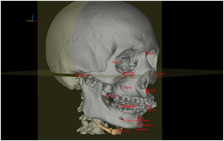
Figure 1. Schematic diagram of bony landmarks in modified MMA. Landmarks of the anterior maxillary segment: A point-subspinale; ANS point-anterior nasal spine; U1 point-mesioincisal angle of the upper central incisor. Landmarks of the posterior maxillary segment: U6-mesiobuccal cusp of maxillary first molar; PNS-posterior nasal spine. Landmarks of the anterior mandible segment: B-point supramental, L1-mesioincisal angle of the lower incisor. Landmarks of the posterior mandible segment: L6 point-mesiobuccal cusp of mandibular first molar; Menton point-the lowest point of mandible; Pogonion point-the most prominent point of the chin; mental foramen point-mental foramen.
Airway measurement
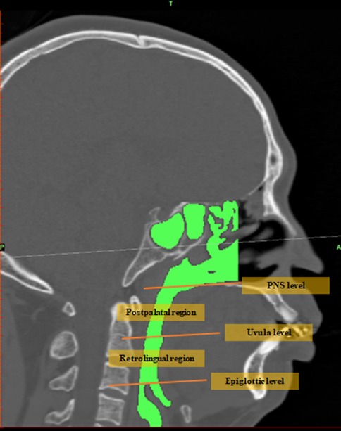
Preoperative and postoperative spiral CT data were imported into the Mimics 20.01 platform to reconstruct the jaw, airway, and hyoid. Image segmentation of these structures was performed based on the threshold values from their DICOM image series. A three-dimensional airway model was created for the region between the nostrils and the infraglottic cavity, excluding the paranasal sinuses. Then, the airway model was smoothed while preserving patient-specific characteristics. The airway was further divided into the nasal airway, postpalatal airway, and postlingual airway using three virtual planes, which were defined as parallel to the FH plane and passing through the PNS, uvula tip, and epiglottic tip, respectively. Preoperative and postoperative jaws, airways, and hyoids were matched by point-based registration of orbital and cranial landmarks. Landmarks (Figure 1) of preoperative and postoperative jaws were analyzed. Pre- and postoperative airway configuration changes were quantified by measuring total airway volume, postpalatal volume, postlingual volume, total airway surface area, postpalatal surface area, postlingual surface area, mean airway cross-sectional area, cross-sectional area at the PNS level, cross-sectional area at the uvula level, and cross-sectional area at the epiglottic level. A schematic diagram of the upper airway segmentation is shown in Figure 2.
Computational fluid dynamics analysis
Preoperative and postoperative airway models were exported as STL files and further smoothed in the Geomagic platform. Each airway was then meshed into numerical computational cells. Laminar and turbulent CFD simulations were performed on the 3D airway models to predict the flow field in the nasal and upper airway of 13 patients before and after MMA. For numerical simulations, the inlet air pressure at the nostrils was set to ambient pressure (1 atm). A steady airflow rate of 700 mL/s, representative of adult inhalation, instead of dynamic tidal air flow, was used to calculate flow fields and pressure distributions. The same airflow rate was used for all cases. The simulation was designed to model human inspiration at rest under atmospheric pressure (1.013 × 10⁵ Pa) and atmospheric temperature (20°C). The coefficients of viscosity (1.822 × 10⁵ Pas) and density (1.205 kg/m3) were provided as fluid data.
Statistical analysis
All statistical analyses were performed using GraphPad Prism 8.0 and SPSS 25.0 software. The Shapiro–Wilk (S-W) test was used to evaluate the normality of data distribution. For comparison between two groups, a two-tailed paired t-test was applied for the data with normally distributed differences, and the Mann–Whitney U-test was used for non-normally distributed ones. To analyze correlations between two groups, Pearson correlation analysis was used for normally distributed continuous data, while Spearman correlation analysis was used for ordinal variables. A p-value <0.05 was considered statistically significant.
Results
Patient characteristics
A total of 13 patients with skeletal class II dentomaxillofacial deformity were enrolled in this study. All patients presented with both mandibular retrusion and sleep apnea. Three of them also reported maxillary protrusion. Patient characteristics are summarized in Table 1. Four patients had undergone tonsillectomy, and two had tried but eventually discontinued CPAP due to intolerance. Snoring was the most frequent symptom, followed by excessive daytime sleepiness, frequent awakenings, morning headaches, and nighttime urination. One patient had systemic diseases secondary to OSA, including hypertension and stroke.
The mean BMI before surgery was 24.58 ± 2.82 kg/m2. Height and weight were remeasured 6 months after surgery when the patients conducted the postoperative PSG. The mean postoperative BMI was 25.10 ± 3.49 kg/m2. No significant differences were observed.
Satisfaction with facial appearance
All patients reported being “satisfied” or “very satisfied” with their postoperative facial appearance (Table 2). Radiographic records of a typical case are shown in Figure 3. None of the patients reported that their appearance had changed to bimaxillary protrusion after surgery.

Figure 3. Lateral cephalometric radiograph, posteroanterior cephalometric radiograph and panoramic radiograph during orthodontic-surgical combined therapy. (A) Before pre-orthognathic orthodontics, (B) 1 week before orthognathic surgery, (C) 1 week after orthognathic surgery, (D) 6 months after orthognathic surgery.
Treatment effectiveness
Results of preoperative and postoperative PSG were presented in Table 3. All preoperative symptoms resolved, with only two patients complaining of slight snoring. Significant improvement was observed after modified MMA. Following the criteria of a “>50% improvement in AHI” or “post-treatment AHI < 15 events/h,” modified MMA achieved successful therapeutic outcomes in all cases. Seven patients achieved complete resolution (AHI < 5 events/h). The average reduction in the AHI was 30.32 events/h. Recovery of the LSAT during PSG was at an average of 8.23%. The ESS score decreased by an average of 8.44.
Surgical movement of the landmarks
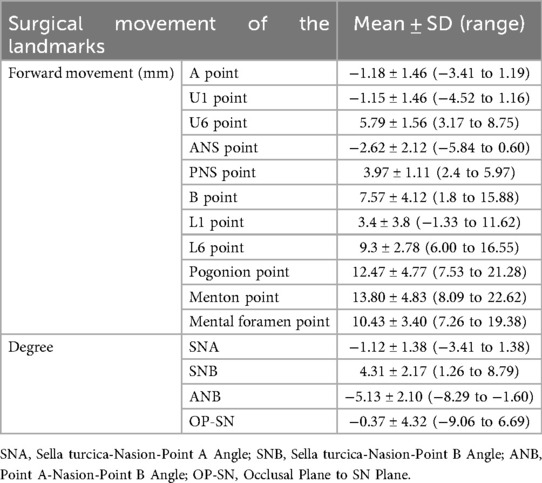
Surgical movement of key landmarks was also measured. The hyoid point was displaced forward by about 5.06 ± 5.09 mm and lifted up by about 4.94 ± 5.51 mm. Other detailed data are presented in Table 4.
Morphological characteristics of the upper airway
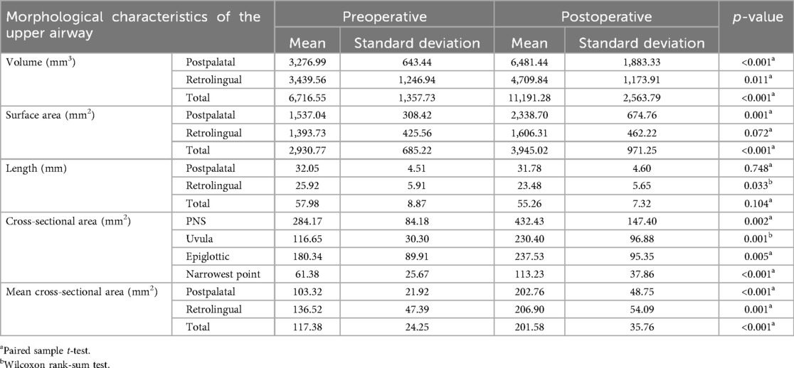
The 3D morphological characteristics of the upper airway are presented in Table 5, and the three-dimensional reconstruction of the upper airway is shown in Figure 4. The volume, surface area, length, and average cross-sectional area of the upper airway were measured. The postpalatal, retrolingual, and total parameters were evaluated separately. In addition, the axial area of the upper airway was evaluated at the PNS level, the uvula level, and the epiglottic level.
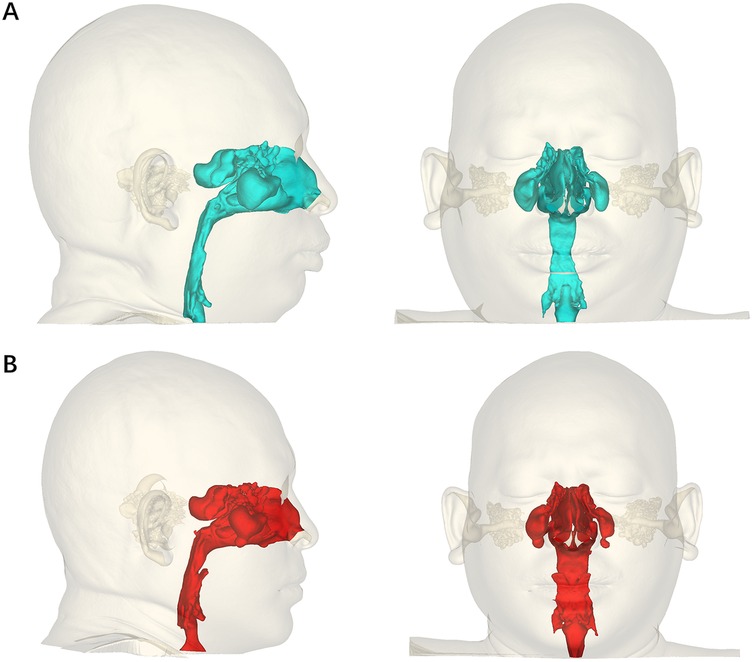
Figure 4. Pre-orthognathic (A) and post-orthognathic (B) three-dimensional reconstruction of the upper airway.
Modified MMA significantly increased the volume, cross-sectional area, and average cross-sectional area of the upper airway. The postpalatal and total surface areas of the upper airway were also significantly increased after surgery. However, no significant differences were observed in the retrolingual level surface area of the upper airway between the preoperative and postoperative measurements. The length of the upper airway tended to be shortened, with no significant difference, except at the retrolingual level.
Aerodynamic characteristics of the upper airway
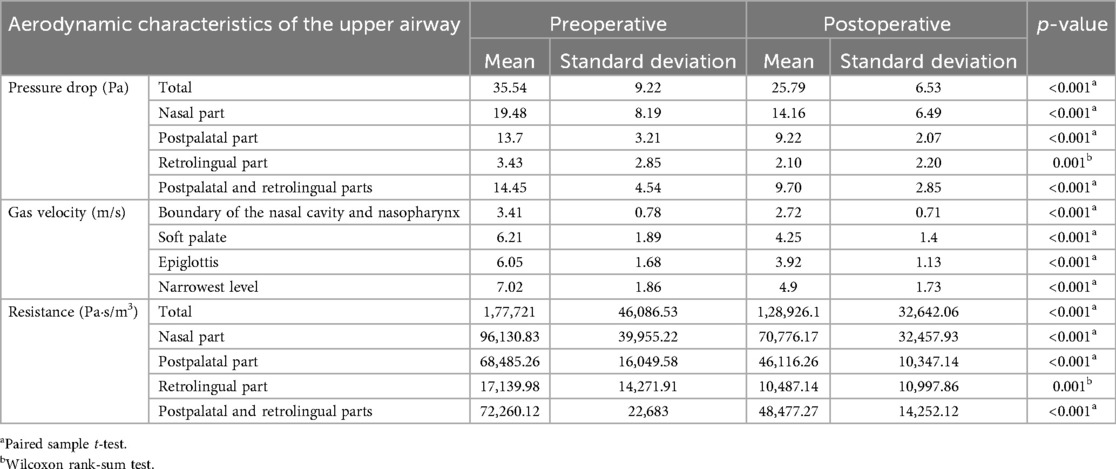
Patient upper airway aerodynamic characteristics during inspiration are summarized in Table 6. Pressure drop, gas velocity, and resistance in the upper airway were significantly decreased after modified MMA. The simulation results of upper airway pressure and gas velocity are shown in Figure 5.
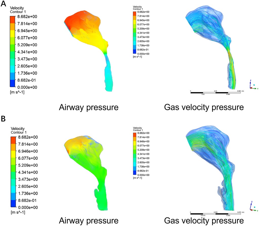
Figure 5. Computational Fluid Dynamics (CFD) of pre-orthognathic (A) and post-orthognathic (B) upper airway. Simulation of upper airway pressure is on the left and simulation of upper airway gas velocity is on the right.
Correlation analysis between surgical movements of bone segments and changes in airway characteristics
A correlation between surgical movements of bone segments and changes in airway morphologic features was observed. In detail, the change in total volume (r = 0.6044, p = 0.0287) and retrolingual surface area (r = 0.6447, p = 0.0174) exhibits a positive correlation with the degree of forward movement of the hyoid bone (Figure 6A). A negative correlation was observed between the upward movement of the hyoid bone and changes in retrolingual length (r = −0.5659, p = 0.0473) and total length (r = −0.6538, p = 0.0182) (Figure 6B). No significant correlation was found between the movement of the anterior maxillary segment and changes in any airway morphological characteristic (p > 0.05). Unexpectedly, aside from postpalatal length (r = −0.5974, p = 0.0311), total surface area (r = 0.6241, p = 0.0226), and retrolingual surface area (r = 0.5582, p = 0.0474), no other morphological feature showed a significant correlation with the movement of the posterior maxillary segment (Figure 6C). A strong correlation was identified between the advanced movement of the anterior mandible section and several airway characteristics, including postpalatal volume (r = 0.6572, p = 0.0147), postpalatal surface area (r = 0.7603, p = 0.0026), postpalatal average cross-sectional area (r = 0.6817, p = 0.0103), and cross-sectional area at the uvula level (r = 0.7205, p = 0.0055). Furthermore, the total average cross-sectional area demonstrated a moderate correlation with the movement of the anterior mandible section (r = 0.5575, p = 0.0487) (Figure 6D). In contrast, weaker correlations were observed between similar airway characteristics and the movement of the posterior mandible segment (Figure 6E). No correlations were found between surgical movement of bone segments and sleep-related indicators or aerodynamic parameters.
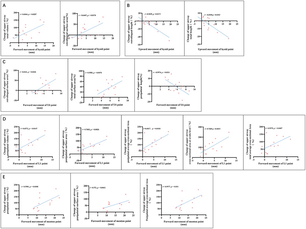
Figure 6. Correlation analysis between change of airway morphological characteristics and forward movement of the hyoid bone (A), upward movement of the hyoid bone (B), posterior maxillary segment (C), anterior mandible segment (D), posterior mandible segment (E).
Discussion
OSA is a very common disorder among adults. Globally, an estimated 936 million adults aged 30–69 years are affected by mild to severe obstructive sleep apnea, while 425 million within the same age range have moderate to severe obstructive sleep apnea (14). High BMI, increasing age, and male gender are regarded as risk factors for the incidence and severity of OSA (3). In our study, potential confounding variables, including sex, age, and BMI, were excluded to minimize their influence on the results.
The aim of OSA treatment is to restore adequate ventilatory function and maintain blood oxygen saturation during sleep (15). Therapeutic options can be categorized into four types: lifestyle modification, mandibular advancement devices (MADs), CPAP, and surgical interventions (4, 16). Lifestyle modifications, such as sleeping in a side-lying position and weight management, are effective for patients with mild OSA. Although CPAP remains the gold standard treatment for OSA, not all patients can tolerate lifelong CPAP use. MADs are becoming increasingly popular due to their effectiveness, convenience, and non-invasiveness; however, their use is limited by potential complications such as temporomandibular joint (TMJ) and periodontal damage. Surgical interventions are considered the last option when the non-invasive treatments fail. The aim of surgical intervention is to increase the upper airway patency at specific anatomical levels, including uvulopalatopharyngoplasty, hyoid suspension, epiglottoplasty, implantable neurostimulation devices, and orthognathic surgery (14). Since craniofacial disharmony is recognized as a predisposing risk factor for OSA, orthognathic surgery is considered the best choice to improve both ventilatory function and facial appearance (6–11).
The OSA patients in our study commonly presented with a normal or protruded maxilla combined with mandibular retraction, which is not rare in East Asia. Traditional MMA has become an effective, safe, and long-term stable treatment option for OSA (17–19). However, for these patients, the MMA is unsuitable because it may result in esthetic dissatisfaction due to postoperative bimaxillary protrusion. To avoid this problem, a balance between esthetic improvement and therapeutic efficacy for OSA could be achieved using a modified MMA technique, taking advantage of the existing edentulous spaces created after the extraction of premolars. Benito Anguita et al. reported that with traditional MMA, the mean preoperative AHI decreased significantly from 48.8 to 12.4, while ESS scores improved from 14.5 to 7.8 (17). In a meta-analysis, the mean reduction in AHI was 39.6 among Caucasians and 42.7 among other populations (18). Previous studies using modified MMA (9–11) reported successful therapeutic outcomes in all cases, with a significant decrease in AHI and marked recovery of OSA symptoms. Meanwhile, all patients reported satisfaction with their postoperative appearance. Furthermore, the surgery-first or early approach significantly shortens the lengthy process of preoperative orthodontic decompensation and prevents worsening of OSA symptoms (20), which may result from decreased oral cavity volume (12). Taken together, modified MMA is a better therapeutic option for Eastern Asian patients with skeletal class II dentomaxillofacial deformity and OSA.
Anatomically, abnormalities in craniofacial skeletal and soft tissue structures contribute to partial or complete obstruction of the upper airway in OSA patients. Neelapu et al. showed strong evidence of reduced pharyngeal airway space, inferiorly positioned hyoid bone, and increased anterior facial height in adult OSA patients compared with control subjects (2). Another study reported that upper airway collapsibility is closely associated with hyoid position, tongue volume, pharyngeal length, and waist circumference (21). A study showed that patients with a higher hyoid position responded better to MMA (19). In our study, modified MMA significantly elevated the hyoid bone and increased upper airway volume, consistent with previous reports. The increase in airway volume was closely associated with the forward movement of the hyoid bone, suggesting that OSA patients might benefit from additional hyoid suspension during modified MMA (22). Notably, movement of the hyoid bone is primarily driven by significant advancement of the mandible. Furthermore, the anterior segment of the mandible, rather than the posterior segment, demonstrates greater predictability in improving airway morphology. This finding implies that the airway enlargement achieved through maxillomandibular advancement may be counteracted if the anterior section of the mandible is set back. While the movement of the anterior maxillary section shows no significant influence on airway morphology, advancement of the posterior maxillary segment contributes to airway improvement to some extent.
Aerodynamically, airflow accelerates in narrowed regions. According to the Bernoulli effect, intraluminal pressure decreases as airflow increases. If the intra-airway pressure is lower than the external pressure, airway collapse occurs, which leads to apnea (23, 24). CFD provides an engineering modeling approach using the Navier–Stokes equation to study airway dynamics. CFD simulations showed that decreasing the airway pressure resulting from MMA decreases the breathing workload (25). It is reported that MMA increases airway volume, with a decrease in airway velocity. Significant correlations have been reported between improvements in apnea–hypopnea index values and both the increase in airway volume and the decrease in maximum airway velocity (26). Our study suggested that after removal of the airway narrow structure through modified MMA, the pressure drop, gas velocity, and resistance in the upper airway were significantly decreased.
Conclusion and limitations
Due to the limitation of sample size, our study provides only a preliminary evaluation of the efficacy of modified MMA in treating moderate to severe OSA. Based on our existing data, modified MMA appears to be an excellent therapeutic method for the Eastern Asian population with skeletal class II dentomaxillofacial deformity suffering from moderate to severe OSA, achieving a balance between esthetic improvement and therapeutic efficacy for OSA both anatomically and aerodynamically. Further studies with larger sample sizes and comparisons with other therapeutic methods are warranted.
Data availability statement
The original contributions presented in the study are included in the article/Supplementary Material, further inquiries can be directed to the corresponding author.
Ethics statement
The studies involving humans were approved by the Hospital of Stomatology, Guanghua School of Stomatology, Sun Yat-sen University. The studies were conducted in accordance with the local legislation and institutional requirements. The participants provided their written informed consent to participate in this study.
Author contributions
KL: Methodology, Conceptualization, Software, Writing – original draft, Funding acquisition, Formal analysis. TZ: Software, Writing – original draft, Investigation, Formal analysis. YL: Writing – original draft, Investigation, Methodology. YC: Conceptualization, Supervision, Project administration, Funding acquisition, Data curation, Writing – review & editing, Validation. XL: Writing – review & editing, Methodology. SG: Writing – review & editing, Formal analysis. WS: Writing – review & editing, Investigation. TW: Writing – review & editing, Investigation. LZ: Writing – review & editing, Methodology. ZF: Writing – review & editing, Supervision, Formal analysis, Data curation, Resources. GZ: Validation, Conceptualization, Resources, Writing – original draft, Project administration, Writing – review & editing, Supervision, Methodology, Visualization, Formal analysis, Data curation, Software.
Funding
The author(s) declare that financial support was received for the research and/or publication of this article. The study was supported by grants from the Fundamental Research Funds for the Central Universities, Sun Yat-sen University (No. 24qnpy197), the Open Funding Number of Guangdong Provincial Key Laboratory of Stomatology (No. KF2024120104), the Sun Yat-Sen University Clinical Research 5010 Program (No. 2023009), and the Natural Science Foundation of GuangDong Province (NSFGP) (No. 2024A1515030198).
Conflict of interest
The authors declare that the research was conducted in the absence of any commercial or financial relationships that could be construed as a potential conflict of interest.
Generative AI statement
The author(s) declare that no Generative AI was used in the creation of this manuscript.
Any alternative text (alt text) provided alongside figures in this article has been generated by Frontiers with the support of artificial intelligence, and reasonable efforts have been made to ensure accuracy, including review by the authors wherever possible. If you identify any issues, please contact us.
Publisher's note
All claims expressed in this article are solely those of the authors and do not necessarily represent those of their affiliated organizations, or those of the publisher, the editors and the reviewers. Any product that may be evaluated in this article, or claim that may be made by its manufacturer, is not guaranteed or endorsed by the publisher.
Supplementary material
The Supplementary Material for this article can be found online at: https://www.frontiersin.org/articles/10.3389/froh.2025.1598511/full#supplementary-material
References
1. Lévy P, Kohler M, McNicholas WT, Barbé F, McEvoy RD, Somers VK, et al. Obstructive sleep apnoea syndrome. Nat Rev Dis Primers. (2015) 1:15015. doi: 10.1038/nrdp.2015.15
2. Neelapu BC, Kharbanda OP, Sardana HK, Balachandran R, Sardana V, Kapoor P, et al. Craniofacial and upper airway morphology in adult obstructive sleep apnea patients: a systematic review and meta-analysis of cephalometric studies. Sleep Med Rev. (2017) 31:79–90. doi: 10.1016/j.smrv.2016.01.007
3. de Araujo Dantas AB, Gonçalves FM, Martins AA, Alves G, Stechman-Neto J, Corrêa CC, et al. Worldwide prevalence and associated risk factors of obstructive sleep apnea: a meta-analysis and meta-regression. Sleep Breath. (2023) 27(6):2083–109. doi: 10.1007/s11325-023-02810-7
4. Lorenzi-Filho G, Almeida FR, Strollo PJ. Treating OSA: current and emerging therapies beyond CPAP. Respirology. (2017) 22(8):1500–7. doi: 10.1111/resp.13144
5. Lin S, Premaraj TS, Gamage PT, Dong P, Premaraj S, Gu L. Upper airway flow dynamics in obstructive sleep apnea patients with various apnea-hypopnea index. Life (Basel). (2022) 12(7):1080. doi: 10.3390/life12071080
6. Riley R, Guilleminault C, Powell N, Derman S. Mandibular osteotomy and hyoid bone advancement for obstructive sleep apnea: a case report. Sleep. (1984) 7(1):79–82. doi: 10.1093/sleep/7.1.79
7. Zaghi S, Holty JE, Certal V, Abdullatif J, Guilleminault C, Powell NB, et al. Maxillomandibular advancement for treatment of obstructive sleep apnea: a meta-analysis. JAMA Otolaryngol Head Neck Surg. (2016) 142(1):58–66. doi: 10.1001/jamaoto.2015.2678
8. Zhou N, Ho JTF, Huang Z, Spijker R, de Vries N, Aarab G, et al. Maxillomandibular advancement versus multilevel surgery for treatment of obstructive sleep apnea: a systematic review and meta-analysis. Sleep Med Rev. (2021) 57:101471. doi: 10.1016/j.smrv.2021.101471
9. Goh YH, Lim KA. Modified maxillomandibular advancement for the treatment of obstructive sleep apnea: a preliminary report. Laryngoscope. (2003) 113(9):1577–82. doi: 10.1097/00005537-200309000-00031
10. Navasumrit S, Chen YA, Hsieh YJ, Yao CF, Chang CS, Chen NH, et al. Skeletal and upper airway stability following modified maxillomandibular advancement for treatment of obstructive sleep apnea in skeletal class I or II deformity. Clin Oral Investig. (2022) 26(3):3239–50. doi: 10.1007/s00784-021-04306-8
11. Liao YF, Chiu YT, Lin CH, Chen YA, Chen NH, Chen YR. Modified maxillomandibular advancement for obstructive sleep apnoea: towards a better outcome for Asians. Int J Oral Maxillofac Surg. (2015) 44(2):189–94. doi: 10.1016/j.ijom.2014.09.013
12. Hartfield PJ, Janczy J, Sharma A, Newsome HA, Sparapani RA, Rhee JS, et al. Anatomical determinants of upper airway collapsibility in obstructive sleep apnea: a systematic review and meta-analysis. Sleep Med Rev. (2023) 68:101741. doi: 10.1016/j.smrv.2022.101741
13. Kim JW, Kwon TG. Why most patients do not exhibit obstructive sleep apnea after mandibular setback surgery? Maxillofac Plast Reconstr Surg. (2020) 42(1):7. doi: 10.1186/s40902-020-00250-x
14. Benjafield AV, Ayas NT, Eastwood PR, Heinzer R, Ip MSM, Morrell MJ, et al. Estimation of the global prevalence and burden of obstructive sleep apnoea: a literature-based analysis. Lancet Respir Med. (2019) 7(8):687–98. doi: 10.1016/S2213-2600(19)30198-5
15. Ilea A, Timuș D, Höpken J, Andrei V, Băbțan AM, Petrescu NB, et al. Oral appliance therapy in obstructive sleep apnea and snoring—systematic review and new directions of development. Cranio. (2021) 39(6):472–83. doi: 10.1080/08869634.2019.1673285
16. Verse T, Hörmann K. The surgical treatment of sleep-related upper airway obstruction. Dtsch Arztebl Int. (2011) 108(13):216–21. doi: 10.3238/arztebl.2010.0216
17. Benito Anguita M, Khayat S, López Martín S, Bravo Quelle N, Navarro Cuéllar I, López López A, et al. Virtual guided and customized orthognathic surgery in patients with obstructive sleep apnea syndrome: accuracy and clinical outcomes. J Clin Med. (2025) 14(11):3780. doi: 10.3390/jcm14113780
18. Nanu DP, Diemer TJ, Nguyen SA, Tremont T, Meyer TA, Abdelwahab M. Racial variations in maxillomandibular advancement for obstructive sleep apnea: a systematic review and meta-analysis. Sleep Breath. (2024) 29(1):55. doi: 10.1007/s11325-024-03211-0
19. Thai KH, Chen YA, Yao CF, Chen NH, Liao YF, Chen YR. Comparison of phenotypic characteristics between responders and non-responders to treatment for obstructive sleep apnea with maxillomandibular advancement in adults. Sleep Med. (2025) 129:346–53. doi: 10.1016/j.sleep.2025.03.006
20. Pearl C, Chubb DWR, Marchena JM, Waite P, Buendia AM, Kau CH. Surgery first: current state of the art orthognathic surgery and its potential as a primary treatment modality in obstructive sleep apnea with concurrent dentofacial deformities. Front Oral Maxillofac Med. (2022) 4:29. doi: 10.21037/fomm-21-61
21. Wen XL, Wu BZ, Li Y, Yi B, Peng X. Analysis of the aerodynamic characteristics of the upper airway in obstructive sleep apnea patients. J Dent Sci. (2024) 19(1):329–37. doi: 10.1016/j.jds.2023.03.013
22. Gillespie MB, Ayers CM, Nguyen SA, Abidin MR. Outcomes of hyoid myotomy and suspension using a mandibular screw suspension system. Otolaryngol Head Neck Surg. (2011) 144(2):225–9. doi: 10.1177/0194599810391711
23. Zheng Z, Liu H, Xu Q, Wu W, Du L, Chen H, et al. Computational fluid dynamics simulation of the upper airway response to large incisor retraction in adult class I bimaxillary protrusion patients. Sci Rep. (2017) 7:45706. doi: 10.1038/srep45706
24. Wang X, Jia L, Xu X, Guo J. The relationship between aerodynamic characteristics of the upper airway and severity of obstructive sleep apnea in adults. Cranio. (2023) 43(4):646–53. doi: 10.1080/08869634.2023.2278958
25. Sittitavornwong S, Waite PD, Shih AM, Cheng GC, Koomullil R, Ito Y, et al. Computational fluid dynamic analysis of the posterior airway space after maxillomandibular advancement for obstructive sleep apnea syndrome. J Oral Maxillofac Surg. (2013) 71(8):1397–405. doi: 10.1016/j.joms.2013.02.022
26. Furundarena-Padrones L, Cabriada-Nuño V, Brunsó-Casellas J, Garcia-Fernandez RI, Castro-Quintas S, Santos-Zorrozúa B, et al. Correlation between polysomnographic parameters and volumetric changes generated by maxillomandibular advancement surgery in patients with obstructive sleep apnea: a fluid dynamics study. J Clin Sleep Med. (2024) 20(3):371–9. doi: 10.5664/jcsm.10874
Keywords: obstructive sleep apnea, skeletal class II dentomaxillofacial deformity, modified maxillomandibular advancement, three-dimensional reconstruction, computational fluid dynamics
Citation: Li K, Zhao T, Liu Y, Cao Y, Li X, Gao S, Sun W, Wang T, Zeng L, Feng Z and Zheng G (2025) Modified maxillomandibular advancement for Eastern Asian patients with moderate or severe OSA: an anatomic and aerodynamic assessment of the upper airway. Front. Oral Health 6:1598511. doi: 10.3389/froh.2025.1598511
Received: 23 March 2025; Accepted: 18 August 2025;
Published: 11 September 2025.
Edited by:
Michele Tepedino, University of L'Aquila, ItalyReviewed by:
Giuseppina Laganà, Università Link Campus, ItalyCarlotta Fanelli, University of Foggia, Italy
Copyright: © 2025 Li, Zhao, Liu, Cao, Li, Gao, Sun, Wang, Zeng, Feng and Zheng. This is an open-access article distributed under the terms of the Creative Commons Attribution License (CC BY). The use, distribution or reproduction in other forums is permitted, provided the original author(s) and the copyright owner(s) are credited and that the original publication in this journal is cited, in accordance with accepted academic practice. No use, distribution or reproduction is permitted which does not comply with these terms.
*Correspondence: Guangsen Zheng, emhlbmdnc0BtYWlsLnN5c3UuZWR1LmNu; Zhicai Feng, ZmVuZ3poY0BtYWlsLnN5c3UuZWR1LmNu
†These authors have contributed equally to this work
 Kan Li
 Tianyu Zhao1,2,†
 Yukun Liu
 Yang Cao
 Xiang Li
 Lingchan Zeng
 Guangsen Zheng