 Ayşen Kübra Atik
 İsmail Can Paylan
- Department of Plant Protection, Faculty of Agriculture, Ege University, Izmir, Turkey
The aim of this study is to identify and update the list of viral pathogens in tomato seeds, which plays an important role for our country's economy, and to control, on behalf of our country, viral factors like TYLCV and PepMV, which were thought not to be existing in the past but were determined by new and sensitive methods in the recent years. It is also the aim, if viral pathogens like TYLCV and PepMV are found, to share this findings with the necessary institutions and to update the viral factors which needs to be tested, and the quarantine lists. Two hundred and thirty-eight seed samples were used in this study. Serological and molecular methods were used in order to identify the viral pathogens in the seed samples. Two hundred and thirty-eight seed samples of different tomato varieties were tested in order to prove the possible existence of Tobacco mosaic tobamovirus (TMV), Tomato mosaic tobamovirus (ToMV), Cucumber mosaic cucumovirus (CMV), Tomato yellow leaf curl geminivirus (TYLCV), Tomato spotted wilt tospovirus (TSWV), Pepino mosaic potexvirus (PepMV), Pepper mild mottle tobamovirus (PMMoV), and Tomato brown rugose fruit tobamovirus (ToBRFV) and as a result 12% of the seeds were founded to be infected with TMV, 23% with ToMV and 8% with CMV. Within the scope of this study it was seen that some seed samples have mixed infections. Sixty-seven seeds of the 238 seeds has been found to be infected in this study. This is a rate of 28% were the seeds are infected at least with one viral pathogen.
Introduction
Population growth in the world also triggers the increase in agricultural production. Agriculture is an indispensable sector all over the world. Therefore, it is closely related to all segments of society. It is possible to divide crop production in agriculture into various sub-branches. The tomato plant, which we include in the category of vegetable and fruit production, is grown in many countries of the world with its various benefits.
Although the origin of the tomato plant (Solanum lycopersicum), which is in the Solanaceae family, Solanum genus, is based on the Andes Mountains, its homeland is Peru, which is a South American country. The main reason why its consumption and spread is delayed is the belief that it is toxic. In the 17th century, it spread from South America through Australia to the Asian continent. In the 19th century, it spread in our country through France and Syria. It first spread to our southern regions and then to all our regions, and today it is grown in almost every region of our country (Güvenç, 2017).
Tomato (Solanum lycopersicum L.) is a vegetable species belonging to the Solanaceae family, which is the most cultivated plant in the world after the potato plant (Peralta and Spooner, 2005). Tomato fruit has high nutritional values and constitutes an important source of income for producers (Anonymous, 2007). An average sized (123 grams) tomato fruit contains 94% water and has 26 kcal energy. Also, an average sized tomato fruit has 6 grams of carbohydrates, 1.4 grams of total fiber, 1 gram of protein, 273 milligrams of Potassium, 23 milligrams of ascorbic acid, 11 milligrams of Sodium, 6 milligrams of Calcium, 0.8 milligrams of niacin, 0.6 milligrams of iron, 0.07 milligrams of thiamine, 0.06 milligrams riboflavin and 766 IU of Vitamin A (Gebhardt and Thomas, 2002).
The fruits of tomato plants are mostly evaluated as raw. Considering that ~95% of the structure of tomato fruit is water, it is not suitable for long-term storage. Therefore, tomato puree has to be processed and evaluated in various ways such as tomato juice, pickles, peeled tomatoes, sliced tomatoes, ketchup and dried tomatoes (Demiray and Tülek, 2008).
In tomato production, China is the largest tomato producing country in the world, followed by India. Turkey, on the other hand, ranks third and is one of the leading countries in vegetable production in the world.
Due to suitable ecological factors, our country has an important potential in terms of vegetable cultivation both under cover and in the open. Tomato is a plant suitable for warm and hot climate cultivation. The fruits of the tomato plant are usually rich in sugar, mature and dark in color in hot and sunny climates. In places where summers are very cloudy and cool, full maturation cannot be achieved (Abak et al., 2000). Tomatoes show their best growth at temperatures between 15 and 28°C. High humidity has a positive effect during the growth period of tomato. In the fruit ripening period, it causes an increase in diseases and pests in the plant (Tigchelaar, 1986).
Tomato plant can be grown in loamy and loamy-sandy soils with a pH of 6.0–6.5, rich in nutrients and organic matter. Disease and pest management is needed in order to obtain maximum efficiency from the tomato plant, which needs regular irrigation and fertilization (Vural et al., 2000).
Today, the tomato plant, in which the presence of ~200 different disease pathogens has been detected, is under threat by many bacteria, fungi, viruses and other microorganisms (Jones et al., 1991).
Virus diseases in vegetables, which are among the main food sources of humanity, can cause great damage. Virus diseases seen in vegetables cause negative effects on both the development and yield of the plant. In addition, the struggle against vectors to prevent the transmission of viral pathogens also causes economic losses. The severity of the diseases caused by viral pathogens in tomatoes may vary from year to year depending on the viral pathogen, the vector of the pathogen, the host of the pathogen and the environmental conditions. In addition, chemical control method cannot be applied against viral pathogens as it is applied against other plant pathogens. The cultural methods applied against viral pathogens are not sufficiently known and used by the producers. This situation causes the damage caused by viral factors to increase gradually (Paylan, 2011).
One of the main components in increasing the yield per decare in plant production and obtaining quality plant products is the seed. The seeds used in production are the starting material for plant production. Production should be increased for the products used for the adequate and balanced nutrition of the world population, which continues to increase day by day. In addition to features such as high yield and quality, the use of healthy production materials and seeds is of great importance in product increase. In addition, nearly 90% of the herbal products produced for food consumption are produced from seeds and this reveals the importance of seed health (Erkan, 1998).
The increase in the worldwide seed trade has increased the risk of various viral pathogens and their vectors entering new plant plantations. In addition, current control methods are insufficient to control new strains of existing virus pathogens as well as new virus pathogens. Changes in the genome, host or vector of the virus may increase the severity of the disease. In addition, changing climate and environmental conditions are also effective in the spread of virus pathogens and vectors (Hanssen et al., 2010).
Seed transmission can be seen at high rates in viral disease pathogens. When we plant seeds contaminated with viral pathogens, they can easily spread through vectors. As a result, in some cases, the occurrence of infection can be observed at a rate of up to 100% (Carroll, 1983).
Some viruses can be carried by seeds in only a single plant, while others can be carried by seeds in multiple hosts. The ability to be transported by seeds has a very important place for virus epidemiology (Sevik, 2012).
Many viral pathogens belonging to different genera can cause infestation in the seeds of tomato plants (Uzunogullai and Gümüş, 2015). Some viral pathogens in our study; Tobacco mosaic tobamovirus (TMV), Tomato mosaic tobamovirus (ToMV), Cucumber mosaic cucumovirus (CMV), Tomato yellow leaf curl geminivirus (TYLCV), Tomato spotted wilt tospovirus (TSWV), Tomato brown rugose fruit tobamovirus (ToBRFV), Pepper mild mottle tobamovirus (PMMoV) and Pepino mosaic potexvirus (PepMV).
With the measures to be taken in the production of tomatoes for seed purposes and in the import of the obtained tomato seeds, it will be possible to obtain high quality and high yields in tomato production by ensuring that certified seeds free from viral factors are used by the producers. Certified seed; Certified seed, whose genetic, physical and biological values have been determined as a result of field and laboratory controls, is allowed to be sold after all kinds of trials and examinations by the Ministry, whose variety purity has been ensured and whose name has been determined.
Variety purity is complete in certified seeds. Since there are no foreign substances and foreign seeds in it, you will get whatever product you have planted.
Certified seeds have high germination ability. In other words, every seed you plant in the field will germinate. In this way, it shows a strong development, which provides up to 25% increase in yield and savings in the amount of seeds to be planted per decare in some plants, reducing the cost of seeds and providing profit to the producer. The seed in question has been bred. Therefore, the ability to benefit from soil moisture and plant nutrients (fertilizer) is higher than uncertified seeds. Since all the sown seeds are produced on the same day (uniform output), there are no fluctuations and gaps in the field, so there is no loss of crops. The development of all plants is in the same period, thus; The benefits of processes such as maintenance, hoeing, irrigation, spraying are fully manifested. This provides time and profit to the producer. Since maturation will occur at the same time in all plants, it provides ease of harvest. Since the cultivar characteristics are known, the planting time can be adjusted exactly. When the variety characteristics are known, the application (maintenance, irrigation, etc.) program to be followed during the development of the product can be determined more easily. Since the products obtained have the same features and quality, their market values are high, which provides ease of sale. It provides higher yield since its yield ability is higher than other seeds.
Since the seeds in question are sprayed as a preventative against some diseases that are present in the soil and carried by seeds, the losses are at minimum level.
In this study, viral disease pathogens in tomato seeds collected from producers and various organizations were investigated. Identification of these factors, presence of identified viral pathogens, and some studies have confirmed that tomato seeds infected with TYLCV can transmit the virus (Kil et al., 2016). In this study, it was aimed to determine and update viral disease factors in tomato seeds, which have an important place in our country's economy, and to control viral pathogens such as TYLCV and PepMV, which were thought not to be found in tomato seeds before. It is aimed to update the viral pathogens and quarantine lists that need to be tested by sharing the results with the necessary institutions if they are found. For this purpose, serological and molecular methods were used.
Materials and methods
Material used in research
Seed samples used in the study
For this research, tomato plant, which is produced in our country and has economic importance, was chosen. Tomato seed samples were taken for the diagnosis of viral origin disease agents TMV, ToMV, CMV, TYLCV, TSWV, ToBRFV, PePMV and PMMoV. Seeds showing specific symptoms for TYLCV were also selected. For this purpose, 238 tomato seed samples, 148 hybrid and 90 standard, obtained from many companies, different organizations and tomato producers were studied. Procured tomato seeds were recorded with their source, cultivar names and characteristics. Analysis studies were carried out for a total of eight viral agents that are intensely seen in tomato seeds and likely to be detected as new.
Material used in serological studies
TMV, ToMV, CMV, TYLCV, TSWV, PePMV found in tomato seeds DAS-ELISA method, one of the serological methods, was applied to the seed samples in order to determine the factors of PMMoV and PMMoV. Commercially available solutions (coating, extraction, conjugate, substrate and washing solutions) and diagnostic kits (antisera, positive and negative samples) were used. Titertek, which can read at a wavelength of 405 nm in the conclusion of the tests carried out in the research carried out using 96 well micro dishes, adjustable micro pipettes, pipette tips and distilled water. Multiskan Plus MK II brand ELISA reader was used.
Material used in molecular studies
The material used in the total nucleic acid extraction step
The plant juices obtained from the seeds of the samples determined to be infected with TMV, ToMV, CMV, TYLCV, TSWV, ToBRFV, PePMV or PMMoV as a result of ELISA tests constituted the material of the study. In total RNA extraction study, sterile pestle and pestle, sterile bag, buffer solutions solutions [extraction containing β- Mercaptoethanol bumper, grinding buffer, 6 M sodium iodide (NaI), sarcosil solution, ethanol, silica solution, ethanol, washing buffer solution, RNAse- free sterile water], homogenizer, Sigma D37520 brand centrifuge, ependorf tubes, adjustable micro pipettes and tips, ETG MBT 250 brand heater, Heidolph Unimax 2010 brand shaker platform and ice machine are used.
The material used in the cDNA synthesis step
In the cDNA synthesis stage of the study, as a result of the previously prepared TNA studies, TMV, ToMV, CMV, TSWV, ToBRFV, PePMV and from seeds infected with PMMoV get made TNA extracts were used. In addition, commercially available cDNA synthesis kits (10X RT Random) Primers, 25X dNTP mix (100 mM), Nuclease -free H 2 O, 10X RT Buffer, MultiScribeTM reverse Transcriptase, RNase Inhibitor), adjustable micropipettes and tips, sterile PCR tubes, warmer, shaker (Heidolph Unimax 2010) and PE Applied Biosystems Gene Amp PCR systems 9700 brand Thermal Cycles were used.
The material used in the PCR step
In the study, cDNAs synthesized at the cDNA stage were used. PE Applied Biosystems Gene Amp PCR Systems 9700 brand thermocycler, nuclear free water, commercially available DreamTaq Green PCR Master Mix and TMV, ToMV, TYLCV, TSWV, ToBRFV, PePMV and to PMMoV to the factors specific primers (10 mM), adjustable micropipettes and pipette tips, sterile PCR tubes were used as materials.
Material used in the analysis of amplified PCR products
At this stage of the study, total RNAs of TMV, ToMV, CMV, TYLCV, TSWV, ToBRFV, PePMV and PMMoV obtained as a result of TNA extraction and PCR products obtained from RT-PCR studies were used as materials. Fermentas brand 100 bp DNA Ladder for preparation and staining of samples and gels, commercially available 6X DNA Loading dye, chemicals used in dyeing, 1X TAE buffer solution, agarose, microwave oven, Owl A2 and Owl B1 brand gel electrophoresis apparatus, Consort E815 brand power supply were used. DNR Bio-imaging in the imaging phase of the obtained gels Systems Minibis Pro brand gel imaging device was used.
Other material used in the study
Scope of work; Systec 2540 autoclave, Memmert brand oven, Bioreba AG sample crushing machine, Sigma microcentrifuge, Sartorius precision balance, Scotsman ice machine, Elga ultrapure water machine, Heidolph Unimax 2010 shaker, microwave oven, Nuve ES 500 refrigerated incubator, Binder Incubator, Elga pure water machine, magnetic stirrer, Heidolp tube mixer, HI 9321 pH meter and Sigma brand centrifuge were used at different stages of the study.
Method
Examination of symptoms in seeds
At this stage of the study, typical symptoms of TMV, ToMV, CMV, TYLCV, TSWV, ToBRFV, PePMV and PMMoV agents were sought in tomato seeds (Figures 1–3). Examples are changes in the color or shape of the seed, wrinkling of the seed coat, decrease in seed development, streaks, necrosis, spots, mottling, etc. it was visually examined to detect the symptoms (Erkan, 1998). At the seed identification stage, plastic bags were numbered for each seed sample, and the source, seed type, date and symptoms were recorded.
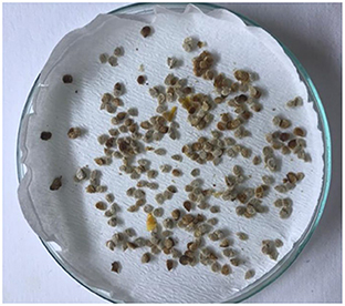
Figure 1. Color changes and signs of disfigurement observed in tomato seeds.
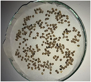
Figure 2. Color changes observed in tomato seeds.
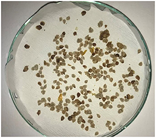
Figure 3. Color changes and signs of wrinkling observed in tomato seeds.
Serological test method (DAS-ELISA)
Seed samples, TMV, ToMV, CMV, TYLCV, TSWV, PePMV and of PMMoV its existence fix about to Clark and Adams (1977) based on the DAS-ELISA test. Table for a total of 238 seed samples DAS-ELISA tests were applied for the viral pathogens given in Table 1.
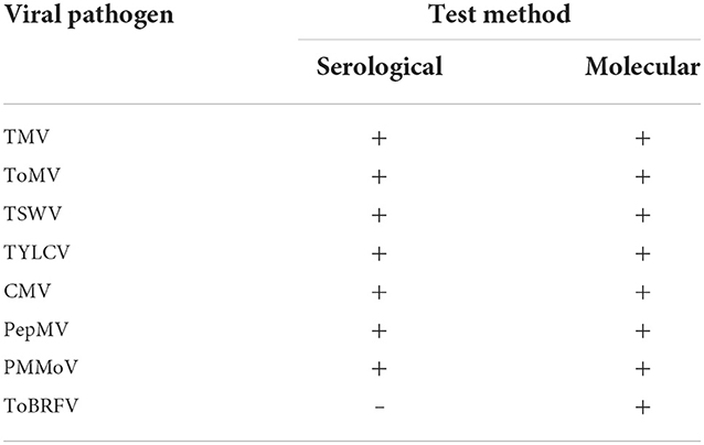
Table 1. Viral pathogens tested by serological and molecular methods in tomato seeds.
DAS-ELISA tests were performed according to the procedure suggested by Bioreba AG. The steps followed in the application of the test are as follows:
1. 100 μl of agent-specific specific IgG was added to each well of the ELISA plates, diluted 1:1,000 in the coating buffer according to the procedure reported by the company, and the plates were covered and incubated at 34°C for 3 h.
2. After the incubation, the liquid in the ELISA plates was drained and then the plates were washed 3 times with washing buffer for 3 min in each wash and allowed to dry on blotting papers.
3. Tomato seed samples collected as study material were crushed in sterile seed crushing bags with a seed crusher by adding 1/10 extraction buffer solution and plant juices were obtained. The extracts, which were put into sterile tubes, were mixed and placed in each well of the ELISA plates in 100 μl amounts and in duplicate. Buffer solution, positive and negative controls of each viral pathogen were added, and the plates were covered and overnight incubated at +4°C.
4. After the incubation, the liquid in the ELISA plates was drained and then the plates were washed 3 times with washing buffer for 3 min in each wash and dried on blotting papers.
5. After washing and drying, the conjugated antibody IgG was diluted 1:1,000 in conjugate buffer, 100 μl was added to each well, the plates were covered and incubated at 34°C for 3 h.
6. After the incubation, the liquid in the ELISA plates was emptied and then washed with washing buffer 3 times for 3 min for each wash and allowed to dry on blotting papers.
7. As a final step, insert into each well of the ELISA plates. Hundred microliter of buffer prepared to be 1 mg/ml of pNPP (para - nitrophenylphosphate) in the substrate solution was added and the plates were covered and incubated for 30–60 min at room temperature and in the dark.
8. In the results, first visual yellow color formation was observed, then the plates were read spectrophotometrically at 405 nm wavelength with an ELISA reader and the absorbance values, which were twice that of the negative controls, were accepted as positive.
Molecular methods
For viral pathogens RT-PCR (Reverse transcriptase), which is the molecular test method, was applied to the seed samples used in the study. polymerase chain reaction) test was applied. The stages of the test are as follows; preparation of seed samples, total nucleic acid extraction (TNA), complementary DNA synthesis (cDNA) and Reverse transcriptase polymerase chain reaction (RT-PCR) was carried out in four steps.
Preparation of tomato seed samples
The tomato seed samples used were obtained from many different institutions, companies and producers. Tomato seed samples were separated into bags as 50–100 seeds by looking at the seed sizes in order to perform the total nucleic acid extraction, which is the first step of the method. The places where the seeds were supplied, the dates and the variety names are written on these bags as labels. In this thesis work Tests were carried out against seed samples within the framework of ISTA standards (ISTA, 2007). The samples were stored at room temperature in the laboratory until TNA extraction was performed.
Total nucleic acid extraction
Silica method was used in TNA extraction (Foissac et al., 2001). For each isolate used, ~1 g of seed sample is placed in a sterile bag and crushed, then 10 ml of grinding containing 1% mercapto-ethanol Homogenization was achieved with buffer. The extract we prepared was taken in 500 μl volume and 100 μl of 10% sarcosil solution was added to it. Tubes are heated at 70°C for 10 min. Five minutes after incubation was kept on ice for a period of time. Afterwards, the seed samples were centrifuged at 14,000 rpm for 10 min. Three hundred microliter of the upper liquid in the tube is taken and a new eppendorf transferred to the tubes. 150 μl of ethanol, 300 μl of 6 M sodium iodide (NaI), 50 μl of silica solution were added to the new tubes we prepared and incubated on a shaker platform for 10 minutes at room temperature. Tubes were centrifuged at 6,000 rpm for 1 min. Then, 500 μl of washing buffer solution was added to remove the remaining silica particles in the tube by discarding the upper liquid. After this process, the tubes are heated at 6,000 rpm. Centrifugation was applied for 1 min. Washing was carried out twice. After washing, the tubes were turned upside down on blotting paper and allowed to dry. After washing, it was dissolved by adding 150 μl of RNAse- free sterile water to the tubes containing silica particles. After thawing, at 70°C for 4 min. Centrifugation was applied to the tubes incubated in the block heater for 3 min at 14,000 rpm. Hundred microliter of the liquid remaining at the top of the tubes is taken and the newly prepared eppendorf transferred to tubes and TNA extraction was completed. The prepared samples were kept in a deep freezer at −20°C until the cDNA synthesis step was carried out (Foissac et al., 2001).
Complementary DNA synthesis
Complementary DNA (cDNA) synthesis was performed with samples from which TNA extraction was performed. Thermo scientific cDNA synthesis kits provided by the company were used and cDNA synthesis processes were carried out according to the procedure specified by the company. Two microliter of total RNA, 1 μl of 10X RT Random Primers were put into sterile PCR tubes sequentially, and the total volume was made up to 12 μl with Nuclease-free HO. After incubation at 70°C for 5 min, the tubes were placed on ice. The total volume was made up to 20 μl by adding 2 μl of 10 mM dNTP mix, 4 μl of 5X reaction buffer, 1 μl of RNase Inhibitor and 1 μl of MultiScribeTM Reverse Transcriptase to the tubes, respectively. The mixture was placed in the Thermal Cycler device and a specific program was applied in accordance with the cDNA procedure of the company. Program 10 min at 25°C, 60 min at 37°C, 5 min at 70°C. cDNA synthesis step was completed (Figure 4).
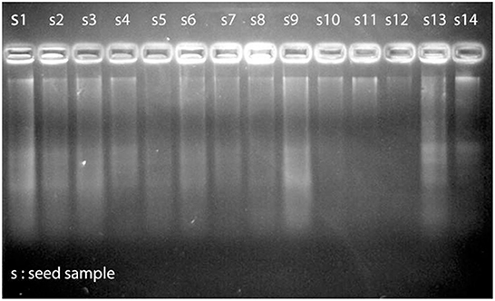
Figure 4. View of nucleic acids (TNA) obtained from tomato seeds in gel imaging system.
Reverse transcriptase polymerase chains reaction
PCR was prepared in a total volume of 50 μl in accordance with the procedure recommended by Thermo Scientific. For PCR applications, 10 μM intermediate stock was created and 1 μl of this intermediate stock was used for 25 μl PCR reaction.
At this stage, 12.5 μl DreamTaq Green PCR Master Mix, 8.5 μl nuclease free water, 1 μl Forward primer, 1 μl Reverse primer and 2 μl cDNA were added to sterile PCR tubes, making the total volume to 25 μl. The replication process was carried out by applying a specific program for the virus to be tested by arranging the tubes in the Thermal Cycler device (Table 2) (Candresse et al., 1995).
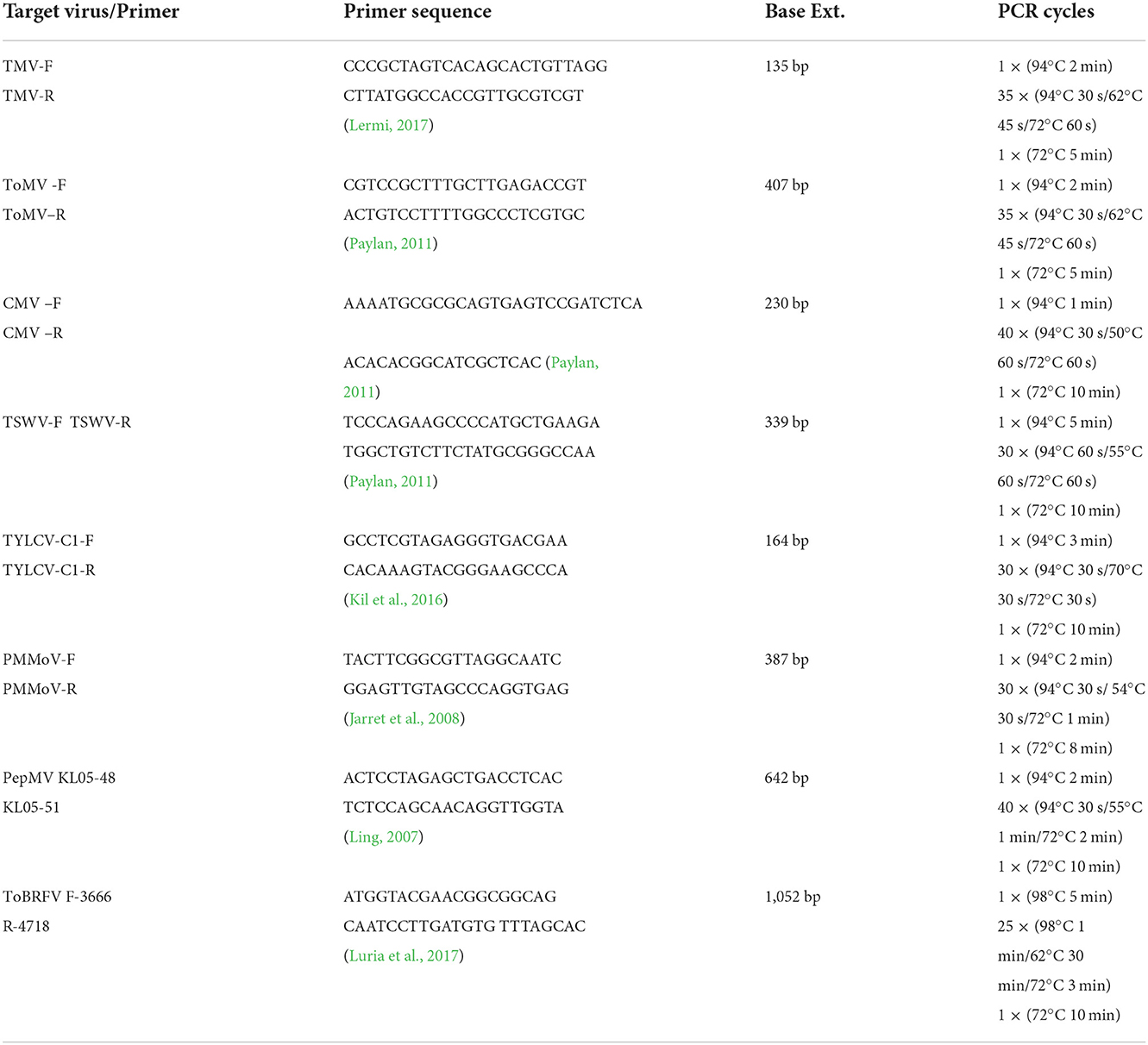
Table 2. Primer pairs used in PCR.
Agarose gel electrophoresis method
One percentage agarose gel was used for the analysis of DNA fragments obtained after the PCR step. A solution was obtained by adding 1.5 g of agarose to 100 ml volume of 1X TAE (Tris-Acetate -EDTA) buffer and leaving the mixture in the microwave for 3 min. The combs were placed on the electrophoresis dish and the gel was poured slowly onto the gel tray on a flat surface. It was waited for 30 min for the polymerization of the agarose, paying attention to the absence of bubbles. After polymerization of the gel, the combs were removed slowly and carefully. The gel was placed in the electrophoresis tank and 1X TAE buffer solution was added to pass the gel 1–2 mm. In order to determine the molecular size of the bands in the first well of the gel, the marker (100 bp DNA Ladder) was loaded in 6 μl volume. Ten microliter of PCR products were taken and mixed with 2 μl of loading buffer (6X DNA Loading dye) and loaded into other wells. Electrophoresis was carried out at 100 V for 60 min in a horizontal setup and in 1X TAE solution.
Evaluation of results on gel imaging device
The bands of the gel, which was stained after the electrophoresis step, it was observed under UV light and photographed with the DNR Bio-Imaging brand Gel Documentation and Analysis System (Israel).
Results
Symptoms observed in seed samples
In the tomato seeds included in this study, as a result of visual inspections in the field and in the laboratory, abnormal symptoms such as seed deformities, darkening of seed color, underdevelopment of seeds, wrinkling and necrosis in the seed coat were observed. While the symptomatological examination of the seeds constitutes the first step for the others of the diagnostic methods, it is known that the abnormalities seen in the seeds may be caused by other pathogens or factors, except the result that may be caused by viral pathogens. In addition, it has been determined as a result of the tests that there may be viral infection in tomato seeds, which are healthy in appearance and no symptoms can be seen as a result of visual inspection.
Results of serological tests
DAS-ELISA test was performed on tomato seed samples, which are included in this study, using commercial diagnostic kits for virus pathogens. This method, It was applied to 238 tomato seed samples. In the tomato seed samples used in the study, TMV, ToMV, TYLCV, CMV, TSWV, PepMV, and PMMoV were tested with serological methods to determine the presence of viral pathogens. Considering the results of serological tests; TMV in three samples, CMV in six samples, ToMV viral pathogen in 22 samples and finally mixed infection in 30 samples were detected. It was determined that 1 or more viral pathogens were found in a total of 67 seed samples out of 238 samples tested. Considering the mixed infection distribution detected in 30 seed samples; It was determined that the viral pathogens of CMV and TMV ToMV were found together in six seed samples. TMV and CMV viral pathogens were found in three seed samples, ToMV and CMV in four seed samples, and TMV and ToMV viral pathogens in 17 seed samples (Table 3).
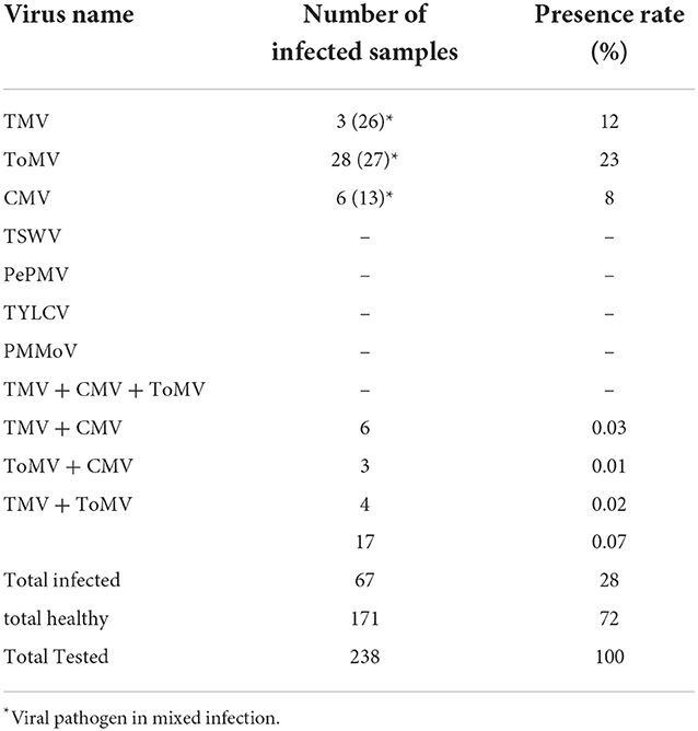
Table 3. Viruses detected as a result of serological tests in tomato seed samples.
Looking at the results of the DAS-ELISA tests, 28% of the tested tomato seeds were found virus infection was determined.
Results of molecular methods
Samples that showed positive results in DAS-ELISA tests and tomato seeds that showed symptoms as a result of visual examinations in the laboratory were selected at a specified rate, and PCR and RT-PCR tests were performed with these selected seeds. Within the scope of the study, tomato seeds without viral infection were also used, except for these selected seeds. Seed samples were tested against TMV, ToMV, CMV, TSWV, PepMV, PMMoV, ToBRFV, and TYLCV viral pathogens by PCR method using RT-PCR method. Unlike the results of previous serological tests in molecular studies, TMV was detected in three seed samples in which the presence of viral pathogens could not be detected by DAS-ELISA method. Except for these three samples, all seeds were found to be infected in RT-PCR tests as well as in DAS-ELISA tests (Figures 5–7). In addition, the tested tomato seed samples showed parallelism with the serological tests, and the presence of TYLCV, PepMV, PMMoV, and ToBRFV viral pathogens could not be detected in the seeds. Tomato seed samples that were not tested for ToBRFV in DAS-ELISA tests were tested by molecular methods to determine the presence of this viral agent, and as a result, ToBRFV viral pathogen could not be detected in 78 samples analyzed.
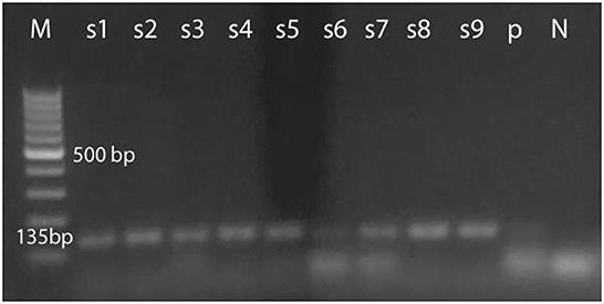
Figure 5. Tobacco in tomato seeds mosaic tobamovirus view of the result of the RT-PCR test applied to the gel imaging system.
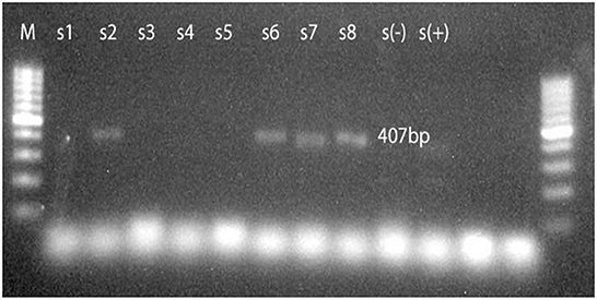
Figure 6. Tomato in tomato seeds mosaic view of the RT-PCR test result for tobamovirus taken on the gel imaging system.
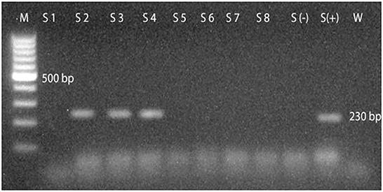
Figure 7. Cucumber in tomato seeds mosaic view of the RT-PCR test result applied for cucumovirus on the gel imaging system.
Conclusion and recommendations
In this study, the detection of viral pathogens in tomato seeds was carried out using serological and molecular methods. In addition, the presence of existing viral pathogens in tomato seeds was determined and updated. As a result of the symptomatological examination, it was observed that there were symptoms such as color changes, mottling, wrinkling, deformities and necrosis in the seeds. It has been concluded through serological and molecular tests that these symptoms in tomato seed samples may be caused by viral pathogens as well as by other pathogens or other factors. In addition, as a result of the studies, it was observed that viral pathogens could be present in tomato seed samples, which did not show any symptoms as a result of visual examinations (Erkan, 1998; Paylan, 2011). In order to determine the presence of TMV, ToMV, CMV, TYLCV, TSWV, PepMV, and PMMoV agents in 238 tomato seed samples obtained from the producer companies within the scope of the research, the samples were tested by DAS-ELISA method. Mixed infection was detected in 30 of the tested seed samples, and 1 or more pathogens were found in a total of 67 samples, 41 of which were standard and 26 of which were hybrid. Pathogen could not be detected in 171 samples.
Within the scope of the research, 238 plant samples, 148 of which are hybrid and 90 of which are standard varieties, were tested. One hundred and twenty-two of 148 hybrid varieties belong to private seed companies. Infection was detected in 18 (15%) of these 122 samples. This rate was lower than the general virus infection (28%), and this may be due to the fact that private seed companies give more importance to certified seed production and seed health tests than local producers. The other 26 hybrid cultivars consist of samples belonging to public institutions such as the ministry and university, and eight samples of 26 samples were determined as virus-infected. The 90 standard cultivars tested belong to local seed producers and individual farmer producers, and viral infection was detected in 41 of these samples. In standard samples, the infection rate (45%) was higher than the general rate (28%). The reason for the high infection rate in standard varieties may be that local producers do not give the necessary importance to seed health, do not have the necessary analyzes done because there is no legal obligation, and uncontrolled seed exchange.
In the study, reliable, sensitive and fast methods such as serological (DAS-ELISA) and molecular (RT-PCR) methods were used for the diagnosis of viral agents in tomato seeds. It is thought that some information such as the procedures and primers used can be a reference in future studies for the detection of viral infection. RT-PCR tests showed 100% success by detecting all positive samples detected in DAS-ELISA tests. In addition, molecular diagnosis has been clarified in a few samples suspected in DAS-ELISA tests. This situation supported the studies stating that the RT-PCR method is a sensitive and safe method in the diagnosis of tomato viruses. However, the need for a large number of chemicals, tools and equipment, the intensity and expensiveness of the test stages limit the use of RT-PCR. However, as a result, parallel results were obtained in both methods used in diagnosis in this study. With the completion of this study, the presence of infectious pathogens in tomato plant seeds was determined. Various viral pathogens (seed-borne) have been detected in tomato seeds in studies carried out on this subject so far. In previous studies on this subject, pathogens such as ToMV, TMV, TRSV, TBRV, and CMV were determined. In this study, it was determined that tomato seeds were infected with TMV, ToMV, and CMV pathogens.
Data availability statement
The original contributions presented in the study are included in the article/supplementary material, further inquiries can be directed to the corresponding author/s.
Author contributions
All authors listed have made a substantial, direct, and intellectual contribution to the work and approved it for publication.
Conflict of interest
The authors declare that the research was conducted in the absence of any commercial or financial relationships that could be construed as a potential conflict of interest.
Publisher's note
All claims expressed in this article are solely those of the authors and do not necessarily represent those of their affiliated organizations, or those of the publisher, the editors and the reviewers. Any product that may be evaluated in this article, or claim that may be made by its manufacturer, is not guaranteed or endorsed by the publisher.
References
Abak, K., Daşgan, H. Y., and Sari, N. (2000). Tomato Cultivation in the Southeastern Anatolia Region. Adana: Tubitak, Tarp Publications, 4–18.
Anonymous (2007). Tomato Spotted Wilt Virüs. Available online at: www.eppo.org/~Quarantine/virus/ (accessed May 15, 2019).
Candresse, T., Lanneau, T., Revers, F., Grasseau, N., Macquare, G., German, S., Malinowsky, T., and Dunez, J. (1995). An immunocapture PCR assay adapted to the detection and the analysis of the molecular variability of apple chlorotic leaf spot virus. Acta Hortic. 386, 136–147. doi: 10.17660/ActaHortic.1995.386.17
Carroll, T. W. (1983). Certification schemes against barley stripe mosaic. Seed Sci. Technol. 11, 1033–1042.
Clark, M. F., and Adams, A. N. (1977). Characteristic of microplate method of enzyme-linked immunosorbent assay for detection of plant viruses. J. Gen. Virol. 34, 475–483. doi: 10.1099/0022-1317-34-3-475
Demiray, E., and Tülek, Y. (2008). The effect of tomato drying technology and drying process on some antioxidant compounds in tomatoes. Electron. J. Food Technol. 9–20.
Erkan, S. (1998). Seed Pathology. Izmir: E. U. Z.F. Department of Plant Protection, Gözdem Office, 275.
Foissac, X., Savalle-Dumas, L., Gentit, P., Dulucq, M. J., and Candresse, T. (2001). Polyvalent detection of fruit tree tricho, capillo and foveaviruses by nested RT-PCR using degenerated and inosine containing primers (PDO RT-PCR). Acta Hortic. 550, 37–43. doi: 10.17660/ActaHortic.2001.550.2
Gebhardt, S. E., and Thomas, R. G. (2002). Nutritive Value of Foods. Washington, DC: USDA Agricultural Research Services, 72.
Güvenç, I. (2017). Vegetable Growing: Basic Information, Conservation and Cultivation. Ankara: Nobel Publications, 288.
Hanssen, I. M., Lapidot, M., and Thomma, B. P. H. J. (2010). Emerging viral diseases of tomato crops. Mol Plant Microbe Interact. 23, 539–548. doi: 10.1094/MPMI-23-5-0539
ISTA (2007). Available online at: http://www.seedtest.org/en/home.html (accessed April 21, 2019).
Jarret, R. L., Gillaspie, A. G., Barkley, N. A., and Pinnow, D. L. (2008). Occurrence and control of pepper mild virus 2008 virus (PMMoV) in the USDA/ARS Capsicum Germ Collection. Seed Technol. 30, 26–36.
Jones, J. B., Jones, J., Stall, R. E., and Zitter, T. A. (1991). Compendium of Tomato Diseases. St. Paul, MN: American Phytopathological Society Press, 73.
Kil, E. J., Kim, S., Lee, Y. J., Byun, H. S., Park, J., Seo, H., et al. (2016). Tomato yellow leaf curl virus (TYLCV-IL): a seed-transmissible geminivirus in tomatoes. Sci. Rep. 6, 19013. doi: 10.1038/srep19013
Lermi, E. D. (2017). Simultaneous Identification of Some Important Viral Disease Factors in Tomato Plants by Multiplex PCr Method (Master thesis), Ege University Institute of Science, Izmir, 63.
Ling, K. S. (2007). Molecular characterization of two pepino mosaic virus variants from imported tomato seed reveals high levels of sequence identity between Chilean And US isolates. Virus Genes 34, 1–8. doi: 10.1007/s11262-006-0003-x
Luria, N., Smith, E., Reingold, V., Bekelman, I., Lapidot, M., Levin, I., et al. (2017). A new Israeli tobamovirus isolate infects tomato plants harboring Tm-22 resistance. Genes. 12, E0170429. doi: 10.1371/journal.pone.0170429
Paylan, I. C. (2011). Research on the Determination of Viruses in the Seeds of Some Vegetable Species and the Inactivation of Viral Infections in Seeds (Doctoral thesis), Ege University Institute of Science and Technology, Izmir, 147.
Peralta, I. E., and Spooner, D. M. (2005). Morphological characterization and relationships of wild tomatoes (Solanum L. section lycopersicon). Monogr. Syst. Bot. Missouri Bot. Garden 104, 227–257.
Tigchelaar, E. C. (1986). “Tomato breeding,” in Breeding Vegetable Crops, ed M. Basset (Westport, CT.: AVI Publishing), 135–171.
Uzunogullai, N., and Gümüş, M. (2015). Detection of cucumber mosaic virus (cucumber Mosaic Virus, CMV) that causes natural infection in some cultural plants in the Marmara Region. Plant Protect. Bull. 16, 9–15.
Keywords: tomato, seed, virus, PCR, ELISA
Citation: Atik AK and Paylan İC (2023) Updating viral agents in tomato seeds with new generation diagnostic technologies. Front. Sustain. Food Syst. 6:945703. doi: 10.3389/fsufs.2022.945703
Received: 16 May 2022; Accepted: 04 October 2022;
Published: 16 February 2023.
Edited by:
Karl Matthews, Rutgers, The State University of New Jersey, United StatesReviewed by:
David Cazol, Kansas State University, United StatesPavor Uslui, University of East London, Pakistan
Copyright © 2023 Atik and Paylan. This is an open-access article distributed under the terms of the Creative Commons Attribution License (CC BY). The use, distribution or reproduction in other forums is permitted, provided the original author(s) and the copyright owner(s) are credited and that the original publication in this journal is cited, in accordance with accepted academic practice. No use, distribution or reproduction is permitted which does not comply with these terms.
*Correspondence: Ayşen Kübra Atik,  YXRpa2F5c2VuQGhvdG1haWwuY29t