Anti-oomycete Activity of Chlorhexidine Gluconate: Molecular Docking and in vitro Studies
- ICAR-Directorate of Coldwater Fisheries Research, Bhimtal, India
Saprolegniosis is one of the most catastrophic oomycete diseases of freshwater fish caused by the members of the genus Saprolegnia. The disease is responsible for huge economic losses in the aquaculture industry worldwide. Until 2002, Saprolegnia infections were effectively controlled by using malachite green. However, the drug has been banned for use in aquaculture due to its harmful effect. Therefore, it has become important to find an alternate and safe anti-oomycete agent that is effective against Saprolegnia. In this study, we investigated the anti-oomycete activity of chlorhexidine gluconate (CHG) against Saprolegnia. Before in vitro evaluation, molecular docking was carried out to explore the binding of CHG with vital proteins of Saprolegnia, such as S. parasitica host-targeting protein 1 (SpHtp1), plasma membrane ATPase, and TKL protein kinase. In silico studies revealed that CHG binds with these proteins via hydrogen bonds and hydrophobic interactions. In an in vitro study, the minimum inhibitory concentration (MIC) and minimum fungicidal concentration (MFC) of CHG against S. parasitica were found to be 50 mg/L. Further, it was tested against S. australis, another species of Saprolegnia, and the MIC and MFC were found to be 100 and 200 mg/L, respectively. At 500 mg/L of CHG, there was complete inhibition of the radial growth of Saprolegnia hyphae. In propidium iodide (PI) uptake assay, CHG treated hyphae had bright red fluorescence of PI indicating the disruption of the cell membrane. The results of the present study indicated that CHG could effectively inhibit Saprolegnia and hence can be used for controlling Saprolegniasis in cultured fish.
Introduction
Saprolegnia is one of the most common oomycetes or fungal-like organisms responsible for devastating diseases in fish. These organisms cause huge losses in aquaculture worldwide and are also considered responsible for the decline in populations of wild fish and amphibians (1–5). The host range of these organisms includes various fish species, amphibians, and crustaceans (6–8). The disease caused by this organism, known as Saprolegniasis, is characterized by white or gray cotton-like mycelial growth at the site of infection. The most common sites of infection are the head, fins, and tail but in severe cases, it spreads further into muscles and blood vessels, and ultimately, the infected fish die due to osmoregulatory failure (9–11).
Earlier, Saprolegnia infections were controlled by using malachite green, but the drug has been banned from use in aquaculture due to its carcinogenic, mutagenic, and teratogenic properties (12–14). Since then, various chemicals have been trialed for their anti-oomycete activity against Saprolegnia infections and found effective but were accompanied by adverse effects. Among the tested chemicals, formalin was effective but reported to be associated with environmental pollution, a health hazard to the handler, and accumulation in fish flesh making it unfit for consumption (14). Similarly, other chemicals, such as hydrogen peroxide, boric acid, and peracetic acid, which demonstrated anti-Saprolegnia activity, are reported to produce negative side effects (15–20). Therefore, it is the need of the hour to discover or identify a chemical that is safe and effective against Saprolegnia.
In this study, the anti-Saprolegnia activity of chlorhexidine gluconate (CHG) was evaluated. It is the gluconate salt form of chlorhexidine. It is a positively charged polybiguanide that can bind to the negatively charged cell membrane of microbes (21, 22). CHG has broad-spectrum antimicrobial activity with rapid action, killing bacteria within 30 s (23, 24). It also has the ability to prevent microbial colonization and the formation of biofilms (25). It exhibits a dose-dependent effect, being bactericidal at high concentrations (26, 27). Apart from bacteria, CHG is also active against enveloped viruses and most fungi (28). It acts by interfering with cell wall integrity, then attacking the cytoplasmic membrane leading to leakage of cell contents and ultimately cell death (23). It is used as an antiseptic for the prevention of surgical wound infection and as a mouthwash in dentistry (29, 30). When used as an antiseptic for skin, CHG was found to have an extremely low potential for dermal reactions (31). Despite its broad-spectrum antimicrobial effect with good safety profile and widespread use, literature on the effect of CHG against Saprolegnia is very limited. There is one patent on a powder formulation developed to treat Saprolegniasis which contains only 1–3 parts of chlorhexidine acetate in combination with other chemicals (Patent no. CN104997805B, China). Therefore, the present study was taken up to evaluate the anti-Saprolegnia activity through in silico and in vitro analysis.
Materials and Methods
Culture of Saprolegnia Species
For evaluation of the anti-oomycete activity of CHG, two species, S. parasitica (Accession no. MT912581) and S. australis (Accession no. MT912582), isolated from rainbow trout (Oncorhynchus mykiss) were used. Both the isolates were cultured on glucose-yeast extract agar (GYA) plates and incubated at 20 ± 1°C. The isolates were maintained by subculturing into new GYA plates after every few days or when the plate has full hyphal growth. Subculture was done by transferring a piece of agar excised from the advancing edges of hyphal growth to the new GYA plate. In all the experiments, 4-day old culture was used to maintain uniformity.
In silico Analysis of the Interaction Between CHG and Vital Proteins of Saprolegnia
The multiple threading approach of I-Tasser was used for homology modeling of S. parasitica proteins, i.e., Tyrosine kinase-like (TKL) protein kinase and the plasma membrane ATPase (32). The fold-based approach of the pDOMTHREADER allowed to find a template for the S. parasitica host targeting protein-1, as reported earlier (33). Using the standalone version of Modeler 9.18, the template with a high score as per pDOMTHREADER was used for homology modeling (34).
Thereafter, the three-dimensional (3D) structures were refined using ModRefiner (https://zhanglab.ccmb.med.umich.edu/ModRefiner/) and the quality was checked by the saves server [SAVESv6.0—Structure Validation Server (ucla.edu)]. In each modeled protein, the binding sites were predicted by the COACH meta server (35). CHG structure was retrieved from Pubchemin in SDF format. The binding energy of ligand-receptor complexes was computed using AutoDockVina software by mimicking the ligand into the active site of the protein (36). There were nine docking poses predicted for each ligand-receptor complex when the exhaustiveness was set to twenty. Among these, the ideal pose of binding between protein and ligand was chosen to explore hydrogen bonding and hydrophobic interactions, with a higher negative docking score (37). PyMOL was used to visualize 3D structures and docking structures, whereas LigPlot 2.1 was used to visualize two-dimensional (2D) structures (38).
Preparation of Chlorhexidine and Resazurin Stock
A stock solution of 10,000 mg/L of CHG was prepared using sterile double-distilled water. Working solutions were prepared by diluting them with sterile double-distilled water. A stock solution of resazurin dye was prepared by dissolving 25 mg in 10 ml of 10 mM sodium phosphate buffer (pH, 7.4). The solution was then vortexed till the dye dissolves completely and sterile filtered using a 0.2 μm syringe filter and stored at −20°C until use. A working solution of resazurin (300 μM) in sodium phosphate buffer was prepared fresh before each experiment.
Determination of Minimum Inhibitory Concentration
The minimum inhibitory concentration (MIC) of CHG was determined against S. parasitica and S. australis, following the published methodologies (39, 40) with some modifications. In a 48-well plate, 400 μl of glucose yeast extract broth (GYB) containing different concentrations (1, 10, 20, 50, 100, and 200 mg/L) of CHG was added. Triplicate wells of each concentration of CHG were used for the assay. Growth control wells without CHG and sterile control wells containing only media were included in the experiment. In each well except the sterile control wells, an agar plug of approximately 4 mm in diameter derived from a GYA plate containing 4-day old culture of Saprolegnia was added. The plate was incubated at 20 ± 1°C for 48 h. Thereafter, 100 μl of freshly prepared resazurin (300 μM) was added to each well. The plate was further incubated and observed for change in color from blue to pink. The minimum concentration of CHG at which the color remained blue was recorded as MIC.
Determination of Minimum Fungicidal Concentration
For the determination of minimum fungicidal concentration (MFC), the experiment was carried out in a 48-well plate similar to that of MIC. Agar plugs containing Saprolegnia hyphae were inoculated in separate wells containing different concentrations of CHG (sub-MIC, MIC, and 2X MIC) and incubated for 24 h at 20 ± 1°C. Growth control wells containing only media and Saprolegnia but no CHG were included in the experiment. After incubation, the agar plugs from control and the CHG treated wells were transferred to GYA plates and incubated for 24 h at 20 ± 1°C. The agar plates were observed for the presence or absence of any visible hyphal growth. The minimum concentration of CHG at which there was no visible hyphal growth was recorded as an MFC value. Further, the effect of CHG on live fish was checked by dip treatment at MIC and MFC for a short period of time.
Radial Growth Inhibition Assay
The effect of CHG on the radial growth of Saprolegnia hyphae was determined in a time series following the protocol of Shin et al. (39). In brief, GYA containing different concentrations of CHG (25, 50, 100, 200, 500, and 1,000 mg/L) was prepared by mixing an equal volume of 2X GYA and 2X CHG to achieve the desired final concentration. The GYA containing CHG was then plated on sterile 90 mm Petri dishes. A growth control plate containing 2X GYA mixed with an equal volume of sterile distilled water was included in the experiment. An agar plug of ~5 mm diameter containing Saprolegnia hyphae was cut from 4-day old culture plate and placed onto the center of the fresh GYA plate. The plates were incubated at 20 ± 1°C, and radial growth was recorded as diameter every 24 h till full mycelial growth was achieved in the growth control plate. The experiment was conducted in triplicates and repeated three times. The percentage inhibition of radial growth (PIRG) was calculated using the formula, PIRG = (DC–DT) ×100/DC, where, DC = diameter of radial growth on the control plate in mm, and DT = diameter of radial growth on CHG treated plate in mm. The values are presented as mean ± SE.
Analysis of Membrane Integrity
A propidium iodide (PI) uptake assay was performed to investigate the effect of CHG on membrane integrity of Saprolegnia following the protocol of Palem et al. (41) and Shin et al. (39) with some alterations. Saprolegnia was inoculated on a GYA plate along with sesame seeds which were used as bait. After 4 days of incubation, the sesame seeds bearing Saprolegnia hyphae were transferred to 48 well plates containing GYB incorporated with MIC and MFC concentration of CHG and incubated at 20 ± 1°C. Growth or negative control wells containing only medium and positive control wells containing medium plus 0.1% triton-X were included in the experiment. After incubation for 24 h, the hypha was washed in PBS and stained with PI (20 μg/ml) for 20 min in dark at room temperature. The PI fluorescence was observed under an inverted fluorescent microscope (Nikon, Japan).
Results
In silico Analysis of the Interaction Between Saprolegnia Proteins and CHG
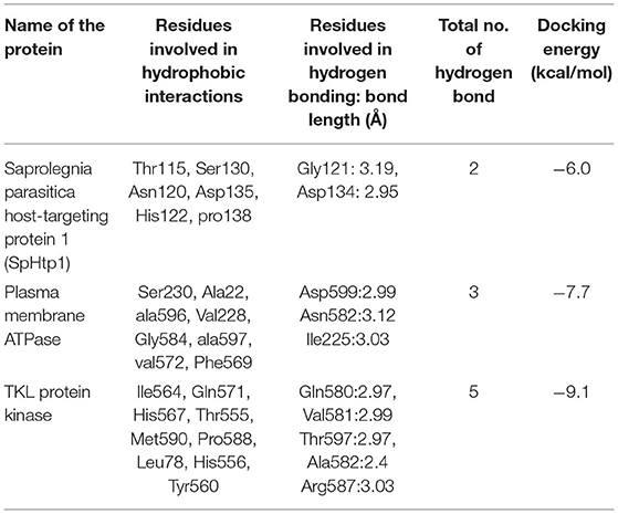
The quality of modeled 3D structure was analyzed through the Ramachandran plot. It was found that about 97% of amino acid residues in the modeled structure were in the most favored region. Details of in silico analysis of the 2D and 3D interactions between the active sites of Saprolegnia proteins and CHG ligand are given in Figure 1, Table 1. Among the proteins studied through molecular docking, the lowest minimum binding energy, −9.1 Kcal/mol, was observed between CHG and TKL protein kinase. CHG was found to bind strongly with TKL protein kinase through five hydrogen bonds and nine hydrophobic interactions. Similarly, the ligand has shown good interaction with plasma membrane ATPase through three hydrogen bonds and eight hydrophobic interactions, with a binding energy of −7.7 Kcal/mol. CHG also interacted with S. parasitica host-targeting protein 1 (SpHtp1) through two hydrogen bonds and six hydrophobic interactions with a binding energy of −6.0 Kcal/mol.

Figure 1. Molecular docking of CHG with proteins of S. parasitica. (A) with S. parasitica host targeting protein, (B) with plasma membrane ATPase, (C) with TKL protein kinase.

Table 1. Details of interactions between Saprolegnia proteins and chlorhexidine gluconate (PubChem CID: 9552079).
Minimum Inhibitory and Fungicidal Concentration
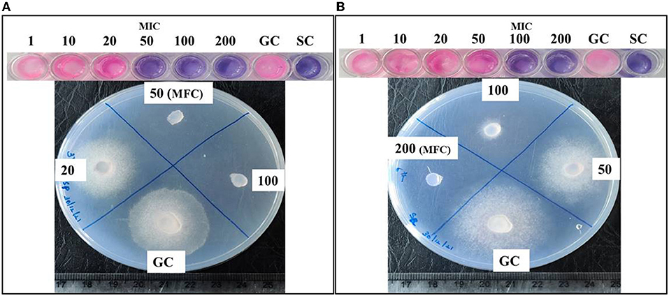
Chlorhexidine gluconate was found to inhibit the growth of S. parasitica and S. australis at different concentrations. The minimum concentration of CHG (MIC), which inhibited the growth of Saprolegnia, observed as an unchanged blue color of resazurin, was found to be 50 and 100 mg/L for S. parasitica and S. australis, respectively. In the case of S. parasitica, MFC was the same as that of MIC, i.e., 50 mg/L. It was evident from the absence of hyphal growth when the CHG treated specimen was inoculated on a fresh GYA plate. However, the MFC of CHG to produce a fungicidal effect in S. australis was found to be 200 mg/L which was two times its MIC value (Figure 2). Fish treated with 100 and 200 mg/L of CHG by dip method showed no abnormal behavior, changes in feed intake, and gross lesions in the skin during the observation of a period of 96 h after treatment.

Figure 2. Determination of minimum inhibitory concentration (MIC) and minimum fungicidal concentration (MFC) of CHG against Saprolegnia. Series of wells with blue and pink color at the top is of resazurin assay for determination of MIC. (A) MIC (blue) and MFC is 50 mg/L for S parasitica, (B) MIC is 100 mg/L and MFC is 200 mg/L for S. australis. Values are concentration of CHG in mg/L. GC, Growth control; SC, Sterile control. In wells of MIC and above, there is no development of pink color. In MFC, hyphal growth is absent.
Radial Growth Inhibition
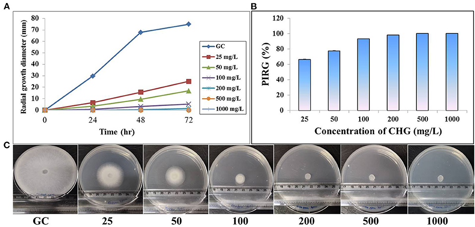
On CHG supplemented GYA, radial growth of Saprolegnia hyphae was inhibited in a dose-dependent manner. The mean diameter of radial hyphal growth decreased with an increased concentration of CHG in the medium. At higher concentrations, 500 and 1,000 mg/L, there was complete inhibition in the radial growth of both the species. At 72 h, the percentage rate of inhibition in radial growth at 25 mg/L was found to be more than 60 and 50% for S. parasitica and S. australis, respectively (Figures 3, 4).

Figure 3. Effect of CHG on radial growth of Saprolegnia parasitica. (A)Time dependent mycelial growth of S. parasitica on glucose yeast extract agar incorporated with different concentrations of chlorhexidine (0, 25, 50, 100, 200, 500 and 1000 mg/L). (B) Estimation of percent inhibition on radial growth of S. parasitica on CHG incorporated glucose yeast extract at 72 hr. (C) Full grown hyphae in growth control (GC) and reduced or absence of hyphal growth in CHG treated plates at 72 hr.

Figure 4. Effect of CHG on radial growth of Saprolegnia australis. (A) Time dependent mycelial growth of S. australis on glucose yeast extract agar incorporated with different concentrations of chlorhexidine (0, 25, 50, 100, 200, 500, and 1,000 mg/L). (B) Estimation of percent inhibition on radial growth of S. australis on CHG incorporated glucose yeast extract at 72 hr. (C) Full grown hyphae in growth control (GC) and reduced or absence of hyphal growth in CHG treated plates at 72 hr.
Disruption of Membrane Integrity
In the PI uptake assay, Saprolegnia hyphae, treated with MIC and MFC of CHG, showed bright red fluorescence indicating loss of membrane integrity. Red fluorescence of high intensity was also observed in Triton-X treated Saprolegnia. The untreated negative control hyphae had no red fluorescence indicating an intact membrane (Figure 5).

Figure 5. PI uptake assay to investigate membrane disruption by chlorhexidine. The first row is of S. australis treated with 0, MIC and MFC of CHG and Triton-X 100. The second row is of S. parasitica treated with 0, MIC/MFC of CHG and Triton-X 100. Scale-100 μm.
Discussion
Saprolegnia species, particularly S. parasitica, have re-emerged as an important pathogen in aquaculture due to a lack of effective control measures. After malachite green has been banned for use in aquaculture, several chemicals have been evaluated for their efficacy to control Saprolegniasis. However, the side effects of those chemicals on the handler, fish, and environment have been the main obstacles for field application in aquaculture (20). This has prompted us to search for new chemicals that are effective against Saprolegnia and safe to use as well. Thus, the current study was undertaken to evaluate the anti-Saprolegnia activity of CHG in vitro. Chlorhexidine is commonly known for its salt forms, such as chlorhexidine acetate, CHG, and chlorhexidine digluconate. CHG is one of the most widely used broad-spectrum antiseptics, effective against various microbial pathogens with low toxicity to the host (27, 42–44). Owing to its low potential for toxicity, CHG has been incorporated in many cosmetic products, antiseptic creams used for wound cleaning, and teat dip in the dairy industry. It is one of the most commonly used chemicals in dentistry and as an adjunct in the treatment of oral yeast infections (45, 46).
Before carrying out the in vitro studies, the probable interaction of CHG with vital proteins of S. parasitica was explored through molecular docking. This bioinformatic approach allows to identification potential inhibitory molecules of the oomycete vital proteins, responsible for virulence (33, 46). Recently, Kumar et al. (20) have also identified several compounds along with Saprolegnia target proteins using the computational proteomics approach. In this study, CHG was found to interact with Saprolegnia proteins (SpHtp1, TKL kinase, and plasma membrane ATPase) through hydrogen bonds and hydrophobic interactions, which revealed the potential of the anti-Saprolegnia activity. SpHtp1 translocates inside fish cells in a tyrosine-O-sulfate–dependent manner and may play a role in saprolegniasis (47, 48). Similarly, TKL protein kinase and plasma membrane ATPase are important proteins that can be targeted for the discovery of antifungal agents (49, 50). The interaction of TKL protein kinase and plasma membrane ATPase with CHG obeyed Lipinski's rule of five (51). It is a rule of thumb to evaluate the drug-likeness of a chemical compound. Based on the findings of molecular docking, CHG was predicted to produce an inhibitory effect against Saprolegnia.
The efficacy of CHG was evaluated in vitro against two species, S. parasitica and S. australis. CHG produced its inhibitory and fungicidal effect at 50 mg/L which corresponds to 0.005% against S. parasitica. Whereas, higher dosage of CHG, i.e., 100 mg/L or 0.01% for growth inhibition and 200 mg/L 0.02% for the killing of S. australis were required. This difference in sensitivity can be corroborated by reports on the variable efficacy of boric acid among Saprolegnia species (52). Such variation in antifungal effect against different species has also been reported in Aspergillus flavus and Aspergillus niger (53). This may be due to differences in the cell wall or membrane component between the species (54). In addition, we have observed such variation in the efficacy of antimicrobials in bacteria among different species of Aeromonas, i.e., Aeromonas sobria, Aeromonas hydrophilla, and Aeromonas salmonicida (37, 55).
Chlorhexidine gluconate was found to produce its growth inhibitory effect against the two tested species of Saprolegnia at 0.005 to 0.01% and its biocidal effect at 0.005 to 0.02%. The effective dose is much lesser than that of common concentrations of CHG used for medicinal purposes. Most widely available chlorhexidine products contain 2% active chlorhexidine in its salt form either gluconate or acetate (56). It was reported that 0.02% chlorhexidine produced no abnormal behavior and no significant changes in hematological and serum biochemical parameters in laboratory rats (57). Wade et al. (58) stated that alcoholic formulations of 4–5% of CHG seem to be safe and effective in preventing infection after clean surgery in adults. This indicates that CHG can produce its growth inhibitory and biocidal effect against Saprolegnia at a safe concentration for the handler.
Chlorhexidine gluconate was found to cause complete inhibition of radial growth at ≥500 mg/L in both the species of Saprolegnia. It is less than the concentration of boric acid required to produce a similar effect (59). Moreover, the MIC and MFC of CHG are also much lesser than that of boric acid. This may be well explained by the mode of action of chlorhexidine. CHG kills bacteria and fungus through disruption in structural organization and integrity of the cytoplasmic membrane, resulting in leakage of intracellular components (23). As expected, in the PI uptake assay, CHG-treated Saprolegnia hypha showed red fluorescence but the un-treated one did not. This suggested that CHG disrupted the cell membrane and allowed the membrane impermeant dye, PI to enter inside the cells, and stained nucleic acid. As per the literature, chlorhexidine can also kill the microbes within a very less time of contact (57) making it suitable for dip treatment. In fish, dip treatments are done by keeping the fish in a strong solution of chemicals for <1 min (60). We also found no gross changes in the fish treated with MIC and MFC of CHG.
With our findings, it is confirmed that CHG can bind strongly with important proteins of Saprolegnia, which depicts its anti-oomycete potential. The inhibitory effect of CHG against Saprolegnia was further demonstrated through various in vitro assays. It produced its inhibitory and fungicidal effect against Saprolegnia species at the sub-therapeutic level. As there is a lack of effective prophylactic and therapeutic measures against Saprolegnia, an effective and eco-friendly chemical is highly essential to control Saprolegniasis. Therefore, CHG, with high efficacy against various microbial pathogens and negligible toxicity can be an ideal candidate for application in aquaculture for the management of Saprolegniasis. Further, CHG can be incorporated as a major component in formulations for the treatment of Saprolegniasis. As downstream work, CHG may be evaluated for its efficacy for disinfection of fish eggs and treatment of Saprolegniasis in adult fish.
Data Availability Statement
The original contributions presented in the study are included in the article/supplementary material, further inquiries can be directed to the corresponding author.
Ethics Statement
The animal study was reviewed and approved by Institutional Animal Care and Use Committee, ICAR-DCFR.
Author Contributions
DT and VCK conceptualized and designed the experiments. DT, VCK, and VP conducted the in vitro assays and wrote the manuscript. RAHB did in silico analysis. AP conducted fluorescent microscopy. RST contributed in culture of Saprolegnia. SC, DT, and VCK conducted the dip treatment of fish. DT, VCK, and PKP edited the manuscript.
Funding
This work was supported by the ICAR Institutional fund under the project “Evaluation of antimicrobial potential of nano and polymer based formulations against saprolegniasis”.
Conflict of Interest
The authors declare that the research was conducted in the absence of any commercial or financial relationships that could be construed as a potential conflict of interest.
Publisher's Note
All claims expressed in this article are solely those of the authors and do not necessarily represent those of their affiliated organizations, or those of the publisher, the editors and the reviewers. Any product that may be evaluated in this article, or claim that may be made by its manufacturer, is not guaranteed or endorsed by the publisher.
References
1. Madrid A, Godoy P, González S, Zaror L, Moller A, Werner E, et al. Chemical characterization and anti-oomycete activity of Laureliopsis philippianna essential oils against Saprolegnia parasitica and S. australis. J Molecules. (2015) 20:8033–47. doi: 10.3390/molecules20058033
2. Neitzel DA, Elston R, Abernethy CS. Prevention of Prespawning Mortality: Cause of Salmon Headburns and Cranial Lesions. Richland, WA: Pacific Northwest National Lab.(PNNL) (2004). doi: 10.2172/15020751
3. Kiesecker JM, Blaustein AR, Miller CL. Transfer of a pathogen from fish to amphibians. J Conserv Biol. (2001) 15:1064–70. doi: 10.1046/j.1523-1739.2001.0150041064.x
4. Fregeneda-Grandes JM, Rodríguez-Cadenas F, Aller-Gancedo JM. Fungi isolated from cultured eggs, alevins and broodfish of brown trout in a hatchery affected by saprolegniosis. J Fish Biol. (2007) 71:510–8. doi: 10.1111/j.1095-8649.2007.01510.x
5. Jiang RH, Bruijn Ide, Haas BJ, Belmonte R, Löbach L, Christie J, et al. Distinctive expansion of potential virulence genes in the genome of the oomycete fish pathogen Saprolegnia parasitica. PLoS Genet. (2013) 9:e1003272. doi: 10.1371/journal.pgen.1003272
6. van West P. Saprolegnia parasitica, an oomycete pathogen with a fishy appetite: new challenges for an old problem. Mycologist. (2006) 20:99–104. doi: 10.1016/j.mycol.2006.06.004
7. Blaustein AR, Hokit DG, O'Hara RK, Holt RA. Pathogenic fungus contributes to amphibian losses in the Pacific Northwest. J Conserv Biol. (1994) 67:251–4. doi: 10.1016/0006-3207(94)90616-5
8. Diéguez-Uribeondo J, Cerenius L, Söderhäll K. Saprolegnia parasitica and its virulence on three different species of freshwater crayfish. Aquaculture. (1994) 120:219–28. doi: 10.1016/0044-8486(94)90080-9
9. Phillips AJ, Anderson VL, Robertson EJ, Secombes CJ, Van West P. New insights into animal pathogenic oomycetes. Trend Microbiol. (2008) 16:13–19. doi: 10.1016/j.tim.2007.10.013
10. Pickering AD, Willoughby LG. Saprolegnia infections of salmonid fish. In: Roberts RJ, editor. Microbial Diseases of Fish. London: Academic Press Inc. (1982). p. 271–97.
11. Hatai K, Hoshiai G. Pathogenicity of saprolegnia parasitica cocker. In: Mueller GJ, editor. Salmon Saprolegniosis. Portland, OR: US Department of Energy, Bonneville Power Administration (1994). p. 87–98.
12. Forneris G, Bellardi S, Palmegiano G, Saroglia M, Sicuro B, Gasco L, et al. The use of ozone in trout hatchery to reduce saprolegniasis incidence. Aquaculture. (2003) 221:157–66. doi: 10.1016/S0044-8486(02)00518-5
13. Culp S, Beland F, Heflich R, Benson R, Blankenship L, Webb P, et al. Mutagenicity and carcinogenicity in relation to DNA adduct formation in rats fed leucomalachite green. Mutat Res. (2002) 506:55–63. doi: 10.1016/S0027-5107(02)00152-5
14. Gieseker C, Serfling S, Reimschuessel R. Formalin treatment to reduce mortality associated with Saprolegnia parasitica in rainbow trout, oncorhynchus mykiss. Aquaculture. (2006) 253:120–9. doi: 10.1016/j.aquaculture.2005.07.039
15. Rach JJ, Valentine JJ, Schreier TM, Gaikowski MP, Crawford TG. Efficacy of hydrogen peroxide to control saprolegniasis on channel catfish (Ictalurus punctatus) eggs. Aquaculture. (2004) 238:135–42. doi: 10.1016/j.aquaculture.2004.06.007
16. Mustafa S, Al-Rudainy A, Al-Faragi J. Assessment of hydrogen peroxide on histopathology and survival rate in common carp, Cyprinus carpio l. infected with saprolegniasis. Iraqi J Agric Sci. (2019) 50:697–704.
17. Acar Ü, Inanan BE, Zemheri F, Kesbiç OS, Yilmaz S. Acute exposure to boron in Nile tilapia (Oreochromis niloticus): Median-lethal concentration (LC50), blood parameters, DNA fragmentation of blood and sperm cells. J Chemosphere. (2018) 213:345–50. doi: 10.1016/j.chemosphere.2018.09.063
18. Öz M, Inanan BE, Dikel S. Effect of boric acid in rainbow trout (Oncorhynchus mykiss) growth performance. J Appl Anim Res. (2018) 46:990–3. doi: 10.1080/09712119.2018.1450258
19. Marchand PA, Phan TM, Straus DL, Farmer BD, Stüber A, Meinelt T. Reduction of in vitro growth in Flavobacterium columnare and Saprolegnia parasitica by products containing peracetic acid. J Aquac Res. (2012) 43:1861–6. doi: 10.1111/j.1365-2109.2011.02995.x
20. Kumar S, Mandal RS, Bulone V, Srivastava V. Identification of growth inhibitors of the fish pathogen saprolegnia parasitica using in silico subtractive proteomics, computational modeling, and biochemical validation. Front Microbiol. (2020) 11:571093. doi: 10.3389/fmicb.2020.571093
21. Leikin J, Paloucek F. Chlorhexidine Gluconate. Poisoning and Toxicology Handbook Informa. (2008). p. 183–4. doi: 10.3109/9781420044805
22. Tanzer J, Slee A, Kamay B. Structural requirements of guanide, biguanide, and bisbiguanide agents for antiplaque activity. Antimicrob Agents Chemother. (1977) 12:721–9. doi: 10.1128/AAC.12.6.721
23. McDonnell G, Russell AD. Antiseptics and disinfectants: activity, action and resistance. Clin Microbiol Rev. (1999) 12:147–79. doi: 10.1128/CMR.12.1.147
24. Genuit T, Bochicchio G, Napolitano LM, McCarter RJ, Roghman MC. Prophylactic chlorhexidine oral rinse decreases ventilator-associated pneumonia in surgical ICU patients. Surg Infect. (2001) 2:5–18. doi: 10.1089/109629601750185316
25. Mohammadi Z, Abbott PV. The properties and applications of chlorhexidine in endodontics. Int Endod J. (2009) 42:288–302. doi: 10.1111/j.1365-2591.2008.01540.x
26. Ferraz GC, ME V, Berber V, Teixeira F, Souza-Filho F. In vitro antimicrobial activity of several concentrations of sodium hypochlorite and chlorhexidine gluconate in the elimination of enterococcus faecalis. J Int Endod. (2001) 34:424–8. doi: 10.1046/j.1365-2591.2001.00410.x
27. Z.M. Kanisavaran. Chlorhexidine gluconate in endodontics: an update review. Int Dent J. (2008) 58:247–57. doi: 10.1111/j.1875-595X.2008.tb00196.x
28. Lim KS, Kam PCA. Chlorhexidine–pharmacology and clinical applications. Anaesth Intensive Care. (2008) 36:502–12. doi: 10.1177/0310057X0803600404
29. Dumville JC, McFarlane E, Edwards P, Lipp A, Holmes A, Liu Z. Preoperative skin antiseptics for preventing surgical wound infections after clean surgery. Cochrane Database Syst Rev. (2015) 4:CD003949. doi: 10.1002/14651858.CD003949.pub4
30. James P, Worthington HV, Parnell C, Harding M, Lamont T, Cheung A, et al. Chlorhexidine mouthrinse as an adjunctive treatment for gingival health. Cochrane Database Syst Rev. (2017) 3:CD008676. doi: 10.1002/14651858.CD008676.pub2
31. Rosenberg A, Alatary SD, Peterson AF. Safety and efficacy of the antiseptic chlorhexidine gluconate. Surg Gynecol Obstet. (1976) 143:789–92.
32. Yang J, Zhang Y. I-TASSER server: new development for protein structure and function predictions. J Nucleic Acid Res. (2015) 43:W174–81. doi: 10.1093/nar/gkv342
33. Tandel RS, Dash P, Bhat RAH, Thakuria D, Sawant PB, Pandey N, et al. Anti-oomycetes and immunostimulatory activity of natural plant extract compounds against Saprolegnia spp: molecular docking and in-vitro studies. Fish Shellfish Immunol. (2021) 114:65–81. doi: 10.1016/j.fsi.2021.04.018
34. Šali A, Blundell TL. Comparative protein modelling by satisfaction of spatial restraints. J Mol Biol. (1993) 234:779–815. doi: 10.1006/jmbi.1993.1626
35. Yang J, Roy A, Zhang Y. Protein–ligand binding site recognition using complementary binding-specific substructure comparison and sequence profile alignment. J Bioinformatics. (2013) 29:2588–95. doi: 10.1093/bioinformatics/btt447
36. Trott O, Olson AJ. AutoDock vina: improving the speed and accuracy of docking with a new scoring function, efficient optimization, and multithreading. J Comput Chem. (2010) 31:455–61. doi: 10.1002/jcc.21334
37. Bhat RAH, Thakuria D, Pant V, Khangembam VC, Tandel RS, Shahi N, et al. Antibacterial and antioomycete activities of a novel designed RY12WY peptide against fish pathogens. Microb Pathog. (2020) 149:104591. doi: 10.1016/j.micpath.2020.104591
38. Laskowski RA, Swindells MB. LigPlot+: Multiple Ligand–Protein Interaction Diagrams For Drug Discovery. J Chem Inf Model. (2011) 51:2778–86. doi: 10.1021/ci200227u
39. Shin S, Kulatunga D, Dananjaya S, Nikapitiya C, Lee J, De Zoysa M. Saprolegnia parasitica isolated from rainbow trout in Korea: characterization, anti-Saprolegnia activity and host pathogen interaction in zebrafish disease model. Microbiology. (2017) 45:297–311. doi: 10.5941/MYCO.2017.45.4.297
40. Chadha S, Kale S. Simple fluorescence-based high throughput cell viability assay for filamentous fungi. Lett Appl Microbiol. (2015) 61:238–44. doi: 10.1111/lam.12460
41. Palem PP, Kuriakose GC, Jayabaskaran CJ. An endophytic fungus, talaromyces radicus, isolated from Catharanthus roseus, produces vincristine and vinblastine, which induce apoptotic cell death. PLoS ONE. (2015) 10:e0144476. doi: 10.1371/journal.pone.0144476
43. Parwani SR, Parwani RN, Chitnis P, Dadlani HP, Prasad SVS. Comparative evaluation of anti-plaque efficacy of herbal and 0.2% chlorhexidine gluconate mouthwash in a 4-day plaque re-growth study. J Indian Soc Periodontol. (2013) 17:72–7. doi: 10.4103/0972-124X.107478
44. Vernon MO, Hayden MK, Trick WE, Hayes RA, Blom DW, Weinstein RA, et al. Chlorhexidine gluconate to cleanse patients in a medical intensive care unit: the effectiveness of source control to reduce the bioburden of vancomycin-resistant enterococci. J Arch Intern Med. (2006) 166:306–12. doi: 10.1001/archinte.166.3.306
45. Karpiński T, Szkaradkiewicz A. Chlorhexidine–pharmaco-biological activity and application. Eur Rev Med Pharmacol Sci. (2015) 19:1321–6.
46. Ellepola AN, Chandy R, Khan ZU. In vitro impact of limited exposure to subtherapeutic concentrations of chlorhexidine gluconate on the adhesion-associated attributes of oral Candida species. J Med Princ. (2016) 25:355–62. doi: 10.1159/000445688
47. Van West P, Bruijn IDe, Minor KL, Phillips AJ, Robertson EJ, Wawra S, et al. The putative RxLR effector protein SpHtp1 from the fish pathogenic oomycete Saprolegnia parasitica is translocated into fish cells. FEMS Microbiol Lett. (2010) 310:127–37. doi: 10.1111/j.1574-6968.2010.02055.x
48. Wawra S, Bain J, Durward E, Bruijn Ide, Minor KL, Matena A, et al. Host-targeting protein 1 (SpHtp1) from the oomycete Saprolegnia parasitica translocates specifically into fish cells in a tyrosine-O-sulphate–dependent manner. Proc Nat Acad Sci. (2012) 109:2096–101. doi: 10.1073/pnas.1113775109
49. Judelson HS, Ah-Fong AM. The kinome of phytophthora infestans reveals oomycete-specific innovations and links to other taxonomic groups. BMC Genomics. (2010) 11:1–20. doi: 10.1186/1471-2164-11-700
50. Kjellerup L, Gordon S, Cohrt KOH, Brown WD, Fuglsang AT, Winther A-ML. Identification of antifungal H+-ATPase inhibitors with effect on plasma membrane potential. Antimicrob Agents Chemother. (2017) 61:e00032–17. doi: 10.1128/AAC.00032-17
51. Lipinski CA. Lead-and drug-like compounds: the rule-of-five revolution. Drug Discov Today Technol. (2004) 1:337–41. doi: 10.1016/j.ddtec.2004.11.007
52. Ali SE, Thoen E, Evensen Ø, Skaar I. Boric acid inhibits germination and colonization of Saprolegnia spores in vitro and in vivo. PLoS ONE. (2014) 9:e91878. doi: 10.1371/journal.pone.0091878
53. Rahmoun N, Boucherit-Otmani Z, Boucherit K, Benabdallah M, Choukchou-Braham N. Antifungal activity of the Algerian Lawsonia inermis (henna). Pharm Biol. (2013) 51:131–5. doi: 10.3109/13880209.2012.715166
54. Escriba PV. Membranes: a meeting point for lipids, proteins and therapies. J Cell Mol Med. (2008) 12:829–75. doi: 10.1111/j.1582-4934.2008.00281.x
55. Bhat RAH, Khangembam VC, Thakuria D, Pant V, Tandel RS, Tripathi G, et al. Antimicrobial activity of an artificially designed peptide against fish pathogens. Microbiol Res. (2022) 260:127039. doi: 10.1016/j.micres.2022.127039
56. Yanong RPE, Erlacher-Reid C. Biosecurity in Aquaculture, Part 1: An Overview. SRAC Publication No. 4707. United States Department of Agriculture, National Institute of Food and Agriculture (2012).
57. Xue Y, Zhang S, Yang Y, Lu M, Wang Y, Zhang T, et al. Acute pulmonary toxic effects of chlorhexidine (CHX) following an intratracheal instillation in rats. Hum Exp Toxicol. (2011) 30:1795–803. doi: 10.1177/0960327111400104
58. Wade RG, Burr NE, McCauley G, Bourke G, Efthimiou O. The comparative efficacy of chlorhexidine gluconate and povidone-iodine antiseptics for the prevention of infection in clean surgery: a systematic review and network meta-analysis. Ann Surg. (2021) 274:e481–8. doi: 10.1097/SLA.0000000000004076
59. Ali SE, Gamil AAA, Skaar I, Evensen Ø, Charo-Karisa H. Efficacy and safety of boric acid as a preventive treatment against Saprolegnia infection in Nile tilapia (Oreochromis niloticus). Sci Rep. (2019) 9:18013. doi: 10.1038/s41598-019-54534-y
Keywords: Saprolegnia, molecular docking, chlorhexidine, anti-oomycete activity, membrane disruption
Citation: Thakuria D, Khangembam VC, Pant V, Bhat RAH, Tandel RS, C S, Pande A and Pandey PK (2022) Anti-oomycete Activity of Chlorhexidine Gluconate: Molecular Docking and in vitro Studies. Front. Vet. Sci. 9:909570. doi: 10.3389/fvets.2022.909570
Received: 31 March 2022; Accepted: 09 May 2022;
Published: 17 June 2022.
Edited by:
Prapansak Srisapoome, Kasetsart University, ThailandReviewed by:
Guihong Fu, Hunan Agricultural University, ChinaJacob Lorenzo-Morales, University of La Laguna, Spain
Copyright © 2022 Thakuria, Khangembam, Pant, Bhat, Tandel, C., Pande and Pandey. This is an open-access article distributed under the terms of the Creative Commons Attribution License (CC BY). The use, distribution or reproduction in other forums is permitted, provided the original author(s) and the copyright owner(s) are credited and that the original publication in this journal is cited, in accordance with accepted academic practice. No use, distribution or reproduction is permitted which does not comply with these terms.
*Correspondence: Victoria C. Khangembam, victoria.chanu@icar.gov.in
†These authors have contributed equally to this work
 Dimpal Thakuria
 Victoria C. Khangembam
 Vinita Pant
 Raja Aadil Hussain Bhat
 Ritesh Shantilal Tandel
 Siva C.
  Amit Pande
 Pramod Kumar Pandey