The Effect of Bone Morphogenetic Protein 2 or Extracellular Signal-Regulated Kinase 1 Silencing on Phosphorus Utilization and Related Parameters in Primary Broiler Osteoblasts
- 1Poultry Mineral Nutrition Laboratory, College of Animal Science and Technology, Yangzhou University, Yangzhou, China
- 2Mineral Nutrition Research Division, Institute of Animal Science, Chinese Academy of Agricultural Sciences, Beijing, China
Two experiments were conducted to study the effect of bone morphogenetic protein 2 (BMP2) or extracellular signal-regulated kinase 1 (ERK1) silencing on phosphorus (P) utilization and related parameters in primary broiler osteoblasts. Experiment 1 was carried out to select the most efficacious siRNAs against BMP2 or ERK1 for the subsequent experiment. In experiment 2, with or without the siRNA against BMP2 or ERK1, primary broiler osteoblasts were incubated in the medium supplemented with 0.0 or 2.0 mmol/L of P as NaH2PO4 for 12 days. The osteoblastic P utilization and related parameters were determined. The results showed that the si980 and si1003 were the most effective (P < 0.05) in inhibiting BMP2 and ERK1 expressions, respectively. The BMP2 silencing reduced (P < 0.004) the osteoblastic P retention rate, alkaline phosphatase (ALP) activity, BMP2 mRNA and protein expressions. Supplemental P increased (P = 0.0008) ALP activity. Significant interactions (P < 0.04) between the gene silencing and supplemental P level were observed in both mineralization formation and bone gal protein (BGP) content. The BMP2 silencing decreased (P < 0.05) mineralization formation at both 0.0 and 2.0 mmol/L of added P levels, but the decreased degree was greater at 2.0 mmol/L of added P level, while BMP2 silencing reduced (P < 0.05) BGP content at only 2.0 mmol/L of added P level. The ERK1 silencing decreased (P < 0.004) mineralization formation, ALP activity, BGP content, ERK1 mRNA, ERK1 and p-ERK1 protein expressions. Supplemental P increased (P < 0.03) mineralization formation, ALP activity, BGP content and p-ERK1 protein expression, but inhibited (P = 0.014) ERK1 protein expression. There was an interaction (P < 0.03) between the gene silencing and supplemental P level in the osteoblastic P retention rate. The ERK1 silencing decreased (P < 0.05) it regardless of 0.0 or 2.0 mmol/L of added P level, but the reduced degree was greater at 2.0 mmol/L of added P level. It was concluded that either BMP2 or ERK1 silencing suppressed P utilization, and thus either of them participated in regulating P utilization in primary broiler osteoblasts.
Introduction
The studies of molecular mechanisms of osteogenic key gene functions in regulating phosphorus (P) utilization in primary broiler osteoblastic models will help to reduce poultry production costs and environmental concerns. The genes of bone morphogenetic protein 2 (BMP2) and activating extracellular signal-regulated kinase 1 (ERK1) play a crucial role in regulating osteoblastic differentiation and P homeostasis (1–4). Alkaline phosphatase (ALP) and bone gal protein (BGP) are the important regulators of bone matrix mineralization and adjust the hydroxyapatite formation (3). The BMP2 is the most potent inducer for the osteoblastic mineralization and stimulating the osteoblast differentiation. Earlier researches indicated that BMP2 was involved in bone signal responses and promoted the expressions of ALP and BGP and osteoblastic mineralization via ERK1 (5–9). Sun et al. (3) reported that overexpression of BMP2 increased the formation of mineralization nodules and promoted ALP activity in bone mesenchymal stem cells of rats. Furthermore, ERK1 inhibitor was shown to suppress the ALP activity, bone mineralization and downregulate the expression of BMP2 mRNA in mice bone mesenchymal stem cells (10). The results from our studies in vivo and in vitro have demonstrated that P utilization in the bone and osteoblasts of broilers might be partially regulated by BMP2 and ERK1 (11, 12). However, it is unknown if BMP2 or ERK1 is directly involved in regulating P utilization in primary broiler osteoblasts. Therefore, we hypothesized that either BMP2 or ERK1 silencing would affect P utilization in primary broiler osteoblasts. The objective of the present study was to investigate the effect of either BMP2 or ERK1 silencing on P retention rate, mineralization formation, ALP activity and BGP content in primary broiler osteoblasts in order to test the above hypothesis.
Materials and Methods
The primary cultured osteoblasts of broiler chicks were used in the present study, and the Animal Care Committee of Yangzhou University approved the care and handling of the broiler chicks from which the tibial osteoblasts were obtained.
Reagents
Dulbecco's Modified Eagle's Medium (DMEM) and 0.25% trypsin were purchased from Maichen Technology (Beijing, China). Dulbeccos phosphate-buffer saline (D-PBS), 5,000 U mL−1 penicillin, 5,000 μg mL−1 streptomycin, fetal bovine serum (FBS), P-free DMEM and L-glutamine were purchased from Thermo Fisher Scientific (Waltham, USA). The 1% Alizarin Red S (ARS) was purchased from Solarbio Technology (Beijing, China). The ALP and BGP assay kits were purchased from Nanjing Jiancheng Bioengineering Institute (Nanjing, China). Dexamethasone and ascorbic acid were purchased from Sigma-Aldrich (Louis, USA). Acetic acid glacial and ammonium hydroxide were purchased from Shanghai Macklin Biochemical Co., Ltd (Macklin, China). The JetPRIME reagent was purchased from Polyplus-transfection® SA (Strasbourg, France).
Isolation, Culture, and Treatments of Primary Broiler Osteoblasts
The primary osteoblasts were isolated by tissue explant method, and their culture and treatments were done as previously described (13). Briefly, the tibiae were obtained from one-day-old chicks (male Arbor Acres broilers, purchased from Jinghai Poultry Group Co., Ltd., Jiangsu, China), scraped cleanly, and then cut into 1 mm2 pieces. Whereafter, the clean pieces were incubated in DMEM with 15% FBS, 1% penicillin/streptomycin and 1% L-glutamine at 37 °C in a humidified atmosphere containing 5% CO2. Once the cells migrated from bone pieces and reached to confluency, the cells were digested with 0.25% trypsin and then sub-cultured in 6-well-plates (5 × 10⁵ cells/well) in the above DMEM complete medium. When the sub-cultured cells grew to 80–90% confluency, different treatments were performed.
Design and Synthesis of siRNAs
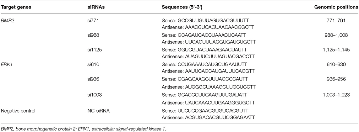
The siRNAs directed against BMP2 (namely si771, si988 and si1125) or ERK1 (namely si610, si936 and si1003) were designed (BMP2, NM_204258; ERK1, NM_204150.1). One negative control siRNA (NC-siRNA), which did not target any known chicken genes, was also designed. All of the siRNAs (Table 1) were synthesized by the Suzhou Genepharma Co., Ltd (Jiangsu, China).

Table 1. Sequences of the synthesized siRNA molecules and their positions in genomic regions of target genes (experiment 1).
Screening of the Most Effective siRNAs Against BMP2 or ERK1 (Experiment 1)
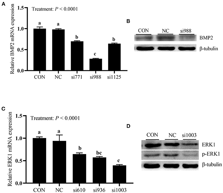
To screen the most effective siRNA against BMP2 or ERK1, the cells were randomly divided into one of eight treatments with three replicates per treatment, including the blank control group (CON), negative control (NC) group, si771 group, si988 group, si1125 group, si610 group, si936 group and si1003 group. The cells were transfected with 80 pmol/well-siRNA (NC-siRNA, si771, si988, si1125, si610, si936, or si1003) using the JetPRIME reagent according to the manufacturer's instructions, and the CON cells were not treated at all. At 48 h of post-transfection, the cells were collected for evaluating the suppressive effects of the siRNAs against BMP2 or ERK1 by using real time quantitative PCR (RT-qPCR) and Western blotting. Either the BMP2-specific siRNAs or ERK1-specific siRNAs shared the same controls (CON and NC). Among the siRNAs we examined, si988 and si1003 were, respectively screened as the most powerful siRNAs for BMP2 and ERK1 suppressions.
Effect of BMP2 or ERK1 Silencing on P Utilization and Related Parameters in Primary Broiler Osteoblasts (Experiment 2)
A completely randomized design involving a 2 × 2 factorial arrangement with two added P levels (0.0 and 2.0 mmol/L of P as NaH2PO4) and two cell types (normal cells and BMP2 or ERK1 silencing cells). There were 4 replicates for each treatment. The normal cells were transfected with the NC-siRNA, and the BMP2 or ERK1 silencing cells were, respectively transfected with si988 and si1003. At 24 h post-transfection, all cells were washed twice with the P-free DMEM medium to remove the residual P. Then, the normal cells and BMP2 or ERK1 silencing cells were incubated in the osteogenic induction medium (OIM) supplemented with 0.0 or 2.0 mmol/L of P for 12 days. Selecting the supplemental P dose of 2 mmol/L in the present study was based on our previous studies (11, 13). The OIM was the P-free DMEM supplemented with 15% FBS, 1% penicillin/streptomycin, 1% L-glutamine, 100 nmol/L Dex and 50 μg/mL ascorbic acid. Without and with supplemental P, the mediums were analyzed to contain 0.68 and 2.57 mmol/L of total P, respectively. Furthermore, during the period of P treatment, transfection was performed every 3 days until samples needed to be collected. During the P treatment period, the old medium was collected and pooled together at every medium change to determine the osteoblastic P retention rate. At the end of P treatment, the cells were collected for analyses of P utilization-related parameters (mineralization formation, ALP activity and BGP content).
Osteoblastic P Retention Rate Determination
The total P contents in the fresh or old medium were determined as previously described (13). Briefly, 1 mL of fresh or old medium was digested with HNO3-HClO4 (5:1) of mixed acid at 200 °C. The digested residues were dissolved in 10 mL of 2% HNO3 and then used for the total P determination. The osteoblastic P retention rate (%) = (V1 × C1-V2 × C2)/V1 × C1, where V1 and C1 represent, respectively total volume (mL) and total P content (mmol/L) of the added fresh medium; V2 and C2 represent, respectively total volume (mL) and total P content (mmol/L) of the pooled old medium.
Detection and Quantification of Mineralization Formation
The mineralization formation was quantified by ARS straining as previously described (14). Briefly, the cells were firstly strained with 1% ARS (pH 4.2) for 10 min. After drying, the content of each well was then solubilized with 10% (v/v) acetic acid, collected into a tube and heated at 85°C for 10 min. The slurry was centrifuged at 20,000 g for 15 min, and the supernatant was neutralized with 10% (v/v) ammonium hydroxide, and then the absorbances read in a 96-well-plate at 405 nm. Finally, the ARS concentrations were calculated based on the ARS standard curve. In the present study, the mineralization formation was represented by the ARS concentration (mmol/L).
Determinations of ALP Activity and BGP Content
The osteoblastic ALP activity was determined by a microplate reader with commercial kits. The osteoblastic BGP content was determined by the ELISA method with commercial kits.
RT-qPCR Analysis
The mRNA expressions of BMP2 and ERK1 in the osteoblasts were determined by the RT-qPCR according to the procedure as previously described (11). The 2−ΔΔCt method was used to calculate the relative mRNA expressions of target genes to reference genes including β-actin and glyceraldehyde-3-phosphate dehydrogenase (GAPDH). The primer sequences used in the present study were shown in Table 2.
Western Blotting Assay
Total and phosphorylated protein expression levels of target genes were analyzed by Western blotting as previously described (11, 15, 16). The primary antibodies used in the experiment included anti-BMP2 (catalog no. A0231; Abclonal, Wuhan, China), anti-EKR1 (catalog no. 16443-1-AP; Proteintech, Wuhan, China), anti-phosphorylated EKR1 (catalog no. AP0234; Abclonal, Wuhan, China) and anti β-tubulin (catalog no. HX1829; Huaxingbio, Beijing, China). Horseradish peroxidase (HRP)-conjugated goat anti-rabbit (catalog no. HX2030; Huaxingbio, Beijing, China) and goat anti-mouse (catalog no. HX2032; Huaxingbio, Beijing, China) were used as secondary antibodies. The membranes were then visualized using the ECL system (Tanon, Shanghai, China).
Statistical Analyses
All statistical analyses were conducted using the GLM procedure of SAS 9.4 (SAS institute Inc., Cary, NC, USA). The data of experiment 1 were subjected to one-way ANOVA, whereas the data of experiment 2 were analyzed by two-way ANOVA, and the statistical model included the added P level, cell type and their interaction. The replicate served as the experimental unit. Different means were separated using the LSD method, and the statistical significance was declared at P ≤ 0.05.
Results
Interference Efficiencies of siRNAs in Primary Broiler Osteoblasts (Experiment 1)
As shown in Figure 1A, the treatment affected (P < 0.0001) BMP2 mRNA expression. Compared with the NC and CON groups, the expressions of BMP2 mRNA in cells transfected with three BMP2-siRNAs decreased (P < 0.05). However, the expression of BMP2 mRNA in the si988 group was the lowest (P < 0.05) with 70% of the inhibition efficiency compared with the CON, and no difference (P > 0.05) between si771 and si1125 groups was observed. As shown in Figure 1C, when compared with the NC and CON groups, the expressions of ERK1 mRNA in cells transfected with three ERK1-siRNAs decreased (P < 0.05). However, the expression of ERK1 mRNA was lower (P < 0.05) in si1003 group than in the si610 group with 60% of the si1003 inhibition efficiency compared with the CON, and no differences (P > 0.05) between si1003 and si936 groups and between si610 and si936 groups were observed. In addition, BMP2, ERK1 and p-ERK1 protein expressions were also clearly inhibited (Figures 1B,D). Taken together, these results indicated that the si988 and si1003 could be selected as the most effective siRNAs for BMP2 and ERK1 to be used in the following experiment 2.

Figure 1. Screening of the most effective siRNAs against BMP2 mRNA and protein (A,B), and ERK1 mRNA and protein (C,D) in primary broiler osteoblasts (experiment 1). Each value represents the means ± SE, n = 3. Lacking the same letters (a, b, c) means significant differences, P < 0.05. BMP2, bone morphogenetic protein 2; ERK1, extracellular signal-regulated kinase 1.
Effect of BMP2 Silencing on P Utilization and Related Parameters in Primary Broiler Osteoblasts (Experiment 2)
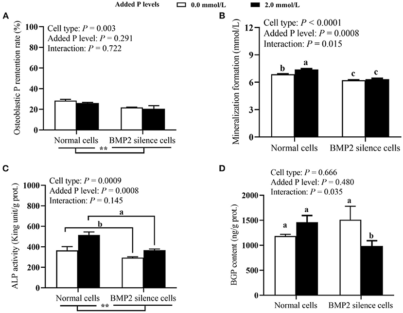
As shown in Figures 2A–D, the BMP2 silencing reduced (P ≤ 0.003) the P retention rate and ALP activity. The osteoblastic ALP activity at 2.0 mmol/L of added P level was higher (P = 0.0008) than that at 0.0 mmol/L of supplemental P level. The BMP2 silencing decreased (P < 0.05) mineralization formation at both 0.0 and 2.0 mmol/L of added P levels, but the reduced degree was greater at 2.0 mmol/L of added P level. The BMP2 silencing reduced (P < 0.05) osteoblastic BGP content supplemented with 2.0 mmol/L P, but had no effect (P > 0.05) on it at 0 mmol/L of added P level. As shown in Figures 3A–C, both BMP2 mRNA and protein levels were decreased (P ≤ 0.004) in BMP2-silenced cells. However, added P level and their interaction had no effects (P > 0.26) on BMP2 mRNA and protein expressions.

Figure 2. Effects of cell types and P levels on the P retention rate (A), mineralization formation (B) ALP activity (C), and BGP content (D) in primary broiler osteoblasts (experiment 2). Each value represents the means ± SE, n = 4. Lacking the same letters (a, b, c) means significant differences, P < 0.05. **P < 0.01. ALP, alkaline phosphatase; BGP, bone gla protein; BMP2, bone morphogenetic protein 2; P, Phosphorus.

Figure 3. Effects of cell types and P levels on the tibial osteoblastic BMP2 mRNA (A) and protein expressions (B,C) in primary broiler osteoblasts (experiment 2). Each value represents the means ± SE, n = 4. **P < 0.01. BMP2, bone morphogenetic protein 2; P, Phosphorus.
Effect of ERK1 Silencing on P Utilization and Related Parameters in Primary Broiler Osteoblasts (Experiment 2)
As shown in Figures 4A–D, both cell type and added P level had significant effects (P ≤ 0.024) on osteoblastic mineralization formation, ALP activity and BGP content. Cell type, added P level and their interaction had significant effects (P ≤ 0.025) on P retention rate of osteoblasts. The ERK1 silencing decreased (P ≤ 0.002) osteoblastic mineralization formation, ALP activity and BGP content. The osteoblast mineralization, ALP activity and BGP content at 2.0 mmol/L added of P level were higher (P ≤ 0.024) than those at 0.0 mmol/L of added P level. The ERK1 silencing decreased (P < 0.05) the P retention rate of osteoblasts regardless of 0.0 or 2.0 mmol/L of added P level, but the reduced degree was greater at 2.0 mmol/L of added P level. As shown in Figures 5A–D, the ERK1 mRNA and protein expressions, as well as p-ERK1 protein expression were decreased (P ≤ 0.004) in ERK1-silenced cells. The P addition had no effect (P > 0.45) on the ERK1 mRNA expression, but inhibited (P = 0.014) its protein expression, and upregulated (P = 0.015) the p-ERK1 protein expression. No interaction (P > 0.08) between cell type and added P level in ERK1 mRNA and protein expressions and p-ERK1 protein expression.

Figure 4. Effects of cell types and P levels on the P retention rate (A), mineralization formation (B) ALP activity (C), and BGP content (D) in primary broiler osteoblasts (experiment 2). Each value represents the means ± SE, n = 4. Lacking the same letters (a, b, c, d) means significant differences, P < 0.05. ** P < 0.01. ALP, alkaline phosphatase; BGP, bone gla protein; ERK, extracellular signal-regulated kinase 1; P, Phosphorus.

Figure 5. Effects of cell types and P levels on the tibial osteoblastic ERK1 mRNA (A), ERK1 and p-ERK1 protein expressions (B–D) in primary broiler osteoblasts (experiment 2). Each value represents the means ± SE, n = 4. Lacking the same letters (a, b) means significant differences, P < 0.05. ** P < 0.01. ERK, extracellular signal-regulated kinase 1; P, Phosphorus.
Discussion
The hypothesis that either BMP2 or ERK1 silencing would affect P utilization in primary broiler osteoblasts has been supported by the results of the current study. In the present study, either BMP2 or ERK1 silencing significantly decreased the osteoblastic P retention rate, mineralization formation, ALP activity and BGP content, indicating that either BMP2 or ERK1 silencing suppressed P utilization, and thus either of them participated in regulating P utilization of primary broiler osteoblasts. The aforementioned new findings have never been reported before, and thus provided a new perspective and idea for developing feasible strategies for improving P utilization in the bone of broiler chickens.
The BMP and ERK1 signaling pathways play a crucial role in the maturation, differentiation and mineralization of osteoblasts (6–8, 17). The BMP family proteins, especially BMP2, are essential molecules for osteoblastic differentiation and key bone regulators (8). It was reported that the activation of BMP2 could further induce the phosphorylation of ERK1, to promote the osteogenic differentiation (1). Zhu et al. (10) reported that ERK1 inhibitor could suppress the ALP activity, bone mineralization and downregulate the expression of BMP2 mRNA in mice bone mesenchymal stem cells. However, p-ERK1 was enhanced by neutralizing anti-BMP2/4 antibodies in hepatocytes, suggesting that BMP2 would exert its function via ERK1 pathway (18). In chickens, previous findings suggested that the BMP2 and ERK1 pathways would have a strong possibility to be involved in bone P utilization in vitro or in vivo (11, 12). As added P level increased, BMP2 mRNA expression increased, while ERK1 mRNA expression decreased in broiler tibia (12). Consistently, the BMP2 mRNA expression increased linearly and quadratically, whereas the ERK1 mRNA expression decreased linearly as added P level increased in primary osteoblasts cells (11). These results suggested that ERK1 expression might be inversely correlated with the BMP2 expression. In the present study, added P level had no effect on BMP2 and ERK1 mRNA expressions and BMP2 protein expressions, but inhibited ERK1 protein expression, and upregulated the p-ERK1 protein expression. Exact reasons and mechanisms for the above disparities remain unclear, and need to be further investigated in the future.
Osteoblasts play a major role in the bone formation and development. In broilers, bone P retention and bone mineralization-related parameters are sensitive indictors that reflect P utilization (12, 13, 15). The ALP and BGP have been demonstrated to participate in the process of hydroxyapatite formation and mineralization in osteoblasts (19). The ALP secreted by osteoblasts, is an enzyme hydrolyzing phosphate ester to provide inorganic P for hydroxyapatite formation (20, 21). Hence, the increased inorganic P resulting from the higher ALP activity implied that the bone tissue underwent more intense osteogenesis. The BGP is necessary for the osteogenic differentiation and is involved in bone density regulation to inhibit the development of osteoclasts. In addition, BGP regulates the hydroxyapatite crystals size and shape in bone growth (15). Our studies in broilers demonstrated that the ALP activities in serum and in the tibia, as well as BGP content in the tibia, decreased as added P level increased (15, 22, 23). Moreover, correlation analyses showed that the P utilization-related parameters were negatively associated with ALP activity and BGP content in broiler tibia (12, 15). In the present study, either BMP2 or ERK1 silencing significantly reduced the tibial osteoblastic ALP activity and BGP content. Added P level increased the tibial osteoblastic ALP activity and BGP content in the ERK1-silenced experiment, and the ALP activity was increased by P addition in the BMP2-silenced experiment. Therefore, the above results further suggested that both BMP2 and ERK1 would be involved in the regulation of the bone P utilization in broilers.
The BMP2 is known to induce the activation of ERK signaling molecules, thus to promote the osteoblastic differentiation and bone formation (10). In addition, P has been recognized as a particular signaling molecule that affects the expression of osteogenesis genes, including BMP2, ALP and BGP through the MAPK pathway (11, 12). However, no in vitro study has been reported on the effect of BMP2 or ERK1 silencing on P utilization of primary broiler osteoblasts. Our results indicated that BMP2 or ERK1 silencing suppressed P retention rate, mineralization formation, ALP activity and BGP content. The above findings will help us better understand the role of BMP2 or ERK1 in regulating P utilization of primary broiler osteoblasts and highlight their potential mechanisms.
Conclusions
Either BMP2 or ERK1 silencing suppressed P utilization, and thus either of them was involved in regulating the P utilization in primary broiler osteoblasts.
Data Availability Statement
The datasets presented in this study can be found in online repositories. The names of the repository/repositories and accession number(s) can be found in the article/supplementary material.
Ethics Statement
The study protocol was approved by the Animal Care Committee of the Department of Animal Science and Technology of Yangzhou University, Yangzhou, China (permit number: SYXK (Su) 2021-0027) and conducted in accordance with the guidelines of the Animal Use Committee of the Chinese Ministry of Agriculture (Beijing, China).
Author Contributions
YG and TL designed the experiments, analyzed the data, and wrote the original draft. YH, XC, BW, LZha, and LZhu performed the experiment. XL supervised and edited the thesis. All authors contributed to the article and approved the submitted version.
Funding
This study was financed by the Key Program of the National Natural Science Foundation of China (Project no. 31630073; Yangzhou, P. R. China), the Natural Science Foundation of Jiangsu Province (Project no. BK20210809; Yangzhou, P. R. China), and Initiation Funds of Yangzhou University for Distinguished Scientists (Yangzhou, P. R. China).
Conflict of Interest
The authors declare that the research was conducted in the absence of any commercial or financial relationships that could be construed as a potential conflict of interest.
Publisher's Note
All claims expressed in this article are solely those of the authors and do not necessarily represent those of their affiliated organizations, or those of the publisher, the editors and the reviewers. Any product that may be evaluated in this article, or claim that may be made by its manufacturer, is not guaranteed or endorsed by the publisher.
References
1. Bokui N, Otani T, Igarashi K, Kaku J, Oda M, Nagaoka T, et al. Involvement of MAPK signaling molecules and Runx2 in the NELL1-induced osteoblastic differentiation. FEBS Lett. (2008) 582:365–71. doi: 10.1016/j.febslet.2007.12.006
2. Halloran D, Durbano HW, Nohe A. Bone morphogenetic protein-2 in development and bone homeostasis. J Dev Biol. (2020) 8:19. doi: 10.3390/jdb8030019
3. Sun J, Li JY Li CC, Yu YC. 2015. Role of bone morphogenetic protein-2 in osteogenic differentiation of mesenchymal stem cells. Mol Med Rep. (2015) 12:4230–7. doi: 10.3892/mmr.2015.3954
4. Hang QL, Zhou Y, Hou SC, Zhang DM, Yang XY, Chen JP, et al. Asparagine-linked glycosylation of bone morphogenetic protein-2 is required for secretion and osteoblast differentiation. Glycobiology. (2014) 24:292–304. doi: 10.1093/glycob/cwt110
5. Shibasaki S, Kitano S, Karasaki M, Tsunemi S, Sano H, Iwasaki T. Blocking c-Met signaling enhances bone morphogenetic protein-2-induced osteoblast differentiation. FEBS Open Bio. (2015) 5:341–7. doi: 10.1016/j.fob.2015.04.008
6. Gallea S, Lallemand F, Atfi A, Rawadi G, Ramez V, Spinella-Jaegle S, et al. Activation of mitogen-activated protein kinase cascades is involved in regulation of bone morphogenetic protein-2-induced osteoblast differentiation in pluripotent C2C12 cells. Bone. (2001) 28:491–8. doi: 10.1016/s8756-3282(01)00415-x
7. Guicheux J, Lemonnier J, Ghayor C, Suzuki A, Palmer G, Caverzasio J. Activation of p38 mitogen-activated protein kinase and c-Jun-NH2-terminal kinase by BMP-2 and their implication in the stimulation of osteoblastic cell differentiation. J Bone Miner Res. (2003) 18:2060–8. doi: 10.1359/jbmr.2003.18.11.2060
8. Kim BS, Kang HJ, Park JY, Lee J. Fucoidan promotes osteoblast differentiation via JNK- and ERK-dependent BMP2-Smad 1/5/8 signaling in human mesenchymal stem cells. Exp Mol Med. (2015) 47:e128. doi: 10.1038/emm.2014.95
9. Lai CF, Cheng SL. Signal transductions induced by bone morphogenetic protein-2 and transforming growth factor-beta in normal human osteoblastic cells. J Biol Chem. (2002) 277:15514–22. doi: 10.1074/jbc.M200794200
10. Zhu D, Deng X, Han XF, Sun XX, Liu YQ. Wedelolactone enhances osteoblastogenesis through erk- and jnk-mediated bmp2 expression and smad/1/5/8 phosphorylation. Molecules. (2018) 23:561. doi: 10.3390/molecules23030561
11. Li TT, Cao SM, Liao XD, Shao YX, Zhang LY, Lu L, et al. The effects of inorganic phosphorus levels on phosphorus utilization, local bone-derived regulators and BMP/MAPK pathway in primary cultured osteoblasts of broiler chicks. Front Vet Sci. (2022) 9:855405. doi: 10.3389/fvets.2022.855405
12. Liao XD, Cao SM, Li TT, Shao YX, Zhang LY, Lu L, et al. Regulation of bone phosphorus retention and bone development possibly by BMP and MAPK signaling pathways in broilersz. J Integr Agr. (2021) in press.
13. Li TT, Geng YQ, Hu Y, Zhang LY, Cui XY, Zhang WY, et al. Dentin matrix protein 1 silencing inhibits phosphorus utilization in primary cultured tibial osteoblasts of broiler chicks. Front Vet Sci. (2022) 9:875140. doi: 10.3389/fvets.2022.875140
14. Gregory CA, Gunn WG, Peister A, Prockop DJ. An alizarin red based assay of mineralization by adherent cells in culture: comparison with cetylpyridinium chloride extraction. Anal Biochem. (2004) 329:77–84. doi: 10.1016/j.ab.2004.02.002
15. Cao SM Li TT, Shao YX, Zhang LY, Lu L, Zhang RJ, et al. Regulation of bone phosphorus retention and bone development possibly by related hormones and local bone-derived regulators in broiler chicks. J Anim Sci Biotechnol. (2021) 12:88. doi: 10.1186/s40104-021-00610-1
16. Shi SR, Wu S, Shen YR, Zhang S, Xiao YQ, He X, et al. Iron oxide nanozyme suppresses intracellular Salmonella enteritidis growth and alleviates infection in vivo. Theranostics. (2018) 8:6149–62. doi: 10.7150/thno.29303
17. Xiao G, Gopalakrishnan R, Jiang D, Reith E, Benson MD, Franceschi RT. Bone morphogenetic proteins, extracellular matrix, and mitogen-activated protein kinase signaling pathways are required for osteoblast-specific gene expression and differentiation in MC3T3-E1 cells. J Bone Miner Res. (2002) 17:101–10. doi: 10.1359/jbmr.2002.17.1.101
18. Chen HY, Choesang TZ Li HH, Sun SM, Pham P, Bao WL, et al. Increased hepcidin in transferrin-treated thalassemic mice correlates with increased liver BMP2 expression and decreased hepatocyte ERK activation. Haematologica. (2016) 101:297–308. doi: 10.3324/haematol.2015.127902
19. Qin Y, Guan J, Zhang C. Mesenchymal stem cells: mechanisms and role in bone regeneration. Postgrad Med J. (2014) 90:643–7. doi: 10.2174/1574888X1205170616101116
20. Shao YX, Sun GM, Cao SM, Lu L, Zhang LY, Liao XD, et al. Bone phosphorus retention and bone development of broilers at different ages. Poult Sci. (2019) 98:2114–21. doi: 10.3382/ps/pey565
21. Xiao YQ, Zhang S, Tong HB, Shi SR. Comprehensive evaluation of the role of soy and isoflavone supplementation in humans and animals over the past two decades. Phytother Res. (2018) 32:384–94. doi: 10.1002/ptr.5966
22. Jiang Y, Lu L, Li SF, Wang L, Zhang LY, Liu SB, et al. An optimal dietary nonphytate phosphorus level of broilers fed a conventional corn-soybean meal diet from 4 to 6 weeks of age. Animal. (2016) 10:1626–34. doi: 10.1017/S1751731116000501
Keywords: chick (Gallus gallus), primary osteoblast, BMP2, ERK1, silencing, P utilization
Citation: Geng Y, Li T, Hu Y, Zhang L, Cui X, Zhu L, Wu B and Luo X (2022) The Effect of Bone Morphogenetic Protein 2 or Extracellular Signal-Regulated Kinase 1 Silencing on Phosphorus Utilization and Related Parameters in Primary Broiler Osteoblasts. Front. Vet. Sci. 9:943864. doi: 10.3389/fvets.2022.943864
Received: 14 May 2022; Accepted: 10 June 2022;
Published: 30 June 2022.
Edited by:
Shourong Shi, Poultry Institute (CAAS), ChinaReviewed by:
Aimin Wu, Sichuan Agricultural University, ChinaGuohua Liu, Feed Research Institute (CAAS), China
Copyright © 2022 Geng, Li, Hu, Zhang, Cui, Zhu, Wu and Luo. This is an open-access article distributed under the terms of the Creative Commons Attribution License (CC BY). The use, distribution or reproduction in other forums is permitted, provided the original author(s) and the copyright owner(s) are credited and that the original publication in this journal is cited, in accordance with accepted academic practice. No use, distribution or reproduction is permitted which does not comply with these terms.
*Correspondence: Xugang Luo, wlysz@263.net
†These authors have contributed equally to this work
 Yanqiang Geng
 Tingting Li
 Yun Hu
 Liyang Zhang2
  Xiaoyan Cui