 Micaela Encinas1Ximena Ferrara Muñiz1Romina Ayelén Sammarruco2
 Victoria Ruiz Menna2
 Carlos Javier Garro2Fernando Delgado2
 Analía Macías3
 Gabriel Magnano3
 Martín José Zumárraga1*Sergio Gabriel Garbaccio2†
 María Emilia Eirin1†
- 1Instituto de Agrobiotecnología y Biología Molecular (IABiMo) UEDD CONICET-INTA, Centro de Investigación en Ciencias Veterinarias y Agronómicas (CICVyA)-CNIA, Hurlingham, Argentina
- 2Instituto de Patobiología Veterinaria (IPVET), UEDD CONICET-INTA, Instituto Nacional de Tecnología Agropecuaria (INTA), INTA-CONICET, Hurlingham, Argentina
- 3Departamento de Patología Animal, Facultad de Agronomía y Veterinaria, Universidad Nacional de Río Cuarto, Río Cuarto, Argentina
Ante-mortem diagnosis of bovine tuberculosis (bTB) is based mainly on the tuberculin skin test (TST) and the ɣ-IFN release assay (IGRA). Some infected animals escape screening tests, thus, limit herd sanitation. Previous reports have suggested a predominant pattern of multi-organ lesions attributable to Mycobacterium bovis (the causative agent of bTB) bacteraemia. A case–control study was conducted to investigate blood PCR as an alternative tool for improving ante-mortem detection of TST false-negative bovines. Cases comprised 70 TST false-negative bovines (cases), which were serology positive, and controls included 81 TST positive bovines; all of them confirmed as infected with M. bovis. Detection of the IS6110 target through touchdown blood-PCR (IS6110 TD-PCR) was performed. The positivity of the blood-PCR was 27.2% in the control group. This performance was similar to the 15% obtained among cases (p = 0.134). Most cases identified by the IS6110 TD-PCR exhibited focalized lesions (p = 0.002). Results demonstrated that blood-PCR could detect TST false-negative cattle, even if they are negative for IGRA. Considering that cases exhibited humoral response to M. bovis, further studies conducted in a pre-serological stage could provide evidence about the real contribution of the technique in herds.
Introduction
Bovine tuberculosis (bTB), a chronic disease disseminated worldwide, impacts on the agricultural countries. Its etiological agent, Mycobacterium bovis, infects a wide range of mammalians, including humans. The control and eradication programs of this disease consist of a “test and slaughter” policy by using the in vivo tuberculin skin test (TST) as the primary screening tool (1). The TST is highly effective but has limitations. The diagnosis maybe compromised by high rates of exposure to non-tuberculous mycobacteria (NTM) (2), and the existence of non-reactors to the screening tests. Previous reports showed the presence of false-negative animals to the screening tests in endemic herds (3–10).
TST false-negative bovines constitute a challenge for herd sanitation since they remain in herds as a source of infection. In Argentina, Garbaccio et al. (5) reported that 76% of TST false-negative dairy cattle were also negative to the IGRA, which suggests that tests based on cellular-mediated immune response partially detect the infection among these animals. In addition, humoral response detects TST false-negative cattle from endemic herds, which is more likely associated to late stages of the disease (4, 5, 10). However, some TST and IGRA false-negative animals could be in a pre-serological stage and therefore, they would not be detected in herds.
TST false-negative cattle presented exacerbated gross lesions with a predominant multi-organ commitment (5), which can occur because of haematogenous dissemination of the bacilli from the primary infection area. This fact suggests that viable mycobacteria could be detectable in blood samples, as previously confirmed in naturally infected cattle (11). Regarding M. bovis detection in blood, previous studies developed different specific PCRs in blood, targeting M. bovis sequences (mpb70, esxB gene, RD4 region, IS6110), with variable results (12–16).
Zumárraga et al. (17) developed a touchdown cycling for PCR, with the IS6110 element as target (IS6110 TD-PCR), which is highly specific for the Mycobacterium tuberculosis complex (MTC) members (17). IS6110 TD-PCR showed an enhanced sensitivity compared to the standard IS6110-PCR and detected M. bovis DNA in different biological samples (tissues, milk, nasal swabs) and colony cultures (colony-PCR), for direct diagnosis in infected M. bovis bovines (17–20). However, the usefulness of IS6110 TD-PCR in blood samples remains unexplored.
The objective of the study was to evaluate the performance of the IS6110 TD-PCR in TST false-negative cattle from bTB endemic herds. Results confirm the presence of M. bovis DNA in blood, and constitute evidence for future studies to evaluate the usefulness of the technique in a pre-serological stage of infection.
Materials and methods
Study design
A case–control study was conducted to assess the performance of the IS6110 TD PCR in blood of Friesian Holstein bovines (n = 151), predominantly older than 2 years old, with confirmed bTB. Animals came from 31 different dairy herds under sanitation programs located in the central productive dairy region of Argentina.
The control group (n = 81) included bovines positive to the TST according to the Argentinean official guidelines. Selection of cases (n = 70) was made among non-reactors to the TST, considering those animals positive for humoral response against M. bovis by Enzyme-Linked Immunosorbent Assay (ELISA) (5). Blood from cases and controls was tested by PCR.
Cases were also tested by IGRA, as an ancillary test to the TST that also evidences cell-mediated immune response.
The pathology pattern was evaluated at the slaughterhouse inspection to characterize the disease presentation among cases. It was recorded as one organ with lesions or more than one organ with lesions (multi-organ lesions).
Animals were bTB confirmed when they were positive for at least one of the following tests: bacteriology, histopathology-ZN staining and tissue-PCR.
Tuberculin skin test
Accredited veterinarians performed the caudal fold TST (CF-TST) according to official guidelines of the Service for National Agri-Food Health and Quality (Law 128/12)1. Because the CF-TST was applied in herds under sanitation, an animal exhibiting a skin-fold thickness increase ≥3 mm was positive (severe interpretation test).
Interferon-gamma release assay (IGRA)
All cases were tested by IGRA. Heparinized blood (200 μL) from the jugular vein was stimulated with 25 μL of the following antigens: Avian Tuberculin PPD 2500 (250 IU/mL), Bovine Tuberculin PPD 3000 (300 IU/mL), sterile Phosphate Buffered saline (PBS) solution 1X and BOVIGAM® Pokeweed Mitogen (5 μg/mL) in a 96-well cell culture plate. Harvested plasma were stored at −20°C for γ-IFN measurement by ELISA using a commercial bovine γ-IFN-microplate ELISA. Blood stimulation, ELISA as well as interpretation of data (standard interpretation, cut-off = 0.1) were performed according to the manufacturer’s instructions (BOVIGAM TB kit, Thermofisher).
Detection of humoral response by ELISA
Humoral ELISA tested sera of non-reactor TST animals. Blood (10 mL) was obtained from the jugular vein 15–20 days’ after CF-TST. Serum (100 μL, 1,100 in PBS 1X solution), was centrifuged at 2500 g and then used to test antibody against bovine protein purified derivative (PPDB). ELISA consisted of a binding step (12 μL of PPDB per well, 1 μg/mL) performed over night at 4°C, a washing step (five times with a solution of PBS 1X-Tween 0.5, 1% of skimmed milk) and an incubation step with sera (100 μL, diluted 1,100 in PBS 1X solution) 1 h at 37°C. The plate was washed with PBS 1X and 100 μL of conjugate anti-Bovine IgG−Peroxidase antibody (Sigma, dilution 1:7000) was added to incubate the samples with this secondary antibody for 1 h at 37°C was added. The plates were washed five times more. Finally, 100 μL of the developing substrate (Citrate Buffer + ABTS (Sigma) + H2O2) were added and incubated for 10 min in the dark. The reaction reading was performed at 405 nm. Optical densities (OD) ≥ 0.42 classified animals as positive (5).
Blood-PCR
Blood (10 mL) anticoagulated with EDTA (0.5 mg/mL) was obtained from the jugular vein. DNA extraction was performed with a commercial kit (PuriPrep-S kit, InbioHighway), following the manufacturer’s instructions. As blood-PCR is not a routine protocol performed in our laboratory, some evaluations were performed before testing the presence of M. bovis DNA in the samples. The quality (A260nm/A280nm ratio) and concentration (A260) were assessed by spectrophotometry (NanodropTM, Thermo Fisher Scientist). The DNA integrity was also analyzed by an electrophoresis in a 0.8% agarose gel (TAE buffer 1X, 80 volts for 60 min.) stained with 5 μg/mL of ethidium bromide (Promega, USA). Amplification of a 450 bp fragment of the endogenous 16S mitochondrial ribosomal RNA gene (16SRNArmt) (21) was also performed by using GoTaq® G2 DNA Polymerase (Promega, USA). The amplification products were visualized by electrophoresis in a 1.2% agarose gel (TAE buffer 1X, 90 volts for 45 min) stained with ethidium bromide, as described above, and with a 100 bp molecular weight marker (100 bp Plus DNA Ladder, Trans, China).
The presence of M. bovis DNA in blood was assessed using an IS6110 specific MCT fragment (IS6110 TD-PCR) using the HotStarTaq® Master Mix kit (Qiagen, USA), as described previously (17). The products were visualized by electrophoresis in a 2% agarose gel (TAE buffer 1X, 80 volts for 45 min) stained with ethidium bromide, as described above, and with a 100 bp molecular weight marker (100 bp Plus DNA Ladder, Trans, China). The expected amplification product was of 245 bp.
Post-mortem inspection and sampling
Official veterinarians performed a detailed inspection searching for lesions compatible with bTB, according to the official slaughterhouse procedures. Sampling of representative portions of the retropharyngeal and submandibular lymph nodes (LN), respiratory (tracheobronchial and mediastinal LN, and lung), digestive (mesenteric and hepatic LN, and liver), and mammary (udder and supra mammary) LN were aseptically collected in individual containers to perform tissue-PCR and bacteriology. In addition, other pieces were disposed in containers with 10% buffered formalin for fixing tissues for histopathology. Regarding cases, after performing the pathology inspection, animals were classified as those exhibiting one organ with macroscopic lesions or those with multi-organ lesions.
Bacteriology and histopathology
For bacteriology, 30 g of each tissue was cut into small pieces with scissors and put into a sterile bag with 20 mL of double-distilled sterile water. Maceration was performed for 3 min. (Basic Masticator, IUL Instruments type 470, Spain) and the homogenate was decontaminated by Petroff’s method (22). An aliquot of 2 mL of each decontaminated sample was inoculated on egg-based Stonebrink solid media at 37°C with biweekly observation, for at least 8 weeks. This inoculation was done in triplicate. Suspected M. bovis colonies were stained with Ziehl-Neelsen (ZN) to identify acid-fast bacteria (23). For histopathology, fixed samples were dehydrated with different alcohol solutions of increasing strength (50, 70, 80, 95% and absolute ethanol), subsequently clarified with xylene, and finally, embedded in paraffin. The paraffin plugs were cut in 5 μm-thick sections (Leica RM2125 RTS, Biosystems), deparaffinized, hydrated and stained with hematoxylin-eosin and ZN staining (23).
Tissue-PCR
Two different PCR were interchangeably used to check the presence of M. bovis DNA in the collected tissues: IS6110 TD-PCR (17), with an expected product of 245 bp, and an adapted touchdown-PCR from a previously described Rv2807 nested-PCR (Rv2807 TD-PCR) (24). DNA from tissues was obtained using a commercial extraction kit (ADN PuriPrep-T Kit, InbioHighway, Argentina) according to the manufacturer’s instructions. GoTaq® G2 DNA Polymerase (Promega, USA) was used according to the manufacturer’s instructions. Briefly, the adaptation step of the Rv2807 TD-PCR consisted of an initial denaturation at 96°C for 3 min, with eight cycles of 96°C for 1 min, with an annealing temperature that was gradually reduced from 72°C to 64°C, and an extension step at 72°C for 1 min. The procedure was followed by 30 cycles including a denaturation step at 96°C for 1 min, annealing at 66°C for 1 min, extension at 72°C for 1 min 45 s, and a final extension at 72°C for 8 min. The expected amplification product was of 443 bp. PCR products were visualized in an 2% agarose gel (TAE buffer 1X), 80 volts for 45 min, stained with 5 μg/mL of ethidium bromide (Promega, USA), and compared to a 100 bp molecular weight marker (100 bp Plus DNA Ladder, Trans, China). Image digitalization was performed in a Geldoc Genetic Analyzer (Bio-Rad Laboratories).
Statistical analysis
The statistical comparison of proportions was performed with EpiDat 3.0 version software (Xunta de Galicia, OPS-OMS). Calculated p values equal to or less than 0.05 were considered statistically significant. The DNA concentration and quality were analyzed to identify significant outliers (p > 0.05) using the Grubbs’ test (25). Data distribution was tested by the Shapiro Wilk test with median and interquartile range of 25–75 (RIQ25–75) as descriptive statistics. Mean comparisons were performed using unpaired Student’s t test (GraphPad Software, Inc.). The Fisher’s exact Test was used to evaluate the association between the IS6110 TD-PCR results in blood and the pathology profile.
Results
The in vitro IGRA test, that represents an additional tool that measures cell-mediated immune response, recorded 55.4% of negativity among cases. This result shows that animals either positive or negative to this ancillary screening test, were similarly represented within the CF-TST false negative bovines (p > 0.05) (Table 1).
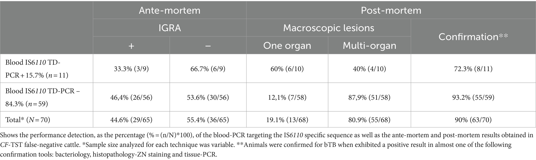
Table 1. Performance of the blood IS6110 TD-PCR in CF-TST false-negative bovines.
At the slaughterhouse inspection, cases exhibited 97.1% of macroscopic lesions compatible with bTB. Of these animals, 80.9% developed multi-organ lesions and, in a lesser proportion, 19.1% of them granulomas limited to one organ. Two animals lacked macroscopic lesions, although with a positive result of histopathology-ZN staining (Table 1).
DNA extraction from blood was performed in all the studied bovines (n = 151). A median genomic DNA concentration of the cases was 5.9 (3.6; 11.2) ng/μL, significantly lower than that obtained in the control group, in which the median concentration was 27.0 (19.0; 56.3) ng/μL (p < 0.0001). Despite this difference, the quality of the templates according to A260nm/A280nm ratio and the positive results for the 16SRNArmt PCR suggested good conditions of the extracted DNA (Supplementary Figure 1). Thus, the template was useful for further downstream determinations such as the IS6110 TD-PCR.
Blood-PCR performed in cases and controls (n = 151), yielded 22% of positivity for the IS6110 TD-PCR. Data stratification revealed that 27.2% of controls were positive for the IS6110 TD-PCR, while a lower proportion (15.7%) of the cases was positive for this test; however, the differences were not significant (p = 0.134).
On the other hand, IS6110 TD-PCR yielded significantly higher positive results in animals with focused lesions (46.2%), compared to animals with multi-organ lesions (7.3%) (p = 0.002). In addition, 66.7% of the IS6110 TD-PCR positive samples were IGRA negative (Table 1).
Discussion
Bovine tuberculosis, a health concern for animals and humans, is an ancient disease distributed worldwide, ranks among the top zoonotic threats (26). Currently, TST false-negative cattle constitute a challenge for diagnosis, because these animals remain in herds spreading the disease. Some of these bovines also fails to react to ancillary diagnosis tools such as the IGRA (3, 5).
In Argentina, our group reported cases of these TST false-negative bovines with repeatedly negative results for the CF-TST and a predominant multi-organ pathology in herds under sanitation, most of these animals were negative for IGRA (5). In Spain, TST and IGRA double negative bovines were identify positive for bacteriology. Researchers reported that these animals yielded a significant higher proportion of positive culture in beef cattle compared to dairy cattle (3).
In experimental infections, M. bovis promotes a predominance of cell-mediated immunity but with low or absent antibodies during the initial stages. The humoral response increases as the infection progresses, which is associated with the exacerbation of the disease pathology (27, 28). TST false-negative cattle exhibit humoral response, and previous studies demonstrated that antibody detection against M. bovis could contribute to identify these animals in naturally endemic herds (5, 6, 10). However, the association of antibodies with later stages of the disease could limit the performance of serology-based tools to detect those false-negative bovines that have not yet reached this humoral stage.
The present study constitutes valuable evidence to understand how to deal with this phenotype in endemic bTB herds. However, it is important to highlight that the results obtained here are biased given the selection criteria to identify CF-TST false-negative bovines in endemic herds. This selection was based on the presence of humoral response. Some questions remain open: Can the double TST and IGRA false-negative cattle be identified before the humoral stage? Are there false-negative animals with lesions but no humoral response, despite a putative advanced infection stage?
M. bovis genome detection in blood samples constitutes an approach poorly developed among positive reactors in the literature, and with no antecedents among the CF-TST false-negative phenotype. In Ethiopia, PCR targeting the M. bovis mpb70 gene in blood samples detected a significantly higher proportion (68%) of positive animals (12) than the obtained in the present research (p < 0.0001). These animals belonged to bovines chronically infected, as screened by the cervical TST. In our study, CF-TST positive bovines came from herds under sanitation, were highly infected bovines are not expected to be present for an extensive period. These differences could limit the sensitivity of IS6110 TD-PCR in blood samples. Conversely, a previous Indian study reported no IS6110-PCR positive blood samples from cervical-TST positive cattle with confirmed infection (14). The researchers used the phenol-chloroform-isoamyl alcohol method to extract DNA, in contrast to the present study and the research of Elsohaby et al., who used optimized commercial kits. Thakur et al. did not perform control checks on the quantity and quality of the extracted DNA, which are essential to ensure the usefulness of a DNA sample for downstream applications, including PCR (29).
By performing the IS6110 TD-PCR in milk of bovines, Zumárraga et al. (20) demonstrated the presence of M. bovis in bTB free herds of a dairy productive area with hot spots of infection in Santa Fe province, Argentina. Based on this study, the authorities incorporated the detection of M. bovis in dairy herds as a surveillance complementary tool in the Regional Plan for the control and eradication of bovine tuberculosis in Santa Fe province (Law 949/12, Ministry of Production of Santa Fe province, Argentina). However, the results of the present study suggest that the use of PCR for blood samples could be useful, not only for dairy cattle, but also for beef cattle, and in every period of the productive cycle.
Despite the predominant multi-organ lesions observed among the CF-TST false-negative bovines, this pathology pattern would not necessary reflect a greater presence of mycobacterial DNA in blood.
A possible explanation would be that circulating naked DNA, viable bacilli associated with bacteraemia or both, could be present in blood but in an intermittent manner.
CF-TST false-negative bovines limit the sanitation of bTB infected herds based on the application of ante-mortem screening tests, supporting the search of improved diagnosis tools. Results presented here, although preliminary, confirm the presence of M. bovis DNA in bovine blood samples of the CF-TST false-negative bovines suggesting the useful of this technique to be considered. However, further studies conducted in a pre-serological stage could provide more evidence about the real contribution of the technique in herds.
Data availability statement
The raw data supporting the conclusions of this article will be made available by the authors, without undue reservation.
Ethics statement
The animal studies were approved by CICUAE Instituto Nacional de Tecnología Agropecuaria, CICVyA. The studies were conducted in accordance with the local legislation and institutional requirements. Written informed consent was not obtained from the owners for the participation of their animals in this study because Study was performed in endemic herds. Animals detected as M. bovis infected must be sent to the slaughterhouse due to the infection in Argentina is a mandatory reportable disease.
Author contributions
MEn: Data curation, Formal analysis, Investigation, Methodology, Writing – original draft, Writing – review & editing. XF: Data curation, Methodology, Writing – review & editing. RS: Data curation, Investigation, Methodology, Writing – review & editing. VR: Data curation, Methodology, Writing – review & editing. CG: Data curation, Methodology, Writing – review & editing. FD: Data curation, Investigation, Methodology, Writing – review & editing. AM: Investigation, Methodology, Writing – review & editing. GM: Investigation, Methodology, Writing – review & editing. MZ: Conceptualization, Funding acquisition, Resources, Supervision, Writing – review & editing. SG: Conceptualization, Data curation, Formal analysis, Funding acquisition, Project administration, Resources, Supervision, Writing – review & editing. MEi: Conceptualization, Data curation, Formal analysis, Funding acquisition, Methodology, Project administration, Resources, Supervision, Writing – original draft, Writing – review & editing.
Funding
The author(s) declare that financial support was received for the research, authorship, and/or publication of this article. This present study was supported by the following funding sources: the Instituto Nacional de Tecnología Agropecuaria PNSA-PDi-113, PICT 2019 04085, PICT Start Up 2019 00038 and PIP 2021-2023 11220200101912CO.
Acknowledgments
The authors thank Julia Sabio y Garcia for the English revision of the manuscript. MEi and MZ are researchers of the National Research Council of Argentina (CONICET).
Conflict of interest
The authors declare that the research was conducted in the absence of any commercial or financial relationships that could be construed as a potential conflict of interest.
Publisher’s note
All claims expressed in this article are solely those of the authors and do not necessarily represent those of their affiliated organizations, or those of the publisher, the editors and the reviewers. Any product that may be evaluated in this article, or claim that may be made by its manufacturer, is not guaranteed or endorsed by the publisher.
Supplementary material
The Supplementary material for this article can be found online at: https://www.frontiersin.org/articles/10.3389/fvets.2024.1359205/full#supplementary-material
FIGURE S1 | Features of DNA templates and IS6110 TD-PCR in CF-TST false-negative bovines. (A) DNA concentration and quality. Scatter plot of concentration (ng/μL) and Quality (proportion A260/A280) of DNA from blood samples from CF-TST false-negative bovines (n = 70) was determined by spectrophotometry. Outliers, previously identified by Grubbs test (GraphPad), were excluded from the statistical analysis. (B) DNA integrity templates obtained from bovine blood samples. Electrophoresis in a 0.8% agarose gel stained with ethidium bromide to evaluate DNA integrity and RNA contamination of samples (loading sample: 5 μL). S1–S9 refers to nine different genomic DNAs extracted from blood samples belonging to CF-TST false-negative bovines. “High molecular weight” represent the region of the gel expected to show DNA not disrupted, as a dense band of high molecular genomic DNA. “Degradation” shows the region of the gel in which genomic DNA degradation, observed as a continued and diffuse band, it is visualized, and finally, the “RNA contamination” indicates the area of the gel in which contaminating RNA that has co-eluted with the genomic DNA during the extraction process is expected to be detected. (C) IS6110 TD-PCR amplification in bovine blood samples. Electrophoresis in a 2% agarose gel to visualize the amplification of IS6110 target in blood samples of CF-TST false-negative bovines. Red arrow indicates a band of 245 bp corresponding to IS6110 amplification sequence. S1–S9 refers to DNA extracted from different animals. PC, positive control (M. bovis DNA, AN5 strain). CC, contamination control (DNase and RNase free water); MWM, molecular weight marker.
References
1. Reis, AC, Ramos, B, Pereira, AC, and Cunha, MV. Global trends of epidemiological research in livestock tuberculosis for the last four decades. Transbound Emerg Dis. (2021) 68:333–46. doi: 10.1111/tbed.13763
2. Jenkins, AO, Gormley, E, Gcebe, N, Fosgate, GT, Conan, A, Aagaard, C, et al. Cross reactive immune responses in cattle arising from exposure to Mycobacterium bovis and non-tuberculous mycobacteria. Prev Vet Med. (2018) 152:16–22. doi: 10.1016/j.prevetmed.2018.02.003
3. Álvarez, J, Perez, A, Marqués, S, Bezos, J, Grau, A, de la Cruz, ML, et al. Risk factors associated with negative in-vivo diagnostic results in bovine tuberculosis-infected cattle in Spain. BMC Vet Res. (2014) 10:14. doi: 10.1186/1746-6148-10-14
4. Casal, C, Díez-Guerrier, A, Álvarez, J, Rodriguez-Campos, S, Mateos, A, Linscott, R, et al. Strategic use of serology for the diagnosis of bovine tuberculosis after intradermal skin testing. Vet Microbiol. (2014) 170:342–51. doi: 10.1016/j.vetmic.2014.02.036
5. Garbaccio, SG, Garro, CJ, Delgado, F, Tejada, GA, Eirin, ME, Huertas, PS, et al. Enzyme-linked immunosorbent assay as complement of intradermal skin test for the detection of Mycobacterium bovis infection in cattle. Tuberculosis. (2019) 117:56–61. doi: 10.1016/j.tube.2019.05.006
6. Griffa, N, Moyano, RD, Canal, AM, Travería, GE, Santangelo, MP, Alonso, N, et al. Development and diagnostic validation of an ELISA based on an antigenic mixture for the detection of bovine tuberculosis. Vet J. (2020) 256:105426. doi: 10.1016/j.tvjl.2020.105426
7. Houlihan, MG, Dixon, FW, and Page, NA. Outbreak of bovine tuberculosis featuring anergy to the skin test, udder lesions and milkborne disease in young calves. Vet Rec. (2008) 163:357–61. doi: 10.1136/vr.163.12.357
8. Raffo, E, Steuer, P, Tomckowiack, C, Tejeda, C, Collado, B, and Salgado, M. More insights about the interfering effect of Mycobacterium avium subsp. paratuberculosis (MAP) infection on Mycobacterium bovis (M. bovis) detection in dairy cattle. Trop Anim Health Prod. (2020) 52:1479–85. doi: 10.1007/s11250-019-02151-2
9. Waters, WR, Buddle, BM, Vordermeier, HM, Gormley, E, Palmer, MV, Thacker, TC, et al. Development and evaluation of an enzyme-linked immunosorbent assay for use in the detection of bovine tuberculosis in cattle. Clin Vacc Immunol. (2011) 18:1882–8. doi: 10.1128/CVI.05343-11
10. Waters, WR, Vordermeier, HM, Rhodes, S, Khatri, B, Palmer, MV, Maggioli, MF, et al. Potential for rapid antibody detection to identify tuberculous cattle with non-reactive tuberculin skin test results. BMC Vet Res. (2017) 13:164. doi: 10.1186/s12917-017-1085-5
11. Maggioli, MF . A bloody evidence: is Mycobacterium bovis bacteraemia frequent in cattle?! Virulence. (2016) 7:748–50. doi: 10.1080/21505594.2016.1213477
12. Elsohaby, I, Mahmmod, YS, Mweu, MM, Ahmed, HA, El-Diasty, MM, Elgedawy, AA, et al. a. Accuracy of PCR, mycobacterial culture and interferon-γ assays for detection of Mycobacterium bovis in blood and milk samples from Egyptian dairy cows using Bayesian modelling. Prev Vet Med. (2020) 181:105054. doi: 10.1016/j.prevetmed.2020.105054
13. Elsohaby, I, Ahmed, HA, El-Diasty, MM, Elgedawy, AA, Mahrous, E, and El Hofy, FI. Serological and molecular evidence of Mycobacterium bovis in dairy cattle and dairy farm workers under the intensive dairy production system in Egypt. J Appl Microbiol. (2020) 129:1207–19. doi: 10.1111/jam.14734
14. Thakur, MK, Sinha, DK, and Singh, BR. Evaluation of complementary diagnostic tools for bovine tuberculosis detection in dairy herds from India. Vet World. (2016) 9:862–8. doi: 10.14202/vetworld.2016.862-868
15. Brahma, D, Narang, D, Chandra, M, and Singh, ST. Comparison of multiplex and ordinary PCR for diagnosis of paratuberculosis and tuberculosis in blood samples (buffy coat) of cattle and buffaloes. Iranian journal of veterinary research. (2020) 21, 52–56.
16. Cezar, RD, Lucena-Silva, N, Filho, AF, Borges, J, de, M, and de Oliveira, PR. Molecular detection of Mycobacterium bovis in cattle herds of the state of Pernambuco. BMC veterinary research. (2016) 12, 31. doi: 10.1186/s12917-016-0656-1
17. Zumárraga, MJ, Meikle, V, Bernardelli, A, Abdala, A, Tarabla, H, Romano, MI, et al. Use of touch-down polymerase chain reaction to enhance the sensitivity of Mycobacterium bovis detection. J Vet Diagnostic Investig. (2005) 17:232–8. doi: 10.1177/104063870501700303
18. Barandiaran, S, Pérez Aguirreburualde, MS, Marfil, MJ, Martínez Vivot, M, Aznar, N, Zumárraga, M, et al. Bayesian assessment of the accuracy of a PCR-based rapid diagnostic test for bovine tuberculosis in swine. Front Vet Sci. (2019) 6:204. doi: 10.3389/fvets.2019.00204
19. Marfil, MJ, Huertas, PS, Garbaccio, SG, Barandiaran, S, Martínez Vivot, M, Garro, C, et al. Detection of viable Mycobacterium bovis in lungs and livers sold in Butchers’ shops in Buenos Aires, Argentina. Foodborne Pathog Dis. (2018) 15:758–62. doi: 10.1089/fpd.2018.2467
20. Zumárraga, MJ, Soutullo, A, García, MI, Marini, R, Abdala, A, Tarabla, H, et al. Detection of Mycobacterium bovis-infected dairy herds using PCR in bulk tank milk samples. Foodborne Pathog Dis. (2012) 9:132–7. doi: 10.1089/fpd.2011.0963
21. Roellig, DM, Gomez-Puerta, LA, Mead, DG, Pinto, J, Ancca-Juarez, J, Calderon, M, et al. Hemi-nested PCR and RFLP methodologies for identifying blood meals of the Chagas disease vector, Triatoma infestans. PLoS One. (2013) 8:e74713. doi: 10.1371/journal.pone.0074713
22. de Kantor, IN . Bacteriología de la Tuberculosis humana y animal. CEPANZO OPS/OMS Serie de Monografías. (1989) 11:63.
23. AAVLD. Manual de Diagnostico de Micobacterias de Importancia en Medicina Veterinaria. Comisión Científica de Micobacterias. (2005):20–8.
24. Araújo, CP, Osório, AL, Jorge, KS, Ramos, CA, Souza Filho, AF, Vidal, CE, et al. Direct detection of Mycobacterium tuberculosis complex in bovine and bubaline tissues through nested-PCR. Braz J Microbiol. (2014) 45:633–40. doi: 10.1590/s1517-83822014000200035
25. Grubbs’ Test GraphPad Software, Inc. (n.d.). Available at: https://www.graphpad.com/quickcalcs/Grubbs1.cfm (Accessed November 28, 2023).
26. Kock, R, Michel, AL, Yeboah-Manu, D, Azhar, EI, Torrelles, JB, Cadmus, SI, et al. Zoonotic tuberculosis – the changing landscape. Int J Infectious Dis. (2021) 113:S68–72. doi: 10.1016/j.ijid.2021.02.091
27. Pollock, JM, McNair, J, Welsh, MD, Girvin, RM, Kennedy, HE, Mackie, DP, et al. Immune responses in bovine tuberculosis. Tuberculosis. (2001) 81:103–7. doi: 10.1054/tube.2000.0258
28. Welsh, MD, Cunningham, RT, Corbett, DM, Girvin, RM, McNair, J, Skuce, RA, et al. Influence of pathological progression on the balance between cellular and humoral immune responses in bovine tuberculosis. Immunology. (2005) 114:101–11. doi: 10.1111/j.1365-2567.2004.02003.x
29. Lucena-Aguilar, G, Sánchez-López, AM, Barberán-Aceituno, C, Carrillo-Ávila, JA, López-Guerrero, JA, and Aguilar-Quesada, R. DNA source selection for downstream applications based on DNA quality indicators analysis. Biopreservation Biobanking. (2016) 14:264–70. doi: 10.1089/bio.2015.0064
30. Garbaccio, S, Barandiaran, S, Fernandez, A, Macias, A, Magnano, G, Vivot, M, et al. Ensayo interlaboratorio: aislamiento de Mycobacterium bovis a partir de lesiones granulomatosas en bovinos [Interlaboratory test: isolation of Mycobacterium bovis from granulomatous lesions in bovine]. Revista Argentina de Microbiologia. (2016) 48:161–5. doi: 10.1016/j.ram.2016.03.004
Keywords: bovine tuberculosis, anergy, diagnosis, IS6110 touchdown-PCR, blood, false-negative, tuberculin skin test
Citation: Encinas M, Ferrara Muñiz X, Sammarruco RA, Ruiz Menna V, Garro CJ, Delgado F, Macías A, Magnano G, Zumárraga MJ, Garbaccio SG and Eirin ME (2024) Limited usefulness of the IS6110 touchdown-PCR in blood for tuberculin skin test false-negative cattle with serological response to Mycobacterium bovis. Front. Vet. Sci. 11:1359205. doi: 10.3389/fvets.2024.1359205
Edited by:
Francisco Javier Salguero, UK Health Security Agency (UKHSA), United KingdomReviewed by:
Gobena Ameni, United Arab Emirates University, United Arab EmiratesAman Ullah Khan, University of Veterinary and Animal Sciences, Pakistan
Copyright © 2024 Encinas, Ferrara Muñiz, Sammarruco, Ruiz Menna, Garro, Delgado, Macías, Magnano, Zumárraga, Garbaccio and Eirin. This is an open-access article distributed under the terms of the Creative Commons Attribution License (CC BY). The use, distribution or reproduction in other forums is permitted, provided the original author(s) and the copyright owner(s) are credited and that the original publication in this journal is cited, in accordance with accepted academic practice. No use, distribution or reproduction is permitted which does not comply with these terms.
*Correspondence: Martín José Zumárraga, enVtYXJyYWdhLm1hcnRpbkBpbnRhLmdvYi5hcg==
†These authors have contributed equally to this work and share last authorship