 Fanny Rachidi1*
 Abass Elhadi1,2
 Lilli Bittner-Schwerda1
 Melanie Schären-Bannert1
 Gábor Köller1
 Jan Schinköthe3
 Anna Majcher3
 Benjamin Diehl3
 Florian Hansmann3
 Tilman Kühn1
 Alexander Starke1
- 1Clinic for Ruminants and Swine, Faculty of Veterinary Medicine, Leipzig University, Leipzig, Germany
- 2Department of Surgery and Anaesthesia, Faculty of Veterinary Medicine, University of Khartoum, Khartoum, Sudan
- 3Faculty of Veterinary Medicine, Institute of Veterinary Pathology, Leipzig University, Leipzig, Germany
The etiological relationship between osteomyelitis (OM) of the ribs and sternum and endocarditis valvularis thromboticans (EVT) in dairy cattle was assessed using clinical, ultrasonographic, bacteriologic, and postmortem findings. Five dairy cows (2nd–6th lactation, 135–304 days in milk, 490–630 kg) were admitted to our clinic because of low production and poor body condition. Clinical examination revealed abnormalities in the circulatory and locomotor system, as well as the ribs and sternum. Ultrasonographic examinations of the heart, ribs, and sternum were performed, and samples were collected for laboratory analyses. The cows underwent an orthopedic examination and bacteriologic testing of blood (N = 5), abscesses of the ribs or sternum (N = 3), and synovial fluid (N = 2). All five cows were euthanized because of a poor prognosis, and a postmortem examination was carried out. During the postmortem examination, samples were collected from endocardial lesions (N = 4) and abscesses of the ribs and/or sternum (N = 2) that had not been accessible intra vitam. The physical condition of the cows suggested a chronic metastatic disease accompanied by pyemia. All cows had OM of the ribs or sternum and EVT, along with other inflammatory diseases, including arthritis (N = 4), tendovaginitis (N = 2), and abscesses (N = 2). These clinical diagnoses were confirmed during the postmortem examination. Bacteriologic examination revealed Trueperella pyogenes in the rib and sternal lesions (N = 5), blood cultures (N = 3), endocardial lesions (N = 3), and samples taken from the uterus, kidney, spleen, and muscle (N = 4). Our results emphasize the importance of ruling out EVT in cattle with lesions of the ribs and sternum. Ultrasonographic and microbiologic examinations support the diagnosis and help determine the extent and severity of the changes. While a definitive primary cause cannot be identified due to the animals’ polymorbid condition, the observed lesions suggest that apostematous inflammation of the ribs or sternum may act as a potential source of infection.
1 Introduction
Rib and sternal lesions in cattle have been observed at the herd level (1–3), in abattoirs (4–7), and during postmortem examination (8, 9). While only externally visible rib swellings have been described at the herd level (2), rib and sternal fractures (5, 7, 9–17), inflammation of the costochondral junction (CCJ) (4, 18, 19), and osteomyelitis (OM) (6, 11, 19) have been documented at the slaughterhouse and during postmortem examinations. In adult cattle, they are assumed to be caused by trauma (20) and various technopathies, especially in tie-stall systems (21). Lame cows may lie down too quickly or awkwardly on poorly designed surfaces (12). Inadequate bone mineralization (16), osteoporosis (17), and behaviors associated with the establishment of social hierarchy within a herd (15) may also play a role. In addition, rib fractures in calves can result from trauma at birth (8, 13). Risk factors for rib fractures in adult cattle include lameness (1, 5, 22, 23), increasing age (1, 5, 23), malnutrition (16, 17), increased calcium mobilization in late pregnancy (14), narrow passageways, and inadequate feed bunk space and cubicle design (1). Rib and sternal lesions can lead to pain and impairment of well-being, behavior, and performance, as well as life-threatening concomitant diseases such as endocarditis valvularis thromboticans (EVT) (10, 11, 14, 15, 18). Although apostematous inflammation of the ribs or sternum and life-threatening concomitant diseases associated with bacteremia have been reported, a potential etiology linking these lesions has not yet been investigated (PubMed search on 12 April 2025, keywords: bacteremia, rib, sternum, adult cattle).
This case series describes five cows with EVT and osteomyelitis of the ribs and/or sternum, aiming to identify the etiology of and relationship between rib and sternal lesions and EVT using clinical, laboratory, diagnostic imaging, and postmortem findings.
2 Case presentation
2.1 Animals and diagnostic procedures
Between 2018 and January 2024, 2,611 cattle were examined at the Clinic for Ruminants and Swine, Faculty of Veterinary Medicine, Leipzig University, and 6,210 animals were evaluated during on-farm visits. Rib abnormalities were documented in 284 cows, of which 66 underwent detailed diagnostic evaluation. EVT was confirmed in 16 of these cases. This case series describes five German Holstein cows (median age 5.5 years…-304) selected from the 16 EVT-positive cows across five different dairy farms. All five cows were at least in their first lactation and had rib or sternal lesions and EVT confirmed by both ultrasonography (performed by the first and last authors) and postmortem examination. The cows were pre-treated by the on-farm veterinarians with anti-inflammatory drugs and antibiotics (N = 5), an intravenous infusion of 0.9% NaCl solution (N = 1), and a ruminal magnet (N = 1). They were admitted to our clinic due to poor production performance and body condition (N = 5), recurrent fever (N = 2), and abortion (N = 1).
The cows underwent thorough clinical (24) and ultrasonographic (25–28) examinations, including lesion assessment and echocardiography. Blood, aspirated fluid, and tissue samples were collected for hematologic, clinical chemistry, and microbiologic analyses. The owners were contacted immediately after the diagnoses were established (in the case of cow #4, after the follow-up examination), and euthanasia was recommended due to the poor prognosis. After euthanasia, a complete postmortem examination was conducted, including tissue fixation and histologic evaluation. A detailed description of the diagnostic procedures is provided in the Supplementary material.
2.2 Results
2.2.1 Clinical findings
At the time of admission, all five cows had a mildly to moderately abnormal general condition, mild lameness, and moderate to poor body condition. The heart rate was higher than the reference range (60–90 beats per min) (24) in four cows, the respiratory rate was elevated in two cows (reference range: 24–36 breaths per min) (24), and the rectal temperature was increased in one cow (reference range: 38–39°C) (24). The general physical condition of all cows suggested a chronic disease process. Based on the results of the clinical examination, abnormalities in the circulatory and musculoskeletal systems were tentatively diagnosed (Supplementary Table 2).
The clinical examination of the circulatory system revealed congested jugular veins, tachycardia, and abnormal heart sounds (systolic endocardial murmur) in three cows. In all three cases, the point of maximal audibility was located in the area of the tricuspid valve. Cow #5 also showed arrhythmia. In cow #4, initial findings of the circulatory system were unremarkable except for a slightly elevated heart rate; however, a follow-up examination 2 months later showed significant cardiovascular deterioration, loss of body condition, and poor performance (Supplementary Table 2). For the other four cows, a long-term follow-up was not feasible due to animal welfare considerations.
The rib and sternal lesions appeared as round to oval swellings without visible skin changes (Figure 1). The five cows had 15 abnormalities in the ribs (N = 11) or in both the ribs and sternum (N = 4). The lesions primarily involved the 8th to 10th ribs and the adjacent sternal region; in cow #2, the 5th to 7th ribs were affected, and in cow #5, the 13th rib was involved in addition to the 8th to 10th ribs. They were located at the level of the elbow (N = 6), the sternum (N = 3), and the shoulder joint (N = 2). The lesions were mostly bilateral, non-mobile, predominantly bony, without crepitation, and did not feel warmer than normal. Pressure elicited a pain response in three cows (Supplementary Table 2).
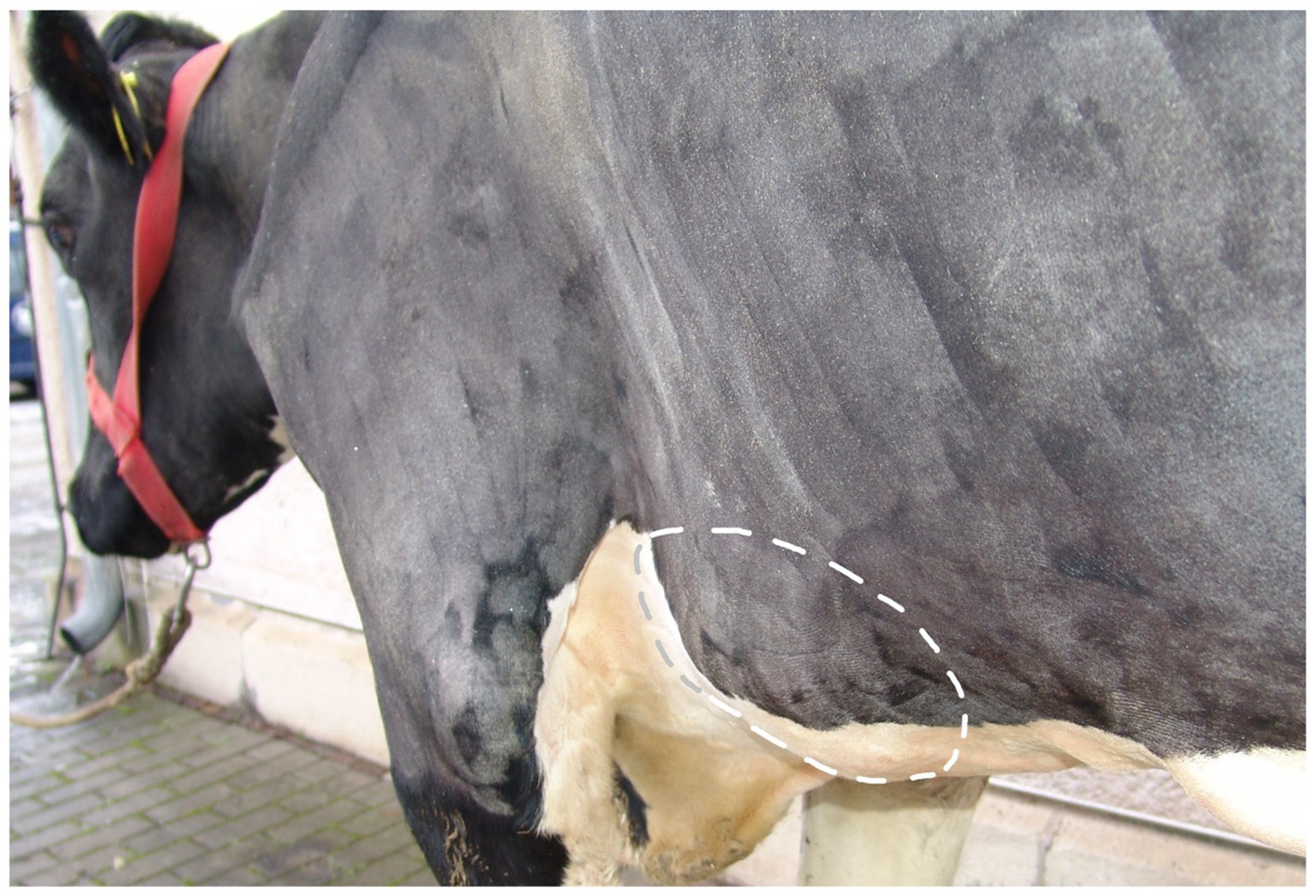
Figure 1. Image of a 4.5-year-old pregnant German Holstein cow (#2, 2nd lactation, 287 days in milk) diagnosed with endocarditis valvularis thromboticans. Caudolateral view from the left side showing the extensively clipped thorax and forelimb. The dashed circle shows an approximately 30 × 20 × 4 cm swelling associated with the 5th to 7th ribs, including the corresponding sternebrae. The skin over the swelling was intact.
The examination of the musculoskeletal system revealed lameness in all cows (grade I to III out of V) (29), with joint abnormalities (N = 3), tendon sheath abnormalities (N = 2), or abscesses in the musculature (N = 2; Supplementary Table 2).
2.2.2 Further diagnostics
Laboratory testing revealed increased inflammatory markers, including leukocytosis, increased total protein, decreased albumin concentration, and a positive glutaraldehyde test. In cow #4, the results were outside the reference ranges at both the initial and follow-up examinations (Supplementary Table 3).
Sonographic examination of the heart revealed hyperechoic, circumferential, cauliflower-like enlargement of the tricuspid valve (N = 5) and bicuspid valve (N = 3). The pericardium, epicardium, and myocardium were unremarkable. In cow #4, no cardiac abnormalities were observed at the initial examination, but bicuspid and tricuspid valves were abnormal at the follow-up examination 2 months later.
The transcutaneous ultrasonography of the ribs and sternum showed unremarkable skin but thickened, irregular musculature with hypoechogenic and hyperechogenic areas in four cows (Figure 2). In all cows, the periosteum was detached from the bone, which appeared irregular and, in two cows, showed step-like interruptions. Widened costochondral junctions were seen in four cows, with exostoses, axial deviations, and fluid-filled cavities in three. These cavities had an echogenic capsule and contained hypoechogenic to anechogenic fluid with echogenic reflexes. Flow phenomena were visualized in two cases (N = 2), and ultrasound-guided aspiration yielded creamy, light yellow exudate in three cases (N = 3; Table 1).
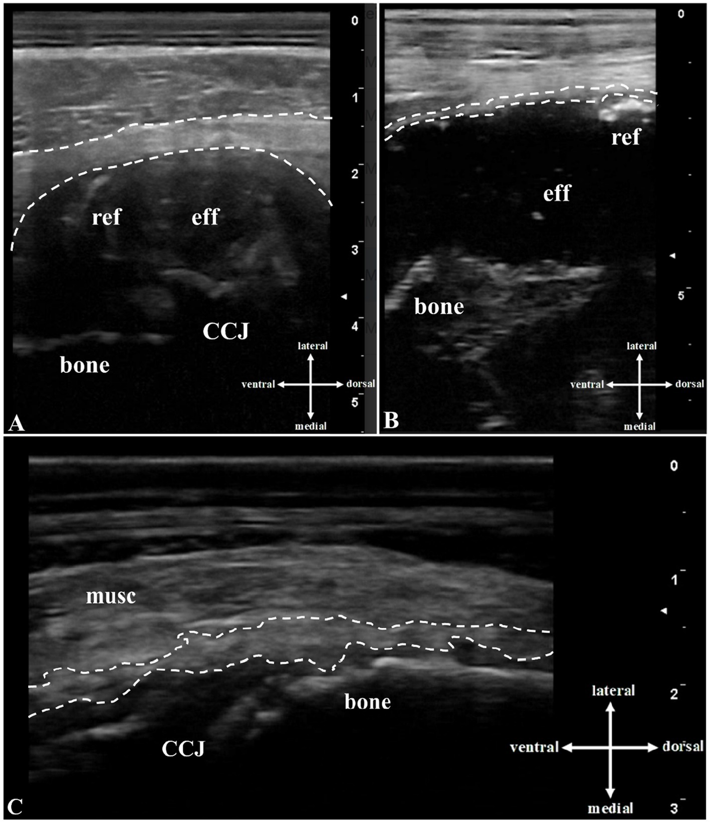
Figure 2. Transcutaneous B-mode ultrasound image (10-MHz linear transducer, penetration depth 3 to 8 cm) of the rib lesions: In the area of the (A) left 4th rib of cow #1: A hypoechogenic, fluid-filled cavity (eff) lateral to the uneven echogenic bone (bone), echogenic reflexes (ref), and an echogenic capsule (space between dashed lines) are seen above the costochondral junction (CCJ). (B) In the area of the left 7th rib of cow #2: A hypoechogenic, fluid-filled cavity (eff) lateral to the irregularly-shaped echogenic bone, echogenic and hyperechogenic reflexes (ref), and an echogenic capsule (space between dashed lines) are evident. (C) In the area of the left 8th rib of cow #5: The musculature is thicker than normal and irregular, with hyperechogenic and hypoechogenic areas (musc). Thickened, elevated echoic periosteum (space between dashed lines) and hyperechogenic irregular bone with hyperechogenic exostoses (bone) near the costochondral junction (CCJ) are present.
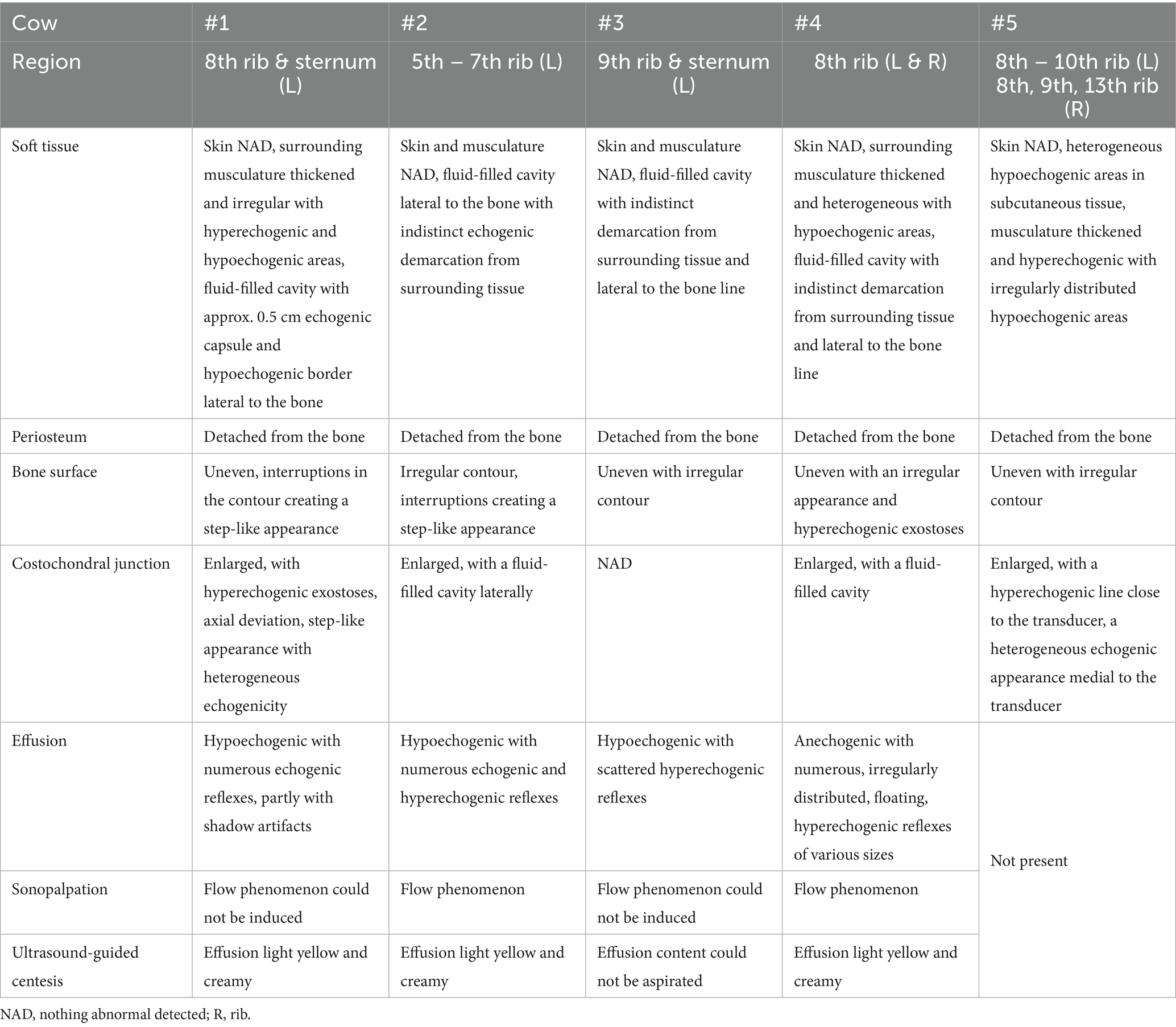
Table 1. Results of the ultrasonographic examination of the ribs in the five cows with swellings associated with the ribs or sternum and endocarditis valvularis thromboticans.
The results of the bacteriologic testing of the aspirate, blood culture, and various tissues are shown in Supplementary Table 4. Trueperella pyogenes was detected in 18 of the 21 positive microbiologic tests. The pathogen was identified in all rib and sternal lesions, in 60% of the blood cultures examined, 75% of the analyzed endocardial lesions, and in all other sampled organs or lesions (stifle joint, tendon sheath, uterus, spleen, kidney, and abscesses). In cow #4, Trueperella pyogenes was cultured from fluid aspirated from the cavity near the rib at both the initial and follow-up examinations. In addition, the enriched blood culture medium tested positive for Helcococcus ovis.
2.3 Clinical diagnosis and outcome
Based on the clinical and ultrasonographic findings, all cows were diagnosed with EVT. Septicemia and osteomyelitis (OM) of the ribs (N = 3) or both the ribs and sternum (N = 2) were tentatively diagnosed based on the clinical findings. In addition, arthritis (N = 4), tendovaginitis (N = 2), and abscess (N = 2) were diagnosed. In cow #4, there was no indication of EVT at the initial examination (clinical and ultrasonographic). Therefore, local treatment of claw disorders and abscesses in both hind limbs was performed, along with systemic antimicrobial and analgesic therapy. After the completion of the treatment, further monitoring was delegated to the owner. At the follow-up examination 2 months later, the cow’s condition had deteriorated, and in addition to the rib lesions and musculoskeletal disorders, EVT was diagnosed. Due to the guarded (restitutio ad vitam) or grave (resitutio ad integrum) prognosis, all cows were euthanized after consultation with the owners. In cow #4, euthanasia was performed at the time of the follow-up examination, whereas the remaining four cows were euthanized immediately after diagnosis at initial presentation. A postmortem examination was performed on all cows.
2.4 Gross and microscopic findings
All cows had suppurative to pyogranulomatous OM and costochondritis in at least one rib, with cow #5 having six affected ribs (Supplementary Table 5). Cow #2 had a large space-occupying extra-and intra-thoracal lesion involving the ribs and sternum that measured up to 17.5 cm (craniocaudal) × 12.5 cm (lateromedial) × 17.5 cm (dorsoventral, Figures 3A,B). The affected areas showed clearly visible macroscopic changes compared to unaffected regions of the thorax (Figure 3C).
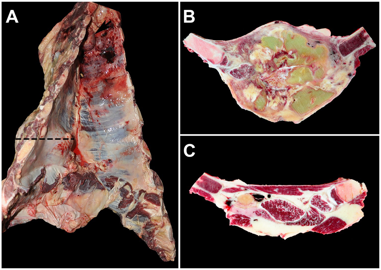
Figure 3. Thorax with severe lesions of the ribs and adjacent sternum (cow #2). (A) A space-occupying mass affecting the left 5th to 7th ribs and extending particularly far into the thoracic cavity (dashed line) with a severely enlarged cranial thoracic lymph node (arrow) is visible. (B) A cross-section of the tissue along the dashed line in image A reveals the mass as severe, chronic, multifocal to confluent, suppurative osteomyelitis and costochondritis with multifocal abscesses. (C) For comparison, the same sectional plane as in image B shows unaltered ribs and musculature on the right side of the thorax.
The cows had lesions suggestive of embolic showering, frequently observed as EVT in bicuspid and tricuspid valves. Severe lesions were seen in the tricuspid valve (Supplementary Figure 1A), in contrast to the mild to moderate lesions observed in the bicuspid valve. Cow #5 had EVT involving the aortic valve. Granulation tissue formation from the adjacent mural endocardium and myocardium with septic emboli containing numerous coccoid bacteria enmeshed in degenerating neutrophils and fibrin clots was observed. Septic embolic showering was also evident as suppurative nephritis (N = 5, Supplementary Figure 1B), vasculocentric suppurative pneumonia (N = 4), and suppurative splenitis (cow #1; Supplementary Figure 1D).
The musculoskeletal system, particularly the joints, was also affected (Supplementary Table 5). Moderate to severe suppurative to fibrin-suppurative arthritis was prominent in the majority of the cows (N = 4), frequently affecting the stifle joints (Supplementary Figure 1C). Cow #4 had severe suppurative myositis with multifocal abscesses in the semitendinosus and semimembranosus muscles. Inflammation of the skin, subcutis, bone, and claw wall was observed in the hind claws of three cows (Supplementary Table 5). Other lesions, which in all likelihood were not embolic, included suppurative mastitis (cow #1), ulcerative dermatitis of the sternal region (cow #2), and moderate suppurative endometritis (cow #5). In addition, in three cows, mild to moderate, multifocal lymphohistioplasmacytic to suppurative meningoencephalitis was seen.
3 Discussion
3.1 Diagnosis based on the results of the clinical and pathologic examinations
To the best of the authors` knowledge, OM of the ribs or sternum, bacteremia, and EVT have not been described in live cows (PubMed search on 12 April 2025, keywords: bacteremia, endocarditis, osteomyelitis, rib, sternum, cattle). In human medicine, septicemia and EVT have been reported as complications of sternal abscesses (30–35), but no cases describe EVT as the primary cause of rib or sternal OM. The disease course in cow #4 suggests a similar sequence: although the initial clinical and ultrasonographic examinations revealed no abnormalities of the cardiovascular system, EVT was diagnosed 2 months later based on both clinical signs and ultrasonographic findings. Whether a compensated EVT with lesions smaller than 0.5 cm, which may have been undetectable by ultrasonography (28), was already present at the time of the initial examination cannot be ruled out definitively. In contrast, the rib and sternal lesions were already evident during the first examination and remained unchanged. Although such longitudinal case tracking has not yet been conducted in cattle, the recommendation to examine the ribs and sternum in slaughtered cows suspected of bacteremia (4), highlighting a potential link between these lesions and bacteremia. Inflammatory changes in other bones have also been described as a cause of EVT in cattle (36–38).
Our results revealed that OM of the ribs and sternum occurred in the cattle with bacteremia and EVT. The course of disease in cow #4 suggests that OM of the ribs and sternum may have preceded the development of EVT and bacteremia. However, given the advanced and polymorbid condition of all cows in this series, a definitive conclusion regarding the temporal and causal relationship between these lesions and systemic infection cannot be drawn. A hematogenous spread originating from other, undetected infectious foci remains a possibility. On the other hand, a comprehensive postmortem examination of all organ systems was carried out in all cases, and such infectious foci would very likely have been identified. Therefore, we consider rib and sternal OM as potential contributors to systemic infection in cattle and recommend their inclusion in diagnostic evaluations, especially in animals presenting with signs of bacteremia or EVT.
3.2 Diagnostic imaging and laboratory tests
Diagnostic imaging and laboratory tests are essential for diagnosing these conditions. Hyperproteinemia, hypergammaglobulinemia, and leukocytosis are typical findings in cows with EVT (38). The glutaraldehyde test, a rapid test for identifying hypergammaglobulinemia (39), was positive in all cows. Chemical and microbiologic analyses of blood and exudate also provided valuable information for the diagnosis of EVT (40, 41) and OM of the ribs and sternum (10). Ultrasonography of the heart is recommended for the diagnosis of EVT in live cattle (42). Endocardial lesions exceeding 0.5 cm can be visualized using ultrasonography (28). In contrast, procedures for diagnosing OM of the ribs and sternum in live cattle are lacking (PubMed search on 12 April 2025, keywords: osteomyelitis, rib, sternum, cattle, ultrasonography). Although cattle with OM of the long bones often exhibit characteristic clinical findings (43), the signs of OM of the ribs and sternum are non-specific, and diagnosis by inspection and palpation alone is difficult (10). Palpation of lesions is limited to external examination, but in some cases, lesions can only be detected internally, such as during abdominal surgery. Diagnostic imaging, primarily ultrasonography and radiography, is required to diagnose OM of the long bones in cattle. Characteristic findings in cattle with OM, which we were also able to visualize in the abnormal rib and sternal areas (Figure 2; Table 1), are soft tissue swelling, thickening and detachment of the periosteum, and irregular rough bone contours that may have a step-like appearance (26). In foals and people with OM of the ribs and sternum, ultrasonography is considered a better diagnostic tool than radiography (44–47). However, ossification of abscess capsules or other structures, as seen in cow #5, can result in soft tissue structures being further away from the transducer and inaccessible because of sound cancelation (48). In all five cows, the swellings associated with the ribs and sternum, which initially appeared benign on clinical examination, turned out to be merely the tip of the iceberg. Therefore, when similar abnormalities are found during clinical examination, a thorough evaluation of the patient, including the cardiovascular system, should be carried out using the diagnostic methods described above. Ultrasonography is particularly useful, providing important information that usually contributes significantly to the diagnosis and prognosis.
3.3 Microbiology
Trueperella pyogenes was identified as the main pathogen in all cases, reinforcing its role in purulent infections such as EVT (40, 49) and OM (43, 50) in cattle. Trueperella pyogenes was reported in only one cow with EVT and abscessation of the costochondral junction of the 3rd rib; however, blood culture was not performed (18). While blood culture has been used to diagnose EVT in cattle (37), it has not been a diagnostic tool for OM (PubMed search on 12 April 2025, keywords: osteomyelitis, blood culture, cattle). Findings in humans with OM of the ribs and sternum show that bacteremia and subsequent EVT occur, and early treatment, facilitated by microbiological diagnostics, can halt disease progression (30). When performed correctly, microbiologic testing is a valuable diagnostic tool. In the present report, conventional sampling, storage, and culture techniques were used successfully. Proper execution of these steps and the timing of collection are crucial (51). In agreement with other studies, the conventional blood culture system used in the present study was effective for blood analysis (52, 53), and Amies medium sample tubes were the appropriate choice for exudates, synovial fluid, and small tissue samples (51). In line with the recommendations for cattle with arthritis and concurrent OM (54), we collected tissue samples rather than synovial fluid for the microbiologic testing. In contrast, there are currently no standardized guidelines for the collection and analysis of blood cultures in cattle, horses, or companion animals (55). In our study, two cows had negative blood culture results. Given that all animals had received antibiotic treatment prior to referral, the absence of detectable pathogens in these cases may have been due to antimicrobial interference or intermittent bacteremia. However, prior antimicrobial therapy should not be considered a contraindication for blood cultures, as repeated sampling and increased blood volume can enhance detection rates even under such conditions (56, 57). In human medicine, when a bloodstream infection is suspected, it is recommended to collect two to four blood culture sets, each containing 20 to 30 mL of blood, ideally obtained several hours apart. In cases of endocarditis, even more frequent blood culture sampling may be necessary (56, 57). However, both approaches require considerable time and financial resources and must be weighed against the clinical benefits in routine settings (55, 58). In our cases, it was not feasible to collect a minimum of three blood cultures at one-hour intervals, as recommended for cattle with EVT (59). To compensate, we collected both aerobic and anaerobic blood cultures from each cow, effectively doubling the total blood volume analyzed. Given that Trueperella pyogenes can grow in both aerobic and anaerobic media, this approach likely improved the diagnostic yield. The isolation of Trueperella pyogenes from the blood cultures, endocardial lesions, and fluid-filled cavities associated with the ribs and sternum suggested a possible link between these abnormalities. Further studies are needed to determine the prevalence of this disease process on a larger scale, the involvement of other pathogens, and the potential virulence factors contributing to the development of OM of the ribs and sternum in cattle infected with Trueperella pyogenes.
3.4 Potential causes
Despite originating from different dairy farms with varying stall designs and levels of cow comfort, all cows exhibited musculoskeletal disorders and lameness in addition to EVT and OM. While lameness prevalence ranged from 24 to 60% across the farms (Supplementary Table 1), only individual affected cows were presented for examination. Although lameness prevalence varied across the herds and appeared relatively high, particularly on farm 5, the recorded rates remained within the typical range reported for dairy farms in the region (60, 61). No prior trauma had been reported in any of the animals, and there were no skin changes over the rib and sternal lesions. Interestingly, skin changes were also absent in affected cows in another report (14), although trauma was the expected cause. This may have been related to the chronicity of the condition. Previous studies have identified lameness as a key risk factor for rib and sternal swellings, likely due to impaired lying behavior on hard surfaces, which leads to rib injuries (1, 12, 23). Rib injuries have also been documented in slaughtered cattle that had collided with gates and other objects (62, 63), as well as in humans, where seatbelt-related injuries increase the risk of rib and sternal fractures (64–66). An analogous scenario is possible in dairy cattle during routine restraint in a hoof-trimming chute that uses only a chest belt or metal brackets to secure the thorax. Although trauma caused by inadequate fixation in a hoof-trimming chute has not been reported (PubMed search on 12 April 2025, keywords: claw trimming, rib fracture, cattle), it remains a potential risk for injury. Visible changes to the skin and hair coat after blunt trauma caused by objects with a smooth surface can be mild. Furthermore, the causative injury may have occurred long before the animal was examined. Thus, the risk posed by restraint in a hoof-trimming chute, as well as by gates and other objects in the housing environment, milking parlor, hoof trimming area, and slaughter facilities, should be examined in future studies.
3.5 Relevance of osteomyelitis of the ribs and sternum
All cows in this case series had severe diseases with a poor prognosis. They were lame and exhibited compromised health status, and three of them had a pain response when pressure was applied to the rib swellings. In all cases, discussions were held with the responsible farm personnel prior to euthanasia regarding the clinical findings, possible etiologies, and farm-specific risk factors. On-farm investigations were conducted, including systems-based analyses of potential environmental and management-related risk factors, as well as locomotion scoring and assessment of rib and sternal swellings. The diagnostic implications, potential causes, and possible consequences for animal health, welfare, and farm economics were discussed in detail with the responsible farm personnel. Previous studies reported rib swellings in 2.3 to 9.6% of the cows examined (1–3, 23), but EVT and OM of the ribs were not diagnosed, likely due to a lack of thorough examination of the cardiovascular system and the ribs and sternum. Reliable prevalence data on OM of the ribs or sternum in cattle are scarce. Endocarditis valvularis thromboticans occurred in 0.15% (67) of slaughtered cows and 9% of autopsied cattle (68). The difficulty in identifying cows with OM of the ribs and EVT raises animal welfare concerns, (13, 17–19), particularly as both rib and sternal lesions (10, 11, 14, 15) and EVT (69) are painful, chronic conditions with a poor prognosis. The clinically deteriorated, polymorbid condition of the cows in this report, along with the presence of decompensated EVT, warranted immediate euthanasia of all five cows (cow #4 at the follow-up examination). However, timely surgical intervention for the OM in cow #4 at the initial presentation might have potentially prevented the development of EVT. Successful therapy is contingent upon early detection and adequate intervention (31). To date, there are no treatment recommendations available for cows with OM of the ribs and sternum (PubMed search on 12 April 2025, keywords: cattle, osteomyelitis, rib, sternum, therapy). In human medicine, such cases have been successfully treated with extended antibiotic therapy, radical surgical intervention, including wound debridement and drainage, often combined with vacuum-assisted wound therapy, and intensive care support (30, 32, 33, 70). However, even in patients with well-healed, uncomplicated rib and sternum fractures, chronic pain has often been reported (71). In our case, such an intensive treatment approach, as described for human patients, would not have been feasible on a commercial dairy farm. However, in the future, early identification of such cases may allow for the development of practical, farm-adapted therapeutic options for cattle.
This case series highlights the importance of rib and sternal lesions and their secondary complications in cattle. The painful lesions and diagnostic challenges emphasize their impact on animal welfare. In addition to inspection and palpation of the ribs and sternum, future studies should include the various diagnostic steps outlined here for both individual cases and herd-level assessments. Early detection of OM of the ribs and sternum could, ideally, facilitate timely intervention and the development of practical treatment strategies, potentially reducing the long-term animal welfare concerns associated with these conditions.
4 Conclusion
Osteomyelitis (OM) of the ribs and sternum is an important animal welfare concern. It leads to chronic disease, may cause pain and suffering, and can be fatal. Clinical diagnosis is difficult in live animals, and thus, diagnosis may be delayed, jeopardizing the well-being of cattle. A thorough clinical examination, along with additional diagnostic tools—particularly ultrasonography and laboratory analyses—is required for diagnosing OM of the ribs and sternum and endocarditis valvularis thromboticans (EVT) in live cows. In the authors’ opinion, these diagnostic tools should be integrated into a standard examination protocol for cows with swellings associated with the ribs or sternum to ensure that OM and EVT are not overlooked. Trueperella pyogenes was the main pathogen in all the lesions examined. This pathogen was cultured using conventional sampling and culture techniques under field conditions. In addition to the factors discussed in the literature, trauma caused by environmental elements such as gates and poor fixation methods in hoof-trimming chutes should be considered potential causes of OM of the ribs and sternum.
Data availability statement
The original contributions presented in the study are included in the article/Supplementary material, further inquiries can be directed to the corresponding author.
Ethics statement
Ethical approval was not required for the studies involving animals in accordance with the local legislation and institutional requirements because they were clinic patients who underwent our standard protocol of clinical examination, further diagnostics and therapy. Written informed consent was obtained from the owners for the participation of their animals in this study. Written informed consent was obtained from the participants for the publication of this case report.
Author contributions
FR: Conceptualization, Data curation, Funding acquisition, Investigation, Methodology, Visualization, Writing – original draft. AE: Data curation, Investigation, Methodology, Writing – review & editing. LB-S: Investigation, Methodology, Validation, Writing – review & editing. MS-B: Investigation, Methodology, Validation, Writing – review & editing. GK: Investigation, Methodology, Writing – review & editing. JS: Data curation, Investigation, Methodology, Writing – original draft. AM: Investigation, Methodology, Writing – review & editing. BD: Investigation, Methodology, Writing – review & editing. FH: Data curation, Investigation, Methodology, Visualization, Writing – review & editing. TK: Investigation, Methodology, Writing – review & editing. AS: Conceptualization, Data curation, Funding acquisition, Investigation, Methodology, Project administration, Resources, Supervision, Validation, Visualization, Writing – review & editing.
Funding
The author(s) declare that financial support was received for the research and/or publication of this article.
Acknowledgments
The authors acknowledge support from the German Research Foundation (DFG) and Leipzig University within the program of Open Access Publishing. The authors thank R. Waelchli and C. Winder for their professional advice. We sincerely acknowledge the support of our colleagues at the Clinic for Ruminants and Swine in Leipzig during the management of these cases.
Conflict of interest
The authors declare that the research was conducted in the absence of any commercial or financial relationships that could be construed as a potential conflict of interest.
Correction note
A correction has been made to this article. Details can be found at: 10.3389/fvets.2025.1681013.
Generative AI statement
The authors declare that no Gen AI was used in the creation of this manuscript.
Publisher’s note
All claims expressed in this article are solely those of the authors and do not necessarily represent those of their affiliated organizations, or those of the publisher, the editors and the reviewers. Any product that may be evaluated in this article, or claim that may be made by its manufacturer, is not guaranteed or endorsed by the publisher.
Supplementary material
The Supplementary material for this article can be found online at: https://www.frontiersin.org/articles/10.3389/fvets.2025.1589472/full#supplementary-material
References
1. Stilwell, G, Azevedo, JM, and Nunes, T. Prevalence and risk factors associated with rib lesions in dairy cows. Animals. (2024) 14:338. doi: 10.3390/ani14020338
2. Abele, GE, Zablotski, Y, Feist, M, Jensen, KC, Stock, A, Campe, A, et al. Prevalence of and factors associated with swellings of the ribs in tie stall housed dairy cows in Germany. PLoS One. (2022) 17:e0269726. doi: 10.1371/journal.pone.0269726
3. Oehm, AW, Jensen, KC, Tautenhahn, A, Mueller, K-E, Feist, M, and Merle, R. Factors associated with lameness in tie stall housed dairy cows in South Germany. Front Vet Sci. (2020) 7:601640. doi: 10.3389/fvets.2020.601640
4. Petersen, JV, Abildgaard, KS, Poulsen, MK, and Alban, L. Investigating ways of detecting and handling findings indicating prior septicaemia in bovines. Food Control. (2022) 137:108901. doi: 10.1016/j.foodcont.2022.108901
5. Blowey, R, and Bell, N. Rib fractures in slaughter cattle. Vet Rec. (2014) 175:231. doi: 10.1136/vr.g5500
6. Biss, ME, Alley, MP, and Hathaway, SC. Lesions in the carcasses and viscera of very young slaughter calves condemned at post-mortem meat inspection. N Z Vet J. (1994) 42:121–7.
8. Schuijt, G . Iatrogenic fractures of ribs and vertebrae during delivery in perinatally dying calves: 235 cases (1978-1988). J Am Vet Med Assoc. (1990) 197:1196–202. doi: 10.2460/javma.1990.197.09.1196
9. Miyashita, M . Neuropathologische Untersuchungen bei BSE-Verdachtsrindern in Bayern. LMU München: Faculty of Veterinary Medicine (2003).
10. Braun, U, Warislohner, S, Hetzel, U, and Nuss, K. Case report: clinical and postmortem findings in four cows with rib fracture. BMC Res Notes. (2017) 10:85. doi: 10.1186/s13104-017-2415-1
11. Braun, U, Keller, A, Muggli, E, Keller, SM, and Nuss, K. Abscessation, osteomyelitis and fracture of the sternum in a cow. Vet Rec. (2009) 164:152–3. doi: 10.1136/vr.164.5.152
12. Blowey, R . Rib dislocation or fracture associated with bovine lameness. Vet Rec. (2007) 160:383–4. doi: 10.1136/vr.160.11.383-a
13. Ahern, BJ, and Levine, DG. Multiple rib fracture repair in a neonatal Holstein calf. Vet Surg. (2009) 38:787–90. doi: 10.1111/j.1532-950X.2009.00567.x
14. Hochsteiner, W, Weinberger, H, Weichselbaum, J, and Baumgartner, W. Infected fracture of the sternum in a dairy cow. Prakt Tierarzt. (2000) 81:234–8.
15. Paton, R . Observations on rib fractures in slaughter cattle. Vet Rec. (2014) 175:123–4. doi: 10.1136/vr.g4881
16. Shupe, JL, Butcher, JE, Call, JW, Olson, AE, and Blake, JT. Clinical signs and bone changes associated with phosphorus deficiency in beef cattle. Am J Vet Res. (1988) 49:1629–36. doi: 10.2460/ajvr.1988.49.09.1629
17. Wehrle-Martinez, A, Lawrence, K, Back, PJ, Rogers, CW, Gibson, M, and Dittmer, KE. Osteoporosis is the cause of spontaneous humeral fracture in dairy cows from New Zealand. Vet Pathol. (2023) 60:88–100. doi: 10.1177/03009858221122500
18. Caivano, D, Marchesi, MC, Boni, P, Passamonti, F, Venanzi, N, and Lepri, E. Mural endocarditis and embolic pneumonia due to Trueperella pyogenes in an adult cow with ventricular septal defect. Vet Sci. (2021) 8:318. doi: 10.3390/vetsci8120318
19. Wyssmann, E . Pyämische Lungen-und Brustbeinabszesse als Folge einer Aderlassfistel bei einer Kuh. Schweiz Arch Tierheilkd. (1912) 54:474–8.
20. Stöber, M . Krankheiten von Brustfell, Brusthöhle, Zwerchfell und Brustwand: Brustbeinfraktur / Brustbeinfistel In: G Dirksen, HD Gründer, and M Stöber, editors. Innere Medizin und Chirurgie des Rindes. Wien: Parey Verlag (2002). 353.
21. Nives, M, and Amon, M, Traumatic lesions in dairy cows caused by housing systems. Proceedings of the 18th World Buiatrics Congress, Bologna, Italy, (1994) 2:991–4.
22. Blowey, R, Bell, N, and Boyling, E, The prevalence of palpable rib swellings and association with lameness in 13 dairy herds. 16th International Symposium and 8th Conference on Lameness in Ruminants. Rotorua, New Zealand (2011) 28.
23. Boyling, E . Estimating the prevalence of rib swellings in dairy cattle. United Kingdom: University of Bristol Bristol (2011).
25. Bitschnau, R, Gehmacher, O, Kopf, A, Scheier, M, and Mathis, G. Ultraschalldiagnostik von Rippen-und Sternumfrakturen. Ultraschall Med. (1997) 18:158–61. doi: 10.1055/s-2007-1000416
26. Kofler, J, Geissbühler, U, and Steiner, A. Diagnostic imaging in bovine orthopedics. Vet Clin North Am Food Anim Pract. (2014) 30:11–53. doi: 10.1016/j.cvfa.2013.11.003
27. Kofler, J . Sonographische Untersuchung des Bewegungsapparats beim Rind. Tierarztl Prax Ausg G. (2011) 39:299–313. doi: 10.1055/s-0038-1623075
28. Hollenberg, C . Echokardiographische Untersuchungen im B-und M-Mode-Verfahren an herzkranken Rindern. Hannover: Veterinary University (2002).
29. Offinger, J, Herdtweck, S, Rizk, A, Starke, A, Heppelmann, M, Meyer, H, et al. Postoperative analgesic efficacy of meloxicam in lame dairy cows undergoing resection of the distal interphalangeal joint. J Dairy Sci. (2013) 96:866–76. doi: 10.3168/jds.2011-4930
30. Munakata, H, Murakami, Y, Mabuni, K, Tsuchiya, H, Shinzato, M, Umemura, T, et al. Successful surgical treatment of traumatic sternal fracture with extensive mediastinal abscess and concomitant mitral valve endocarditis: a case report. J Cardiothorac Surg. (2016) 11:1–4. doi: 10.1186/s13019-016-0507-y
31. Mallinson, RH, Tremlett, CH, Payne, BV, and Richards, JE. Sternal osteomyelitis after cardiopulmonary resuscitation. J R Soc Med. (1999) 92:87. doi: 10.1177/014107689909200213
32. Tamburrini, A, Ahmed, H, Talbot, T, Harrison, O, Khan, M, Tilley, S, et al. Delayed post-traumatic presentation of severe sternal osteomyelitis: a strong multidisciplinary effort and a novel reconstruction technique for a challenging case. Trauma Case Rep. (2020) 27:100305. doi: 10.1016/j.tcr.2020.100305
33. Patrício, C, Ribeiro, R, Malheiro, R, and Da Silva, FP. A chest wall pulsating mass. BMJ Case Rep. (2015) 2015:207972. doi: 10.1136/bcr-2014-207972
34. Kurihara, I, Kashiura, M, Moriya, T, and Sugawara, H. Sternal osteomyelitis and infective endocarditis after old trivial chest trauma in untreated diabetes mellitus: a case report. J Gen Fam Med. (2020) 21:188–90. doi: 10.1002/jgf2.347
35. Ghassemi, M, Agger, WA, Vanscoy, RE, and Howe, GB. Chronic sternal wound infection and endocarditis with Coxiella burnetii. Clin Infect Dis. (1999) 28:1249–51. doi: 10.1086/514797
36. Buczinski, S, Francoz, D, Fecteau, G, and DiFruscia, R. A study of heart diseases without clinical signs of heart failure in 47 cattle. Can Vet J. (2010) 51:1239.
37. Houe, H, Eriksen, L, Jungersen, G, Pedersen, D, and Krogh, HV. Sensitivity, specificity and predictive value of blood cultures from cattle clinically suspected of bacterial endocarditis. Vet Rec. (1993) 133:263–6. doi: 10.1136/vr.133.11.263
39. Liberg, P, Pehrson, B, and Sandholm, M. The value of the glutaraldehyde and formaldehyde tests in evaluation of the globulin level in bovine blood. Acta Vet Scand. (1975) 16:236–43. doi: 10.1186/BF03546678
40. Schiele, R, Amtsberg, G, and Meier, C. Diagnostic studies in endocarditic cattle using cultured blood samples. Dtsch Tierarztl Wochenschr. (1984) 91:15–8.
41. Mohamed, T, and Buczinski, S. Clinicopathological findings and echocardiographic prediction of the localisation of bovine endocarditis. Vet Rec. (2011) 169:180. doi: 10.1136/vr.d4346
42. Buczinski, S . Cardiovascular ultrasonography in cattle. Vet Clin North Am Food Anim Pract. (2009) 25:611–32. doi: 10.1016/j.cvfa.2009.07.010
43. Verschooten, F, Vermeiren, D, and Devriese, L. Bone infection in the bovine appendicular skeleton: a clinical, radiographic, and experimental study. Vet Radiol Ultrasound. (2000) 41:250–60. doi: 10.1111/j.1740-8261.2000.tb01488.x
44. Neil, KM, Charman, RE, and Vasey, JR. Rib osteomyelitis in three foals. Aust Vet J. (2010) 88:96–100. doi: 10.1111/j.1751-0813.2009.00547.x
45. Cesarini, C, Macieira, S, Girard, C, Drolet, R, d'Anjou, M-A, and Jean, D. Costochondral junction osteomyelitis in 3 septic foals. Can Vet J. (2011) 52:772–7.
46. Griffith, JF, Rainer, TH, Ching, AS, Law, KL, Cocks, RA, and Metreweli, C. Sonography compared with radiography in revealing acute rib fracture. Am J Roentgenol. (1999) 173:1603–9. doi: 10.2214/ajr.173.6.10584808
47. Schleifer, J, Liteplo, AS, and Kharasch, S. Point-of-care ultrasound in a child with chest wall pain and rib osteomyelitis. J Emerg Med. (2019) 57:550–3. doi: 10.1016/j.jemermed.2019.03.036
48. Kofler, J . Ultrasonography of the bovine musculoskeletal system: Indications, examination protocols, findings. Hannover: Schlütersche (2021).
49. Kutzer, P, Schulze, C, Engelhardt, A, Wieler, LH, and Nordhoff, M. Helcococcus ovis, an emerging pathogen in bovine valvular endocarditis. J Clin Microbiol. (2008) 46:3291–5. doi: 10.1128/JCM.00867-08
50. Firth, EC, Kersjes, AW, Dik, KJ, and Hagens, FM. Haematogenous osteomyelitis in cattle. Vet Rec. (1987) 120:148–52. doi: 10.1136/vr.120.7.148
51. Neumeister, B, Geiss, HK, Braun, R, and Kimmig, P. Mikrobiologische Diagnostik: Bakteriologie-Mykologie-Virologie-Parasitologie. Stuttgart: Georg Thieme Verlag (2009).
52. Gareis, M, Seidel, KE, and Diehl, T. Erfahrungen mit dem Einsatz eines Blutkultursystems zum Nachweis klinisch relevanter Bakterien in der veterinärmedizinischen Diagnostik. Tierarztl Prax. (1996) 24:419–25.
53. Fecteau, G, van Metre, DC, Paré, J, Smith, BP, Higgins, R, Holmberg, CA, et al. Bacteriological culture of blood from critically ill neonatal calves. Can Vet J. (1997) 38:95–100.
54. Heppelmann, M, Staszyk, C, Rehage, J, and Starke, A. Arthrotomy for the treatment of chronic purulent septic gonitis with subchondral osteolysis in two calves. N Z Vet J. (2012) 60:310–4. doi: 10.1080/00480169.2012.682956
55. Giancola, S, and Hart, KA. Equine blood cultures: can we do better? Equine Vet J. (2023) 55:584–92. doi: 10.1111/evj.13891
56. Lamy, B, Dargère, S, Arendrup, MC, Parienti, J-J, and Tattevin, P. How to optimize the use of blood cultures for the diagnosis of bloodstream infections? A state-of-the-art. Front Microbiol. (2016) 7:697. doi: 10.3389/fmicb.2016.00697
57. Miller, JM, Binnicker, MJ, Campbell, S, Carroll, KC, Chapin, KC, Gilligan, PH, et al. A guide to utilization of the microbiology laboratory for diagnosis of infectious diseases: 2018 update by the Infectious Diseases Society of America and the American Society for Microbiology. Clin Infect Dis. (2018) 67:e1–e94. doi: 10.1093/cid/ciy381
58. Buczinski, S, Tsuka, T, and Tharwat, M. The diagnostic criteria used in bovine bacterial endocarditis: a meta-analysis of 460 published cases from 1973 to 2011. Vet J. (2012) 193:349–57. doi: 10.1016/j.tvjl.2012.02.012
59. Kasari, TR, and Roussel, AJ. Bacterial endocarditis in large animals. Part l. pathophysiologic, diagnostic, and therapeutic considerations. Compend Contin Educ Pract Vet. (1989) 11:769–74.
60. Rachidi, F, Černá, A, Zenker, M, Starke, A, and Ullrich, E. Infektiöse Klauenerkrankungen des Dermatitis-Digitalis-Komplexes: Untersuchung und Bewertung der Haupteinflussfaktoren auf die Entstehung von infektiösen Klauenerkrankungen des Dermatitis - Digitalis - Komplexes. Dresden: Landesamt für Umwelt, Landwirtschaft und Geologie (2021).
61. Jensen, KC, Oehm, AW, Campe, A, Stock, A, Woudstra, S, Feist, M, et al. German farmers' awareness of lameness in their dairy herds. Front Vet Sci. (2022) 9:1–14. doi: 10.3389/fvets.2022.866791
62. Strappini, AC, Metz, JH, Gallo, C, Frankena, K, Vargas, R, de Freslon, I, et al. Bruises in culled cows: when, where and how are they inflicted? Animal. (2013) 7:485–91. doi: 10.1017/S1751731112001863
63. Blackshaw, JK, Blackshaw, AW, and Kusano, T. Cattle behaviour in a saleyard and its potential to cause bruising. Aust J Exp Agric. (1987) 27:753–7. doi: 10.1071/EA9870753
64. Arajärvi, E, and Santavirta, S. Chest injuries sustained in severe traffic accidents by seatbelt wearers. J Trauma Acute Care Surg. (1989) 29:38–42. doi: 10.1097/00005373-198901000-00007
65. Porter, RS, and Zhao, N. Patterns of injury in belted and unbelted individuals presenting to a trauma center after motor vehicle crash: seat belt syndrome revisited. Ann Emerg Med. (1998) 32:418–24. doi: 10.1016/S0196-0644(98)70169-6
66. Sharma, OP, Oswanski, MF, Kaminski, BP, Issa, NM, Duffy, B, Stringfellow, K, et al. Clinical implications of the seat belt sign in blunt trauma. Am Surg. (2009) 75:822–7. doi: 10.1177/000313480907500914
67. Kogka, E, Larsen, MH, Poulsen, MK, Petersen, JV, Vester, CT, and Alban, L. Assessing the value of bacteriological examination as a diagnostic tool in relation to meat inspection in cattle. Int J Food Microbiol. (2021) 338:108997. doi: 10.1016/j.ijfoodmicro.2020.108997
68. Müller, M, Platz, S, Ehrlein, J, Ewringmann, T, Mölle, G, and Weber, A. Bakteriell bedingte Thrombembolie bei Milchkühen--eine retrospektive Auswertung von 31 Sektionsfällen unter besonderer Berücksichtigung des Ursachenkomplexes. Berl Munch Tierarztl Wochenschr. (2005) 118:121–7.
69. Bexiga, R, Mateus, A, Philbey, AW, Ellis, K, Barrett, DC, and Mellor, DJ. Clinicopathological presentation of cardiac disease in cattle and its impact on decision making. Vet Rec. (2008) 162:575–80. doi: 10.1136/vr.162.18.575
70. Patel, NN, Murphy, GJ, and Hamilton, M. Traumatic sternal abscess with mediastinal involvement. Ann Thorac Surg. (2008) 86:1997. doi: 10.1016/j.athoracsur.2008.03.054
Keywords: osteomyelitis, endocarditis, Trueperella pyogenes , bacteriology, blood culture, ultrasonography, postmortem examination, cattle
Citation: Rachidi F, Elhadi A, Bittner-Schwerda L, Schären-Bannert M, Köller G, Schinköthe J, Majcher A, Diehl B, Hansmann F, Kühn T and Starke A (2025) Case Report: Clinical, laboratory, and pathological findings in cows with osteomyelitis of the ribs and sternum and endocarditis valvularis thromboticans. Front. Vet. Sci. 12:1589472. doi: 10.3389/fvets.2025.1589472
Edited by:
Michael Jaffe, Mississippi State University, United StatesReviewed by:
Vincent Dore, Montreal University, CanadaSarah Raabis, Colorado State University, United States
Copyright © 2025 Rachidi, Elhadi, Bittner-Schwerda, Schären-Bannert, Köller, Schinköthe, Majcher, Diehl, Hansmann, Kühn and Starke. This is an open-access article distributed under the terms of the Creative Commons Attribution License (CC BY). The use, distribution or reproduction in other forums is permitted, provided the original author(s) and the copyright owner(s) are credited and that the original publication in this journal is cited, in accordance with accepted academic practice. No use, distribution or reproduction is permitted which does not comply with these terms.
*Correspondence: Fanny Rachidi, ZmFubnkuZWJlcnRAdW5pLWxlaXB6aWcuZGU=