 Carolina Frizzo-Ramos1,2
 Pavlos G. Doulidis2
 Iwan Anton Burgener2
 Christa Horvath Ungerböck2Veronika Einspieler2Ulrike Weiser2Lucia Panakova2
 Franziska Roth-Walter1,3*
- 1Department of Biological Sciences and Pathobiology, University of Veterinary Medicine Vienna, Vienna, Austria
- 2Department for Companion Animals and Horses, Clinical Unit of Small Animals Internal Medicine, Dermatology, University of Veterinary Medicine Vienna, Vienna, Austria
- 3ViaLym FlexCo, Tulln, Austria
Introduction: Micronutritional deficits are linked to increased morbidity and mortality. Canine atopic dermatitis (CAD) often presents with iron depletion and subclinical inflammation, despite their typical meat-based diets, suggesting widespread micronutritional malabsorption. This study aimed to determine if a complementary lymph food—enriched with whey protein, vitamins, minerals, and antioxidants, and designed to bypass mucosal malabsorption barriers—could improve CAD clinical signs and blood parameters.
Animals: Thirty-eight dogs diagnosed with canine atopic dermatitis were included in the study.
Methods: In a 112-day, double-blinded, randomized study, 38 CAD dogs daily received 10 g of either the active lymph food (n = 19) or a hydrolyzed food placebo (n = 19). Owners weekly recorded pruritus using the Pruritus Visual Analog Scale (PVAS) and logged medication use (Janus kinase inhibitors, corticosteroids, antihistamines, cyclosporine, and Lokivetmab). Veterinarians monthly assessed skin lesions via the Canine Atopic Dermatitis Extent and Severity Index (CADESI-4). Blood samples were collected at baseline and at the study's end. Treatment success was defined as a decrease of ≥2 on the PVAS and a ≥50% reduction in CADESI-4.
Results: The complementary lymph food was well-accepted by the dogs. The active feed group showed significantly greater reductions in both CADESI-4 scores (−55%) and PVAS scores (−1.8) compared to the placebo group (+26%, p < 0.0003 and −0.05%, p = 0.0074, respectively), indicating an added benefit to standard care treatment. Medication use significantly declined in the active group. Furthermore, red blood cell counts, packed cell volume (PCV), and serum iron increased in the active group but not in the placebo group. A threefold significantly greater proportion of dogs in the active group achieved treatment success compared to the placebo group.
Conclusion: Lymphatic nutrient feeding significantly improved CAD symptoms, suggesting a causative role of nutritional deficiencies in driving skin inflammation. This study strongly suggests a beneficial role for targeted lymphatic nutrient delivery in CAD management.
1 Introduction
Canine atopic dermatitis (CAD) is a common, genetically influenced inflammatory skin condition in dogs, primarily recognized by its characteristic itchiness and skin lesions (1, 2). The International Contact Dermatitis Association (ICADA) recently redefined CAD as a hereditary, T-cell-driven inflammatory skin disease that typically causes itching, stemming from a complex interplay of skin barrier abnormalities, allergen sensitization, and microbial imbalances (3). While the precise mechanisms that encompass CAD are still under investigation, the role of nutrition in its development and progression is increasingly recognized (4–20).
Similar to human eczema, CAD has been linked to anemia (4, 5, 17–22) and broader nutritional imbalances, such as deficiencies in essential minerals (17–21), vitamins, and antioxidants (19, 20). In humans, iron-deficiency (17–20) (and vitamin A-deficiency) is an independent factor for “all-cause” mortality and morbidity (23, 24), with “anemia of chronic inflammation” resulting from reduced nutrient utilization and malabsorption. Consequently, clinical guidelines for conditions like heart failure already address the lower bioavailability of nutrients (25) or “mucosal block” (26–30) by administering nutrients intravenously or going via the lymph (20, 31–36).
Numerous veterinary studies have already evaluated the use of vitamin D (15), polyunsaturated fatty acids (37–39), and diets enriched in antioxidants (14, 40–43) to reduce medication reliance and improve CAD symptoms. While some studies have shown encouraging results (14, 16), success rates are often limited (37, 39–44). A significant challenge is that while the systemic bioavailability of nutrients, such as iron and polyunsaturated fatty acids, is normal in healthy dogs, they may not reach the target organs in sub-clinical inflammation (39).
In this study, we aimed to address malabsorption and mineral-/vitamin deficiencies in CAD patients. We achieved this by providing a supplementary feed rich in proteins known to bypass the “mucosal block” (21) through lymphatic uptake. These proteins are also known carriers for vitamins, antioxidants, and minerals. Unlike many studies that solely assess clinical symptoms, we also monitored blood parameters, particularly iron levels, as a direct means to gauge the compensation of nutritional deficits and ultimately assess the systemic health outcomes in canine patients throughout the intervention.
2 Methods
2.1 Canine participants and study design
We designed a randomized, double-blinded, controlled study with two groups that were supplemented for a period of 4 months either daily with 10 g lymph food, containing a micronutrient, antioxidant, and whey protein-rich formula, or 10 g hydrolyzed hypoallergenic placebo. Blood samples were collected at enrollment and end of the study. During the treatment course, the pet owners recorded weekly their medication use as well as assessed symptoms via Pruritus Visual Analog Scale (PVAS). At the monthly visits, dogs were evaluated via PVAS by their owners and via Canine Atopic Dermatitis Extent and Severity Index (CADESI-4) scores by the veterinarians. Dog owners provided written informed consent. All methods were carried out accordance with Austrian guidelines and regulations, and approval was given by the Ethics Committee of the University of Veterinary Medicine Vienna and the Austrian Federal Ministry of Science and Research (Ref: BMBWF 2023-0.618.811).
We screened 39 dogs diagnosed with CAD. One patient was excluded at study entry as a splenic hemangiosarcoma was diagnosed, which required immediate surgical treatment. We randomized 38 patients and assigned them to two arms (n = 19/group). After clinical evaluation, instruction on the study protocol (supplementation, weekly assessment of PVAS, and medication), and blood sampling, the containers with the study food were given to the dog owners.
In the active group, one patient was withdrawn from the study after 3 months because the dog suffered from persistent vomiting. These symptoms persisted 1 month after discontinuation of the supplement, as reported in a follow-up call. In the placebo group, two dogs were withdrawn from the study: one after 2 weeks due to persistent vomiting and diarrhea and another after 6 weeks due to repeated vomiting and increased itching during participation in the study. As such, 18 in the active and 17 in the placebo arm finished the study per protocol (PP). All data were analyzed according to intention-to-treat (ITT), such as data of all participants until study withdrawal, and per protocol (PP), containing only data of those who completed the study.
2.2 Randomization, masking, and sample size
Randomization was performed in a block of four design with a computerized system by an investigator, who had no contact with the study participants or dog owners. A randomization list was generated in the form of an excel-list and randomized numbers were assigned for containers with active or placebo food. Each dog received study food in the form of a powder for the 16-week intervention in two containers of about 600 g each and a 10 g measuring spoon. The clinical study team, which was blinded, assigned the randomized vials to dog owners and recorded the assigned randomization number of study participants in a study case report form. Ten participants demanded refills of the study food during the study course (four in placebo and six in the active group). In these cases, the randomization code of the dog in need of a refill was provided to the investigator who performed the randomization, and another container with 600 g of study food was provided.
The participants, the personnel who administered the study interventions, and assessed outcomes were blinded to the group assignment. Unblinding was performed after outcome assessment, sample processing, and the last visit. There was no case of accidental unblinding. Sample size was estimated using an online statistical tool (https://clincalc.com/stats/samplesize.aspx) based on prior data, assuming α = 0.05 and power = 0.9, resulting in 26 dogs per group, including allowance for dropouts.
2.3 Inclusion criteria
To be included in the study, dogs had to be client-owned and diagnosed with CAD. Their CAD diagnosis was established through a documented history of compatible clinical signs and a thorough exclusion of other pruritic dermatoses, which involved a clinician-determined diagnostic regimen to eliminate bacterial, fungal, ectoparasites, or flea allergic dermatitis, demonstrating no improvement after ectoparasitic treatment. Patients suffering from overlapping food allergies were allowed, provided their symptoms were stable at the time of enrollment.
At the time of study enrollment, eligible dogs were required to be older than 12 months of age and weigh between 3 and 60 kg, presenting with active CAD confirmed by an owner-assessed PVAS score of at least 2 points and a clinician-assessed CADESI-4 score of at least 6 points, assessed while on their current treatment. Dogs with comorbidities were permitted to enroll, provided their existing treatment had remained unchanged for a minimum of 6 weeks.
Stability of ongoing CAD treatment was also a key criterion. Patients were either not allowed to receive any antipruritic medication or the following medications were permitted: antihistamines and topical treatments needed to be unchanged for at least 4 weeks; immunosuppressants such as ciclosporin or glucocorticoids for at least 8 weeks; and oclacitinib (Apoquel) for a minimum of 2 weeks with a constant dose. The use of lokivetmab (Cytopoint) was allowed without a washout period, but the patient must have received it at least 4 weeks prior to the start of the study. Additionally, if receiving allergen immunotherapy (AIT), it must have been initiated at least 12 months prior to enrollment with a consistent dosage and frequency for the preceding 3 months.
Participants were also required to maintain a stable diet, unchanged from 1 month prior to study initiation, and be on approved regular ectoparasite prophylaxis (e.g., isoxazoline). During the study, local antiseptic treatment with shampoos or wipes was recommended, and local corticosteroid-containing products (spray, ear drops) could be administered as needed with accurate documentation. Any requirement for systemic antibiotics or antifungals for skin disease had to be documented, with local treatment being the recommended approach for superficial secondary infections.
2.4 Exclusion criteria
Dogs with evidence of malignant neoplasia, those receiving systemic antimicrobial therapy for bacterial or fungal skin infections at the time of enrollment, as well as pregnant or lactating bitches were excluded.
2.5 Study food
The supplement used in this study was an officially approved and verified product by the Austrian Federal Office for Food Safety (BAES) and complied with all relevant EU feed registry standards.
For administration, the study feed consisted of a mixture of three parts lymph-supportive feed and one part hypoallergenic feed containing a hydrolyzed protein source, corresponding to a 3:1 ratio (lymph feed:hypoallergenic feed). The lymph-supportive component contained a blend of whey proteins fortified with essential minerals (iron, zinc, copper, and manganese) and vitamins (A, D3, E, and C), together with antioxidants derived from rose hip, apple, strawberry, turmeric, and honey tree flower.
Dogs in the active group were fed the prepared 3:1 mixture, while the control group received only the hypoallergenic feed.
The complementary feed mixture contained 27.5% crude protein, 6.3% crude fat, and 1.1% crude fiber. Its nutritional additives per kilogram were as follows: Vitamin A: 45,000 IU; Vitamin D3: 1,200 IU; Vitamin E: 260 IU; Vitamin C: 3 g; Iron (Fe): 460 mg; Zinc (Zn): 100 mg; Copper (Cu): 4 mg; and Manganese (Mn): 17 mg.
All additive concentrations were within the recommended ranges defined by EU Regulation (EC) No 1831/2003 and well-below the 100-fold upper limits permitted for complementary feeds according to the FEDIAF Nutritional Guidelines (2024).
The plant-derived ingredients contributed a broad range of naturally occurring antioxidants, such as vitamin C, carotenoids, and diverse polyphenolic compounds such as flavonoids (flavonols, flavan-3-ols, and anthocyanins), phenolic acids, and curcuminoids. Dog owners were instructed to administer approximately one measuring spoon (≈ 10 g) of the study feed daily, mixed with the dog's regular food throughout the study. For dogs weighing < 10 kg, a half-spoon dose was recommended.
This fixed daily dose was chosen for practicability and because, in nutritional applications, dosing is commonly based on size categories rather than exact body weight, as metabolic and absorptive differences are less pronounced than for pharmacological agents(45, 46).
To facilitate contextual comparison of nutrient levels, a supplementary table was added summarizing the recommended daily intakes for complete feeds based on FEDIAF (2024) (47–49) and European Commission feed-additive references (50–52). To facilitate contextual interpretation of nutrient levels, the daily recommended intakes for complete feeds according to FEDIAF (2024) and European Commission feed-additive references are provided in Supplementary Table S3.
2.6 Clinical evaluation
2.6.1 PVAS and CADESI-4
Pruritus was assessed by quantifying owner-assessed pruritus using a 10-cm PVAS given at study entry (V1) and the monthly follow-up visits (V2–V5), where owners marked a line indicating their dog's pruritus severity (0 = no itching, 10 = extremely severe constant itching); the score was then measured from the “no itching” end to their mark. Additionally, dog owners assessed weekly throughout the study period the itching behavior via PVAS of their dog virtually via Limesurvey.
Skin lesions were assessed by the veterinarian via CADESI-4, a validated clinician-assessed scoring system designed to quantify both the extent and severity of skin changes. This involved scoring the severity (0–3) of erythema, lichenification, and alopecia/excoriation across 20 specific body sites, with individual scores summed to provide a total index reflective of disease extent and severity.
2.6.2 Medication score and combined symptom medication score CSMS
Medication was scored by expanding the scoring system of the publication by Kasper et al. (53) and also giving points for the use of lokivetmab, as shown in Table 1.
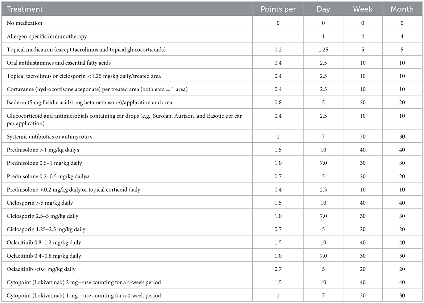
Table 1. Detailed medication score.
To better evaluate clinical signs with the level of applied medication, a combined Symptom Medication Score (CSMS) was determined, similar to the one already used in human medicine for allergen immunotherapy (54). For this, a daily Medication score dMS was determined as follows: dMS = sum of the medication intake of the last 4 weeks (according to questionnaire)/(number of days, i.e., usually 30 days), with the CSMS evaluated as follows: CADESI 4 + dMS.
2.6.3 Treatment efficacy
Treatment efficacy was primarily defined by two key criteria: (1) an absolute reduction of at least 2 points in the owner-assessed PVAS score from study entry to the final visit, and (2) a relative reduction of at least 50% in the clinician-assessed CADESI-4 score. These thresholds follow previously published studies and manufacturer data for oclacitinib (Apoquel®, Zoetis) that define clinically meaningful improvement in pruritus and skin lesion severity in canine atopic dermatitis.
Secondary analyses included weekly absolute and relative changes in PVAS scores compared to baseline, as well as absolute and relative changes in medication scores and blood values over the course of the study.
2.7 Collected parameters
All datasets were analyzed per protocol, including only those who completed the study as well as with all randomized dogs as intention-to-treat (ITT)-group. Baseline data, PVAS, and CADESI4 scores were collected on the day of enrollment and at the monthly visits, whereas via Limesurvey dog owners reported during the study period weekly PVAS and medication use. Blood samples were obtained at the beginning and the end of the study.
2.8 Collected blood analysis
Complete blood cell count, such as serum iron, total protein, albumin, C-reactive protein (CRP), and creatinine, was measured in the certified diagnostic laboratory of the university.
2.9 Statistical analysis
The study was designed before participants were enrolled. Data were examined for normal and log-normal distribution using Anderson-Darling tests. Normally distributed data sets were compared with the Student t-test. The non-parametric Mann–Whitney U test was applied to determine differences between the study arms. The same analyses were employed for the data per protocol (pp) and intention-to-treat (ITT). Matched data over time were analyzed. For parametric parameters, RM-one-way ANOVA with Geissner–Greenhouse correction and uncorrected Fisher's LSD were used, whereas matched non-parametric parameters were compared with the Friedman test and uncorrected Dunn's test. In cases where data were missing, mixed analysis was used.
When comparing weekly entry of the active and placebo arm side-by-side, RM-2way ANOVA, followed by uncorrected Fisher's LSD was used. To test treatment efficacy, chi-squared test was used. All tests were two-sided; p-values less than 0.05 were considered significant.
Data and graphs were analyzed with GraphPad Software Prism 10.4.2 (GraphPad Software, San Diego, CA).
3 Results
3.1 Study participants
Of the 39 screened dogs, 38 were eligible and randomized into two equal groups (n = 19 per group). The trial profile is presented in Figure 1. All randomized dogs were included in the ITT population (n = 19 in both groups).
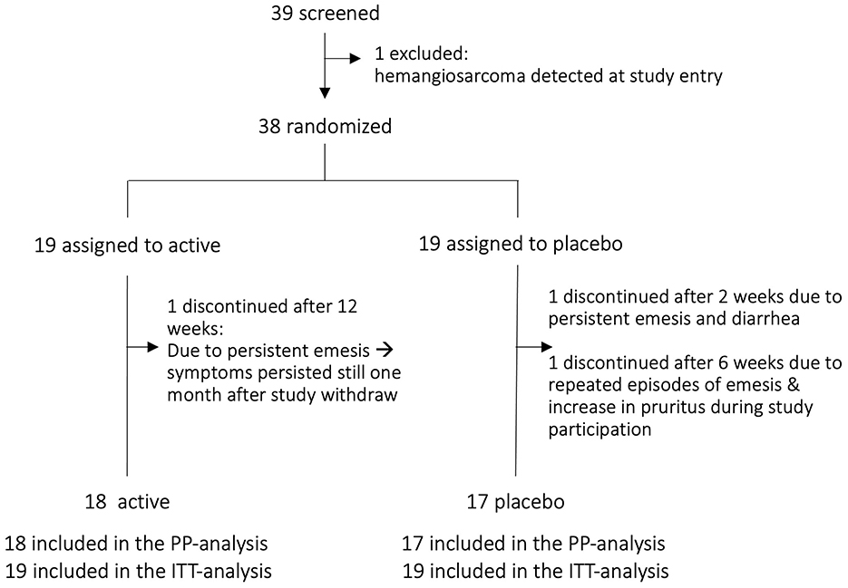
Figure 1. Trial profile. PP per protocol, ITT, intention-to-treat.
The study was completed by 35 dogs who were included in the PP analysis (18 in the active arm, 17 in the placebo arm). One dog was excluded during screening due to a newly detected splenic hemangiosarcoma requiring immediate care. Three dogs discontinued supplementation: one in the active group after approximately 3 months due to intermittent episodes of postprandial regurgitation, yet screening tests performed due to the clinical manifestation revealed no abnormalities. A follow-up of 30 days after discontinuation of the supplement showed that the signs persisted regardless of the withdrawal. Two dogs from the placebo arm discontinued participation, one after 2 weeks and the other after 6 weeks, both reporting persistent episodes of emesis and an increase in pruritus.
Baseline demographics and clinical characteristics were comparable between the ITT groups, showing no significant differences in gender, weight, PVAS, or CADESI-4 scores (Table 2). In the PP analysis, the initial CADESI scoring was higher in the active group compared to placebo, while weight, gender, and itching behavior (PVAS) were similar at study entry.
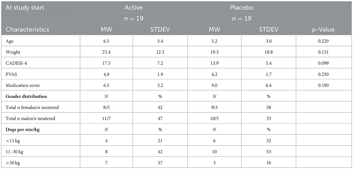
Table 2. Dogs'characteristics at day 0 in ITT-group.
3.2 Significant improvement in active arm for canine atopic dermatitis
This study assessed the efficacy of an active treatment for CAD, utilizing both veterinary assessments (CADESI-4) and owner-reported pruritus (PVAS). Assessments were conducted at baseline and monthly intervals to track changes in clinical signs.
While both groups experienced some improvement, possibly due to detailed and regular rechecks, the active treatment arm showed markedly superior results (Figures 2A, B). Specifically, the average CADESI-4 score in the active group improved by −9.278 points (95% CI: −12.77 to −5.787), representing a 55% relative improvement. In contrast, the placebo group saw a more modest −2.7-point improvement (95% CI: −5.261 to −0.2688), a 26% relative improvement.
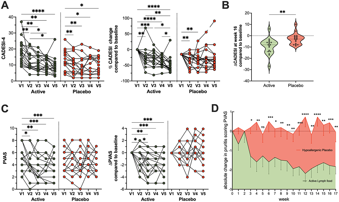
Figure 2. Clinical improvement in the active treatment arm. Significant improvement in clinical signs and pruritus was observed in the active treatment group compared to the placebo group throughout the study period. (A) Monthly absolute and relative veterinarian-assessed CADESI scores for individual dogs in both groups from baseline visit 1 (V1) to study end visit 5 (V5), (B) Comparison of absolute changes from baseline in CADESI scores between the active and placebo groups. (C) Monthly owner-assessed Pruritus Visual Analog Scale (PVAS) scores and their changes to baseline for both groups. (D) Weekly owner-assessed PVAS scores, showing the average (mean ± SEM) of all dogs per study arm over the entire observation period. For statistical analysis, data in A and C were analyzed via repeated measures one-way ANOVA with Geisser–Greenhouse correction and uncorrected Fisher's LSD test (single pooled variance), whereas B was compared using an unpaired t-test. Weekly PVAS changes in D were analyzed by a mixed-effects model and uncorrected Fisher's LSD. All data underwent normality testing via Anderson-Darling. Significance levels are denoted as: n.s. Not significant, *p < 0.05, **p < 0.01, ***p < 0.001; ****p < 0.0001.
The impact on owner-assessed pruritus was even more striking. Dogs in the active group experienced a significant and sustained reduction in pruritus (PVAS scores, Figure 2C) from the second visit onwards, which persisted or deepened over 4 months. The placebo group, however, showed no consistent improvement in PVAS scores throughout the study. Weekly pruritus assessments corroborated these findings, with stable PVAS in the placebo arm and a consistent, significant decline in the active arm (Figure 2D). Representative images of affected areas in dogs before and after 4 months of complementary lymph food are shown in Figure 3.
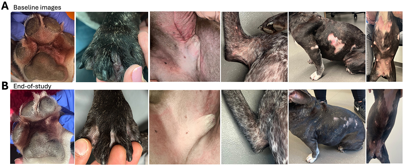
Figure 3. Clinical outcomes of lymph food supplementation in dogs (active treatment arm). Representative images illustrate the effects of daily 10 g lymph food supplementation. The upper row shows dogs at study start (baseline), while the lower row displays their condition at study end. Specific improvements in the active treatment arm include: reduced swelling and redness (first column), resolution of granulomas (second column, black paw), and significant improvement in skin barrier integrity, characterized by decreased redness, skin thickening, and hyperpigmentation (third column). Moreover, improvements in wounds, alopecia, and redness were observed, linked to reduced itching (fourth to sixth columns)
3.3 Less medication usage in the active arm
Prior to study enrollment (Table 3), approximately half of the active group (47%) and two-thirds of the placebo group (68%) had previously used Janus kinase-1 (JAK) inhibitor (oclacitinib).
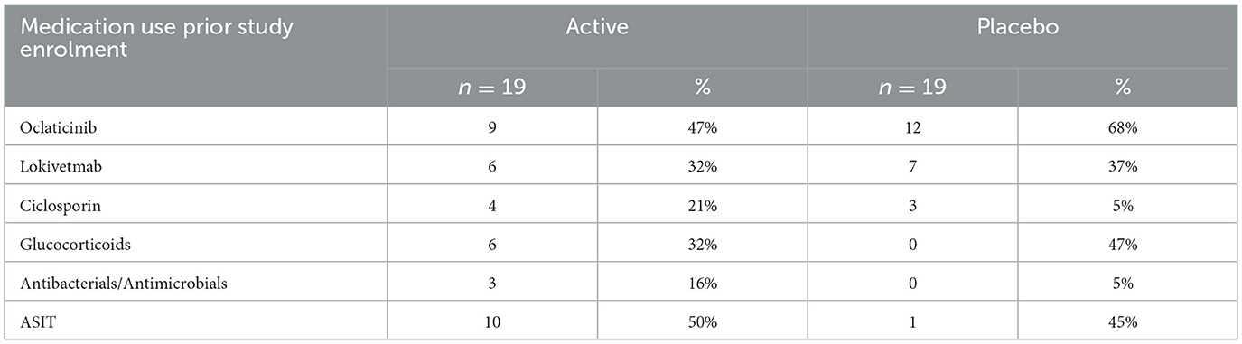
Table 3. History of medication use prior study enrollment.
About a third of both groups had experience with anti-IL-31 therapy (lokivetmab), with 32% in the active arm and 37% in the placebo arm. Ciclosporin use was more prevalent in the active arm (21%) compared to the placebo arm (5%).
Reported glucocorticoid (GC) use differed notably, with 32% of the active group having a history of use, while 47% of the placebo group had prior GC exposure. Antibacterials/antimicrobials were less common, with 16% in the active group and 5% in the placebo group reporting prior study enrollment.
Finally, allergen-specific immunotherapy (ASIT) was ongoing in about half of the active arm (50%) and 45% of the placebo arm at the time of study entry.
In the active arm, a notable decline in the overall use of certain symptomatic medication was observed over the 16-week study period (Tables 4, 5, and Figure 4). Specifically, the use of glucocorticoids decreased significantly, with the number of subjects receiving them dropping from 12 (67%) at week 1 to 6 (33%) at week 16, and the average dose declining from 3.8 ± 3.8 to 0.9 ± 1.7. Similarly, antibacterial/antimicrobial use also declined, with the number of subjects dropping from 4 (22%) at week 1 to just 1 (6%) at week 16. Oclacitinib, Lokivetmab, Ciclosporin, and ASIT were still administered, though the average dose of oclacitinib markedly decreased from 6.7 ± 1.9 at start to 4.0± 2.6 at the end of the study. This overall reduction in concomitant medication use suggests an improved control of symptoms.
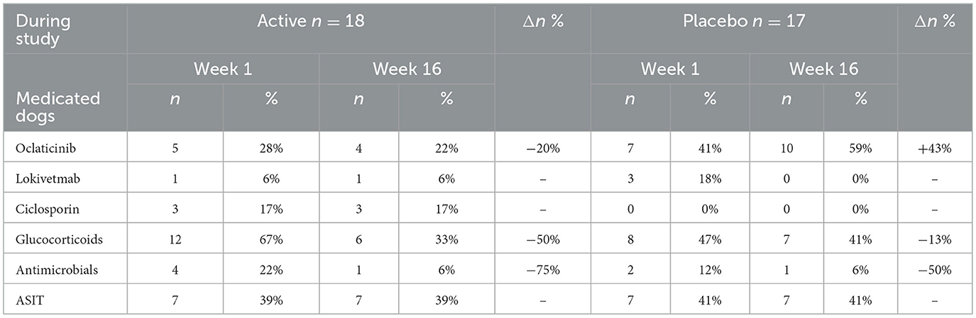
Table 4. Medicated dogs during study period per medication type.
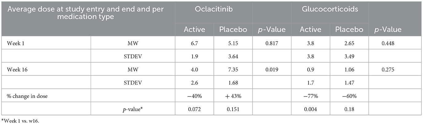
Table 5. Average dose (expressed as points) per dog at study entry and end per medication type.
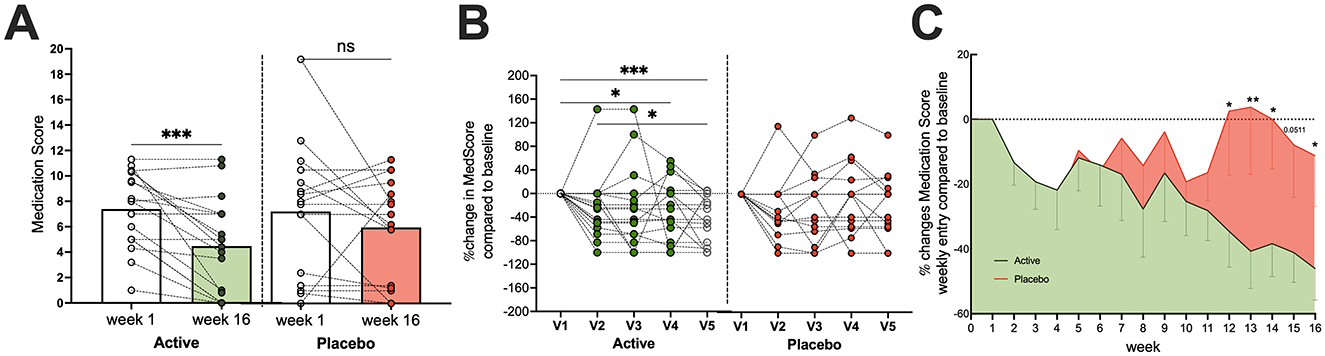
Figure 4. Reduced medication use in the active treatment arm. Dogs in the active treatment group required significantly less medication throughout the study compared to the placebo group. (A) Significant drop in absolute medication scores for active-treated dogs, which was not observed in the placebo arm. (B) Relative monthly changes in medication scored for individual dogs in the active and placebo group. (C) Relatively weekly changes in medication use for both arms. For statistical analysis, data in (A) were compared by paired t-test, data in (B) were analyzed by RM one-way ANOVA with Geisser–Greenhouse correction and uncorrected Fisher's LSD test (single pooled variance), whereas RM two-way ANOVA and uncorrected Fisher's LSD were employed for panel (C). All data were tested for normality using Anderson–Darling. Significance levels are indicated as: n.s. Not significant, *p < 0.05, **p < 0.01, ***p < 0.001.
Conversely, in the placebo arm, an increase in oclacitinib use was observed, rising from seven subjects (41%) at week 1 to 10 subjects (59%) by week 16, with the average points for the dose applied increasing from 5.2 ± 3.6 to 7.4 ± 1.7 at study end. Use of other medications, such as glucocorticoids, antibacterials/antimicrobials, and ASIT, remained stable or showed minor changes in the placebo group. As depicted in Figure 4, frequency and dose decreased significantly in the active arm but did not differ in the placebo arm from the study entry to the end.
3.4 Greater effectiveness in dogs with complementary lymph food and standard care treatment
Dogs receiving lymph food supplementation alongside standard care demonstrated significantly greater treatment success compared to those on a hypoallergenic placebo. This success was defined as previously reported by achieving at least a 50% reduction in CADESI-4 scores and/or a 2-point reduction in owner-assessed PVAS scores from baseline (55–57).
By the end of the study, 61.1% of dogs supplemented with lymph food achieved this definition of treatment success based on veterinarian-assessed CADESI-4 scores compared to only 17.6% of dogs in the placebo group. This indicates that the likelihood of improvement was approximately 3.5 times greater in the lymph food arm (p = 0.0087; RR: 3.46, 95% CI: 1.318–10.34).
Similarly, owner-assessed PVAS scores showed that 55% of dogs in the active group met the criteria for success vs. 17% in the placebo arm. This translates to an approximately threefold greater impact on symptom amelioration in the active group, as perceived by owners (p = 0.0204; RR: 3.15, 95% CI: 1.170–9.513), as shown in Table 6.
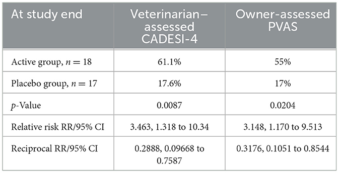
Table 6. Estimated proportion of dogs with treatment success.
3.5 Lymph food supplementation boosts blood parameters in dogs under standard care
Our 4-month study revealed that dogs supplemented with lymph food, in addition to standard care, experienced improved blood parameters (Figure 5). This included significant increases in red blood cell counts, packed cell volume (PCV), and serum iron, suggesting better iron utilization. Intriguingly, in dogs with CAD, baseline red blood cell counts were found to account for roughly 14% and PCV to approximately 11% of the variation in their CAD skin lesion severity, as measured by CADESI-04 scores. The observed improvements in red blood cell parameters in the lymph food group are therefore particularly noteworthy.
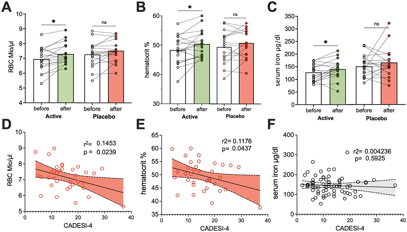
Figure 5. Lymph food supplementation significantly improves erythroid parameters and iron status in dogs. Blood parameters were assessed in dogs at study entry and end. Panels A, B, and C compare “before and after” values (A) red blood cell counts, (B) hematocrit, and (C) serum iron levels, respectively, in dogs supplemented with lymph food (green bars) or a hypoallergenic placebo (red bars). Paired t-tests were used to compare “before and after” values. Panels D, E, and F illustrate linear regression analyses of (D) red blood cell counts, (E) hematocrit, and (F) serum iron, respectively, with CADESI-04 scores. Significant negative dependencies from linear regressions are shown in red, while data points for non-significant dependencies are in gray. *p < 0.05.
4 Discussion
Canine atopic dermatitis is linked to specific nutritional deficiencies, particularly in lipids (16), minerals, such as iron and zinc (44), and various vitamins (15, 21, 58). These parallels are striking and reflect similar deficits observed in human patients with atopic dermatitis (54, 59–66). Our previous research has already demonstrated that despite consuming an iron-rich, meat-based diet, dogs with CAD often suffer from iron deficiency. Importantly, a lack of iron can act as a critical “danger signal” for immune cells, triggering their activation and driving systemic inflammation (17–20).
This situation is particularly challenging because the body's defensive state inherently impairs its ability to absorb and effectively utilize dietary micronutrients. During immune responses, nutrients are often sequestered to “safer harbors,” such as the liver and mononuclear cells, a protective mechanism designed to prevent pathogens from accessing vital resources. This phenomenon of impaired dietary absorption, termed mucosal block, was first identified in dogs (19, 26).
While this mucosal block provides a vital short-term strategic advantage by limiting the spread of viral, bacterial, and fungal pathogens (67–73), its persistence becomes detrimental in chronic conditions like atopic dermatitis. In CAD, an inciting pathogen is often absent, yet the body's protective mechanism inadvertently prevents it from maintaining adequate micronutrient levels. In humans, these micronutrient deficiencies, particularly of iron and vitamin A, are directly linked to immune activation and are independent predictors of overall morbidity and mortality (5, 17, 18, 74–96).
Fortunately, the mucosal block is not absolute. Although micronutrient uptake into the bloodstream is compromised, the lymphatic system remains a viable alternative route. Here, immune cells in the lymphatic system and lymph nodes actively monitor micronutrients before their eventual release into the bloodstream (19, 20).
Building on this understanding, our study investigated the potential benefits of a supplementary food where micronutrients were carried by digestion-resistant transport proteins predominantly from whey. These proteins are well-documented to be absorbed via the lymphatics (97–101) and are established carriers for essential nutrients, such as lipids (102–105), minerals (106, 107), vitamins (108–112), and antioxidants (113–116).
In this randomized, double-blinded, controlled pilot study, we demonstrate for the first time that for dogs undergoing standard care for CAD—such as medication to control pruritus (lokivetmab, oclacitinib, ciclosporin, and glucocorticoids) and, when necessary, topical or antimicrobial treatment—the addition of this complementary lymph-targeted food led to significantly superior outcomes across all measured clinical parameters. The improvement observed was approximately three times greater than that in the placebo-supplemented group. Concurrently, medication usage significantly declined, and systemic blood parameters improved. This finding is particularly noteworthy given that the veterinarian-assessed skin lesions were partly correlated with insufficient red blood cell counts, suggesting a broader systemic impact and indicating a physiological link between systemic iron handling, inflammation, and disease progression.
Chronic inflammation is known to disrupt iron homeostasis through upregulation of hepcidin, which inhibits iron export from enterocytes and macrophages, resulting in functional iron deficiency and impaired erythropoiesis—the hallmarks of anemia of inflammation (17–20, 117, 118). The improvement in blood parameters during supplementation suggests a restoration of iron utilization and erythroid balance. The supplement provided bioavailable iron, zinc, copper, and vitamin C, alongside plant-derived polyphenols, carotenoids, and curcuminoids. Vitamin C enhances intestinal iron absorption and reduces oxidative stress that limits iron mobilization. In addition, polyphenols contribute to iron solubility, improve lipid metabolism and membrane fluidity, and favor macrophage polarization toward an anti-inflammatory (M2-like) phenotype (19, 118). Together, these mechanisms may suppress hepcidin signaling, enhance iron recycling, and reduce inflammatory iron sequestration, thereby normalizing hematological indices and contributing to clinical improvement.
When analyzing outcomes based on breed disposition, brachycephalic dogs were similarly distributed between the active and placebo groups, but appeared to benefit particularly from the lymph-targeted food, possibly due to their inherent challenges with breathing and feeding.
It's important to acknowledge that while our study was randomized to ensure an even distribution of participants, after accounting for drop-outs, dogs in the active treatment arm were, at the study's inception, significantly sicker, exhibiting higher CADESI scores and often presenting with co-morbidities, such as arthrosis and a history of previous surgeries. While this baseline difference may partly explain the more dramatic improvements observed in this group, it could also suggest a greater need for highly bioavailable nutrients in these more compromised animals. Interestingly, the lymph-targeted food also appeared to specifically promote fur growth, with many patients experiencing years of bald patches showing remarkable recovery.
Crucially, the improved health outcomes observed were consistently matched by an improved red blood cell parameter status. This highlights that simply providing bioavailable micronutrients—which were measurable in individual dogs—was sufficient to significantly ameliorate atopic dermatitis symptoms. We hypothesize that this superiority, compared to the use of highly effective symptomatic drugs alone (119–121), stems from the fact that these conventional medications primarily suppress symptoms without addressing an underlying trigger for atopic dermatitis. Consequently, the inflammatory trigger may persist, leading to symptom exacerbation once drug effects wane. In contrast, the lymph-targeted food appears to compensate for apparent nutritional deficits, thereby potentially removing one key trigger for inflammation if these deficiencies were indeed driving the condition.
While acknowledging the limitations of this study, including the initial baseline differences in disease severity, the consistent improvements across clinical and hematological parameters, coupled with reduced medication burden, are highly encouraging. The apparent breed-specific benefits and the striking promotion of regrowth also highlight areas for future investigation.
In conclusion, our results suggest a paradigm shift toward integrating targeted nutritional support as a fundamental component of CAD management. This approach not only significantly improves clinical signs but also offers the potential to reduce dependence on pharmaceutical interventions. Indeed, the improved health outcomes were directly matched by improved blood parameters, indicating that the targeted nutritional deficits were effectively compensated. Given these promising findings from our pilot study, we strongly encourage independent replication of this study to confirm these benefits and further elucidate the precise mechanisms by which lymph-targeted nutritional supplementation contributes to improved outcomes in canine atopic dermatitis.
Data availability statement
The original contributions presented in the study are included in the article/Supplementary material, further inquiries can be directed to the corresponding author.
Ethics statement
The animal studies were approved by Ethics Committee of the University of Veterinary Medicine Vienna and the Austrian Federal Ministry of Science and Research (Ref: BMBWF 2023-0.618.811). The studies were conducted in accordance with the local legislation and institutional requirements. Written informed consent was obtained from the owners for the participation of their animals in this study.
Author contributions
CR: Funding acquisition, Formal analysis, Visualization, Writing – review & editing, Data curation, Investigation. PD: Methodology, Investigation, Writing – review & editing. IB: Writing – review & editing, Resources, Methodology. CH: Writing – review & editing, Investigation. VE: Investigation, Writing – review & editing. UW: Writing – review & editing, Investigation. LP: Methodology, Funding acquisition, Writing – review & editing, Supervision. FR-W: Investigation, Resources, Project administration, Visualization, Writing – original draft, Methodology, Funding acquisition, Formal analysis, Conceptualization.
Funding
The author(s) declared that financial support was received for this work and/or its publication. The study was supported by the European Society of Veterinarian Dermatology and by ViaLym FlexCo, Tulln, Austria. The study concept, experimental design, and clinical protocol were developed independently in an academic setting prior to the establishment and funding of ViaLym FlexCo. The funder had no role in the study design; collection, analysis, or interpretation of data; manuscript preparation; or the decision to submit the manuscript for publication. Open Access funding provided by University of Veterinary Medicine Vienna.
Acknowledgments
We thank all dog owners and study participants that made this study possible.
Conflict of interest
CFR received personal fees from ViaLym FlexCo. FRW is a founder and shareholder of ViaLym FlexCo (Tulln, Austria).
The remaining author(s) declared that the research was conducted in the absence of any commercial or financial relationships that could be construed as a potential conflict of interest.
The author(s) declared that they were an editorial board member of Frontiers, at the time of submission. This had no impact on the peer review process and the final decision.
Generative AI statement
The author(s) declared that generative AI was used in the creation of this manuscript. For text improvement and shortening of the abstract, Generative AI tools (gemini 2.5 flash) was used.
Any alternative text (alt text) provided alongside figures in this article has been generated by Frontiers with the support of artificial intelligence and reasonable efforts have been made to ensure accuracy, including review by the authors wherever possible. If you identify any issues, please contact us.
Publisher's note
All claims expressed in this article are solely those of the authors and do not necessarily represent those of their affiliated organizations, or those of the publisher, the editors and the reviewers. Any product that may be evaluated in this article, or claim that may be made by its manufacturer, is not guaranteed or endorsed by the publisher.
Supplementary material
The Supplementary Material for this article can be found online at: https://www.frontiersin.org/articles/10.3389/fvets.2025.1657869/full#supplementary-material
References
1. Cheema B, Chokshi A, Orimoloye O, Ardehali H. Intravenous iron repletion for patients with heart failure and iron deficiency. JACC. (2024) 83:2674–89. doi: 10.1016/j.jacc.2024.03.431
2. Outerbridge CA, Jordan TJM. Current knowledge on canine atopic dermatitis: pathogenesis and treatment. Adv Small Anim Care. (2021) 2:101–15. doi: 10.1016/j.yasa.2021.07.004
3. Eisenschenk MC, Hensel P, Saridomichelakis MN, Tamamoto-Mochizuki C, Pucheu-Haston CM, Santoro D, et al. Introduction to the ICADA 2023 canine atopic dermatitis pathogenesis review articles and updated definition. Vet Dermatol. (2024) 35:3–4. doi: 10.1111/vde.13183
4. Yang L, Sato M, Saito-Abe M, Miyaji Y, Shimada M, Sato C, et al. Allergic disorders and risk of anemia in Japanese children: findings from the Japan Environment and Children's Study. Nutrients. (2022) 14:4335. doi: 10.3390/nu14204335
5. Rhew K, Oh JM. Association between atopic disease and anemia in pediatrics: a cross-sectional study. BMC Pediatr. (2019) 19:455. doi: 10.1186/s12887-019-1836-5
6. Stojkovic A, Radlovic N, Vuletic B, Nestorovic B, Lekovic Z, Obradovic S, et al. Presentation of an infant with nutritional deficiency dermatitis as the initial manifestation of cystic fibrosis. Srp Arh Celok Lek. (2013) 141:810–3. doi: 10.2298/SARH1312810S
7. Thomas L, Naidoo K, Darne S. Zinc deficiency and severe protein-energy malnutrition in a child with atopic eczema. Clin Exp Dermatol. (2019) 44:215–7. doi: 10.1111/ced.13634
8. Vollbracht C, Raithel M, Krick B, Kraft K, Hagel AF. Intravenous vitamin C in the treatment of allergies: an interim subgroup analysis of a long-term observational study. J Int Med Res. (2018) 46:3640–55. doi: 10.1177/0300060518777044
9. Tromp IIM, Franco OH, van den Hooven EH, Heijboer AC, Jaddoe VWV, Duijts L, et al. 25-Hydroxyvitamin D concentrations, asthma and eczema in childhood: the generation R study. Clin Nutr. (2018) 37:169–76. doi: 10.1016/j.clnu.2016.11.019
10. El-Heis S, Crozier SR, Robinson SM, Harvey NC, Cooper C, Inskip HM, et al. Higher maternal serum concentrations of nicotinamide and related metabolites in late pregnancy are associated with a lower risk of offspring atopic eczema at age 12 months. Clin Exp Allergy. (2016) 46:1337–43. doi: 10.1111/cea.12782
11. Amin MN, Liza KF, Sarwar MS, Ahmed J, Adnan MT, Chowdhury MI, et al. Effect of lipid peroxidation, antioxidants, macro minerals and trace elements on eczema. Arch Dermatol Res. (2015) 307:617–23. doi: 10.1007/s00403-015-1570-2
12. Witzel-Rollins A, Murphy M, Becvarova I, Werre SR, Cadiergues MC, Meyer H. Non-controlled, open-label clinical trial to assess the effectiveness of a dietetic food on pruritus and dermatologic scoring in atopic dogs. BMC Vet Res. (2019) 15:220. doi: 10.1186/s12917-019-1929-2
13. van Beeck FL, Watson A, Bos M, Biourge V, Willemse T. The effect of long-term feeding of skin barrier-fortified diets on the owner-assessed incidence of atopic dermatitis symptoms in Labrador retrievers. J Nutr Sci. (2015) 4:e5. doi: 10.1017/jns.2014.61
14. de Santiago MS, Arribas JLG, Llamas YM, Becvarova I, Meyer H. Randomized, double-blind, placebo-controlled clinical trial measuring the effect of a dietetic food on dermatologic scoring and pruritus in dogs with atopic dermatitis. BMC Vet Res. (2021) 17:354. doi: 10.1186/s12917-021-03063-w
15. Klinger CJ, Hobi S, Johansen C, Koch HJ, Weber K, Mueller RS. Vitamin D shows in vivo efficacy in a placebo-controlled, double-blinded, randomised clinical trial on canine atopic dermatitis. Vet Rec. (2018) 182:406. doi: 10.1136/vr.104492
16. McFadden RA, Heinrich NA, Haarstad AC, Tomlinson DJ. A double-blinded, randomized, controlled, crossover evaluation of a zinc methionine supplement as an adjunctive treatment for canine atopic dermatitis. Vet Dermatol. (2017) 28:569–e138. doi: 10.1111/vde.12466
17. Peroni DG, Hufnagl K, Comberiati P, Roth-Walter F. Lack of iron, zinc, and vitamins as a contributor to the etiology of atopic diseases. Front Nutr. (2022) 9:1032481. doi: 10.3389/fnut.2022.1032481
18. Roth-Walter F. Iron-deficiency in atopic diseases: innate immune priming by allergens and siderophores. Front Allergy. (2022) 3:859922. doi: 10.3389/falgy.2022.859922
19. Roth-Walter F, Berni Canani R, O'Mahony L, Peroni D, Sokolowska M, Vassilopoulou E, et al. Nutrition in chronic inflammatory conditions. Bypassing the mucosal block for micronutrients. Allergy. (2024) 79:353–83. doi: 10.1111/all.15972
20. Vassilopoulou E, Venter C, Roth-Walter F. Malnutrition and allergies: tipping the immune balance towards health. J Clin Med. (2024) 13:4713. doi: 10.3390/jcm13164713
21. Ramos CF, Doulidis PG, Polakova N, Burgener IA, Jensen-Jarolim E, Cimarelli G, et al. Iron deficiency in dogs suffering from atopic dermatitis. BMC Vet Res. (2024) 20:506. doi: 10.1186/s12917-024-04350-y
22. Sato S. Iron deficiency: structural and microchemical changes in hair, nails, and skin. Semin Dermatol. (1991) 10:313–9.
23. Schrage B, Rubsamen N, Schulz A, Munzel T, Pfeiffer N, Wild PS, et al. Iron deficiency is a common disorder in general population and independently predicts all-cause mortality: results from the Gutenberg Health Study. Clin Res Cardiol. (2020) 109:1352–7. doi: 10.1007/s00392-020-01631-y
24. Sun Y, Zhang H, Qi G, Tian W. Nutrient deficiency patterns and all-cause and cardiovascular mortality in older adults with hypertension: a latent class analysis. BMC Public Health. (2024) 24:1551. doi: 10.1186/s12889-024-19003-w
25. Mah E, Sapper TN, Chitchumroonchokchai C, Failla ML, Schill KE, Clinton SK, et al. α-Tocopherol bioavailability is lower in adults with metabolic syndrome regardless of dairy fat co-ingestion: a randomized, double-blind, crossover trial. Am J Clin Nutr. (2015) 102:1070–80. doi: 10.3945/ajcn.115.118570
26. Crosby WH. Mucosal block. An evaluation of concepts relating to control of iron absorption. Semin Hematol. (1966) 3:299–313.
27. Krishnan S, Bhuyan UN, Talwar GP, Ramalingaswami V. Effect of vitamin A and protein-calorie undernutrition on immune responses. Immunology. (1974) 27:383–92.
28. Golden MH, Jackson AA, Golden BE. Effect of zinc on thymus of recently malnourished children. Lancet. (1977) 2:1057–9. doi: 10.1016/S0140-6736(77)91888-8
29. Harvey J, French T, Meyer D. Chronic iron deficiency anemia in dogs. J Am Anim Hosp Assoc. (1982) 2. doi: 10.2460/javma.1982.181.11.1402
30. Nathanson MH, Muir A, McLaren GD. Iron absorption in normal and iron-deficient beagle dogs: mucosal iron kinetics. Am J Physiol. (1985) 249:G439–48. doi: 10.1152/ajpgi.1985.249.4.G439
31. Kocher A, Bohnert L, Schiborr C, Frank J. Highly bioavailable micellar curcuminoids accumulate in blood, are safe and do not reduce blood lipids and inflammation markers in moderately hyperlipidemic individuals. Mol Nutr Food Res. (2016) 60:1555–63. doi: 10.1002/mnfr.201501034
32. Elz AS, Trevaskis NL, Porter CJH, Bowen JM, Prestidge CA. Smart design approaches for orally administered lipophilic prodrugs to promote lymphatic transport. J Control Release. (2022) 341:676–701. doi: 10.1016/j.jconrel.2021.12.003
33. von der Weid PY. Lymphatic vessel pumping. Adv Exp Med Biol. (2019) 1124:357–77. doi: 10.1007/978-981-13-5895-1_15
34. Trevaskis NL, Kaminskas LM, Porter CJ. From sewer to saviour - targeting the lymphatic system to promote drug exposure and activity. Nat Rev Drug Discov. (2015) 14:781–803. doi: 10.1038/nrd4608
35. Han S, Quach T, Hu L, Lim SF, Zheng D, Leong NJ, et al. Increasing linker chain length and intestinal stability enhances lymphatic transport and lymph node exposure of triglyceride mimetic prodrugs of a model immunomodulator mycophenolic acid. Mol Pharm. (2023) 20:2675–85. doi: 10.1021/acs.molpharmaceut.3c00099
36. Abdallah M, Mullertz OO, Styles IK, Morsdorf A, Quinn JF, Whittaker MR, et al. Lymphatic targeting by albumin-hitchhiking: applications and optimisation. J Control Release. (2020) 327:117–28. doi: 10.1016/j.jconrel.2020.07.046
37. Muller MR, Linek M, Lowenstein C, Rothig A, Doucette K, Thorstensen K, et al. Evaluation of cyclosporine-sparing effects of polyunsaturated fatty acids in the treatment of canine atopic dermatitis. Vet J. (2016) 210:77–81. doi: 10.1016/j.tvjl.2015.11.012
38. Harvey RG. A blinded, placebo-controlled study of the efficacy of borage seed oil and fish oil in the management of canine atopy. Vet Rec. (1999) 144:405–7. doi: 10.1136/vr.144.15.405
39. Mueller RS, Fettman MJ, Richardson K, Hansen RA, Miller A, Magowitz J, et al. Plasma and skin concentrations of polyunsaturated fatty acids before and after supplementation with n-3 fatty acids in dogs with atopic dermatitis. Am J Vet Res. (2005) 66:868–73. doi: 10.2460/ajvr.2005.66.868
40. Schafer L, Thom N. A placebo-controlled, double-blind study evaluating the effect of orally administered polyunsaturated fatty acids on the oclacitinib dose for atopic dogs. Vet Dermatol. (2024) 35:408–17. doi: 10.1111/vde.13246
41. Ferguson EA, Littlewood JD, Carlotti DN, Grover R, Nuttall T. Management of canine atopic dermatitis using the plant extract PYM00217: a randomized, double-blind, placebo-controlled clinical study. Vet Dermatol. (2006) 17:236–43. doi: 10.1111/j.1365-3164.2006.00523.x
42. Nagle TM, Torres SM, Horne KL, Grover R, Stevens MT. A randomized, double-blind, placebo-controlled trial to investigate the efficacy and safety of a Chinese herbal product (P07P) for the treatment of canine atopic dermatitis. Vet Dermatol. (2001) 12:265–74. doi: 10.1046/j.0959-4493.2001.00267.x
43. Marsella R, Messinger L, Zabel S, Rosychuck R, Griffin C, Cronin PO, et al. A randomized, double-blind, placebo-controlled study to evaluate the effect of EFF1001, an Actinidia arguta (hardy kiwi) preparation, on CADESI score and pruritus in dogs with mild to moderate atopic dermatitis. Vet Dermatol. (2010) 21:50–7. doi: 10.1111/j.1365-3164.2009.00773.x
44. Franco J, Rajwa B, Gomes P, HogenEsch H. Local and systemic changes in lipid profile as potential biomarkers for canine atopic dermatitis. Metabolites. (2021) 11:670. doi: 10.3390/metabo11100670
45. American Animal Hospital Association. 2021 AAHA nutrition and weight-management guidelines for dogs and cats. J Am Anim Hosp Assoc. (2021) 57:153–78.
46. Montoya M, Péron F, Hookey T, Morrison J, German AJ, Gaillard V. et al. Overweight and obese body condition in ~49 million dogs and ~13 million cats seen at primary practices across the USA: prevalences by life stage from early growth to senior. Prevent Vet Med. (2025) 235:106398. doi: 10.1016/j.prevetmed.2024.106398
47. FEDIAF. Nutritional Guidelines for Complete and Complementary Pet Food for Cats and Dogs. Brussels, Belgium: FEDIAF EuropeanPetFood (2024).
48. FEDIAF. FEDIAF Announces Updated 2024 Nutritional Guidelines. Brussels, Belgium: FEDIAF EuropeanPetFood (2024).
49. FEDIAF. Nutritional Guidelines (FEDIAF: overview and downloads). Brussels, Belgium: FEDIAF EuropeanPetFood (2025).
51. European Commission Directorate-General for Health and Food Safety. Legislation on feed additives - Food Safety. European Commission (2003).
52. European Commission Directorate-General for Health and Food Safety. Feed additives - Food Safety (EU Register overview). European Commission (2023).
53. Kasper B, Zablotski Y, Mueller RS. Long-term use of lokivetmab in dogs with atopic dermatitis. Vet Dermatol. (2024) 35:683–93. doi: 10.1111/vde.13286
54. Bartosik T, Jensen SA, Afify SM, Bianchini R, Hufnagl K, Hofstetter G, et al. Ameliorating atopy by compensating micronutritional deficiencies in immune cells: a double-blind placebo-controlled pilot study. J Allergy Clin Immunol Pract. (2022) 10:1889–902. e9 doi: 10.1016/j.jaip.2022.02.028
55. Forster S, Boegel A, Despa S, Trout C, King S. Comparative efficacy and safety of ilunocitinib and oclacitinib for the control of pruritus and associated skin lesions in dogs with atopic dermatitis. Vet Dermatol. (2025) 36:165–76. doi: 10.1111/vde.13319
56. Olivry T, Bensignor E, Favrot C, Griffin CE, Hill PB, Mueller RS, et al. Development of a core outcome set for therapeutic clinical trials enrolling dogs with atopic dermatitis (COSCAD'18). BMC Vet Res. (2018) 14:238. doi: 10.1186/s12917-018-1569-y
57. Lee S, Yun T, Koo Y, Chae Y, Lee D, Choi D, et al. Clinical efficacy of oclacitinib and lokivetmab in dogs with canine atopic dermatitis. J Vet Clin. (2021) 38:127–34. doi: 10.17555/jvc.2021.38.3.127
58. Plevnik Kapun A, Salobir J, Levart A, Tavcar Kalcher G, Nemec Svete A. Vitamin E supplementation in canine atopic dermatitis: improvement of clinical signs and effects on oxidative stress markers. Vet Rec. (2014) 175:560. doi: 10.1136/vr.102547
59. Fortes C, Mastroeni S, Mannooranparampil TJ, Di Lallo D. Pre-natal folic acid and iron supplementation and atopic dermatitis in the first 6 years of life. Arch Dermatol Res. (2019) 311:361–7. doi: 10.1007/s00403-019-01911-2
60. Guarneri F, Guarneri C, Cannavo SP. Oral iron therapy and chronic idiopathic urticaria: sideropenic urticaria? Dermatol Ther. (2014) 27:223–6. doi: 10.1111/dth.12122
61. Al Senaidy AM. Serum vitamin A and beta-carotene levels in children with asthma. J Asthma. (2009) 46:699–702. doi: 10.1080/02770900903056195
62. Fabian E, Poloskey P, Kosa L, Elmadfa I, Rethy LA. Nutritional supplements and plasma antioxidants in childhood asthma. Wien Klin Wochenschr. (2013) 125:309–15. doi: 10.1007/s00508-013-0359-6
63. Wood LG, Garg ML, Smart JM, Scott HA, Barker D, Gibson PG. Manipulating antioxidant intake in asthma: a randomized controlled trial. Am J Clin Nutr. (2012) 96:534–43. doi: 10.3945/ajcn.111.032623
64. Allen S, Britton JR, Leonardi-Bee JA. Association between antioxidant vitamins and asthma outcome measures: systematic review and meta-analysis. Thorax. (2009) 64:610–9. doi: 10.1136/thx.2008.101469
65. Yang H, Chen JS, Zou WJ, Tan Q, Xiao YZ, Luo XY, et al. Vitamin A deficiency exacerbates extrinsic atopic dermatitis development by potentiating type 2 helper T cell-type inflammation and mast cell activation. Clin Exp Allergy. (2020) 50:942–53. doi: 10.1111/cea.13687
66. Xiang J, Wang H, Li T. Comorbidity of vitamin A and vitamin D deficiency exacerbates the severity of atopic dermatitis in children. Dermatology. (2019) 235:196–204. doi: 10.1159/000496603
67. Gwamaka M, Kurtis JD, Sorensen BE, Holte S, Morrison R, Mutabingwa TK, et al. Iron deficiency protects against severe Plasmodium falciparum malaria and death in young children. Clin Infect Dis. (2012) 54:1137–44. doi: 10.1093/cid/cis010
68. Muriuki JM, Mentzer AJ, Kimita W, Ndungu FM, Macharia AW, Webb EL, et al. Iron status and associated malaria risk among African children. Clin Infect Dis. (2019) 68:1807–14. doi: 10.1093/cid/ciy791
69. Nyakeriga AM, Troye-Blomberg M, Dorfman JR, Alexander ND, Back R, Kortok M, et al. Iron deficiency and malaria among children living on the coast of Kenya. J Infect Dis. (2004) 190:439–47. doi: 10.1086/422331
70. Cegielski JP, Arab L, Cornoni-Huntley J. Nutritional risk factors for tuberculosis among adults in the United States, 1971-1992. Am J Epidemiol. (2012) 176:409–22. doi: 10.1093/aje/kws007
71. Boelaert JR, Vandecasteele SJ, Appelberg R, Gordeuk VR. The effect of the host's iron status on tuberculosis. J Infect Dis. (2007) 195:1745–53. doi: 10.1086/518040
72. McDermid JM, Hennig BJ, van der Sande M, Hill AV, Whittle HC, Jaye A, et al. Host iron redistribution as a risk factor for incident tuberculosis in HIV infection: an 11-year retrospective cohort study. BMC Infect Dis. (2013) 13:48. doi: 10.1186/1471-2334-13-48
73. Isanaka S, Mugusi F, Urassa W, Willett WC, Bosch RJ, Villamor E, et al. Iron deficiency and anemia predict mortality in patients with tuberculosis. J Nutr. (2012) 142:350–7. doi: 10.3945/jn.111.144287
74. Luo J, Wang X, Yuan L, Guo L. Iron deficiency, a risk factor of thyroid disorders in reproductive-age and pregnant women: a systematic review and meta-analysis. Front Endocrinol (Lausanne). (2021) 12:629831. doi: 10.3389/fendo.2021.629831
75. Chang R, Chu KA, Lin MC, Chu YH, Hung YM, Wei JC. Newly diagnosed iron deficiency anemia and subsequent autoimmune disease: a matched cohort study in Taiwan. Curr Med Res Opin. (2020) 36:985–92. doi: 10.1080/03007995.2020.1748585
76. Drury KE, Schaeffer M, Silverberg JI. Association between atopic disease and anemia in US children. JAMA Pediatr. (2016) 170:29–34. doi: 10.1001/jamapediatrics.2015.3065
77. Rhew K, Brown JD, Oh JM. Atopic disease and anemia in Korean patients: cross-sectional study with propensity score analysis. Int J Environ Res Public Health. (2020) 17 doi: 10.3390/ijerph17061978
78. Petje LM, Jensen SA, Szikora S, Sulzbacher M, Bartosik T, Pjevac P, et al. Functional iron-deficiency in women with allergic rhinitis is associated with symptoms after nasal provocation and lack of iron-sequestering microbes. Allergy. (2021) 76:2882–6. doi: 10.1111/all.14960
79. Krishna MT, Subramanian A, Adderley NJ, Zemedikun DT, Gkoutos GV, Nirantharakumar K. Allergic diseases and long-term risk of autoimmune disorders: longitudinal cohort study and cluster analysis. Eur Respir J. (2019) 54:1900476. doi: 10.1183/13993003.00476-2019
80. Batchelor EK, Kapitsinou P, Pergola PE, Kovesdy CP, Jalal DI. Iron deficiency in chronic kidney disease: updates on pathophysiology, diagnosis, and treatment. J Am Soc Nephrol. (2020) 31:456–68. doi: 10.1681/ASN.2019020213
81. Susantitaphong P, Alqahtani F, Jaber BL. Efficacy and safety of intravenous iron therapy for functional iron deficiency anemia in hemodialysis patients: a meta-analysis. Am J Nephrol. (2014) 39:130–41. doi: 10.1159/000358336
82. Albaramki J, Hodson EM, Craig JC, Webster AC. Parenteral versus oral iron therapy for adults and children with chronic kidney disease. Cochrane Database Syst Rev. (2012) 1:CD007857. doi: 10.1002/14651858.CD007857.pub2
83. Reinhold J, Papadopoulou C, Baral R, Vassiliou VS. Iron deficiency for prognosis in acute coronary syndrome - A systematic review and meta-analysis. Int J Cardiol. (2021) 328:46–54. doi: 10.1016/j.ijcard.2020.12.021
84. Osman M, Syed M, Balla S, Kheiri B, Faisaluddin M, Bianco C, et al. Meta-analysis of intravenous iron therapy for patients with iron deficiency and heart failure. Am J Cardiol. (2021) 141:152–3. doi: 10.1016/j.amjcard.2020.11.025
85. Zhang J, Hu S, Jiang Y, Zhou Y. Efficacy and safety of iron therapy in patients with chronic heart failure and iron deficiency: a systematic review and meta-analysis based on 15 randomised controlled trials. Postgrad Med J. (2020) 96:766–76. doi: 10.1136/postgradmedj-2019-137342
86. Nickol AH Frise MC, Cheng HY, McGahey A, McFadyen BM, Harris-Wright T, et al. A cross-sectional study of the prevalence and associations of iron deficiency in a cohort of patients with chronic obstructive pulmonary disease. BMJ Open. (2025) 5:e007911. doi: 10.1136/bmjopen-2015-007911
87. Cloonan SM, Mumby S, Adcock IM, Choi AMK, Chung KF, Quinlan GJ. The “Iron”-y of Iron overload and iron deficiency in chronic obstructive pulmonary disease. Am J Respir Crit Care Med. (2017) 196:1103–12. doi: 10.1164/rccm.201702-0311PP
88. Pizzini A, Aichner M, Sonnweber T, Tancevski I, Weiss G, Loffler-Ragg J. The significance of iron deficiency and anemia in a real-life COPD cohort. Int J Med Sci. (2020) 17:2232–9. doi: 10.7150/ijms.46163
89. Wyart E, Hsu MY, Sartori R, Mina E, Rausch V, Pierobon ES, et al. Iron supplementation is sufficient to rescue skeletal muscle mass and function in cancer cachexia. EMBO Rep. (2022) 23:e53746. doi: 10.15252/embr.202153746
90. Ludwig H, Evstatiev R, Kornek G, Aapro M, Bauernhofer T, Buxhofer-Ausch V, et al. Iron metabolism and iron supplementation in cancer patients. Wien Klin Wochenschr. (2015) 127:907–19. doi: 10.1007/s00508-015-0842-3
91. Escobar Alvarez Y, de Las Penas Bataller R, Perez Altozano J, Ros Martinez S, Sabino Alvarez A, Blasco Cordellat A, et al. SEOM clinical guidelines for anaemia treatment in cancer patients (2020). Clin Transl Oncol. (2021) 23:931–9. doi: 10.1007/s12094-021-02580-2
92. Teng IC, Tseng SH, Aulia B, Shih CK, Bai CH, Chang JS. Can diet-induced weight loss improve iron homoeostasis in patients with obesity: a systematic review and meta-analysis. Obes Rev. (2020) 21:e13080. doi: 10.1111/obr.13080
93. Zhao L, Zhang X, Shen Y, Fang X, Wang Y, Wang F. Obesity and iron deficiency: a quantitative meta-analysis. Obes Rev. (2015) 16:1081–93. doi: 10.1111/obr.12323
94. Klip IT, Comin-Colet J, Voors AA, Ponikowski P, Enjuanes C, Banasiak W, et al. Iron deficiency in chronic heart failure: an international pooled analysis. Am Heart J. (2013) 165:575–82. e3 doi: 10.1016/j.ahj.2013.01.017
95. Guedes M, Muenz D, Zee J, Lopes MB, Waechter S, Stengel B, et al. Serum biomarkers of iron stores are associated with worse physical health-related quality of life in nondialysis-dependent chronic kidney disease patients with or without anemia. Nephrol Dial Transplant. (2021) 36:1694–703. doi: 10.1093/ndt/gfab050
96. Ruiter G, Lanser IJ, de Man FS. van der Laarse, WJ, Wharton J, Wilkins MR, et al. Iron deficiency in systemic sclerosis patients with and without pulmonary hypertension. Rheumatology. (2014) 53:285–92. doi: 10.1093/rheumatology/ket331
97. Dzieciatkowska M, D'Alessandro A, Moore EE, Wohlauer M, Banerjee A, Silliman CC, et al. Lymph is not a plasma ultrafiltrate: a proteomic analysis of injured patients. Shock. (2014) 42:485–98. doi: 10.1097/SHK.0000000000000249
98. Li X, Wei L, Jia L, Li M, Zhu L, Liu L, et al. Identification and characterization of cow's milk proteins from the rat intestinal lymph using a proteomic strategy. Proteomics. (2013) 13:2649–56. doi: 10.1002/pmic.201300097
99. Roth-Walter F, Berin MC. Arnaboldi, P, Escalante, CR, Dahan, S, Rauch, J, et al. Pasteurization of milk proteins promotes allergic sensitization by enhancing uptake through Peyer's patches. Allergy. (2008) 63:882–90. doi: 10.1111/j.1398-9995.2008.01673.x
100. Fukushima Y, Kawata Y, Onda T, Kitagawa M. Long-term consumption of whey hydrolysate formula by lactating women reduces the transfer of beta-lactoglobulin into human milk. J Nutr Sci Vitaminol. (1997) 43:673–8. doi: 10.3177/jnsv.43.673
101. Kilshaw PJ, Cant AJ. The passage of maternal dietary proteins into human breast milk. Int Arch Allergy Appl Immunol. (1984) 75:8–15. doi: 10.1159/000233582
102. Missaoui K, Gonzalez-Klein Z, Pazos-Castro D, Hernandez-Ramirez G, Garrido-Arandia M, Brini F, et al. Plant non-specific lipid transfer proteins: an overview. Plant Physiol Biochem. (2022) 171:115–27. doi: 10.1016/j.plaphy.2021.12.026
103. Shewry PR, Beaudoin F, Jenkins J, Griffiths-Jones S, Mills EN. Plant protein families and their relationships to food allergy. Biochem Soc Trans. (2002) 30(Pt 6):906–10. doi: 10.1042/bst0300906
104. Radauer C, Breiteneder H. Evolutionary biology of plant food allergens. J Allergy Clin Immunol. (2007) 120:518–25. doi: 10.1016/j.jaci.2007.07.024
105. Wei X, Kim W-S, Song B, Oehrle NW, Liu S, Krishnan HB. Soybean mutants lacking abundant seed storage proteins are impaired in mobilization of storage reserves and germination. ACS Omega. (2020) 5:8065–75. doi: 10.1021/acsomega.0c00128
106. Permyakov EA. α-lactalbumin, amazing calcium-binding protein. Biomolecules. (2020) 10:1210. doi: 10.3390/biom10091210
107. Lu J, Stewart AJ, Sadler PJ, Pinheiro TJ, Blindauer CA. Albumin as a zinc carrier. properties of its high-affinity zinc-binding site. Biochem Soc Trans. (2008) 36(Pt 6):1317–21. doi: 10.1042/BST0361317
108. Hufnagl K, Afify SM, Braun N, Wagner S, Wallner M, Hauser M, et al. Retinoic acid-loading of the major birch pollen allergen Bet v 1 may improve specific allergen immunotherapy: in silico, in vitro and in vivo data in BALB/c mice. Allergy. (2020) 75:2073–7. doi: 10.1111/all.14259
109. Hufnagl K, Ghosh D, Wagner S, Fiocchi A, Dahdah L, Bianchini R, et al. Retinoic acid prevents immunogenicity of milk lipocalin Bos d 5 through binding to its immunodominant T-cell epitope. Sci Rep. (2018) 8:1598. doi: 10.1038/s41598-018-19883-0
110. Hufnagl K, Jensen-Jarolim E. Vitamin A and D in allergy: from experimental animal models and cellular studies to human disease. Allergo J Int. (2018) 27:72–8. doi: 10.1007/s40629-018-0054-2
111. Hufnagl K, Jensen-Jarolim E. Does a carrot a day keep the allergy away? Immunol Lett. (2019) 206:54–8. doi: 10.1016/j.imlet.2018.10.009
112. Hufnagl K, Kromp L, Bianchini R, Afify SM, Wiederstein M, Redegeld FA, et al. Bet v 1 from birch pollen is a hypoallergen with vitamin D3 in the pocket. Allergy. (2021) 76:3801–4. doi: 10.1111/all.15052
113. Regner A, Szepannek N, Wiederstein M, Fakhimahmadi A, Paciosis LF, Blokhuis BR, et al. Binding to iron quercetin complexes increases the antioxidant capacity of the major birch pollen allergen Bet v 1 and reduces its allergenicity. Antioxidants. (2022) 12:42. doi: 10.3390/antiox12010042
114. Roth-Walter F, Afify SM, Pacios LF, Blokhuis BR, Redegeld F, Regner A, et al. Cow's milk protein beta-lactoglobulin confers resilience against allergy by targeting complexed iron into immune cells. J Allergy Clin Immunol. (2021) 147:321–34. e4 doi: 10.1016/j.jaci.2020.05.023
115. Roth-Walter F, Gomez-Casado C, Pacios LF, Mothes-Luksch N, Roth GA, Singer, et al. Bet v 1 from birch pollen is a lipocalin-like protein acting as allergen only when devoid of iron by promoting Th2 lymphocytes. J Biol Chem. (2014) 289:17416–21. doi: 10.1074/jbc.A114.567875
116. Roth-Walter F, Pacios LF, Gomez-Casado C, Hofstetter G, Roth GA, Singer J, et al. The major cow milk allergen Bos d 5 manipulates T-helper cells depending on its load with siderophore-bound iron. PLoS ONE. (2014) 9:e104803. doi: 10.1371/journal.pone.0104803
117. Xia Y, Li Y, Wu X, Zhang Q, Chen S, Ma X, et al. Ironing out the details: how iron orchestrates macrophage polarization. Front Immunol. (2021) 12:669566. doi: 10.3389/fimmu.2021.669566
118. Scarano A, Laddomada B, Blando F, De Santis S, Verna G, Chieppa M, et al. The chelating ability of plant polyphenols can affect iron homeostasis and gut microbiota. Antioxidants. (2023) 12:630. doi: 10.3390/antiox12030630
119. Olivry T, DeBoer DJ, Favrot C, Jackson HA, Mueller RS, Nuttall T, et al. 2015 updated guidelines from the International Committee on Allergic Diseases of Animals (ICADA). BMC Vet Res. (2015) 11:210. doi: 10.1186/s12917-015-0514-6
120. Steffan J, Parks C, Seewald WG. North American Veterinary Dermatology Cyclosporine Study Clinical trial evaluating the efficacy and safety of cyclosporine in dogs with atopic dermatitis. J Am Vet Med Assoc. (2005) 226:1855–63. doi: 10.2460/javma.2005.226.1855
121. Cosgrove SB, Wren JA, Cleaver DM, Walsh KF, Follis SI, King VI, et al. A blinded, randomized, placebo-controlled trial of the efficacy and safety of the Janus kinase inhibitor oclacitinib (Apoquel(R)) in client-owned dogs with atopic dermatitis. Vet Dermatol. (2013) 24:587–97. e141-2 doi: 10.1111/vde.12088
Keywords: canine atopic dermatitis, inflammation, CADESI, pruritus, malabsorption, veterinary complementary feed, lymph food, nutrient deficiency
Citation: Frizzo-Ramos C, Doulidis PG, Burgener IA, Horvath Ungerböck C, Einspieler V, Weiser U, Panakova L and Roth-Walter F (2025) Lymph food to improve canine atopic dermatitis: a randomized, double-blinded, controlled trial in dogs with standard-care treatment. Front. Vet. Sci. 12:1657869. doi: 10.3389/fvets.2025.1657869
Received: 11 July 2025; Revised: 24 October 2025; Accepted: 24 November 2025;
Published: 19 December 2025.
Edited by:
Regiane R. Santos, Schothorst Feed Research, NetherlandsReviewed by:
Elena Dalle Vedove, CIAM S.r.l, ItalyChristopher Thomas, Cardiff University, United Kingdom
Copyright © 2025 Frizzo-Ramos, Doulidis, Burgener, Horvath Ungerböck, Einspieler, Weiser, Panakova and Roth-Walter. This is an open-access article distributed under the terms of the Creative Commons Attribution License (CC BY). The use, distribution or reproduction in other forums is permitted, provided the original author(s) and the copyright owner(s) are credited and that the original publication in this journal is cited, in accordance with accepted academic practice. No use, distribution or reproduction is permitted which does not comply with these terms.
*Correspondence: Franziska Roth-Walter, ZnJhbnppc2thLnJvdGgtd2FsdGVyQHZldG1lZHVuaS5hYy5hdA==