 Long Liu1,2
 Qiusheng Shi
 Rong Wu
- 1Department of Ultrasound, Shanghai General Hospital of Nanjing Medical University, Shanghai, China
- 2Department of Ultrasound, Shanghai General Hospital, Shanghai Jiao Tong University School of Medicine, Shanghai, China
Objective: To construct a nomogram based on preoperative clinical and ultrasound indicators to predict aggressiveness of solitary papillary thyroid carcinoma (PTC).
Methods: Preoperative clinical and ultrasound data from 709 patients diagnosed with solitary PTC between January 2017 and December 2020 were analyzed retrospectively. Univariate and multivariate logistic regression analyses were performed to identify the factors associated with PTC aggressiveness, and these factors were used to construct a predictive nomogram. The nomogram’s performance was evaluated in the primary and validation cohorts.
Results: The 709 patients were separated into a primary cohort (n = 424) and a validation cohort (n = 285). Univariate analysis in the primary cohort showed 13 variables to be associated with aggressive PTC. In multivariate logistic regression analysis, the independent predictors of aggressive behavior were age (OR, 2.08; 95% CI, 1.30-3.35), tumor size (OR, 4.0; 95% CI, 2.17-7.37), capsule abutment (OR, 2.53; 95% CI, 1.50-4.26), and suspected cervical lymph nodes metastasis (OR, 2.50; 95% CI, 1.20-5.21). The nomogram incorporating these four predictors showed good discrimination and calibration in both the primary cohort (area under the curve, 0.77; 95% CI, 0.72-0.81; Hosmer–Lemeshow test, P = 0.967 and the validation cohort (area under the curve, 0.72; 95% CI, 0.66-0.78; Hosmer–Lemeshow test, P = 0.251).
Conclusion: The proposed nomogram shows good ability to predict PTC aggressiveness and could be useful during treatment decision making.
Advances in knowledge: Our nomogram—based on four indicators—provides comprehensive assessment of aggressive behavior of PTC and could be a useful tool in the clinic.
Introduction
Papillary thyroid cancer (PTC) accounts for 80%-85% of follicular cell–derived thyroid cancers (1). It generally follows an indolent course, with five-year survival rate >90% (2, 3), but may sometimes show aggressive behavior and present with capsular invasion (CAI), extrathyroidal extension (ETE), vascular invasion, and regional and distant metastasis (DM) (4–9). Early awareness of aggressive behavior can help clinicians select the appropriate management approach (10) but, unfortunately, the aggressive nature of the tumor is usually recognized only after surgery. It is therefore important to develop methods for preoperative prediction of aggressiveness.
Preoperative computed tomography and magnetic resonance imaging can display ETE and extent of invasion of larynx, trachea, and esophagus (11), but these modalities have disadvantages. Computed tomography exposes the patient to ionizing radiation and is not as accurate as ultrasonography for judging ETE or diagnosing lateral cervical lymph node metastasis (CLNM) (12). Magnetic resonance imaging can assess the spread of PTC but is susceptible to respiratory movement artifacts and is not as sensitive as ultrasonography for diagnosis of lymph node metastasis (13–16). Ultrasonography, the most commonly used imaging modality for preoperative differentiation of benign from malignant thyroid nodules (11, 17, 18), is useful for determining the extent of CAI, ETE, and CLNM (19–21), but conventional ultrasound (CUS) may not be able to show CAI and ETE when the nodule is situated close to the posterior thyroid capsule. CUS has high accuracy in the diagnosis of lateral CLNM, but it has poor sensitivity (only 30%) for central CLNM (due to intervening bone and gas) (22). Contrast-enhanced ultrasound (CEUS) has been shown to be more accurate than CUS for judging ETE and CLNM (23–25).
The aim of this study was to identify the clinical and ultrasonographic indicators of aggressive behavior of PTC and to use them to construct a nomogram for preoperative prediction of aggressiveness of solitary PTC.
Materials and methods
This retrospective study was performed in accordance with the principles outlined in the Declaration of Helsinki and was approved by the Institutional Review Board of our Hospital. The need for informed consent was waived as this was a retrospective analysis of anonymized data.
Patients
We reviewed the medical records of 1359 patients who underwent surgeries for suspected thyroid cancer based on preoperative fine needle aspiration cytology findings at our hospital between January 2017 and December 2020. All patients underwent CUS and CEUS examinations before surgery. A total of 709 patients with solitary PTC who met the eligibility criteria were selected for the study. The flowchart in Figure 1 shows the inclusion and exclusion criteria and the patient selection process.
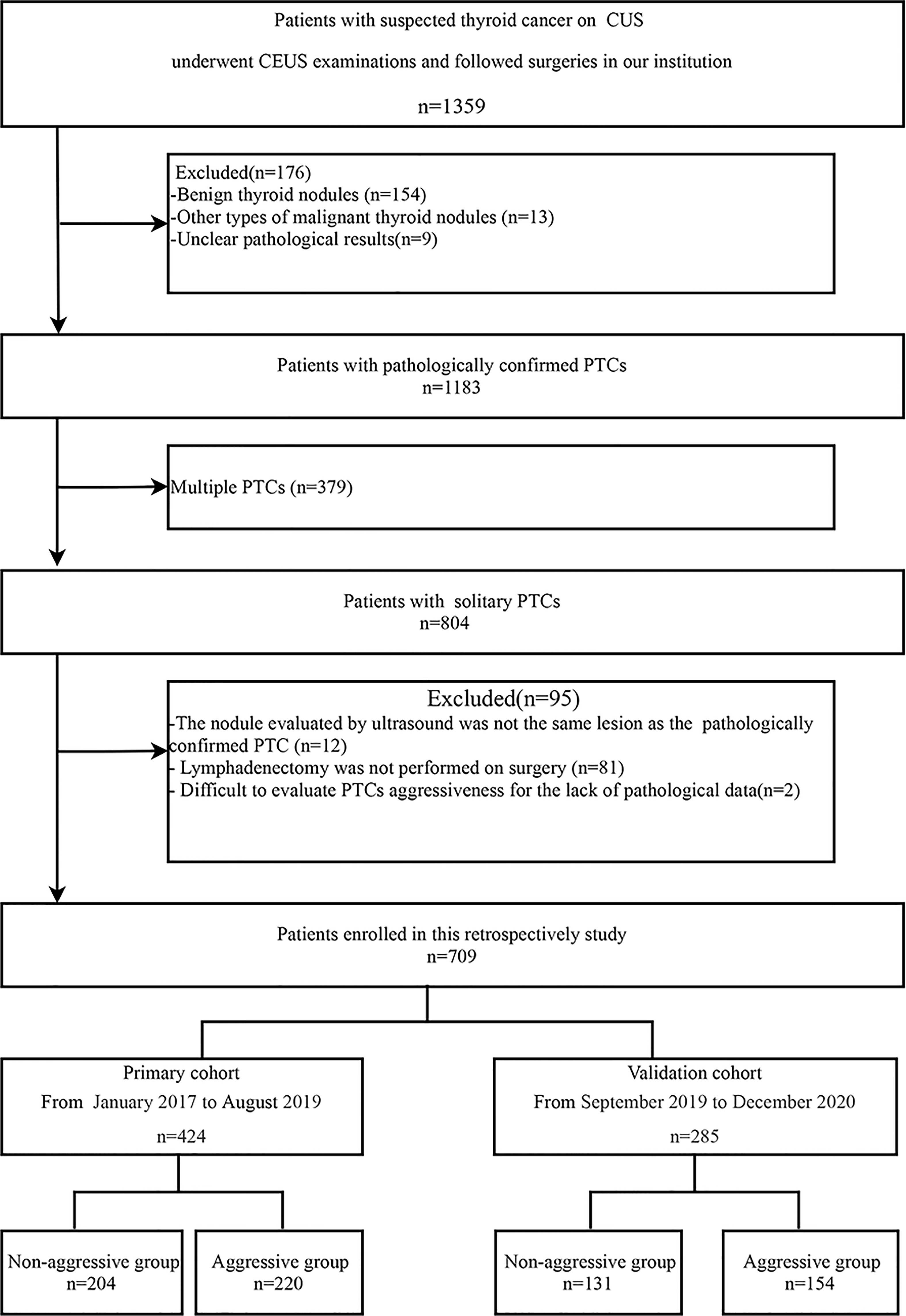
Figure 1 Flow chart showing patient selection process. PTC, papillary thyroid carcinoma; CUS, conventional ultrasound; CEUS, contrast enhanced ultrasound.
Ultrasound examination
Ultrasound examinations were performed using an Aplio 500 (TOSHIBA, Japan) system equipped with a 7-14MHz linear probe and an Aplio i900 (Canon, Japan) system equipped with a 5-18MHZ probe. CEUS was performed using a low mechanical index mode (mechanical indices were 0.12 in the Aplio 500 and 0.04 in the Aplio i900).
On CUS, the following features of PTCs were evaluated: 1) size (≤10 mm or >10 mm); 2) margin (well defined or poorly defined); 3) shape (regular or irregular); 4) aspect ratio (height divided by width on transverse views); 5) calcification (no calcification, microcalcification, macrocalcification, or mixed micro- and macrocalcification); 6) capsule abutment (CA; defined as tumor located adjacent to the capsule with no intervening normal thyroid tissue); 7) loss of echogenic capsule (defined as absence of the normally detected echogenic rim of the thyroid); 8) suspected cervical lymph node metastasis (SCLNM; indicated by loss of the fatty hilum, peripheral vascular flow on color Doppler imaging, increased cortical echogenicity, and a rounded shape [long axis to short axis ratio of <1.5], cystic changes, and microcalcification); and 9) tumor vascularity (classified as types I, II, III, or IV (26)).
On CEUS images, the following features were evaluated: 1) time to tumor enhancement, which was classified as early enhancement (i.e., tumor enhancement before enhancement of surrounding thyroid parenchyma), late enhancement (tumor enhancement after enhancement of surrounding thyroid parenchyma), or simultaneous enhancement (simultaneous enhancement of tumor and surrounding thyroid parenchyma); 2) enhancement intensity of tumor relative to enhancement intensity of surrounding thyroid parenchyma at peak enhancement (classified as hyper-enhancement, iso-enhancement, or hypo-enhancement); 3) homogeneity of tumor enhancement (homogeneous or heterogeneous); 4) discontinuous capsule enhancement (discontinuity in the hyperechoic thyroid capsule).
Image analysis
Two radiologists (each with more than 5 years’ experience in thyroid US and CEUS) blinded to the pathological results evaluated the CUS and CEUS images independently; both radiologists were not otherwise involved in this study. Discrepancies were resolved by consensus or by consultation with a senior radiologist (with more than 20 years’ experience in thyroid CUS and 10 years’ experience in thyroid CEUS). Inter-reader agreement was assessed.
Pathological evaluation
The tumors were classified as aggressive or non-aggressive based on pathologic findings, radiological results, or nuclear imaging findings. Aggressive PTCs were those with one or more of the following: CAI, ETE, lymphatic or vascular invasion, CLNM, and distant metastases. Non-aggressive tumors were those with none of the above features. Pathological indicators were evaluated by pathologists with 10 years of experience in diagnosis of thyroid pathology.
Risk factor identification and nomogram development
First, in the primary cohort, univariate analysis was used to identify the variables significantly associated (at P < 0.05) with aggressive behavior. The identified variables were then entered into multivariable binary logistic regression analysis to identify those independently associated with aggressive behavior. The identified independent risk factors were used to construct the predictive nomogram.
Statistical analysis
Statistical analysis was performed with SPSS 23.0 (IBM Corp., Armonk, NY, USA) and the rms, pROC, and ResourceSelection statistical packages in R 4.1.0 (The R Foundation for Statistical Computing, Vienna, Austria). Quantitative data were expressed as means ± standard deviation or medians (with upper and lower quartiles) according to the normality of the distribution and compared between groups using the t test or the nonparametric Mann–Whitney U test, respectively. Categorical data were summarized as percentages and compared between groups using the Pearson chi-square test or the Fisher exact test. Cohen’s κ analysis was used to evaluate inter-reader agreement.
The performance of the nomogram was assessed by evaluating discrimination and calibration. Receiver operating characteristic curve (ROC) analysis was used to evaluate the discrimination performance of the nomogram, and the area under the curve (AUC) was calculated. Calibration curves were plotted via bootstrapping with 1000 resamples. Finally, the performance of the model was tested in the validation cohort. The Hosmer–Lemeshow test was used to evaluate the goodness of fit of the nomogram. The variance inflation factor was used to evaluate the collinearity of variables included in the nomogram. A variance inflation factors greater than 10 indicates multicollinearity. All statistical tests were two-tailed, and P < 0.05 was considered to indicate a statistically significant difference. The code for model development and evaluation is displayed in the Appendix.
Results
Patient characteristics
A total of 709 patients were included in the study: 180 men (mean age, 42.7 years; age range, 18-80 years) and 529 women (mean age, 44.4 years; age range, 19-79 years). While 424 patients formed the primary cohort, the other 285 patients formed the validation cohort. In the primary cohort, there were 204 patients in the non-aggressive group and 220 in the aggressive group. In the validation cohort, there were 131 patients in the non-aggressive group and 154 patients in the aggressive group. Table 1 shows the baseline characteristics of the patients.
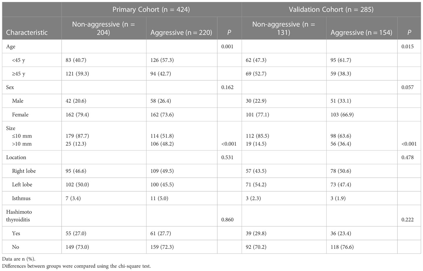
Table 1 Baseline characteristics of patients in primary and validation cohorts.
In both the primary cohort and the validation cohort, patients in the aggressive group tended to be younger and to have larger tumor size than patients in the non-aggressive group (all P < 0.05). Sex ratio, tumor location, and prevalence of Hashimoto thyroiditis were comparable between the aggressive group and the non-aggressive group in both the primary cohort and the validation cohort (all P > 0.05).
Ultrasound features in the primary cohort
In the primary cohort, tumor shape was not significantly different between the non-aggressive group and the aggressive group (P = 0.125), but all other indices (i.e., margin, aspect ratio, calcification, CA, loss of echogenic capsule, SCLNM, vascularity, time to enhancement, intensity of enhancement, homogeneity of enhancement, and discontinuous capsule enhancement) were significantly different between the two groups (all P < 0.05; Table 2).
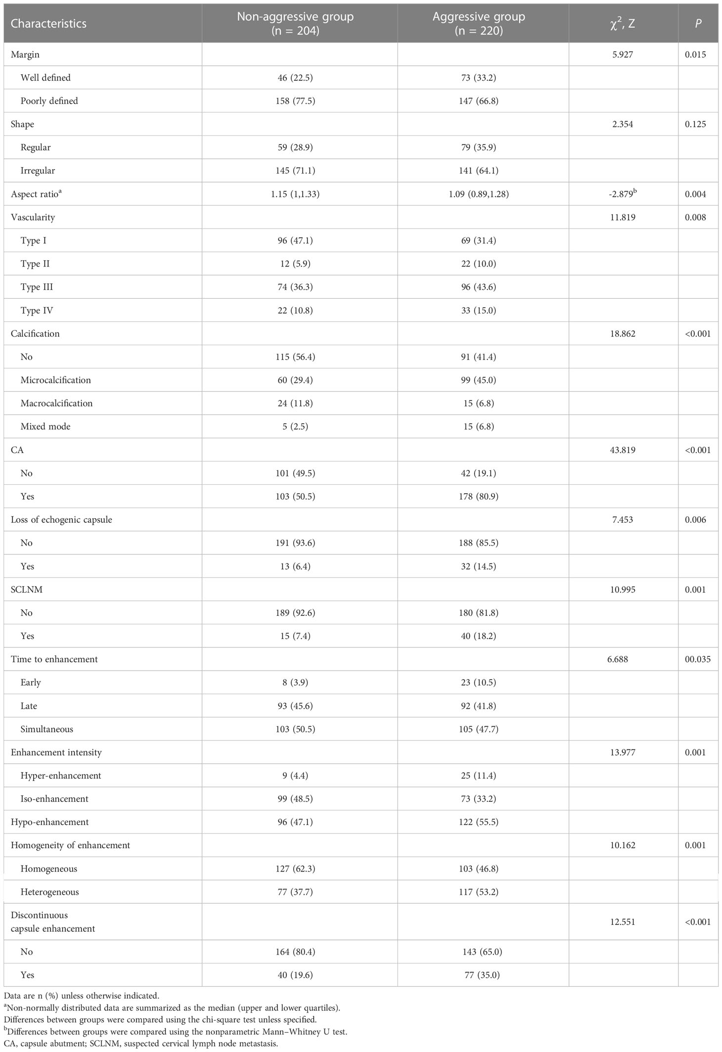
Table 2 Comparison of ultrasound features between the non-aggressive group and aggressive group in the primary cohort.
Multivariate logistic regression analysis and development of the nomogram
Table 3 presents the results of multivariate logistic regression analysis. Age <45 years (OR, 2.08; 95% CI, 1.30-3.35), tumor size > 10mm (OR, 4.00; 95% CI, 2.17-7.37), CA (OR, 2.53, 95% CI, 1.50-4.26), and SCLNM (OR, 2.50; 95% CI, 1.20-5.21) were the variables independently associated with PTC aggressiveness in the primary cohort (all P < 0.05). These four variables were used to construct the predictive nomogram (Figure 2).
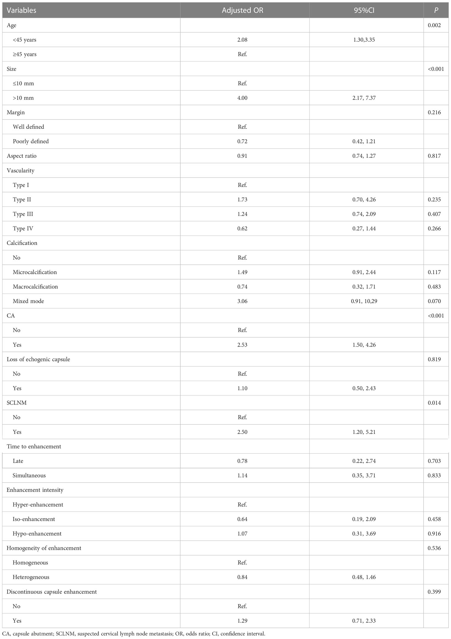
Table 3 Multivariable logistic regression analysis in the primary cohort.
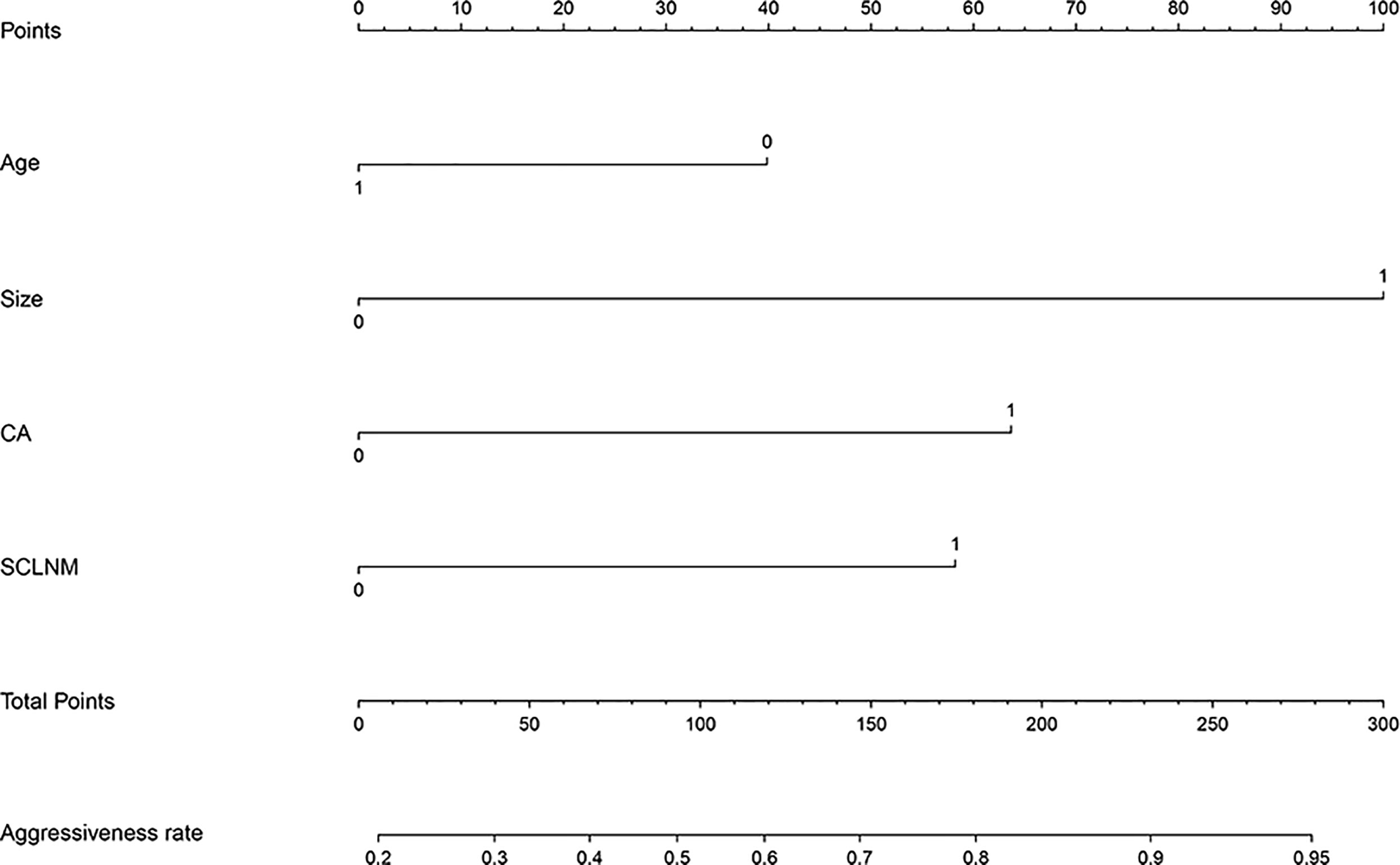
Figure 2 Nomogram to predict aggressiveness of papillary thyroid carcinoma (PTC). The nomogram incorporated patient age, tumor size, capsule abutment (CA), and suspected cervical lymph node metastasis (SCLNM).
Performance of the nomogram in the primary cohort and validation cohort
In ROC analysis, the optimum cutoff value was identified as 0.5. In the primary cohort, the AUC of the nomogram was 0.77 (95% CI, 0.72-0.81) and the sensitivity and specificity were 71.8% and 68.1%, respectively. The Hosmer-Lemeshow test showed good calibration (P = 0.967). The favorable discrimination and calibration were confirmed in the validation cohort, in which the AUC was 0.72 (95% CI, 0.66-0.78) and the sensitivity and specificity were 71.4% and 62.6%, respectively; the Hosmer–Lemeshow test yielded a P value of 0.251 (Figure 3).
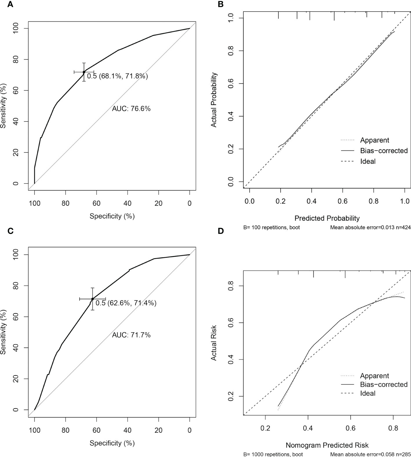
Figure 3 Performance of nomogram for predicting aggressiveness of papillary thyroid carcinoma. In the primary cohort, the performance of the predictive nomogram was evaluated by receiver operating curve analysis (A) and the calibration curve (B). In validation cohort, the model was evaluated by the discrimination (C) and calibration (D) curves.
Inter-reader agreement of CUS and CEUS features
Margin, vascularity, CA, loss of echogenic capsule, SCLNM, time to enhancement, enhancement intensity, and discontinuous capsule enhancement showed substantial inter-reader agreement (κ coefficients = 0.774, 0.667, 0.773, 0.792, 0.697, 0.741, and 0.711, respectively), and shape, calcification and the homogeneity of enhancement showed almost perfect inter-reader agreement (κ coefficients =0.821, 0.907, and 0.878, respectively).
Examples of using the nomogram to predict PTC aggressiveness
The predicted risk of aggressiveness in patient 1 (age, >45 years; size, <10 mm, CA) was less than 0.4. Pathological assessments revealed a non-invasive PTC (Figure 4). For patient 2 (age, <45 years; size >10 mm, CA, SCLNM), the predicted probability of aggressive PTC using the nomogram was greater than 90%, and the tumor was confirmed as an aggressive PTC by pathology (Figure 5).
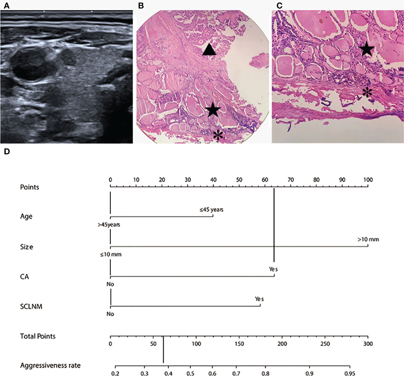
Figure 4 Example of nomogram usage for predicting non-aggressive papillary thyroid carcinoma. In patient 1 (male, 47 years old), preoperative fine needle aspiration cytology confirmed papillary thyroid carcinoma in the right middle part of the thyroid. (A) Conventional ultrasound showed the size was 4.3 mm, with capsule abutment (CA) and no suspected cervical lymph node metastasis (SCLNM). Pathological assessment revealed no capsular invasion. (B) and (C) showed normal thyroid tissue (pentagram) between papillary thyroid carcinoma (triangle) and the capsular (star) at different magnifications (HE staining, 100 ×and 200×, respectively). On the nomogram (D), this patient showed a total score of approximately 62 and the predicted risk of aggressiveness was less than 0.4.
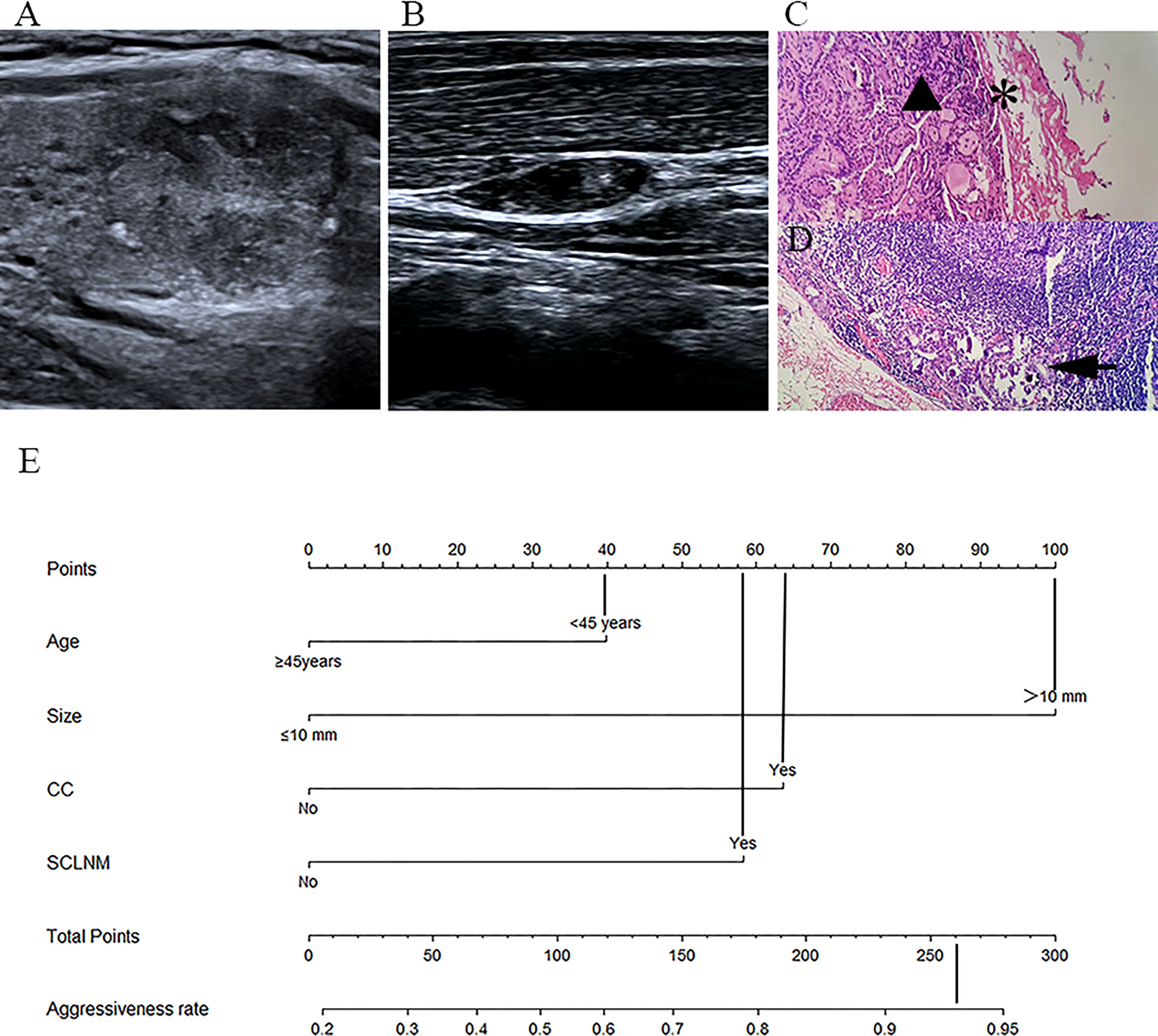
Figure 5 Example of nomogram usage for predicting aggressive papillary thyroid carcinoma. In patient 2 (female, 24 years old), preoperative fine needle aspiration cytology confirmed papillary thyroid carcinoma in the right lower part of the thyroid. (A) Conventional ultrasound showed that the tumor size was 17.8 mm, with capsule abutment (CA). (B) Conventional ultrasound on the right lateral neck level IV indicated suspected cervical lymph node metastasis (SCLNM) with loss of the fatty hilum and microcalcifications. Postoperative pathological assessments (C, D) revealed PTC (triangular) infiltrating the thyroid capsule (stellate) and metastasis (arrow) of the ipsilateral lateral neck lymph nodes (HE staining, 400×). On the nomogram (E), the total score was approximately 260, and the predicted probability of aggressiveness was greater than 0.9.
Discussion
This study aimed to identify the factors associated with aggressiveness of PTC and to use these to construct a predictive nomogram for use in the clinic. We identified four variables—tumor size, patient age, CA, and SCLNM—that were independently associated with PTC aggressiveness. The nomogram constructed using these four variables showed good performance in external validation. Previous predictive models have included only one of the risk factors (e.g., ETE, CLNM) (27–30); our model, incorporating four predictive factors, will enable more accurate stratification of PTC according to aggressiveness. We tried to include CEUS indicators in this model; however, we found no significant differences in CEUS indices between non-aggressive and aggressive groups.
Correct evaluation of PTC aggressiveness is essential for treatment planning. Currently, ATA guidelines recommend active surveillance (AS) as an option in carefully selected patients, the aim being to avoid potential overtreatment while still identifying tumors that would continue to progress (11, 31). Reports from different countries have shown favorable outcomes with AS, but 1%-2% of patients will have progressive tumor growth and ETE (32–34). Therefore, more stringent selection of patients suitable for AS is needed. Stratification of PTC by aggressiveness is especially important for treatment selection in PTC patients with low recurrence risk. Current ATA guidelines recommend lateral lobectomy rather than total thyroidectomy for PTC patients with unifocal tumor of 1-4 cm size and no clinical evidence of lymph node metastasis and DM (11). However, the incidence of PTC recurrence in patients undergoing lobectomy is higher than that in patients undergoing total thyroidectomy, so some patients may have to undergo total thyroidectomy after the initial partial thyroidectomy (35). Thus, careful selection of the surgical approach is necessary for management of low-risk patients.
In this study, tumor size was the strongest independent predictor of PTC aggressiveness. Tumor size is an important prognostic indicator for PTC patients in clinical practice and is incorporated in the American Joint Committee on Cancer (AJCC) TNM staging system and other grading systems such as AGES, AMES, and MACIS. In the studies by de Castro et al. (36) and Sorrenti et al. (37), tumor size was associated with persistence and recurrence of PTC. In our study, tumors >10 mm were more likely to exhibit aggressive characteristics than tumors ≤10 mm. This is consistent with previous reports showing higher incidence of CLNM in patients with large PTC (>1 cm) than in patients with papillary microcarcinoma (38, 39). Lee et al. (40) reported that ETE is more commonly associated with larger PTCs (>10 mm) than with papillary microcarcinomas. Khan et al. (41) found that tumor size >2 cm was an independent risk factor for DM in patients with well-differentiated thyroid cancer. No patient in our study had DM, but our results confirmed that larger PTCs (>1 cm) were more likely to be aggressive. The results of this study suggest that clinicians could consider more conservative management for papillary thyroid microcarcinoma. This conclusion is consistent with the 2015 ATA recommendation of AS for “very low risk” PTC ≤1 cm in size.
CA was found to be an independent predictor of PTC aggressiveness in the present study. Intrathyroidal PTC could be considered to have a relatively lower risk of aggressive behavior. Seong et al. (42) reported that smaller distance between the PTC and the thyroid capsule is associated with higher incidence of CAI and ETE. Lee et al. (40) analyzed the degree of CA and found that increasing the CA ration could predict the ETE. In this study, we did not carry out quantitative analysis of CA parameters; we only defined CA as present or absent. This classification method helps improve the sensitivity of prediction of PTC invasiveness, because this can include those PTCs with only CAI on pathology. The results of this study suggest that PTC patients with CA should be managed more aggressively, while patients with PTC limited to the gland may be considered for more conservative treatment.
We found SCLNM on preoperative CUS to be another independent predictor of aggressiveness of PTC. PTC patients with SCLNM detected by preoperative ultrasound have greater risk of CLNM, which also implies greater risk of aggressiveness. Ultrasound examination of the neck can accurately identify lateral CLNM. Al-Hilli et al. (43) reported that ultrasound had sensitivity, positive predictive value, and accuracy of 78%, 89%, and 75%, respectively, for diagnosis of SCLNM. In addition, for PTC >2 cm in size, the accuracy of identification of multiple CLNM has been found to be as high as 89%. Wu et al. (44) established a machine-learning algorithm to predict central CLNM of PTC and found that SCLNM was an independent predictor of central CLNM. In another study, SCLNM was found to be associated with lymphatic vessel infiltration in the postoperative period (45). Thus, the results of this study suggest that SCLNM before surgery could be considered an indication for lateral neck lymph node resection in PTC patients.
Patient age was another independent predictor of PTC aggressiveness in our study. Aggressive PTC tended to be more common in patients aged <45 years than in patients >45 years. Our result was consistent with the report of Xu et al. (46). Tuttle et al. (33) also found that younger age at diagnosis was independently associated with the likelihood of tumor growth (hazard ratio per year, 0.92). Although PTC patients aged >45 years have less aggressive tumors, in the AJCC staging system for PTC, older age indicates a worse prognosis. This contradiction may be explained by the difference between the aggressiveness and patients’ outcome during tumor progression. In younger patients, the aggressive tumor may grow rapidly, but prognosis remains excellent even if metastasis occurs (47–49); meanwhile, in older PTC patients, less aggressive tumors will be associated with poor prognosis. The results of this study indicate that more aggressive management should be applied in young patients (< 45 years).
This study attempted to construct a nomogram to predict the risk of invasiveness of solitary PTC, providing a new method for clinicians dealing with solitary PTC and may provide a reference for screening appropriate patients for AS. In fact, multiple PTCs are very common in clinical practice, and multifocality in thyroid cancer is a significant risk factor for disease progression and increases the risk of disease recurrence. Patients with multiple PTC are more likely to have ETE and CLNM (50, 51), and the use of AS for multiple PTC is still controversial (52, 53). DM of PTC is uncommon, but is associated with PTC aggressiveness, and DM in thyroid cancer significantly reduces survival in PTC patients (41, 54, 55). On the basis of the guidelines of the American Thyroid Association, DM-PTC is considered to indicate a high risk and usually requires more aggressive treatments (11). Hence, more studies involving DM-PTCs should be carried out in the future. In this study, no statistically significant difference was found in the distribution of Hashimoto thyroiditis between the aggressive and non-aggressive groups. This result is consistent with Cappellacci et al. (56), who found that, although Hashimoto thyroiditis was an independent predictor of differentiated thyroid cancer, the distribution of Hashimoto thyroiditis was similar in patient groups with different aggressive features (e.g., gross ETE, microscopic ETE, vascular invasion, aggressive PTC variants, and extensive CLNM).
This study has several limitations. First, this was a single-center study with a small sample size; our results need to be confirmed in large multi-center studies. Second, no patient had DM in our study; the clinical and ultrasound characteristics of these rare cases need further study. Third, this study only considered solitary PTCs; the findings may not be applicable in patients with multiple PTCs.
Conclusion
The nomogram constructed using these four preoperative indices appears to reliably predict the risk of aggressiveness of solitary PTC. The nomogram might be helpful for management strategy selection in patients with PTC.
Data availability statement
The raw data supporting the conclusions of this article will be made available by the authors, without undue reservation.
Ethics statement
The studies involving human participants were reviewed and approved by The ethics committee of Shanghai General Hospital. Written informed consent for participation was not required for this study in accordance with the national legislation and the institutional requirements.
Author contributions
LL and RW conceived the idea and designed the whole study. LL, CJ, GL participated in the data collection and image analysis. LL performed the statistical analysis. LL participated in manuscript preparation. QS, LD participated in methodology support. All authors contributed to the article and approved the submitted version.
Funding
This work was supported by the National Natural Science Foundation of China (Grants No. 82071931), the interdisciplinary program of Shanghai Jiaotong university(ZH2018ZDA17), the program from Science and Technology Commission of Shanghai Municipality (No. 20Y11912400), and the 2019 clinical research innovation team of Shanghai General Hospital (No. CTCCR-2019B05).
Conflict of interest
The authors declare that the research was conducted in the absence of any commercial or financial relationships that could be construed as a potential conflict of interest.
Publisher’s note
All claims expressed in this article are solely those of the authors and do not necessarily represent those of their affiliated organizations, or those of the publisher, the editors and the reviewers. Any product that may be evaluated in this article, or claim that may be made by its manufacturer, is not guaranteed or endorsed by the publisher.
References
1. Dralle H, Machens A, Basa J, Fatourechi V, Franceschi S, Hay ID, et al. Follicular cell-derived thyroid cancer. Nat Rev Dis Primers (2015) 1:15077. doi: 10.1038/nrdp.2015.77
2. Ito Y, Miyauchi A, Kihara M, Fukushima M, Higashiyama T, Miya A. Overall survival of papillary thyroid carcinoma patients: A single-institution long-term follow-up of 5897 patients. World J Surg (2018) 42(3):615–22. doi: 10.1007/s00268-018-4479-z
3. Milano AF. Thyroid cancer: 20-year comparative mortality and survival analysis of six thyroid cancer histologic subtypes by age, sex, race, stage, cohort entry time-period and disease duration (Seer*Stat 8.3.2) a systematic review of 145,457 cases for diagnosis years 1993-2013. J insurance Med (New York NY) (2018) 47(3):143–58. doi: 10.17849/insm-47-03-143-158.1
4. Youngwirth LM, Adam MA, Scheri RP, Roman SA, Sosa JA. Extrathyroidal extension is associated with compromised survival in patients with thyroid cancer. Thyroid (2017) 27(5):626–31. doi: 10.1089/thy.2016.0132
5. Falvo L, Catania A, D'Andrea V, Marzullo A, Giustiniani MC, De Antoni E. Prognostic importance of histologic vascular invasion in papillary thyroid carcinoma. Ann Surg (2005) 241(4):640–6. doi: 10.1097/01.sla.0000157317.60536.08
6. Wada N, Duh QY, Sugino K, Iwasaki H, Kameyama K, Mimura T, et al. Lymph node metastasis from 259 papillary thyroid microcarcinomas: Frequency, pattern of occurrence and recurrence, and optimal strategy for neck dissection. Ann Surg (2003) 237(3):399–407. doi: 10.1097/01.Sla.0000055273.58908.19
7. Tam S, Boonsripitayanon M, Amit M, Fellman BM, Li Y, Busaidy NL, et al. Survival in differentiated thyroid cancer: Comparing the ajcc cancer staging seventh and eighth editions. Thyroid (2018) 28(10):1301–10. doi: 10.1089/thy.2017.0572
8. Eustatia-Rutten CF, Corssmit EP, Biermasz NR, Pereira AM, Romijn JA, Smit JW. Survival and death causes in differentiated thyroid carcinoma. J Clin Endocrinol Metab (2006) 91(1):313–9. doi: 10.1210/jc.2005-1322
9. Bulfamante AM, Lori E, Bellini MI, Bolis E, Lozza P, Castellani L, et al. Advanced differentiated thyroid cancer: A complex condition needing a tailored approach. Front Oncol (2022) 12:954759. doi: 10.3389/fonc.2022.954759
10. Davies L, Hoang JK. Thyroid cancer in the USA: Current trends and outstanding questions. Lancet Diabetes Endocrinol (2021) 9(1):11–2. doi: 10.1016/s2213-8587(20)30372-7
11. Haugen BR, Alexander EK, Bible KC, Doherty GM, Mandel SJ, Nikiforov YE, et al. 2015 American Thyroid association management guidelines for adult patients with thyroid nodules and differentiated thyroid cancer: The American thyroid association guidelines task force on thyroid nodules and differentiated thyroid cancer. Thyroid (2016) 26(1):1–133. doi: 10.1089/thy.2015.0020
12. Jeong HS, Baek CH, Son YI, Choi JY, Kim HJ, Ko YH, et al. Integrated 18f-fdg Pet/Ct for the initial evaluation of cervical node level of patients with papillary thyroid carcinoma: Comparison with ultrasound and contrast-enhanced ct. Clin Endocrinol (2006) 65(3):402–7. doi: 10.1111/j.1365-2265.2006.02612.x
13. Paudyal R, Lu Y, Hatzoglou V, Moreira A, Stambuk HE, Oh JH, et al. Dynamic contrast-enhanced mri model selection for predicting tumor aggressiveness in papillary thyroid cancers. NMR biomedicine (2020) 33(1):e4166. doi: 10.1002/nbm.4166
14. Hu S, Zhang H, Wang X, Sun Z, Ge Y, Li J, et al. Can diffusion-weighted Mr imaging be used as a tool to predict extrathyroidal extension in papillary thyroid carcinoma? Acad Radiol (2021) 28(4):467–74. doi: 10.1016/j.acra.2020.03.005
15. Song B, Wang H, Chen Y, Liu W, Wei R, Dai Z, et al. Magnetic resonance imaging in the prediction of aggressive histological features in papillary thyroid carcinoma. Medicine (2018) 97(26):e11279. doi: 10.1097/md.0000000000011279
16. Kaplan SL, Mandel SJ, Muller R, Baloch ZW, Thaler ER, Loevner LA. The role of Mr imaging in detecting nodal disease in thyroidectomy patients with rising thyroglobulin levels. AJNR Am J neuroradiology (2009) 30(3):608–12. doi: 10.3174/ajnr.A1405
17. Fresilli D, David E, Pacini P, Del Gaudio G, Dolcetti V, Lucarelli GT, et al. Thyroid nodule characterization: How to assess the malignancy risk. update of the literature. Diagnostics (Basel Switzerland) (2021) 11(8):1374. doi: 10.3390/diagnostics11081374
18. Sorrenti S, Dolcetti V, Radzina M, Bellini MI, Frezza F, Munir K, et al. Artificial intelligence for thyroid nodule characterization: Where are we standing? Cancers (2022) 14(14):3357. doi: 10.3390/cancers14143357
19. Kuo EJ, Thi WJ, Zheng F, Zanocco KA, Livhits MJ, Yeh MW. Individualizing surgery in papillary thyroid carcinoma based on a detailed sonographic assessment of extrathyroidal extension. Thyroid (2017) 27(12):1544–9. doi: 10.1089/thy.2017.0457
20. Jiao WP, Zhang L. Using ultrasonography to evaluate the relationship between capsular invasion or extracapsular extension and lymph node metastasis in papillary thyroid carcinomas. Chin Med J (2017) 130(11):1309–13. doi: 10.4103/0366-6999.206339
21. Zhao H, Li H. Meta-analysis of ultrasound for cervical lymph nodes in papillary thyroid cancer: Diagnosis of central and lateral compartment nodal metastases. Eur J Radiol (2019) 112:14–21. doi: 10.1016/j.ejrad.2019.01.006
22. Hwang HS, Orloff LA. Efficacy of preoperative neck ultrasound in the detection of cervical lymph node metastasis from thyroid cancer. Laryngoscope (2011) 121(3):487–91. doi: 10.1002/lary.21227
23. Zhang Y, Zhang X, Li J, Cai Q, Qiao Z, Luo YK. Contrast-enhanced ultrasound: A valuable modality for extracapsular extension assessment in papillary thyroid cancer. Eur Radiol (2021) 31(7):4568–75. doi: 10.1007/s00330-020-07516-y
24. Chen L, Chen L, Liu J, Wang B, Zhang H. Value of qualitative and quantitative contrast-enhanced ultrasound analysis in preoperative diagnosis of cervical lymph node metastasis from papillary thyroid carcinoma. J ultrasound Med (2020) 39(1):73–81. doi: 10.1002/jum.15074
25. Sorrenti S, Dolcetti V, Fresilli D, Del Gaudio G, Pacini P, Huang P, et al. The role of ceus in the evaluation of thyroid cancer: From diagnosis to local staging. J Clin Med (2021) 10(19):4559. doi: 10.3390/jcm10194559
26. Ivanac G, Brkljacic B, Ivanac K, Huzjan R, Skreb F, Cikara I. Vascularisation of benign and malignant thyroid nodules: Cd us evaluation. Ultraschall der Med (Stuttgart Germany 1980) (2007) 28(5):502–6. doi: 10.1055/s-2007-963023
27. Wang X, Agyekum EA, Ren Y, Zhang J, Zhang Q, Sun H, et al. A radiomic nomogram for the ultrasound-based evaluation of extrathyroidal extension in papillary thyroid carcinoma. Front Oncol (2021) 11:625646. doi: 10.3389/fonc.2021.625646
28. Tian X, Song Q, Xie F, Ren L, Zhang Y, Tang J, et al. Papillary thyroid carcinoma: An ultrasound-based nomogram improves the prediction of lymph node metastases in the central compartment. Eur Radiol (2020) 30(11):5881–93. doi: 10.1007/s00330-020-06906-6
29. Jiang M, Li C, Tang S, Lv W, Yi A, Wang B, et al. Nomogram based on shear-wave elastography radiomics can improve preoperative cervical lymph node staging for papillary thyroid carcinoma. Thyroid (2020) 30(6):885–97. doi: 10.1089/thy.2019.0780
30. Yu J, Deng Y, Liu T, Zhou J, Jia X, Xiao T, et al. Lymph node metastasis prediction of papillary thyroid carcinoma based on transfer learning radiomics. Nat Commun (2020) 11(1):4807. doi: 10.1038/s41467-020-18497-3
31. Scharpf J. Achieving active surveillance for thyroid cancer-not a euphemism for watching a ticking time bomb. JAMA otolaryngology– Head Neck Surg (2017) 143(10):1021–2. doi: 10.1001/jamaoto.2017.1453
32. Oh HS, Ha J, Kim HI, Kim TH, Kim WG, Lim DJ, et al. Active surveillance of low-risk papillary thyroid microcarcinoma: A multi-center cohort study in Korea. Thyroid (2018) 28(12):1587–94. doi: 10.1089/thy.2018.0263
33. Tuttle RM, Fagin JA, Minkowitz G, Wong RJ, Roman B, Patel S, et al. Natural history and tumor volume kinetics of papillary thyroid cancers during active surveillance. JAMA otolaryngology– Head Neck Surg (2017) 143(10):1015–20. doi: 10.1001/jamaoto.2017.1442
34. van Gerwen M, Alpert N, Sinclair C, Kale M, Genden E, Taioli E. Assessing non-aggressiveness of untreated, local and regional, papillary thyroid cancer. Oral Oncol (2020) 105:104674. doi: 10.1016/j.oraloncology.2020.104674
35. Choi JB, Lee SG, Kim MJ, Kim TH, Ban EJ, Lee CR, et al. Oncologic outcomes in patients with 1-Cm to 4-Cm differentiated thyroid carcinoma according to extent of thyroidectomy. Head Neck (2019) 41(1):56–63. doi: 10.1002/hed.25356
36. de Castro TP, Waissmann W, Simões TC, de Mello RC, Carvalho DP. Predictors for papillary thyroid cancer persistence and recurrence: A retrospective analysis with a 10-year follow-up cohort study. Clin Endocrinol (2016) 85(3):466–74. doi: 10.1111/cen.13032
37. Sorrenti S, Carbotta G, Di Matteo FM, Catania A, Pironi D, Tartaglia F, et al. Evaluation of clinicopathological and molecular parameters on disease recurrence of papillary thyroid cancer patient: A retrospective observational study. Cancers (2020) 12(12):3637. doi: 10.3390/cancers14143357
38. So YK, Kim MJ, Kim S, Son YI. Lateral lymph node metastasis in papillary thyroid carcinoma: A systematic review and meta-analysis for prevalence, risk factors, and location. Int J Surg (London England) (2018) 50:94–103. doi: 10.1016/j.ijsu.2017.12.029
39. Zhao H, Huang T, Li H. Risk factors for skip metastasis and lateral lymph node metastasis of papillary thyroid cancer. Surgery (2019) 166(1):55–60. doi: 10.1016/j.surg.2019.01.025
40. Lee CY, Kim SJ, Ko KR, Chung KW, Lee JH. Predictive factors for extrathyroidal extension of papillary thyroid carcinoma based on preoperative sonography. J ultrasound Med (2014) 33(2):231–8. doi: 10.7863/ultra.33.2.231
41. Khan U, Al Afif A, Aldaihani A, MacKay C, Rigby MH, Rajaraman M, et al. Patient and tumor factors contributing to distant metastasis in well-differentiated thyroid cancer: A retrospective cohort study. J Otolaryngol - Head Neck Surg = Le J d'oto-rhino-laryngologie chirurgie cervico-faciale (2020) 49(1):78. doi: 10.1186/s40463-020-00469-8
42. Seong CY, Chai YJ, Lee SM, Kim SJ, Choi JY, Lee KE, et al. Significance of distance between tumor and thyroid capsule as an indicator for central lymph node metastasis in clinically node negative papillary thyroid carcinoma patients. PloS One (2018) 13(7):e0200166. doi: 10.1371/journal.pone.0200166
43. Al-Hilli Z, Strajina V, McKenzie TJ, Thompson GB, Farley DR, Richards ML. The role of lateral neck ultrasound in detecting single or multiple lymph nodes in papillary thyroid cancer. Am J Surg (2016) 212(6):1147–53. doi: 10.1016/j.amjsurg.2016.09.014
44. Wu Y, Rao K, Liu J, Han C, Gong L, Chong Y, et al. Machine learning algorithms for the prediction of central lymph node metastasis in patients with papillary thyroid cancer. Front Endocrinol (2020) 11:577537. doi: 10.3389/fendo.2020.577537
45. Sezer A, Celik M, Yilmaz Bulbul B, Can N, Tastekin E, Ayturk S, et al. Relationship between lymphovascular invasion and clinicopathological features of papillary thyroid carcinoma. Bosnian J basic Med Sci (2017) 17(2):144–51. doi: 10.17305/bjbms.2017.1924
46. Xu Y, Xu L, Wang J. Clinical predictors of lymph node metastasis and survival rate in papillary thyroid microcarcinoma: Analysis of 3607 patients at a single institution. J Surg Res (2018) 221:128–34. doi: 10.1016/j.jss.2017.08.007
47. Ito Y, Miyauchi A, Kihara M, Takamura Y, Kobayashi K, Miya A. Relationship between prognosis of papillary thyroid carcinoma patient and age: A retrospective single-institution study. Endocrine J (2012) 59(5):399–405. doi: 10.1507/endocrj.ej12-0044
48. Huang H, Wu L, Liu W, Liu J, Liu Y, Xu Z. Long-term outcomes of patients with papillary thyroid cancer who did not undergo prophylactic central neck dissection. J Cancer Res Ther (2020) 16(5):1077–81. doi: 10.4103/jcrt.JCRT_620_19
49. Ito Y, Miyauchi A, Ito M, Yabuta T, Masuoka H, Higashiyama T, et al. Prognosis and prognostic factors of differentiated thyroid carcinoma after the appearance of metastasis refractory to radioactive iodine therapy. Endocrine J (2014) 61(8):821–4. doi: 10.1507/endocrj.ej14-0181
50. Joseph KR, Edirimanne S, Eslick GD. Multifocality as a prognostic factor in thyroid cancer: A meta-analysis. Int J Surg (London England) (2018) 50:121–5. doi: 10.1016/j.ijsu.2017.12.035
51. HîȘu L, Ștefan PA, Piciu D. Total tumor diameter and unilateral multifocality as independent predictor factors for metastatic papillary thyroid microcarcinoma. J Clin Med (2021) 10(16):3707. doi: 10.3390/jcm10163707
52. Nagaoka R, Ebina A, Toda K, Jikuzono T, Saitou M, Sen M, et al. Multifocality and progression of papillary thyroid microcarcinoma during active surveillance. World J Surg (2021) 45(9):2769–76. doi: 10.1007/s00268-021-06185-2
53. Brito JP, Ito Y, Miyauchi A, Tuttle RM. A clinical framework to facilitate risk stratification when considering an active surveillance alternative to immediate biopsy and surgery in papillary microcarcinoma. Thyroid (2016) 26(1):144–9. doi: 10.1089/thy.2015.0178
54. Sugitani I, Fujimoto Y, Yamamoto N. Papillary thyroid carcinoma with distant metastases: Survival predictors and the importance of local control. Surgery (2008) 143(1):35–42. doi: 10.1016/j.surg.2007.06.011
55. Wang LY, Palmer FL, Nixon IJ, Thomas D, Patel SG, Shaha AR, et al. Multi-organ distant metastases confer worse disease-specific survival in differentiated thyroid cancer. Thyroid (2014) 24(11):1594–9. doi: 10.1089/thy.2014.0173
Keywords: papillary thyroid carcinoma, thyroid, ultrasound, nomogram, aggressiveness
Citation: Liu L, Jia C, Li G, Shi Q, Du L and Wu R (2023) Nomogram incorporating preoperative clinical and ultrasound indicators to predict aggressiveness of solitary papillary thyroid carcinoma. Front. Oncol. 13:1009958. doi: 10.3389/fonc.2023.1009958
Received: 02 August 2022; Accepted: 16 January 2023;
Published: 31 January 2023.
Edited by:
Salvatore Sorrenti, Department of Surgical Sciences, Sapienza University of Rome, ItalyReviewed by:
Jinhua Yu, Fudan University, ChinaEleonora Lori, Sapienza University of Rome, Italy
Pietro Giorgio Calo’, University of Cagliari, Italy
Alessandro Sanguinetti, Università degli Studi Perugia, Italy
Copyright © 2023 Liu, Jia, Li, Shi, Du and Wu. This is an open-access article distributed under the terms of the Creative Commons Attribution License (CC BY). The use, distribution or reproduction in other forums is permitted, provided the original author(s) and the copyright owner(s) are credited and that the original publication in this journal is cited, in accordance with accepted academic practice. No use, distribution or reproduction is permitted which does not comply with these terms.
*Correspondence: Rong Wu, d3Vyb25nNzExMUAxNjMuY29t