 Yohann Dabi1,2*
 Amelia Favier1,2Léo Razakamanantsoa3,4
 Stéphane Suisse5Yannick Marie6Cyril Touboul1,2
 Clément Ferrier1,2Sofiane Bendifallah1,2Emile Daraï1,2
- 1Sorbonne University, Department of Obstetrics and Reproductive Medicine, Hôpital Tenon, Paris, France
- 2Clinical Research Group (GRC) Paris 6: Centre Expert Endométriose (C3E), Sorbonne University (GRC6 C3E SU), Paris, France
- 3Sorbonne University, Inserm UMR S 938, Centre de recherche de saint Antoine (CRSA), Hôpital Saint Antoine, Paris, France
- 4Department of Radiology imaging and Interventional speciality imaging, Tenon Hospital, Paris, France
- 5Ziwig, Lyon, France
- 6Gentoyping and Sequencing core facility, iGenSeq, Institut du Cerveau et de la Moelle épinière, Institut du Cerveau et de la Moelle (ICM), Hôpital Pitié-Salpêtrière, Paris, France
Cervical cancer (CC) is the fourth cancer in women and is the leading cause of cancer death in 42 countries. Lymph node metastasis is a determinant prognostic factor, as underlined in the latest FIGO classification. However, assessment of lymph node status remains difficult, despite the progress of imaging such as PET-CT and MRI. In the specific setting of CC, all data underlined the need for new biomarkers easily available to assess lymph node status. Previous studies have underlined the potential value of ncRNA expression in gynecological cancers. In this review, we aimed to evaluate the contribution of ncRNAs in tissue and biofluid samples to determine lymph node status in CC with potential impact on both surgical and adjuvant therapies. In tissue samples, our analysis found that there are arguments to support the role of ncRNAs in physiopathology, differential diagnosis from normal tissue, preinvasive and invasive tumors. In biofluids, despite small studies especially concerning miRNAs expression, promising data opens up new avenue to establish a non-invasive signature for lymph node status as well as a tool to predict response to neo- and adjuvant therapies, thus improving management algorithm of patients with CC.
Introduction
Cervical cancer (CC) is the fourth cancer in women and is the leading cause of cancer death in 42 countries (1). World Health Organization (WHO) estimated that 570 000 new cases are diagnosed per year causing 311 000 deaths in 2018, with 90% of them observed in low-income and middle-income countries (1, 2). CC is linked to infection by high-risk human papillomavirus (HPV) genotypes but almost 90% of incident HPV infections are cleared within the two years after initial infection and persist only in 10% of women (3–5). However, a debate persists on whether the virus is completely cleared from basal cells or remains latent with potential reactivation. Moreover, it is important to note that the development of cancer takes 10 to 30 years from preinvasive to invasive tumor underlining the crucial role of prevention policies (6). Primary policy is based on HPV vaccination launched in 2006 with three HPV vaccines currently available (bi, quadri, and nonavalent) highly efficient against high-risk HPV 16/18 and 31/33/45/52/58 genotypes causing 70% and 18.5% of all CC, respectively (5). Secondary prevention consists in the detection and treatment of cervical intra-epithelial neoplasia (CIN). This screening is based on Pap smears at regular intervals imposing substantial medical resources while exhibiting a poor sensitivity, and lack in quality assurance results (7). In contrast HPV-based screening has high sensitivity and accuracy and better reproducibility explaining that most of national and international guidelines recommend primary HPV-based screening over conventional Pap smears (8).
Since the 2018 revised classification of the International Federation of Gynecology and Obstetrics (FIGO), imaging and pathological findings have been incorporated in CC staging (Table 1) (4). The FIGO stage is directly corelated to prognosis ranging from almost 100% 5-year disease-free survival rates for stage IA to 5–15% for stage IV. Moreover, FIGO stage allows determining patients’ allocation in different treatment regimens (26). In addition to classic CC prognostic factors including lymphovascular space invasion (LVSI), age, comorbidities (e.g., anemia, HIV infection), high-risk histological subtypes such as adenosquamous and neuroendocrine carcinomas that are easily available on biopsy and conventional imaging, lymph node status remains relatively difficult to evaluate (27–29). This issue is crucial as for early stages the risk of lymph node metastasis (LNM) is approximately 3.7 to 21.7%, and the 5-year overall survival (OS) decreases from 80% to 53% in case of LNM+ (30–33). Therefore, there is a need of adequate tools to assess pelvic and paraaortic LNM not only for early but also for locally advanced CC (IB3-IV stages). Conventional MRI and CT are widely recommended although both exhibited low sensitivity and specificity; 37–71 and 83–93, and 31–58 and 92–97, respectively (34, 35). Despite the use of specific MRI sequences, its low accuracy could be related to size and morphologic criteria used to diagnose LNM. In this specific context, previous studies demonstrated higher relevance of FDG PET-CT and FDG PET-MRI with respective sensitivity and specificity of 34–82 and 93–100, and 83–91 and 90–94 (36). However, PET-CT has also some limits as its ability to detect metastatic lymph nodes is depending on lymph node size with respective sensitivity of 100%, 67%, and 13% for metastatic nodes ≥ 10 mm, 5–9 mm, and ≤ 4 mm (37). Although the rate of lymph node involvement is about 20% even for locally advanced CC, these limits explain why recent ESGO-ESTRO-ESP guidelines recommend paraaortic lymph node dissection in locally advanced CC in patients with negative paraaortic lymph nodes on imaging due to its major impact on therapies (38).
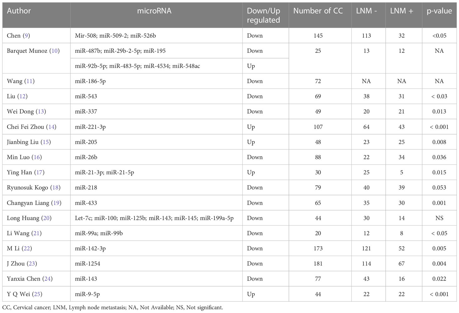
Table 1 Series evaluating the correlation between miRNAs and lymph nodes invasion in patients with cervical cancer.
In the specific setting of CC, all data underlined the need for new biomarkers easily available to assess lymph node status avoiding surgical risks even if para-aortic lymphadenectomy is feasible by mini-invasive surgery.
Numerous studies support that non-coding RNAs (ncRNA) are essential for tumorigenesis by regulating the expression of tumour-related genes. ncRNAs can regulate gene expression primarily by acting as transcription factors, regulating chromatin remodelling or participating in post-transcriptional regulation (39). Moreover, ncRNAs act by guiding DNA synthesis or gene rearrangement, and protecting the genome from foreign nucleic acids (40). In gynaecological cancers, previous studies have underlined the potential value of ncRNA expression in tissue samples to assess prognosis and especially lymph node status in endometrial carcinoma (41). Among ncRNA, miRNAs have been extensively studied showing that they can function as oncogenes and/or tumor suppressor genes depending on the function of their target genes. For CC, although abnormal expression of miRNAs has been demonstrated to be linked to cancer biology, including proliferation, differentiation, apoptosis, migration, invasion, and metastatic angiogenesis (42–49), few reports focused on their relevance to determine lymph node status. Moreover, some studies support that miRNAs emerge as potential tools to differentiate benign from preinvasive and invasive tumours and to determine prognosis (50, 51).
Therefore, the aim of the present review was to evaluate the contribution of ncRNAs in tissue and biofluid samples to determine lymph node status in CC with potential impact on both surgical and medical therapies such as brachy and chemotherapy (52).
ncRNAs
ncRNAs represent about 98% of the transcriptome. Among ncRNAs, arbitrarily those with less than 50 nucleotides are defined as small RNAs (sncRNAs) including microRNAs (miRNAs), Piwi interacting RNAs (piRNAs), transfer RNAs (tRNAs), small nuclear RNAs (snRNAs), and small interfering RNAs (siRNAs) (53, 54). ncRNAs with more than 200 nucleotides are defined as long RNAs (lncRNAs), although an overlapping in nucleotide length exists between snc- and lncRNAs, including intergenic ncRNAs (lincRNAs), some circular RNAs (circRNAs), and ribosomal RNAs (rRNAs) (55).
miRNA
miRNAs are small intracellular RNAs, 18-25 nucleotides long, capable of inducing the extinction (silencing) of gene expression by post-transcriptional regulatory mechanisms, by binding in a targeted manner to the 3’ untranslated parts (3’UTR) mRNAs thus causing translational blockage or degradation (56). However, another mechanism is binding miRNAs to the 5’UTR regions inducing either activation or repression of translation.
miRNA expression in CC tissue samples
In contrast to an abundant literature on miRNAs implicated in the pathogenesis and signaling pathways involved in CC, less data is available allowing correlating their expression with lymph node status (57–60). Moreover, most series focused on one or a limited panel of miRNA mainly using paired tissue samples comparing the expression between CC tissue and adjacent normal appearance tissue (Table 1).
Using The Cancer Genome Atlas (TCGA) database, Chen Q et al, analyzed the expression of 422 miRNAs in 145 patients with early-stage CC (32 LNM+ and 113 LNM−) showing that 75 miRNAs were differentially expressed between the groups (9). After multivariate analysis, miRNA-508, miRNA-509-2, and miRNA-526b were associated with LNM+ status with target genes implicated in the MAPK, cAMP, PI3K/Akt, mTOR, and estrogen cancer signaling pathways. In the same way, using TCGA database on a small series, Y.Q. Wei et al. among 14 miRNAs differentially expressed in CC with LNM+ (miRNA-9-3p, miRNA-191-5p, miRNA-9-5p, miRNA-873-5p, miRNA-378a-3p, miRNA-624-5p, miRNA-149-5p, miRNA-425-5p, miRNA-519a-5p, miRNA-375, miRNA-151b, miRNA-874-3p, miRNA-92b-5p, miRNA-3605-3p) evaluated the relation between miRNA-9-5p and found a significant overexpression (p=0.0002) in patients with LNM+ (25).
Recently, in a small study using microarray, Barquet-Munãoz et al. reported 36 miRNAs differentially expressed between patients with and without LNM including, 17 over-, and 19 underexpressed miRNAs (10). Among them, 10 exhibited high fold change (FC); miRNAs, miR-487b (FC = −3.2, p = 0.0003), miR-194 (FC = −2.8, p = 0.006), miR-34c-5p (FC = −2.46, p = 0.007), miR-29b-2-5p (FC = −2.3, p = 0.007), and miR-195 (FC = −2.07, p = 0.001), miR-548ac (FC = 2.74, p = 0.0003), miR-4534 (FC = 2.47, p = 0.001), miR-483-5p (FC = 2.21, p = 0.002), miR-564 (FC = 2.01, p = 0.006), and miR-92b-5p (FC = 1.82, p = 0.005). This biological miRNA signature allowed to correctly classify 91.6% patients with LNM (11/12) and 92.3% patients without LNM (12/13). However, after qRT-PCR, only seven miRNAs were validated. In a series of 44 CC, Long Huang et al, reported a decrease of a panel of six miRNAs (let-7c, miRNA-100, miRNA-125b, miRNA-143, miRNA-145 and miRNA-199a-5p) correlated with FIGO stage but not with LNM. In a series of 79 CC, R. Kogo et al. reported only a trend for a lower miRNA-218 expression in patients with lymph node metastasis (p=0.053). However, the authors reported a relation between a decrease in miRNA-218 and the occurrence of lymph node recurrence both in pelvic (p=0.032) and para-aortic areas (0.013) (18). In a series of 72 CC vs adjacent normal tissue, A. Wang observed that miR-186-5p was down regulated in patients with LNM+ but with missing data on CC characteristics (11). In a series of 69 CC, Xiaoying Liu et al. found a relation between down-regulation of miRNA-543 and LNM+ (12). Interestingly, we noted that two authors reported a douwnregulation of miRNA-143 correlated with LNM+ justifying further evaluation (20, 24).
miRNA expression in CC biofluids samples
When considering CC onset and progression, the role of HPV has been highlighted demonstrating increased EGFR levels secondary to HPV16E6/E7 and the link with let-7i-5p, miRNA-181a-2-3p (61, 62).
In a series of 182 CC and 12 healthy controls, Wei Jiang et al. noted that low miRNA-101 serum level was associated with LNM+ (p=0.001) (63). Similarly, P. Liu et al. evaluated the serum miRNA-196a by qRT-PCR in 105 CC patients, 86 CIN patients, and 50 healthy controls (64). They demonstrated that serum miRNA-196a levels were higher in CC patients (p< 0.01) and CIN (p< 0.05) compared to healthy controls. Moreover, serum miRNA-196a was associated with LNM+ (p=0.018). Chen-Fei Zhou et al. also demonstrated that miR-221-3p overexpression was correlated with peritumoral lymphangiogenesis and LNM+ status (14). Moreover, they investigated whether miR-221-3p was detected in peripheral plasma of CC patients. In a subpopulation of 40 patients with stage I–II CC (20 with LNM+ and 20 with LNM-) an overexpression of exosomal miRNA-221-3p was observed in patients with LNM+ suggesting its relevance as a potential new biomarker of lymph node status. Comparing CC patients with and without LNM, Zhang Liang et al, observed a low miRNA-378a-3p serum expression in patients LNM+ (p=0.022) (65). Moreover, Qiu et al. have proposed a circulating miRNA signature for the diagnosis and prognosis of early-stage CC based on a series of 112 CC patients, 45 patients with CIN and 90 healthy subjects (66). They found a relation between LNM+ and high serum miRNA-21 associated with low serum miRNA-125b and miRNA-370. Among 17 patients with this signature the rate of LNM+ was 53% while for the 21 patients with simultaneously low serum miRNA-21 and high serum miRNA-125b and high serum miRNA- 370, none exhibited LNM. Although all aforementioned studies confirm the relation between some miRNAs and LNM status, all failed in determining a usable threshold in current practice.
More interesting, in a cohort of 80 patients with FIGO stages I-IIA CC, Zhao et al. (67) analyzed the expression of miRNA-20a and miRNA-203 in serum collected before surgery and treatment. Serum miRNA-20a was higher in patients with LNM+ with an area under the curve (AUC) of 0.734 ± 0.058, sensitivity of 75%, specificity of 72.5%, and a cut-off value of 3.0 as a marker for metastasis. Serum miRNA-203 was less relevant with an AUC of 0.658 ± 0.061, sensitivity of 65%, specificity of 62.5%, and a cut-off value of 0.13 (67). In a series of 100 patients (40 CC patients with LNM+, 40 CC patients with LNM-, and 20 healthy controls), among 89 miRNAs, J. Chen et al. (68) observed that 22 were upregulated and none downregulated. Using qRT-PCR, a panel of five miRNAs (miRNA-1246, miRNA-20a, miRNA-2392, miRNA-3147, miRNA-3162-5p, and miRNA-4484) was predictive of LNM with an AUC of 0.932, sensitivity of 85.6%, and specificity of 85.0%. For miRNA in tissue, the respective AUC, sensitivity and specificity were 0.992, 96.7%, and 95.0%. Moreover, on the same serum samples, the authors evaluated the relevance of SCC antigen as a predictor for lymph node metastasis showing a low relevance compared to the miRNA panel with an AUC of 0.713, sensitivity of 61.2%, and specificity of 70.0%. As previously reported by Zhao et al, these results confirm the relevance of serum miRNA-20a as a potential marker of lymph node status (67).
CircRNA
CircRNA is a class of ncRNAs characterized by covalently closed loop without any 5’-3’ polarity or a polyadenylated tail (69). Thanks to New Generation Sequencing (NGS) and bioinformatic, more circRNAs have been identified (70) demonstrating their implication in both physiological and pathological processes such tumor cell proliferation, invasion and metastasis in various cancers (71). CircRNA regulate gene expression mainly as ceRNAs at various levels; epigenetic, transcriptional and post-transcriptional levels of protein-coding mRNAs (72–74).
A recent preliminary study in three HPV16 positive cervical cancer cases identified 99 deregulated circRNAs (58 over expressed and 41 under expressed circRNAs) (75). However, the data are too scant to evaluate the interaction between HPV and circ-RNAs in CC.
Using GSE30656 and TCGA, among 156 miRNAs, 5,321 mRNAs, and 75 circRNAs, Yuexiong Yi et al. identified regulatory circRNA-miRNA-mRNA networks with 7 hubgenes (RRM2, CEP55, CHEK1, KIF23, RACGAP1, ATAD2 and KIF11) implicated in CC (76). These hubgenes network including 5 circRNAs (circRNA-000596, circRNA-104315, circRNA-400068, circRNA-101958 and circRNA-103519) play a crucial role in chemosensitivity. In contrast to miRNAs expression in CC, little is known on the relation between circRNA expression and LNM (77). In a series of 68 CC, Huang Ma et al. noted an upregulation of circ-0005576 associated with LNM+ by sponging miR-153-3p (78). Recently, Tie-Fang Song et al. reported that upregulated circRNA-101996, binding to miR-1236-3p, was associated to LNM+ (p=0.038) (79). More interesting, in a series of 25 CC, Guo et al. have evaluated the interaction between circ-0023404 and miRNA-5047 showing an upregulation of circ-0023404 correlated to lymphatic angiogenesis associated with VEGFA upregulation (r=0.5448, p=0.0049). and downregulation of miRNA-5047 (r=0.7159, p<0.0001). The authors noted that circ-0023404 by regulating miRNA-5047 enhances metastasis and contributes to chemoresistance to cisplatin (80).
LncRNA and cervical cancer
lncRNAs are now recognized as playing crucial roles in numerous cellular processes, including cell cycle (81), differentiation (82), metabolism (83), and in disease (84). lncRNAs can modulate transcription, epigenetic modifications, protein/RNA stability, translation, and posttranslational modifications by interacting with DNA (85), RNAs (86) and/or proteins (87). While lncRNAs do not encode protein, most of them lncRNAs are transcribed by RNA polymerase II (Pol II) and are capped and polyadenylated (88). Moreover, most lncRNAs are stabilized through polyadenylation (89). Although, lncRNA expression levels are typically lower than that of mRNAs (90), they display stronger tissue-specific expression patterns, suggesting integral roles in cell type–specific processes (91, 92). As proteins, lncRNAs act in different subcellular compartments by direct local molecular interactions maintaining cellular homeostasis.
Furthermore, recent studies have underline the potential distinction between HPV positive and negative CC in terms of prognostic. Indeed, Liu et al. in their review on lncRNAs stated HPV-negative cervical cancers are more likely diagnosed at non-squamous type, older ages, more advanced stage and metastases, and associated with poorer prognosis as compared to HPV-positive cervical cancer (93). All these data could justify combined evaluation of lncRNAs and HPV to evaluate their prognostic relevance.
lncRNA expression in CC tissue samples
lncRNAs are involved in multiple physiological pathways and in cancer development including a role in CC progression, invasion and metastasis. In a review on lncRNA in CC, Tornesello et al. reported that HOTAIR, H19, MALAT1, CCAT2 SPRY4-IT1, CCHE1, PVT1, LINC00675, C5orf66-AS1, FAM83H-AS1, CCAT1, NOC2L-4.1, RSU1P2 have an oncogenic function while EBIC MEG3 GAS5, LET, PAX8 AS1 act as tumor suppressor (77).
In a meta-analysis on HOTAIR including six series with 535 patients, Shasha Liu et al. found that for the three series evaluating the relation between HOTAIR expression and LNM+, a high expression of HOTAIR was associated with positive lymph node status with an OR of 7.52 (95% CI: 3.72–15.20; P < 0.00001) suggesting its relevance as a new biomarker (94). Although analysis of I2 heterogeneity showed that p = 0.401, I2 = 0%, it is important to underlined that among the 365 patients with CC, lymph node status was available in only 180 cases (49.3%) and that HOTAIR expression analysis was performed using the median value compared to normal cervical tissue in two of the three series. Ping Chen et al. found that a high expression of lncRNA TTN-AS1 acting by sponging miRNA-573 was associated with LNM+ (95). In a series of 92 CC, Chun-Ling Yu et al. observed that high LINC00511 was associated with LNM+ (p<0.001) (96). In a series of 39 CC including 19 patients with pelvic LNM, Chunliang Shang et al. found 234 lncRNAs differentially expressed according to LNM status. LncRNA MIR100HG and AC024560.2 exhibited a respective AUC of 0.801 and 0.837 (97) (Table 2).
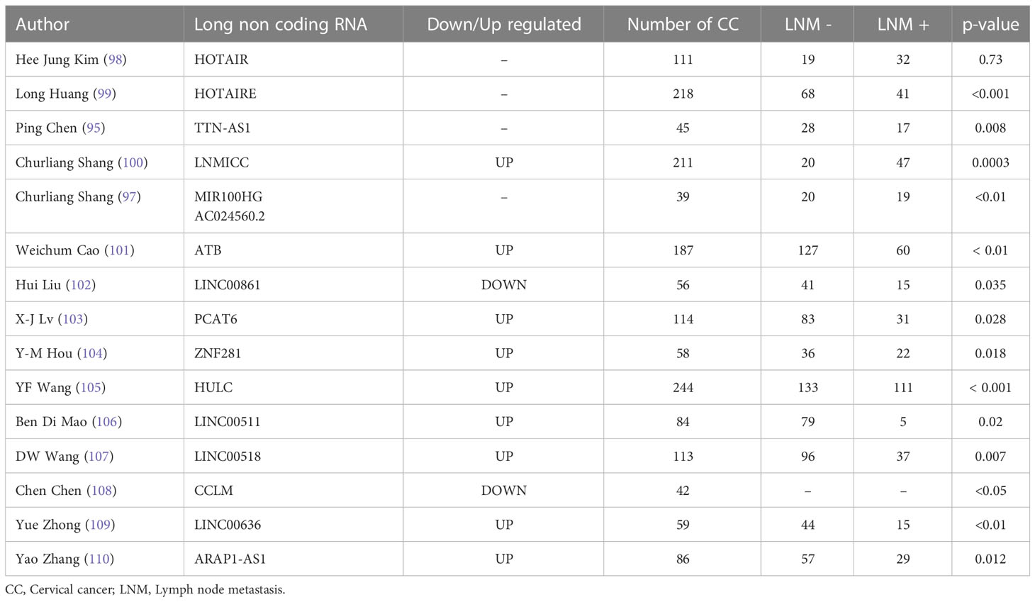
Table 2 Series evaluating the correlation between lncRNAs and lymph nodes invasion in patients with cervical cancer.
From the theragnostic point of view, Mao et al. reported an upregulated expression of LIN00511 in CC with LNM+ associated with paclitaxel (PTX) resistance. Moreover, they observed a relation between LIN00511 and multidrug resistance protein 1 (MRP1) and P-glycoprotein (P-GP) with potential therapeutic target by silencing LINC00511 (106).
lncRNA expression in CC biofluids samples
In a series of 284 patients, Xian-zhen Ding et al. evaluated serum exosomal lncRNA DLX6-AS1 in 114 patients with CC, 60 patients with CIN and 110 healthy women (111). Serum exosomal lncRNA DLX6-AS1 level was significantly elevated in CC patients compared with CIN patients and healthy controls. Based on the median serum value, CC subjects were divided into high and low exosomal lncRNA DLX6-AS expression. High serum exosomal lncRNA DLX6-AS1 levels were correlated with LNM+ (p=0.0071). In addition to a high tissue expression of LNMICC with prometastatic effects suppressed by miR-190 in CC, Chunliang Shang et al. observed a relation between a high serum LNMICC expression and LNM+ (p=0.0003) (97). Similarly, Yue Zhong et al. observed both an overexpression of LINC00636 in tissue samples and in serum with a correlation with LNM+ (p<0.001) (109). In addition to tissue analysis of ARAP1-AS1 in 86 CC patients, Yao Zhang et al. compared serum ARAP1-AS1 expression in 37 CC patients and 20 healthy controls and observed an overexpression with an AUC value of 0.8953 (95% CI: 0.8147-0.9758, p< 0.001) (110). Moreover, serum ARAP1-AS1 expression was correlated with LNM+ (p=0.012) with HR of 2.161 (95% CI:1.354-3.148, p=0.036) (Table 3).
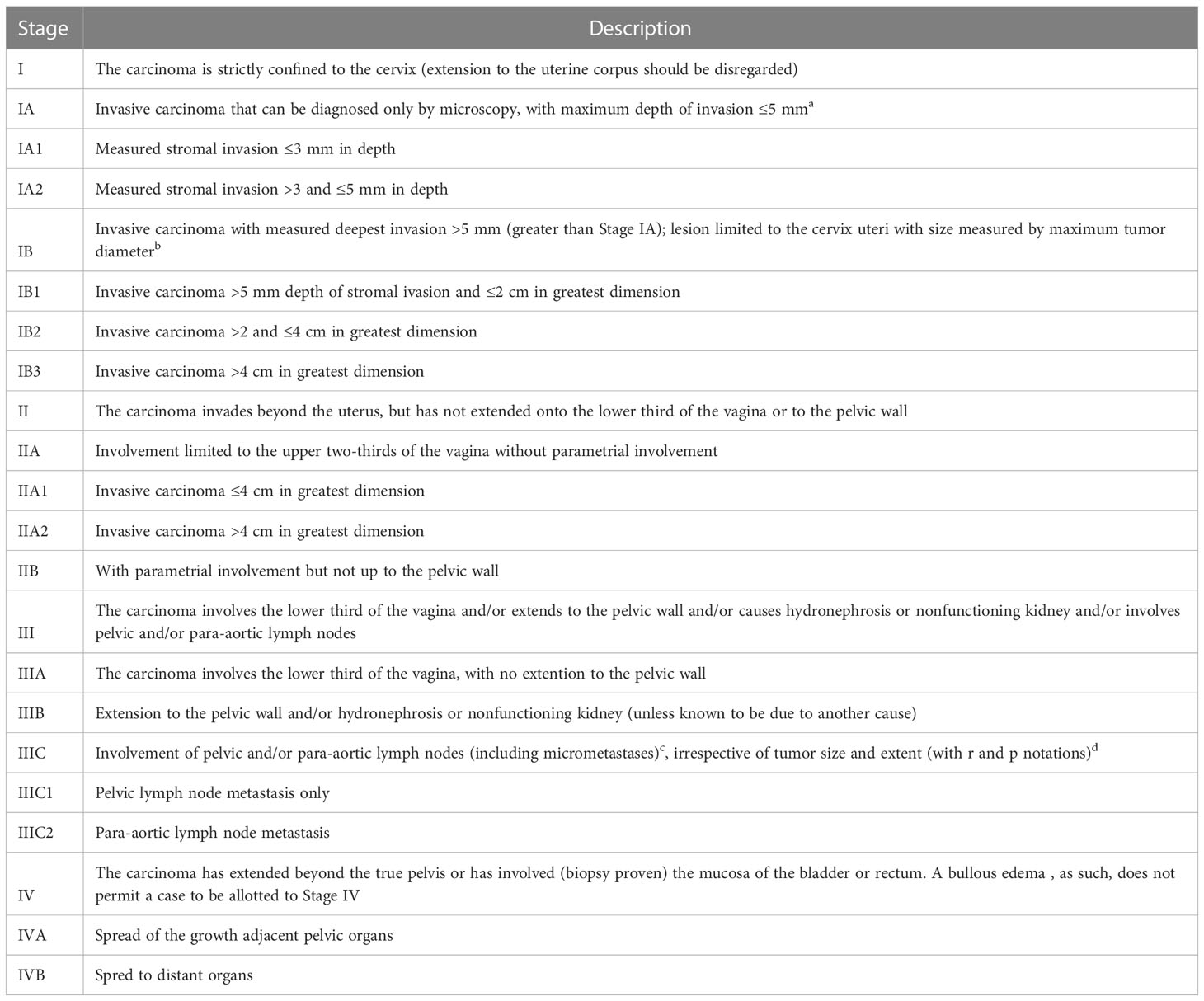
Table 3 2018 FIGO classification of the uterine cervical cancer (112).
Perspectives
From the analysis of the literature on ncRNAs in tissue and biofluids in patients with CC, it appears that there are arguments to support their role in physiopathology, in the differential diagnosis from normal tissue, preinvasive and invasive tumors. However, these analyzes were mainly performed by microarray with validation by qRT-PCR with potential biases linked to the methodology such as the comparison between tumoral sample and adjacent normal appearance tissue and by the preselection of some miRNAs as proven in other context (113). In addition, most studies are from China raising the issue on variations of HPV genotypes according to ethnicity thus on ncRNA expression (114). Moreover, the relatively small number of studies with limited sample size focusing on early CC stages especially concerning miRNAs expression in biofluids of patients with CC limit their potential clinical utility. Indeed, as previously underlined (115) miRNAs are abundant in tissues but often rare in plasma and serum. Moreover, it has been also pointed out that for the quantification of miRNAs in plasma, as well as in other biofluids such as urine and saliva, there is a need to use high-sensitivity platforms such as Next Generation Sequencing (NGS) and Bioinformatic. So far, whatever tissue or biofluids samples, no study using NGS and bioinformatic has been published to support their role in the management of CC. However, all these studies underlined a true relation between miRNA, circRNA and lncRNA with LNM status with a potential non-invasive signature and a relation with response to neo- and adjuvant therapies with a potential contribution in a new algorithm to manage cervical cancer.
Author contributions
Methodology and Design: YD, CT, ED, SB. Data collection: YD, AF, LR, YM. Analysis: YD, CF, SS, AF. Data Interpretation: YD, LR, CF, SB, CT. Manuscript writing: AF, SS, CT, YM. All authors reviewed the manuscript for critical intellectual content. All authors contributed to the article and approved the submitted version.
Conflict of interest
The authors declare that the research was conducted in the absence of any commercial or financial relationships that could be construed as a potential conflict of interest.
Publisher’s note
All claims expressed in this article are solely those of the authors and do not necessarily represent those of their affiliated organizations, or those of the publisher, the editors and the reviewers. Any product that may be evaluated in this article, or claim that may be made by its manufacturer, is not guaranteed or endorsed by the publisher.
References
1. Bray F, Ferlay J, Soerjomataram I, Siegel RL, Torre LA, Jemal A. Global cancer statistics 2018: GLOBOCAN estimates of incidence and mortality worldwide for 36 cancers in 185 countries. CA Cancer J Clin (2018) 68:394–424. doi: 10.3322/caac.21492
2. Arbyn M, Weiderpass E, Bruni L, de Sanjosé S, Saraiya M, Ferlay J, et al. Estimates of incidence and mortality of cervical cancer in 2018: a worldwide analysis. Lancet Glob Health (2020) 8:e191–203. doi: 10.1016/S2214-109X(19)30482-6
3. Abbas M, Mehdi A, Khan FH, Verma S, Ahmad A, Khatoon F, et al. Role of miRNAs in cervical cancer: a comprehensive novel approach from pathogenesis to therapy. J Gynecol Obstet Hum Reprod (2021) 50:102159. doi: 10.1016/j.jogoh.2021.102159
4. Bhatla N, Aoki D, Sharma DN, Sankaranarayanan R. Cancer of the cervix uteri: 2021 update. Int J Gynaecol Obstet (2021) 155(Suppl 1):28–44. doi: 10.1002/ijgo.13865
5. de Sanjosé S, Serrano B, Tous S, Alejo M, Lloveras B, Quirós B, et al. Burden of human papillomavirus (HPV)-related cancers attributable to HPVs 6/11/16/18/31/33/45/52 and 58. JNCI Cancer Spectr (2018) 2:pky045. doi: 10.1093/jncics/pky045
6. Burmeister CA, Khan SF, Schäfer G, Mbatani N, Adams T, Moodley J, et al. Cervical cancer therapies: current challenges and future perspectives. Tumour Virus Res (2022) 13:200238. doi: 10.1016/j.tvr.2022.200238
7. Sankaranarayanan R. Screening for cancer in low- and middle-income countries. Ann Glob Health (2014) 80:412–7. doi: 10.1016/j.aogh.2014.09.014
8. Maver PJ, Poljak M. Primary HPV-based cervical cancer screening in Europe: implementation status, challenges, and future plans. Clin Microbiol Infect (2020) 26:579–83. doi: 10.1016/j.cmi.2019.09.006
9. Chen Q, Zeng X, Huang D, Qiu X. Identification of differentially expressed miRNAs in early-stage cervical cancer with lymph node metastasis across the cancer genome atlas datasets. Cancer Manag Res (2018) 10:6489–504. doi: 10.2147/CMAR.S183488
10. Barquet-Muñoz SA, Pedroza-Torres A, Perez-Plasencia C, Montaño S, Gallardo-Alvarado L, Pérez-Montiel D, et al. microRNA profile associated with positive lymph node metastasis in early-stage cervical cancer. Curr Oncol (2022) 29:243–54. doi: 10.3390/curroncol29010023
11. Wang A, Wang Y, Zhan J, Chen J. MicroRNA-186-5p inhibits the metastasis of cervical cancer by targeting FZD3. J BUON (2021) 26:677–83.
12. Liu X, Gan L, Zhang J. miR-543 inhibites cervical cancer growth and metastasis by targeting TRPM7. Chem Biol Interact (2019) 302:83–92. doi: 10.1016/j.cbi.2019.01.036
13. Dong W, Li B, Wang J, Song Y, Zhang Z, Fu C. MicroRNA-337 inhibits cell proliferation and invasion of cervical cancer through directly targeting specificity protein 1. Tumour Biol (2017) 39. doi: 10.1177/1010428317711323. 1010428317711323.
14. Zhou C-F, Ma J, Huang L, Yi H-Y, Zhang Y-M, Wu X-G, et al. Correction to: cervical squamous cell carcinoma-secreted exosomal miR-221-3p promotes lymphangiogenesis and lymphatic metastasis by targeting VASH1. Oncogene (2022) 41:1231–3. doi: 10.1038/s41388-021-02165-x
15. Liu J, Li Y, Chen X, Xu X, Zhao H, Wang S, et al. Upregulation of miR-205 induces CHN1 expression, which is associated with the aggressive behaviour of cervical cancer cells and correlated with lymph node metastasis. BMC Cancer (2020) 20:1029. doi: 10.1186/s12885-020-07478-w
16. Luo M, Shen D, Wang W, Xian J. Aberrant expression of microRNA-26b and its prognostic potential in human cervical cancer. Int J Clin Exp Pathol (2015) 8:5542–8.
17. Han Y, Xu G-X, Lu H, Yu D-H, Ren Y, Wang L, et al. Dysregulation of miRNA-21 and their potential as biomarkers for the diagnosis of cervical cancer. Int J Clin Exp Pathol (2015) 8:7131–9.
18. Kogo R, How C, Chaudary N, Bruce J, Shi W, Hill RP, et al. The microRNA-218~Survivin axis regulates migration, invasion, and lymph node metastasis in cervical cancer. Oncotarget (2015) 6:1090–100. doi: 10.18632/oncotarget.2836
19. Liang C, Ding J, Yang Y, Deng L, Li X. MicroRNA-433 inhibits cervical cancer progression by directly targeting metadherin to regulate the AKT and β-catenin signalling pathways. Oncol Rep (2017) 38. doi: 10.3892/or.2017.6049
20. Huang L, Lin J-X, Yu Y-H, Zhang M-Y, Wang H-Y, Zheng M. Downregulation of six microRNAs is associated with advanced stage, lymph node metastasis and poor prognosis in small cell carcinoma of the cervix. PLoS One (2012) 7:e33762. doi: 10.1371/journal.pone.0033762
21. Wang L, Chang L, Li Z, Gao Q, Cai D, Tian Y, et al. miR-99a and -99b inhibit cervical cancer cell proliferation and invasion by targeting mTOR signaling pathway. Med Oncol (2014) 31:934. doi: 10.1007/s12032-014-0934-3
22. Li M, Li B-Y, Xia H, Jiang L-L. Expression of microRNA-142-3p in cervical cancer and its correlation with prognosis. Eur Rev Med Pharmacol Sci (2017) 21:2346–50.
23. Zhou J, Liu X, Wang C-H, Wang D, Du J-J. Decreased expression of miR-1254 is associated with cancer aggressiveness and predicts poor outcome in cervical cancer. Eur Rev Med Pharmacol Sci (2018) 22:2997–3001. doi: 10.26355/eurrev_201805_15056
24. Chen Y, Ma C, Zhang W, Chen Z, Ma L. Down regulation of miR-143 is related with tumor size, lymph node metastasis and HPV16 infection in cervical squamous cancer. Diagn Pathol (2014) 9:88. doi: 10.1186/1746-1596-9-88
25. Wei Y-Q, Jiao X-L, Zhang S-Y, Xu Y, Li S, Kong B-H. MiR-9-5p could promote angiogenesis and radiosensitivity in cervical cancer by targeting SOCS5. Eur Rev Med Pharmacol Sci (2019) 23:7314–26. doi: 10.26355/eurrev_201909_18837
26. Pfaendler KS, Tewari KS. Changing paradigms in the systemic treatment of advanced cervical cancer. Am J Obstet Gynecol (2016) 214:22–30. doi: 10.1016/j.ajog.2015.07.022
28. Farley JH, Hickey KW, Carlson JW, Rose GS, Kost ER, Harrison TA. Adenosquamous histology predicts a poor outcome for patients with advanced-stage, but not early-stage, cervical carcinoma. Cancer (2003) 97:2196–202. doi: 10.1002/cncr.11371
29. Hou H, Dai Y, Liang S, Wang Z, Wang J. Sentinel lymph node biopsy is feasible in cervical cancer laparoscopic surgery: a single-center retrospective cohort study. J Oncol (2021) 2021:5510623. doi: 10.1155/2021/5510623
30. Delgado G, Bundy B, Zaino R, Sevin BU, Creasman WT, Major F. Prospective surgical-pathological study of disease-free interval in patients with stage IB squamous cell carcinoma of the cervix: a gynecologic oncology group study. Gynecol Oncol (1990) 38:352–7. doi: 10.1016/0090-8258(90)90072-s
31. Suprasert P, Charoenkwan K, Khunamornpong S. Pelvic node removal and disease-free survival in cervical cancer patients treated with radical hysterectomy and pelvic lymphadenectomy. Int J Gynaecol Obstet (2012) 116:43–6. doi: 10.1016/j.ijgo.2011.08.001
32. Togami S, Kamio M, Yanazume S, Yoshinaga M, Douchi T. Can pelvic lymphadenectomy be omitted in stage IA2 to IIB uterine cervical cancer? Int J Gynecol Cancer (2014) 24:1072–6. doi: 10.1097/IGC.0000000000000163
33. Tanaka Y, Sawada S, Murata T. Relationship between lymph node metastases and prognosis in patients irradiated postoperatively for carcinoma of the uterine cervix. Acta Radiol Oncol (1984) 23:455–9. doi: 10.3109/02841868409136048
34. Selman TJ, Mann C, Zamora J, Appleyard T-L, Khan K. Diagnostic accuracy of tests for lymph node status in primary cervical cancer: a systematic review and meta-analysis. CMAJ (2008) 178:855–62. doi: 10.1503/cmaj.071124
35. Haldorsen IS, Lura N, Blaakær J, Fischerova D, Werner HMJ. What is the role of imaging at primary diagnostic work-up in uterine cervical cancer? Curr Oncol Rep (2019) 21:77. doi: 10.1007/s11912-019-0824-0
36. Grueneisen J, Beiderwellen K, Heusch P, Gratz M, Schulze-Hagen A, Heubner M, et al. Simultaneous positron emission tomography/magnetic resonance imaging for whole-body staging in patients with recurrent gynecological malignancies of the pelvis: a comparison to whole-body magnetic resonance imaging alone. Invest Radiol (2014) 49:808–15. doi: 10.1097/RLI.0000000000000086
37. Kitajima K, Murakami K, Yamasaki E, Kaji Y, Sugimura K. Accuracy of integrated FDG-PET/contrast-enhanced CT in detecting pelvic and paraaortic lymph node metastasis in patients with uterine cancer. Eur Radiol (2009) 19:1529–36. doi: 10.1007/s00330-008-1271-8
38. Cibula D, Pötter R, Planchamp F, Avall-Lundqvist E, Fischerova D, Haie Meder C, et al. The European society of gynaecological Oncology/European society for radiotherapy and Oncology/European society of pathology guidelines for the management of patients with cervical cancer. Radiother Oncol (2018) 127:404–16. doi: 10.1016/j.radonc.2018.03.003
39. Yang L, Froberg JE, Lee JT. Long noncoding RNAs: fresh perspectives into the RNA world. Trends Biochem Sci (2014) 39:35–43. doi: 10.1016/j.tibs.2013.10.002
40. Cech TR, Steitz JA. The noncoding RNA revolution-trashing old rules to forge new ones. Cell (2014) 157:77–94. doi: 10.1016/j.cell.2014.03.008
41. Canlorbe G, Wang Z, Laas E, Bendifallah S, Castela M, Lefevre M, et al. Identification of microRNA expression profile related to lymph node status in women with early-stage grade 1-2 endometrial cancer. Mod Pathol (2016) 29:391–401. doi: 10.1038/modpathol.2016.30
42. Calin GA, Croce CM. MicroRNA signatures in human cancers. Nat Rev Cancer (2006) 6:857–66. doi: 10.1038/nrc1997
43. Calin GA, Dumitru CD, Shimizu M, Bichi R, Zupo S, Noch E, et al. Frequent deletions and down-regulation of micro- RNA genes miR15 and miR16 at 13q14 in chronic lymphocytic leukemia. Proc Natl Acad Sci USA (2002) 99:15524–9. doi: 10.1073/pnas.242606799
44. Bonci D, Coppola V, Musumeci M, Addario A, Giuffrida R, Memeo L, et al. The miR-15a-miR-16-1 cluster controls prostate cancer by targeting multiple oncogenic activities. Nat Med (2008) 14:1271–7. doi: 10.1038/nm.1880
45. Miao J, Regenstein JM, Xu D, Zhou D, Li H, Zhang H, et al. The roles of microRNA in human cervical cancer. Arch Biochem Biophys (2020) 690:108480. doi: 10.1016/j.abb.2020.108480
46. Ribeiro J, Sousa H. MicroRNAs as biomarkers of cervical cancer development: a literature review on miR-125b and miR-34a. Mol Biol Rep (2014) 41:1525–31. doi: 10.1007/s11033-013-2998-0
47. Qin W, Dong P, Ma C, Mitchelson K, Deng T, Zhang L, et al. MicroRNA-133b is a key promoter of cervical carcinoma development through the activation of the ERK and AKT1 pathways. Oncogene (2012) 31:4067–75. doi: 10.1038/onc.2011.561
48. Park S, Kim J, Eom K, Oh S, Kim S, Kim G, et al. microRNA-944 overexpression is a biomarker for poor prognosis of advanced cervical cancer. BMC Cancer (2019) 19:419. doi: 10.1186/s12885-019-5620-6
49. Yang D, Zhang Q. miR-152 may function as an early diagnostic and prognostic biomarker in patients with cervical intraepithelial neoplasia and patients with cervical cancer. Oncol Lett (2019) 17:5693–8. doi: 10.3892/ol.2019.10233
50. Iorio MV, Visone R, Di Leva G, Donati V, Petrocca F, Casalini P, et al. MicroRNA signatures in human ovarian cancer. Cancer Res (2007) 67:8699–707. doi: 10.1158/0008-5472.CAN-07-1936
51. Oliveira DNP, Carlsen AL, Heegaard NHH, Prahm KP, Christensen IJ, Høgdall CK, et al. Diagnostic plasma miRNA-profiles for ovarian cancer in patients with pelvic mass. PLoS One (2019) 14:e0225249. doi: 10.1371/journal.pone.0225249
52. Mazzola R, Ricchetti F, Fiorentino A, Levra NG, Fersino S, Di Paola G, et al. Weekly cisplatin and volumetric-modulated arc therapy with simultaneous integrated boost for radical treatment of advanced cervical cancer in elderly patients: feasibility and clinical preliminary results. Technol Cancer Res Treat (2017) 16:310–5. doi: 10.1177/1533034616655055
53. Shahrouki P, Larsson E. The non-coding oncogene: a case of missing DNA evidence? Front Genet (2012) 3:170. doi: 10.3389/fgene.2012.00170
54. Green D, Fraser WD, Dalmay T. Transfer RNA-derived small RNAs in the cancer transcriptome. Pflugers Arch (2016) 468:1041–7. doi: 10.1007/s00424-016-1822-9
55. Gibb EA, Brown CJ, Lam WL. The functional role of long non-coding RNA in human carcinomas. Mol Cancer (2011) 10:38. doi: 10.1186/1476-4598-10-38
56. Bartel DP. MicroRNAs: target recognition and regulatory functions. Cell (2009) 136:215–33. doi: 10.1016/j.cell.2009.01.002
57. Lai EC. Micro RNAs are complementary to 3’ UTR sequence motifs that mediate negative post-transcriptional regulation. Nat Genet (2002) 30:363–4. doi: 10.1038/ng865
58. Li B, Yang X-X, Wang D, Ji H-K. MicroRNA-138 inhibits proliferation of cervical cancer cells by targeting c-met. Eur Rev Med Pharmacol Sci (2016) 20:1109–14.
59. Su Y, Xiong J, Hu J, Wei X, Zhang X, Rao L. MicroRNA-140-5p targets insulin like growth factor 2 mRNA binding protein 1 (IGF2BP1) to suppress cervical cancer growth and metastasis. Oncotarget (2016) 7:68397–411. doi: 10.18632/oncotarget.11722
60. Zhang J, Zheng F, Yu G, Yin Y, Lu Q. miR-196a targets netrin 4 and regulates cell proliferation and migration of cervical cancer cells. Biochem Biophys Res Commun (2013) 440:582–8. doi: 10.1016/j.bbrc.2013.09.142
61. Akerman GS, Tolleson WH, Brown KL, Zyzak LL, Mourateva E, Engin TS, et al. Human papillomavirus type 16 E6 and E7 cooperate to increase epidermal growth factor receptor (EGFR) mRNA levels, overcoming mechanisms by which excessive EGFR signaling shortens the life span of normal human keratinocytes. Cancer Res (2001) 61:3837–43.
62. Chhabra R. Let-7i-5p, miR-181a-2-3p and EGF/PI3K/SOX2 axis coordinate to maintain cancer stem cell population in cervical cancer. Sci Rep (2018) 8:7840. doi: 10.1038/s41598-018-26292-w
63. Jiang W, Pan JJ, Deng YH, Liang MR, Yao LH. Down-regulated serum microRNA-101 is associated with aggressive progression and poor prognosis of cervical cancer. J Gynecol Oncol (2017) 28:e75. doi: 10.3802/jgo.2017.28.e75
64. Liu P, Xin F, Ma CF. Clinical significance of serum miR-196a in cervical intraepithelial neoplasia and cervical cancer. Genet Mol Res (2015) 14:17995–8002. doi: 10.4238/2015.December.22.25
65. Zhang L, Wu ZA. MicroRNA-378a-3p downregulation as a novel biomarker with poor clinical outcomes in cervical cancer. BioMed Environ Sci (2021) 34:213–21. doi: 10.3967/bes2021.026
66. Qiu H, Liang D, Liu L, Xiang Q, Yi Z, Ji Y. A novel circulating MiRNA-based signature for the diagnosis and prognosis prediction of early-stage cervical cancer. Technol Cancer Res Treat (2020) 19. doi: 10.1177/1533033820970667. 1533033820970667.
67. Zhao S, Yao D, Chen J, Ding N. Circulating miRNA-20a and miRNA-203 for screening lymph node metastasis in early stage cervical cancer. Genet Test Mol Biomarkers (2013) 17:631–6. doi: 10.1089/gtmb.2013.0085
68. Chen J, Yao D, Li Y, Chen H, He C, Ding N, et al. Serum microRNA expression levels can predict lymph node metastasis in patients with early-stage cervical squamous cell carcinoma. Int J Mol Med (2013) 32. doi: 10.3892/ijmm.2013.1424
69. Hentze MW, Preiss T. Circular RNAs: splicing’s enigma variations. EMBO J (2013) 32:923–5. doi: 10.1038/emboj.2013.53
70. Glažar P, Papavasileiou P, Rajewsky N. circBase: a database for circular RNAs. RNA (2014) 20:1666–70. doi: 10.1261/rna.043687.113
71. Liu H, Chen D, Bi J, Han J, Yang M, Dong W, et al. Circular RNA circUBXN7 represses cell growth and invasion by sponging miR-1247-3p to enhance B4GALT3 expression in bladder cancer. Aging (Albany NY) (2018) 10:2606–23. doi: 10.18632/aging.101573
73. Huang S, Wu S, Ding J, Lin J, Wei L, Gu J, et al. MicroRNA-181a modulates gene expression of zinc finger family members by directly targeting their coding regions. Nucleic Acids Res (2010) 38:7211–8. doi: 10.1093/nar/gkq564
74. Ørom UA, Nielsen FC, Lund AH. MicroRNA-10a binds the 5’UTR of ribosomal protein mRNAs and enhances their translation. Mol Cell (2008) 30:460–71. doi: 10.1016/j.molcel.2008.05.001
75. Wang H, Zhao Y, Chen M, Cui J. Identification of novel long non-coding and circular RNAs in human papillomavirus-mediated cervical cancer. Front Microbiol (2017) 8:1720. doi: 10.3389/fmicb.2017.01720
76. Yi Y, Liu Y, Wu W, Wu K, Zhang W. Reconstruction and analysis of circRNA−miRNA−mRNA network in the pathology of cervical cancer. Oncol Rep (2019) 41:2209–25. doi: 10.3892/or.2019.7028
77. Tornesello ML, Faraonio R, Buonaguro L, Annunziata C, Starita N, Cerasuolo A, et al. The role of microRNAs, long non-coding RNAs, and circular RNAs in cervical cancer. Front Oncol (2020) 10:150. doi: 10.3389/fonc.2020.00150
78. Ma H, Tian T, Liu X, Xia M, Chen C, Mai L, et al. Upregulated circ_0005576 facilitates cervical cancer progression via the miR-153/KIF20A axis. BioMed Pharmacother (2019) 118:109311. doi: 10.1016/j.biopha.2019.109311
79. Song T-F, Xu A-L, Chen X-H, Gao J-Y, Gao F, Kong X-C. Circular RNA circRNA_101996 promoted cervical cancer development by regulating miR-1236-3p/TRIM37 axis. Kaohsiung J Med Sci (2021) 37:547–61. doi: 10.1002/kjm2.12378
80. Guo J, Chen M, Ai G, Mao W, Li H, Zhou J. Hsa_circ_0023404 enhances cervical cancer metastasis and chemoresistance through VEGFA and autophagy signaling by sponging miR-5047. BioMed Pharmacother (2019) 115:108957. doi: 10.1016/j.biopha.2019.108957
81. Kitagawa M, Kitagawa K, Kotake Y, Niida H, Ohhata T. Cell cycle regulation by long non-coding RNAs. Cell Mol Life Sci (2013) 70:4785–94. doi: 10.1007/s00018-013-1423-0
82. Ballarino M, Morlando M, Fatica A, Bozzoni I. Non-coding RNAs in muscle differentiation and musculoskeletal disease. J Clin Invest (2016) 126:2021–30. doi: 10.1172/JCI84419
83. Sirey TM, Roberts K, Haerty W, Bedoya-Reina O, Rogatti-Granados S, Tan JY, et al. The long non-coding RNA Cerox1 is a post transcriptional regulator of mitochondrial complex I catalytic activity. Elife (2019) 8:e45051. doi: 10.7554/eLife.45051
84. Esteller M. Non-coding RNAs in human disease. Nat Rev Genet (2011) 12:861–74. doi: 10.1038/nrg3074
85. Arora R, Lee Y, Wischnewski H, Brun CM, Schwarz T, Azzalin CM. RNaseH1 regulates TERRA-telomeric DNA hybrids and telomere maintenance in ALT tumour cells. Nat Commun (2014) 5:5220. doi: 10.1038/ncomms6220
86. Kleaveland B, Shi CY, Stefano J, Bartel DP. A network of noncoding regulatory RNAs acts in the mammalian brain. Cell (2018) 174:350–362.e17. doi: 10.1016/j.cell.2018.05.022
87. Ahn J-H, Lee H-S, Lee J-S, Lee Y-S, Park J-L, Kim S-Y, et al. nc886 is induced by TGF-β and suppresses the microRNA pathway in ovarian cancer. Nat Commun (2018) 9:1166. doi: 10.1038/s41467-018-03556-7
88. Derrien T, Johnson R, Bussotti G, Tanzer A, Djebali S, Tilgner H, et al. The GENCODE v7 catalog of human long noncoding RNAs: analysis of their gene structure, evolution, and expression. Genome Res (2012) 22:1775–89. doi: 10.1101/gr.132159.111
89. Beaulieu YB, Kleinman CL, Landry-Voyer A-M, Majewski J, Bachand F. Polyadenylation-dependent control of long noncoding RNA expression by the Poly(A)-binding protein nuclear 1. PloS Genet (2012) 8:e1003078. doi: 10.1371/journal.pgen.1003078
90. Mukherjee N, Calviello L, Hirsekorn A, de Pretis S, Pelizzola M, Ohler U. Integrative classification of human coding and noncoding genes through RNA metabolism profiles. Nat Struct Mol Biol (2017) 24:86–96. doi: 10.1038/nsmb.3325
91. Cabili MN, Dunagin MC, McClanahan PD, Biaesch A, Padovan-Merhar O, Regev A, et al. Localization and abundance analysis of human lncRNAs at single-cell and single-molecule resolution. Genome Biol (2015) 16:20. doi: 10.1186/s13059-015-0586-4
92. Zuckerman B, Ulitsky I. Predictive models of subcellular localization of long RNAs. RNA (2019) 25:557–72. doi: 10.1261/rna.068288.118
93. Liu Y, Liu H, Sheng B, Pan S, Wang Z-W, Zhu X. The functions of lncRNAs in the HPV-negative cervical cancer compared with HPV-positive cervical cancer. Apoptosis (2022) 27:685–96. doi: 10.1007/s10495-022-01761-w
94. Liu S, Zhang M, Qu P. Expression level and clinical significance of HOX transcript antisense intergenic RNA in cervical cancer: a meta-analysis. Sci Rep (2016) 6:38047. doi: 10.1038/srep38047
95. Chen P, Wang R, Yue Q, Hao M. Long non-coding RNA TTN-AS1 promotes cell growth and metastasis in cervical cancer via miR-573/E2F3. Biochem Biophys Res Commun (2018) 503:2956–62. doi: 10.1016/j.bbrc.2018.08.077
96. Yu C-L, Xu X-L, Yuan F. LINC00511 is associated with the malignant status and promotes cell proliferation and motility in cervical cancer. Biosci Rep (2019) 39:BSR20190903. doi: 10.1042/BSR20190903
97. Shang C, Zhu W, Liu T, Wang W, Huang G, Huang J, et al. Characterization of long non-coding RNA expression profiles in lymph node metastasis of early-stage cervical cancer. Oncol Rep (2016) 35:3185–97. doi: 10.3892/or.2016.4715
98. Kim HJ, Lee DW, Yim GW, Nam EJ, Kim S, Kim SW, et al. Long non-coding RNA HOTAIR is associated with human cervical cancer progression. Int J Oncol (2015) 46:521–30. doi: 10.3892/ijo.2014.2758
99. Huang L, Liao L-M, Liu A-W, Wu J-B, Cheng X-L, Lin J-X, et al. Overexpression of long noncoding RNA HOTAIR predicts a poor prognosis in patients with cervical cancer. Arch Gynecol Obstet (2014) 290:717–23. doi: 10.1007/s00404-014-3236-2
100. Shang C, Wang W, Liao Y, Chen Y, Liu T, Du Q, et al. LNMICC promotes nodal metastasis of cervical cancer by reprogramming fatty acid metabolism. Cancer Res (2018) 78:877–90. doi: 10.1158/0008-5472.CAN-17-2356
101. Cao W, Peng T, Zhou Y. Long noncoding RNA activated by transforming growth factor-β promotes cancer development and is a prognostic marker in cervical cancer. J Cancer Res Ther (2017) 13:801–6. doi: 10.4103/jcrt.JCRT_256_17
102. Liu H, Zhang L, Ding X, Sui X. LINC00861 inhibits the progression of cervical cancer cells by functioning as a ceRNA for miR−513b−5p and regulating the PTEN/AKT/mTOR signaling pathway. Mol Med Rep (2021) 23:24. doi: 10.3892/mmr.2020.11662
103. Lv X-J, Tang Q, Tu Y-Q, Yan D-D, Wei Q-C. Long noncoding RNA PCAT6 regulates cell growth and metastasis via wnt/β-catenin pathway and is a prognosis marker in cervical cancer. Eur Rev Med Pharmacol Sci (2019) 23:1947–56. doi: 10.26355/eurrev_201903_17233
104. Hou Y-M, Wang X-P, Shen C-C, Chen L-T, Zheng X-X. Cervical carcinoma progression is aggravated by lncRNA ZNF281 by binding KLF15. Eur Rev Med Pharmacol Sci (2021) 25:5610–8. doi: 10.26355/eurrev_202109_26780
105. Wang Y-F, Zhang S, Li X-Q, Wang Y. Expression of lncRNA HULC in cervical cancer and its correlation with tumor progression and patient survival. Eur Rev Med Pharmacol Sci (2016) 20:3987–91.
106. Mao B-D, Xu P, Xu P, Zhong Y, Ding W-W, Meng Q-Z. LINC00511 knockdown prevents cervical cancer cell proliferation and reduces resistance to paclitaxel. J Biosci (2019) 44:44.
107. Wang D-W, You D, Dong J, Liu T-F. Knockdown of long non-coding RNA LINC00518 inhibits cervical cancer proliferation and metastasis by modulating JAK/STAT3 signaling. Eur Rev Med Pharmacol Sci (2019) 23:496–506. doi: 10.26355/eurrev_201901_16861
108. Chen C, Shen N, Chen Y, Jiang P, Sun W, Wang Q, et al. LncCCLM inhibits lymphatic metastasis of cervical cancer by promoting STAU1-mediated IGF-1 mRNA degradation. Cancer Lett (2021) 518:169–79. doi: 10.1016/j.canlet.2021.07.005
109. Zhong Y, Lu Q, Qiu W, Luo Y. LINC00636 promotes lymph node metastasis and cervical cancer through targeting NM23. Biosci Rep (2020) 40:BSR20200367. doi: 10.1042/BSR20200367
110. Zhang Y, Wu D, Wang D. Long non-coding RNA ARAP1-AS1 promotes tumorigenesis and metastasis through facilitating proto-oncogene c-myc translation via dissociating PSF/PTB dimer in cervical cancer. Cancer Med (2020) 9:1855–66. doi: 10.1002/cam4.2860
111. Ding X-Z, Zhang S-Q, Deng X-L, Qiang J-H. Serum exosomal lncRNA DLX6-AS1 is a promising biomarker for prognosis prediction of cervical cancer. Technol Cancer Res Treat (2021) 20. doi: 10.1177/1533033821990060. 1533033821990060.
112. Abstracts of the XXII FIGO world congress of gynecology & obstetrics. Int J Gynaecol Obstet (2018) 143(Suppl 3):43–991. doi: 10.1002/ijgo.12584
113. Bendifallah S, Dabi Y, Suisse S, Jornea L, Bouteiller D, Touboul C, et al. A bioinformatics approach to MicroRNA-sequencing analysis based on human saliva samples of patients with endometriosis. Int J Mol Sci (2022) 23:8045. doi: 10.3390/ijms23148045
114. Si D, Yao Y, Chen X, Qiu J. Ethnicity-stratified analysis of the association between P53 rs1042522 polymorphism and women HPV infection: a meta-analysis. Microb Pathog (2021) 161:105099. doi: 10.1016/j.micpath.2021.105099
Keywords: cervical cancer, lymph node metastasis, non-coding RNA, microRNA, long non coding RNA, biomarker
Citation: Dabi Y, Favier A, Razakamanantsoa L, Suisse S, Marie Y, Touboul C, Ferrier C, Bendifallah S and Daraï E (2023) Value of non-coding RNAs to assess lymph node status in cervical cancer. Front. Oncol. 13:1144672. doi: 10.3389/fonc.2023.1144672
Received: 14 January 2023; Accepted: 18 April 2023;
Published: 10 May 2023.
Edited by:
Francesco Fanfani, Agostino Gemelli University Polyclinic (IRCCS), ItalyReviewed by:
Anirban Roychowdhury, Virginia Commonwealth University, United StatesFrancesco Ricchetti, Sacro Cuore Don Calabria Hospital (IRCCS), Italy
Copyright © 2023 Dabi, Favier, Razakamanantsoa, Suisse, Marie, Touboul, Ferrier, Bendifallah and Daraï. This is an open-access article distributed under the terms of the Creative Commons Attribution License (CC BY). The use, distribution or reproduction in other forums is permitted, provided the original author(s) and the copyright owner(s) are credited and that the original publication in this journal is cited, in accordance with accepted academic practice. No use, distribution or reproduction is permitted which does not comply with these terms.
*Correspondence: Yohann Dabi, eW9oYW5uLmRhYmlAZ21haWwuY29t