 Yuzhu Cai
 Lingli Zhang
- Department of Anesthesiology, Anhui Provincial Children’s Hospital, Hefei, China
Lymphatic malformations (LMs) are rare congenital anomalies that can cause life-threatening airway obstruction in neonates, particularly when located in the cervicofacial region. The coexistence of lymphatic malformations with an epiglottic cyst further exacerbates airway compromise, posing significant diagnostic and therapeutic challenges. We report the case of a 27-day-old neonate with extensive cervical lymphatic malformations and an epiglottic cyst causing severe airway obstruction. Timely intervention, including laser ablation of the epiglottic cyst and sclerotherapy for the lymphatic malformations, was successfully performed under general anesthesia. Perioperative management emphasized safe airway control and meticulous surgical planning. Postoperative outcomes were favorable, with significant reduction in lymphatic malformations size and resolution of airway obstruction after additional sclerotherapy sessions. This case underscores the importance of a multidisciplinary approach, combining advanced imaging, innovative airway management techniques, and minimally invasive procedures, to achieve optimal outcomes in complex neonatal airway and vascular anomalies.
Introduction
Lymphatic malformations (LMs) are rare congenital vascular anomalies arising from abnormal lymphatic system development (1). They most commonly affect the cervicofacial region (2), varying in size and complexity, and are often characterized by large cystic masses. In neonates, extensive LMs can cause significant complications by compressing adjacent structures, particularly in the head and neck region, potentially leading to airway obstruction, feeding difficulties, and other life-threatening conditions (3). The coexistence of LMs with epiglottic cysts presents unique diagnostic and therapeutic challenges due to their critical impact on airway management.
Epiglottic cysts is a rare but potentially lethal supraglottic airway pathology in infants and are caused by ductal obstruction of either mucous glands or minor salivary glands in the vallecula and base of the tongue (4).While congenital epiglottic cysts are non-malignant growths, in neonates and infants, substantial cysts located in the epiglottis can potentially obstruct the airway, leading to either respiratory impediments or instances of food inhalation that might result in foreign body obstruction of the trachea. Such occurrences elevate the potential peril of death by asphyxia (5). Their presence exacerbates airway compression symptoms caused by extensive cervical LMs, necessitating prompt evaluation and intervention. Advances in imaging techniques, such as computed tomography (CT), play a critical role in diagnosing and characterizing these complex lesions, while airway endoscopy provides vital insights into the extent and nature of airway involvement. Multidisciplinary collaboration is essential for devising personalized and comprehensive management strategies.
Management of neonatal LMs and associated epiglottic cysts often involves a combination of surgical and minimally invasive interventions. Laser ablation of epiglottic cysts provides rapid relief of airway obstruction (6), whereas sclerotherapy offers a minimally invasive alternative for reducing LMs size (7). Optimal perioperative airway management is crucial, as neonates with compromised airways are at high risk of complications during anesthesia induction and intubation.
This report details a rare case of a 27-day-old neonate with extensive cervical LMs and an associated epiglottic cyst causing severe airway obstruction. The patient underwent successful perioperative management under general anesthesia, including laser ablation of the epiglottic cyst and localized sclerotherapy for the LMs.
Case report
The patient, a 27-day-old female neonate, measured 52 cm in length and weighed 4.15 kg at the time of admission. Prenatal monitoring revealed a multilocular cystic mass in the neck and submandibular region. At 27 days postpartum, the patient presented with respiratory distress and positive signs of retractions (Figure 1A). Born at 39 weeks + 1 day gestation with a birth weight of 3.75 kg, she was admitted for further evaluation. Treatment included nasal oxygen, thermal support, vitamin K1 supplementation, monitoring of blood pressure and glucose, anti-infective therapy, formula feeding, and intravenous nutritional support.
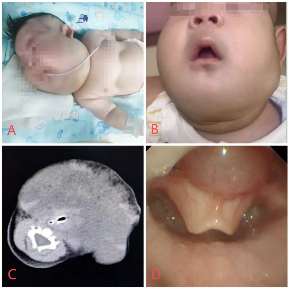
Figure 1. Patient photographs and imaging data. (A) Presence of suprasternal, supraclavicular, and intercostal retractions upon admission. (B) The patient's 8-month-old cervical lymphatic malformation has significantly reduced in size. (C) Cervical and Head CT Image of the Patient. (D) Bronchoscopic view of an epiglottic cyst.
The computed tomography (CT) scan of the head and neck was performed to delineate the extent and nature of the lymphatic malformations (LMs) and the epiglottic cyst in the neonate. The scan revealed extensive hypodense areas in the bilateral maxillofacial and submandibular regions, with scattered hyperdense signals, indicative of hemorrhagic LMs. The lesions measured approximately 62 mm × 45 mm × 44 mm. (Figure 1C). The presence of large hypodense areas suggested the presence of cystic structures typical of LMs. The scattered hyperdense signals within these areas were consistent with hemorrhagic changes. Bronchoscopy revealed an epiglottic cyst and mild tracheomalacia in the upper trachea (Figure 1D). A distinct cystic structure was identified at the base of the epiglottis. This cyst was visualized using bronchoscopy, confirming its location and the degree of airway obstruction it caused. The CT scan provided crucial information for the multidisciplinary team (MDT) to plan the surgical intervention. The extent and location of the LMs and the epiglottic cyst were meticulously assessed to determine the most appropriate approach for laser ablation and sclerotherapy. The MDT decided on an emergency laser ablation of the epiglottic cyst to alleviate immediate airway obstruction, followed by intralesional sclerotherapy for the LMs to reduce their size and prevent future complications.
Following peripheral venous access in the ward, the patient was transferred to the operating room at 14:30, with baseline monitoring showing SpO2 of 94%, HR of 135 bpm, and BP of 82/49 mmHg. The neonate was positioned laterally with a 30° elevation to optimize respiratory function. Preoxygenation with 6 L/min oxygen was administered via face mask for 5 min. Before induction, the airway cart was checked, including rescue equipment such as supraglottic airway devices (I-Gel), with an otolaryngologist on standby for emergency tracheotomy.
Anesthesia was induced using sevoflurane, and intubation was performed using a 3.5 mm uncuffed endotracheal tube and a video laryngoscope (Insight iS2). The epiglottic cyst was visualized and lifted with the laryngoscope for clear exposure of the glottis. Intubation was successful without complications. Cisatracurium (0.5 mg) was administered for muscle relaxation, and anesthesia was maintained with 1.5% sevoflurane and continuous remifentanil infusion (0.2–0.25 µg/kg/min). Dexamethasone (1 mg) was administered prophylactically to prevent postoperative airway edema.
The oxygen concentration was reduced to 40% during surgery to minimize fire risk during laser ablation of the cyst. However, to ensure patient safety with this reduced oxygen level, arterial blood gas analysis was performed every 10 min, and the oxygenation index was continuously monitored and adjusted according to the patient's condition to ensure adequate tissue oxygenation. Laser ablation was performed using a CO2 laser (UltraPulse®, Lumenis, Santa Clara, CA, USA) with a power of 10 W and a spot size of 1 mm. The procedure was performed transorally under bronchoscopic guidance. The procedure lasted 20 min, followed by 26 min of sclerotherapy under interventional guidance. for the LMs. Sclerotherapy was performed using Bleomycin at a dose of 0.5 mg/kg, injected directly into the cystic spaces of the lymphatic malformations. The neonate was transferred intubated to the PICU postoperatively and successfully extubated 24 h later. She was discharged on postoperative day five in stable condition.
The patient underwent three additional sclerotherapy treatments under general anesthesia at 3 months, 5 months, and 8 months of age, with significant reduction in LMs size and favorable outcomes (Figure 1B).
Discussion
This case highlights the successful management of a rare combination of extensive cervical LMs and epiglottic cyst in a neonate. The severe airway obstruction posed a significant challenge, requiring a multidisciplinary approach emphasizing meticulous preoperative planning, timely surgical intervention, and careful perioperative management.
Imaging modalities, particularly CT, were instrumental in delineating the extent of the lesions and guiding interventions. The use of a video laryngoscope facilitated safe airway management and direct visualization of the epiglottic cyst, guiding laser ablation (8). Laser ablation, as a minimally invasive technique, effectively relieved airway obstruction while minimizing collateral tissue damage. Special precautions were taken to reduce intraoperative oxygen concentrations, as high oxygen levels increase the risk of airway fire.Lasing in more than 50% oxygen is comparatively dangerous and can cause airway fire in less than 5 s (9). For every 10% increase in oxygen concentration above 60% the risk of flame increased by a factor of 2.3 (10).
Sclerotherapy provided a valuable minimally invasive option for LMs management, significantly reducing lesion size without the morbidity associated with open surgical excision (11). Perioperative measures, including preoxygenation, equipment readiness, and dexamethasone administration, minimized the risk of catastrophic airway complications and postoperative edema.
In this case, we did not perform drainage or place a drainage tube for the lymphatic malformations. The MDT concluded that the primary cause of airway obstruction was the epiglottic cyst, rather than the cervical lymphatic malformations. Ultrasonography revealed that the lymphatic malformations were predominantly multicystic in nature. Placing a drainage tube might not have resolved the issue and could have potentially led to intracystic hemorrhage, worsening airway obstruction. However, if the cervical lymphatic malformations had posed a direct threat to airway safety, primary drainage would have been the preferred option.
The patient's favorable outcomes underscore the importance of MDT collaboration involving anesthesiologists, pediatric surgeons, otolaryngologists, and interventional radiologists (12). Long-term follow-up is essential, as ongoing sclerotherapy treatments may be required for sustained lesion reduction.
Conclusion
This case highlights the importance of a multidisciplinary approach in managing rare neonatal conditions such as extensive cervical LMs coexisting with an epiglottic cyst, which posed a significant airway obstruction risk. The combination of timely surgical interventions, including laser ablation of the epiglottic cyst and sclerotherapy for the LMs, along with meticulous perioperative airway management, ensured a favorable outcome. This report underscores the critical role of careful preoperative planning, innovative airway management techniques, and long-term follow-up in achieving successful treatment outcomes for complex neonatal airway and vascular anomalies.
Data availability statement
The original contributions presented in the study are included in the article/Supplementary Material, further inquiries can be directed to the corresponding author.
Ethics statement
Written informed consent was obtained from the individual(s), and minor(s)' legal guardian/next of kin, for the publication of any potentially identifiable images or data included in this article.
Author contributions
YC: Writing – original draft, Writing – review & editing. LZ: Writing – review & editing. JW: Writing – review & editing. YS: Writing – review & editing.
Funding
The author(s) declare that no financial support was received for the research and/or publication of this article.
Conflict of interest
The authors declare that the research was conducted in the absence of any commercial or financial relationships that could be construed as a potential conflict of interest.
Generative AI statement
The author(s) declare that no Generative AI was used in the creation of this manuscript.
Any alternative text (alt text) provided alongside figures in this article has been generated by Frontiers with the support of artificial intelligence and reasonable efforts have been made to ensure accuracy, including review by the authors wherever possible. If you identify any issues, please contact us.
Publisher's note
All claims expressed in this article are solely those of the authors and do not necessarily represent those of their affiliated organizations, or those of the publisher, the editors and the reviewers. Any product that may be evaluated in this article, or claim that may be made by its manufacturer, is not guaranteed or endorsed by the publisher.
References
1. Bouwman FCM, Klein WM, de Blaauw I, Woiski MD, Verhoeven BH, Botden SMBI. Lymphatic malformations adjacent to the airway in neonates: risk factors for outcome. J Pediatr Surg. (2021) 56(10):1764–70. doi: 10.1016/j.jpedsurg.2021.03.011
2. Kronfli AP, McLaughlin CJ, Moroco AE, Grant CN. Lymphatic malformations: a 20-year single institution experience. Pediatr Surg Int. (2021) 37(6):783–90. doi: 10.1007/s00383-021-04859-5
3. Honnorat M, Viremouneix L, Ayari S, Guibaud L, Coste K, Claris O, et al. Early adjuvant medication with the mTOR inhibitor sirolimus in a preterm neonate with compressive cystic lymphatic malformation. Front Pediatr. (2020) 8:418. doi: 10.3389/fped.2020.00418
4. Mulcahy CF, Reddy SK, Wikner EE, Mudd PA. Neonatal airway anomaly: vallecular cyst. BMJ Case Rep. (2017) 2017:bcr2017223082. doi: 10.1136/bcr-2017-223082
5. Guo S, Cao C, Feng C, Li Z, Zhou F, Ye H, et al. Case reports of low-temperature plasma radiofrequency treatment for congenital epiglottic cysts in neonates and infants. Ear Nose Throat J. (2023). doi: 10.1177/01455613231199699
6. Chen D, Duan M. Clinical effect of CO2 laser resection of the epiglottic cyst under micro-laryngoscope suspension. Acta Otolaryngol. (2022) 142(5):443–7. doi: 10.1080/00016489.2022.2079717
7. Bouwman FCM, Kooijman SS, Verhoeven BH, Schultze Kool LJ, van der Vleuten CJM, Botden SMBI, et al. Lymphatic malformations in children: treatment outcomes of sclerotherapy in a large cohort. Eur J Pediatr. (2021) 180(3):959–66. doi: 10.1007/s00431-020-03811-4
8. Meng X, Wen Q, Gu J, Wang Y. Videolaryngoscope-assisted coblation of epiglottic cysts. Eur Arch Otorhinolaryngol. (2020) 277(4):1129–32. doi: 10.1007/s00405-020-05804-3
9. Stuermer KJ, Ayachi S, Gostian AO, Beutner D, Hüttenbrink KB. Hazard of CO₂ laser-induced airway fire in laryngeal surgery: experimental data of contributing factors. Eur Arch Otorhinolaryngol. (2013) 270(10):2701–7. doi: 10.1007/s00405-013-2521-1
10. Huang L, Badenoch A, Vermeulen M, Ullah S, Woods C, Athanasiadis T, et al. Risk of airway fire with the use of KTP laser and high flow humidified oxygen delivery in a laryngeal surgery model. Sci Rep. (2022) 12(1):543. doi: 10.1038/s41598-021-04636-3
11. Hu F, Ma F, Liu X, Yu J. Sclerothrapy of giant lymphatic malformation in neonates. J Perinatol. (2025) 45(2):213–7. doi: 10.1038/s41372-024-02113-z
Keywords: lymphatic malformations, epiglottic cyst, neonatal airway obstruction, laser ablation, sclerotherapy, general anesthesia
Citation: Cai Y, Zhang L, Wang J and Sun Y (2025) Multidisciplinary management of extensive cervical lymphatic malformations and epiglottic cyst in a neonate: a case report and review of perioperative strategies. Front. Pediatr. 13:1543250. doi: 10.3389/fped.2025.1543250
Received: 11 December 2024; Accepted: 22 August 2025;
Published: 8 September 2025.
Edited by:
Simonetta Costa, Casilino General Hospital, ItalyReviewed by:
Takahisa Tainaka, Nagoya University, JapanLei Guo, Jinan Children’s Hospital, China
Harsali Fransiscus Lampus, Sam Ratulangi University, Indonesia
Copyright: © 2025 Cai, Zhang, Wang and Sun. This is an open-access article distributed under the terms of the Creative Commons Attribution License (CC BY). The use, distribution or reproduction in other forums is permitted, provided the original author(s) and the copyright owner(s) are credited and that the original publication in this journal is cited, in accordance with accepted academic practice. No use, distribution or reproduction is permitted which does not comply with these terms.
*Correspondence: Yingying Sun, c3VueXkxNjhAMTI2LmNvbQ==