 Yun-Long Li
 He-Wen Wang*
- Department of Pediatrics, Rizhao People's Hospital, Shandong, China
We report an 8-year-old girl presenting with an 8-day history of cough, 5-day fever, and recent-onset conjunctival hyperemia and oral mucosal erosions. Laboratory testing confirmed Mycoplasma pneumoniae infection (IgM antibody and DNA positive). Chest x-ray demonstrated bilateral increased and blurred lung markings, consistent with acute bronchitis. Initial treatment with azithromycin and methylprednisolone sodium succinate (MPSS) improved respiratory symptoms but triggered a progressive rash involving the trunk, perineum, and anus. Adjuvant therapy with intravenous immunoglobulin (IVIG, 22.5 g) and continued immunomodulation with MPSS led to resolution of mucocutaneous manifestations by hospital day 10. This case highlights the potential for M. pneumoniae to induce systemic immune-mediated reactions and underscores the importance of multimodal therapy.
Introduction
Mycoplasma pneumoniae is a common pathogen responsible for community-acquired pneumonia in children, often manifesting with respiratory symptoms (1). Extrapulmonary manifestations, including dermatological involvement, are increasingly recognized. Mycoplasma pneumoniae-induced rash and mucositis (MIRM) is a distinct clinical entity characterized by prominent mucositis (oral, ocular, genital) with variable cutaneous involvement, often mimicking but distinct from Stevens-Johnson syndrome (SJS) and toxic epidermal necrolysis (TEN) (2). MIRM typically presents with less severe skin necrosis and a stronger association with M. pneumoniae infection compared to SJS/TEN (3). Diagnosis relies on recognizing the clinical pattern, confirming M. pneumoniae infection, and excluding other severe drug reactions. This case report describes the presentation, diagnostic journey, and successful management of MIRM in a child.
Case report
An 8-year-old girl presented to the pediatric ward with an 8-day history of cough, 5 days of fever, and 2 days of bilateral conjunctival congestion and painful lip erosions (Figure 1). Physical examination confirmed conjunctival hyperemia and significant erosions of the lips. Initial laboratory investigations confirmed acute Mycoplasma pneumoniae infection (positive serum IgM antibodies and PCR detection of M. pneumoniae DNA). Chest radiography revealed bilateral increased and blurred lung markings, leading to a diagnosis of acute bronchitis.
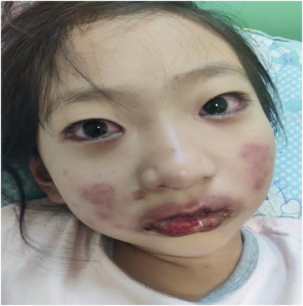
Figure 1. Mouth and Eye.
Treatment was initiated with intravenous azithromycin (10 mg/kg/day) and intravenous methylprednisolone sodium succinate (MPSS, 2 mg/kg/day). While this regimen resulted in improvement of respiratory symptoms (cough and fever), the mucocutaneous condition deteriorated unexpectedly. A progressive maculopapular rash developed on the trunk (Figure 2), extending to involve the perineum (Figure 3) and perianal region (Figure 4). The lip erosions worsened, and ocular symptoms persisted.
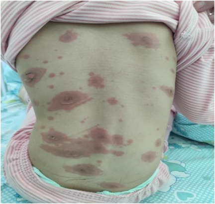
Figure 2. Trunk.
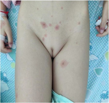
Figure 3. Perineum.
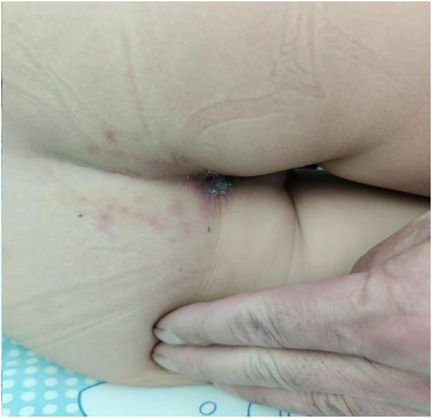
Figure 4. Anus.
Given the confirmed M. pneumoniae infection and the evolving clinical picture characterized by severe mucositis (oral, ocular, genital) with a widespread but non-necrotic rash, the diagnosis was revised to Mycoplasma pneumoniae-induced Rash and Mucositis (MIRM). Based on the skin and mucosal manifestations, MIRM is classified into three types: the classic type (few bullous eruptions or atypical target lesions), the non-bullous type, and the severe type (generalized disseminated bullous eruptions or atypical target lesions) (4). This patient presented with numerous atypical target lesions and bullous eruptions, which was classified as severe-type MIRM. This diagnosis was made after careful differentiation from SJS and TEN, which typically present with more extensive epidermal detachment and necrosis, and are often more strongly linked to drug reactions.
Subsequently, the therapeutic management was intensified with a multi-pronged approach. Immunomodulation was achieved through intravenous immune globulin (IVIG) administered at a total dose of 22.5 g (approximately 1 g/kg) over one day, while intravenous methylprednisolone (MPSS) was maintained at 2 mg/kg/day. No additional dose of IVIG was administered due to the child's rapid clinical improvement. Concurrently, topical mucosal repair agents were implemented, utilizing Kangfuxin Solution for oral lesions and Beifuxin Gel for genital areas to promote healing. Ocular care involved the administration of tobramycin and fluorometholone eye drops to manage conjunctival inflammation and prevent secondary infection. Furthermore, comprehensive supportive care was provided, including adequate hydration, nutritional support, and systematic analgesic management.
The response to this combined therapy was favorable. The conjunctival hyperemia resolved, the mucositis significantly improved, and the widespread rash gradually subsided. By hospital day 10, the mucocutaneous lesions showed substantial healing, and the patient was discharged in stable condition with outpatient follow-up arranged. Oral steroids were tapered over the subsequent weeks.
Discussion
MIRM was historically often categorized under Stevens-Johnson syndrome (SJS). However, due to significant differences in their treatment and prognosis, Canavan et al. proposed the concept of MIRM in 2015 (5). MIRM is caused by immunoglobulin production from B-cell clones, leading to the deposition of immune complexes in the skin and promoting activation of the complement cascade. This mechanism differs from that of SJS and toxic epidermal necrolysis (TEN), which are type IV hypersensitivity reactions mediated by Fas ligand cytotoxicity (5).
Reactive infectious mucocutaneous eruption (RIME) is a condition characterized by mucositis and skin lesions of varying severity triggered by various infections. Oral involvement is universally present, including hemorrhagic crusting of the lips and erosions of the tongue and buccal mucosa (5, 6). Most patients also have ocular and urogenital involvement. Literature reports indicate that various respiratory pathogens can lead to RIME, including Mycoplasma pneumoniae, Chlamydia, influenza B virus, adenovirus, etc. (7–11). The pathogenesis of RIME remains unclear and is thought to involve an immune response triggered by a distant infection, leading to damage of mucocutaneous tissues.
This case illustrates the classic biphasic presentation of MIRM: an initial respiratory phase followed by a mucocutaneous phase. The diagnosis of MIRM hinges on recognizing this pattern, confirming M. pneumoniae infection, and demonstrating the characteristic features: prominent mucositis affecting at least two sites (oral, ocular, urogenital) and a sparse, often polymorphic (maculopapular, targetoid, vesicular), non-necrotic rash, typically involving the trunk and extremities, with frequent genital involvement (2, 12).
Our patient exhibited severe oral and ocular mucositis, perineal/perianal involvement, and a widespread truncal rash, aligning perfectly with MIRM criteria. The timing of the rash after the initiation of antibiotics and steroids initially raised suspicion of a drug reaction. However, the strong evidence of M. pneumoniae infection, the specific mucocutaneous pattern (less epidermal detachment than typical SJS/TEN), and the excellent response to immunomodulation strongly supported MIRM over SJS/TEN (13). Currently, there are no treatment guidelines for MIRM. Therapeutic approaches include anti-Mycoplasma pneumoniae therapy (macrolides, tetracyclines, or quinolones), anti-inflammatory treatment (such as corticosteroids, intravenous immunoglobulin, etc.), and symptomatic supportive care (pain management, intravenous fluid replacement, and mucosal care, etc.) (14).
While macrolides like azithromycin are the mainstay for treating the underlying M. pneumoniae infection, the immune-mediated mucocutaneous manifestations of MIRM often require systemic immunomodulatory therapy (6). Corticosteroids are frequently used first-line, as in this case. However, the progression of mucocutaneous symptoms despite steroid initiation prompted the addition of IVIG, which is increasingly recognized as beneficial in moderate to severe MIRM, potentially halting the immune response more effectively (4, 15). The synergistic effect of continued steroids and IVIG, combined with meticulous supportive care (including targeted mucosal healing agents and ocular therapy), led to a rapid and favorable outcome in this patient. There have also been reports showing that MIRM patients with severe mucosal involvement have responded to other immunomodulatory therapies, including cyclosporine (16), intravenous immunoglobulin (IVIG), and anti-tumor necrosis factor (TNF) agents (17, 18).Therefore, we believe that immunoglobulin therapy is necessary for patients who show a poor response to early corticosteroid treatment or for those with severe MIRM.
This case underscores the importance of considering MIRM in children with M. pneumoniae infection who develop significant mucositis and rash, even after starting antimicrobial therapy. Since there are no formal guidelines, this strategy is based on established management principles for this specific condition. The key is to simultaneously address the underlying infectious trigger, control the exaggerated immune response, and provide intensive supportive care.
Data availability statement
The datasets presented in this article are not readily available because of ethical restrictions and patient privacy concerns. Requests to access the datasets should be directed to the corresponding author.
Ethics statement
Ethical approval was waived by the Institutional Review Board of Rizhao People's Hospital for this case report. Written informed consent was obtained from the minor's legal guardian/next of kin for the publication of any potentially identifiable images or data included in this article.
Author contributions
Y-LL: Formal analysis, Writing – original draft. H-WW: Formal analysis, Writing – review & editing.
Funding
The author(s) declare that no financial support was received for the research and/or publication of this article.
Conflict of interest
The authors declare that the research was conducted in the absence of any commercial or financial relationships that could be construed as a potential conflict of interest.
Generative AI statement
The author(s) declare that no Generative AI was used in the creation of this manuscript.
Any alternative text (alt text) provided alongside figures in this article has been generated by Frontiers with the support of artificial intelligence and reasonable efforts have been made to ensure accuracy, including review by the authors wherever possible. If you identify any issues, please contact us.
Publisher's note
All claims expressed in this article are solely those of the authors and do not necessarily represent those of their affiliated organizations, or those of the publisher, the editors and the reviewers. Any product that may be evaluated in this article, or claim that may be made by its manufacturer, is not guaranteed or endorsed by the publisher.
References
1. Marchello C, Dale AP, Thai TN, Han DS, Ebell MH. Prevalence of atypical pathogens in patients with cough and community-acquired pneumonia: a meta-analysis. Ann Fam Med. (2016) 14(6):552–66. doi: 10.1370/afm.1993
2. Mo X, Wang M, Jiang X, Jin L. Case report: Mycoplasma pneumoniae-induced rash and mucositis in a child. Front Med (Lausanne). (2025) 12:1607970. doi: 10.3389/fmed.2025.1607970
3. Lu H, Zhang B. Mycoplasma-induced rash and mucositis. N Engl J Med. (2023) 389(17):1601. doi: 10.1056/NEJMicm2305301
4. Lofgren D, Lenkeit C. Mycoplasma pneumoniae-induced rash and mucositis: a systematic review of the literature. Spartan Med Res J. (2021) 6(2):25284. doi: 10.51894/001c.25284
5. Canavan TN, Mathes EF, Frieden I, Shinkai K. Mycoplasma pneumoniae-induced rash and mucositis as a syndrome distinct from Stevens-Johnson syndrome and erythema multiforme: a systematic review. J Am Acad Dermatol. (2015) 72(2):239–45. doi: 10.1016/j.jaad.2014.06.026
6. Prindaville B, Newell BD, Nopper AJ, Horii KA. Mycoplasma pneumonia–associated mucocutaneous disease in children: dilemmas in classification. Pediatr Dermatol. (2014) 31(6):670–5. doi: 10.1111/pde.12482
7. Song A, Nicholson C, Maguiness S. Recurrent reactive infectious mucocutaneous eruption (RIME) in two adolescents triggered by several distinct pathogens including SARS-CoV-2 and influenza A. Pediatr Dermatol. (2021) 38(5):1222–5. doi: 10.1111/pde.14780
8. Meyer Sauteur PM, Theiler M, Buettcher M, Seiler M, Weibel L, Berger C. Frequency and clinical presentation of mucocutaneous disease due to Mycoplasma pneumoniae infection in children with community-acquired pneumonia. JAMA Dermatol. (2020) 156(2):144–50. doi: 10.1001/jamadermatol.2019.3602
9. Umapathi KK, Tuli J, Menon S. Chlamydia pneumonia-induced mucositis. Pediatr Neonatol. (2019) 60(6):697–8. doi: 10.1016/j.pedneo.2019.06.005
10. Goyal A, Hook K. Two pediatric cases of influenza B-induced rash and mucositis: Stevens-Johnson syndrome or expansion of the Mycoplasma pneumoniae-induced rash with mucositis (MIRM) spectrum? Pediatr Dermatol. (2019) 36(6):929–31. doi: 10.1111/pde.13921
11. Gámez-González LB, Peña-Varela C, Ramírez-López JM, Yamazaki-Nakashimada MA. Adenoviral-induced rash and mucositis: expanding the spectrum of reactive infectious mucocutaneous eruption. Pediatr Dermatol. (2021) 38(1):306–8. doi: 10.1111/pde.14419
12. Meyer Sauteur PM, Theiler M, Buettcher M, Seiler M, Weibel L, Berger C. Frequency and clinical presentation of mucocutaneous disease due to Mycoplasma pneumoniae infection in children. JAMA Dermatol. (2020) 156(2):144–50. doi: 10.1001/jamadermatol.2019.3602
13. Li K, Haber RM. Stevens-Johnson syndrome without skin lesions (fuchs syndrome): a literature review of adult cases with Mycoplasma cause. Arch Dermatol. (2012) 148:963–4. doi: 10.1001/archdermatol.2012.681
14. Zão I, Ribeiro F, Rocha V, Neto P, Matias C, Jesus G. Mycoplasma pneumoniae-associated mucositis: a recently described entity. Eur J Case Rep Intern Med. (2018) 5(11):000977. doi: 10.12890/2018_000977
15. Song T, Hu H, Yu RH, Xiao P, Li XL, Zhang T, et al. Clinical analysis of 6 cases of Mycoplasma pneumoniae-induced rash and mucositis in children. Zhonghua Er Ke Za Zhi. (2025) 63(2):190–4. doi: 10.3760/cma.j.cn112140-20240730-00529
16. Li HO, Colantonio S, Ramien ML. Treatment of Mycoplasma pneumoniae-induced rash and mucositis with cyclosporine [formula: see text]. J Cutan Med Surg. (2019) 23(6):608–12. doi: 10.1177/1203475419874444
17. Santos RP, Silva M, Vieira AP, Brito C. Mycoplasma pneumoniae-induced rash and mucositis: a recently described entity. BMJ Case Rep. (2017) 2017:bcr2017220768. doi: 10.1136/bcr-2017-220768
Keywords: mycoplasma pneumoniae, rash, mucositis, child, diagosis
Citation: Li Y-L and Wang H-W (2025) Case Report: Mycoplasma pneumoniae-induced rash and mucositis (MIRM) in an 8-year-old girl: concise review of a challenging case.. Front. Pediatr. 13:1697245. doi: 10.3389/fped.2025.1697245
Received: 2 September 2025; Accepted: 27 October 2025;
Published: 13 November 2025.
Edited by:
Mario Barreto, Sapienza University of Rome, ItalyReviewed by:
Suneel Kumar Pooboni, Mediclinic City Hospital, United Arab EmiratesLeigh Stubbs, Cook Children’s Medical Center, United States
Copyright: © 2025 Li and Wang. This is an open-access article distributed under the terms of the Creative Commons Attribution License (CC BY). The use, distribution or reproduction in other forums is permitted, provided the original author(s) and the copyright owner(s) are credited and that the original publication in this journal is cited, in accordance with accepted academic practice. No use, distribution or reproduction is permitted which does not comply with these terms.
*Correspondence: He-Wen Wang, MjQ5ODE2NTYyQHFxLmNvbQ==