 Klaylton Lopes1,2
 Juliana Oliveira3
 Fabio J. C. Sousa-Junior2
 Túlio da F. Santos3
 Débora Andrade2
 Sara L. Andrade4
 Washington L. Pereira4
 Paulo Wender P. Gomes3
 Marta C. Monteiro1
 Consuelo Y. Yoshioka e Silva1,3
 Milton Nascimento da Silva1,3
 Cristiane F. Maia1,2
 Enéas A. Fontes-Júnior1,2*
- 1Programa de Pós-Graduação em Ciências Farmacêuticas, Instituto de Ciências da Saúde, Universidade Federal do Pará, Belém, Brazil
- 2Laboratório de Farmacologia da Inflamação e do Comportamento, Universidade Federal do Pará, Belém, Brazil
- 3Laboratório de Cromatografia Líquida, Instituto de Ciências Exatas e Naturais, Universidade Federal do Pará, Belém, Brazil
- 4Programa de Pós-Graduação em Saúde e Produção Animal na Amazônia, Universidade Federal Rural da Amazônia, Belém, Brazil
Varronia multispicata (Cham.) Borhidi (Cordiaceae), an herbaceous plant distributed in tropical and subtropical regions is native of Brazil and widely used in folk medicine to treat respiratory and digestive diseases, inflammation, and some types of infections. Thus, this study aimed to investigate acute oral toxicity, antinociceptive, and anti-inflammatory activities of dry aqueous extract of V. multispicata (AEVm) and to identify its compounds. Extract was obtained by lyophilized leaf infusion and its composition was analyzed by ultra-performance liquid chromatography-high resolution mass spectrometry (LC-MS). Acute oral toxicity was evaluated in female rats treated with AEVm (2,000 mg/kg) in a single oral dose. Mortality, body weight changes, feed and water intake, organ weights, histological and biochemical parameters were screened for 14 days. Antinociceptive activity was evaluated by writhing (WT), formalin (FT), and hot plate (HP) tests in male mice while anti-inflammatory activity was performed by carrageenan (CPE) and dextran (DPE)-induced paw edema tests and carrageenan-induced peritonitis (CP) test in male rats. Additionally, spontaneous open-field (OF) locomotion was evaluated. LC-MS analysis revealed the presence of flavonoids with biological activity. In toxicity evaluation, extract did not cause deaths in dose of 2,000 mg/kg, and there were no significant behavioral or biochemical alterations. Additionally, evidence of hepatoprotective and antioxidant activity was observed. In pharmacological evaluation AEVm showed dose-dependent antinociceptive activity in WT, with a median effective dose of 146.89 mg/kg, which showed selectivity by inflammatory base processes (FT first phase; p < 0.001), showing no activity in neuropathic nociception components (FT second phase and HP) or about consciousness and locomotion in OF. AEVm also showed significant anti-inflammatory activity, inhibiting CPE (p < 0.001) and cell migration (p < 0.05) and nitric oxide (NO) production (p < 0,01) in CP test. These data demonstrate that AEVm has low oral toxicity—with evidence of hepatoprotective and antioxidant properties—antinociceptive and anti-inflammatory activity, supporting V. multispicata traditional use, possibly related to flavonoids present in its constitution.
Introduction
Inflammation and pain are clinical conditions present in most pathologies, being among main sources of dysfunctional and disabling conditions, requiring pharmacological intervention (Nathan and Ding, 2010; Henschke et al., 2015). Currently the most commonly used medications are steroidal and non-steroidal anti-inflammatory drugs (NSAIDs), and central-acting analgesics (Conforti et al., 2009; Kunanusorn et al., 2009).
In fact, NSAIDs are among most widely used medications due to their efficacy for a wide range of pain and inflammatory conditions. However, their long-term administration may induce several adverse effects such gastro-intestinal ulcers, hepatotoxicity, bleeding, renal disorders, and immunosuppression (Henzen, 2003; Wirtha et al., 2006). Opiates are most effective in cases of moderate to severe pain, although requires the clinical management of risks associated with side effects, abuse and dependence (Rosenblum et al., 2008). Therefore, development of more powerful and safe anti-inflammatory and analgesic drugs is still needed as alternatives to these drugs limitations (Dharmasiri et al., 2003; Kumara, 2001).
Plants inserted in traditional medicine have interested scientific community as a source of new bioactive substances discovery for human disorders treatment (Novais et al., 2003; Samy et al., 2008). In this sense, plants with therapeutic potential are a promising strategy for the development of anti-inflammatory drugs in search of a better therapy, reinforcing the importance of ethnopharmacological knowledge (Gupta et al., 2006; Phanse et al., 2012; Hegde et al., 2014).
The Varronia P. Browne genus was originally attributed to Boraginaceae as a subgenus of Cordia L. (Schlechtendal and Chamisso, 1830; Gottschling et al., 2005; Gurke, 1893; Miller and Wood, 2008), however, morphological and molecular studies have indicated Varronia as a sister-like of Cordia, thus recognizing it as a distinct genus (Gottschling et al., 2005; Miller and Gottschling, 2007). In addition, phylogenetic evidence defined Boraginaceae as non-monophyletic, altering the classification between subfamilies and family, as well as their intrinsic species (Gottschling et al., 2005; Miller and Gottschling, 2007). Even after segregation, Varronia remains one of the largest genera of Cordiaceae (Boraginales), comprising about 130 neotropical species distributed from Mexico to central regions of Southern America (Miller and Gottschling, 2007). In Brazil, there are about 30 of Varronia species widely distributed in Amazon Forest, Atlantic Forest, Cerrado, and Caatinga vegetation (Stapf, 2015).
Varronia multispicata (Cham.) Borhidi (family: Boraginaceae Juss; Genus: Varronia P. Browne), synonyms Cordia multispicata Cham. (basionym), Cordia bahiensis DC., and Lithocardium multispicatum (Cham.) Kuntze, is an herbaceous plant, native of Brazil and mainly distributed in the Amazon area (Rapisarda et al., 1997; Vieira and Silva, 1997; Kuroyanagi et al., 2001; Stapf, 2015; Forzza et al., 2016). This plant is locally known as “maria-preta,” “carucaá,” and “Cordia” (Zoghbi et al., 2010). In folk medicine, tea is widely used as an expectorant, for digestive diseases, as a drug for contusion and for some types of infection (Kuroyanagi et al., 2001). Pharmacological and phytochemical studies have reported several ursane-, oleanane-, and dammarane-type triterpenes isolated as potential antiandrogen constituents in the ethyl acetate fraction of the methanolic extract from the leaves of this species (Kuroyanagi et al., 2001; Kuroyanagi et al., 2003). In addition, sesquiterpenes were the predominant constituent class in the oils of V. multispicata and other Varronia syn. Cordia species (Santos et al., 2006; Oliveira et al., 2007; Diniz et al., 2008; Zoghbi et al., 2010; Rodrigues et al., 2012).
Several studies have shown in other species of Varronia a diversity of constituents and properties, revealing ethnopharmacological and chemotaxonomic importance of this genus. Phytochemical reports indicated presence of monoterpenes (Fun et al., 1990car), triterpenes (Kuroyanagi et al., 2001; Kuroyanagi et al., 2003), sesquiterpenes (Zoghbi et al., 2010; Rodrigues et al., 2012), tannins, and flavonoids (Hormaza et al., 2014). In addition, investigations led to the isolation of larvicidal and antifungal meroterpenoid naphthoquinones from roots of Varronia linnaei (Stearn) J.S. Mill. (syn. Cordia linnaei Stearn) and Varronia curassavica Jacq. (syn. Cordia curassavica (Jacq.) Roem. & Schult. or Cordia verbenacea DC.) (Ioset et al., 1998; Ioset et al., 2000), cytotoxic meroterpenoid benzoquinones against several cancer cell lines from Varronia globosa Jacq. [syn. Cordia globosa (Jacq.) Kunth] (Menezes et al., 2005), and anti-inflammatory sesquiterpenes isolated from the essential oil of Varronia curassavica, that became the basis of a phytotherapic drug (Passos et al., 2007; Dutra et al., 2016).
Based on its use in traditional medicine, present study aimed to investigate chemical composition and effects of dry aqueous extract of leaves of V. multispicata in standard models of toxicity, nociception, and inflammation in rodents.
Materials and Methods
Chemicals and Reagents
Chromatographic-grade acetonitrile was supplied by Tedia (Fairfield, OH, USA) and formic acid from Thermo Fisher Scientific Inc. (Waltham, MA, USA). Ultrapure water obtained by a Direct-Q 5 system from Millipore (Merck, Darmstadt, Germany). TBA, DTNB, ABTS, and Trolox were obtained from Sigma-Aldrich (Darmstadt, Germany) and ALT, AST, GGT, and ALP activity assay kits were obtained from VIDA Biotecnologia (MG, Brazil). All chemicals used were of analytical grade. Acetic acid and formaldehyde (Vetec Química Fina, RJ, Brazil); indomethacin, carrageenan, dextran, cyproheptadine hydrochloride (Sigma-Aldrich, MO, USA); morphine sulfate, and naloxone hydrochloride (Cristália, RJ, Brazil).
Plant Collection, Identification, and Preparation of Dry Aqueous Extract
V. multispicata (Cham.) Borhidi leaves were collected at São Francisco of Pará, Pará state, Brazil (1°18’27.3”S, 47°45’15.0”W), in January 2013 at 24°C and 85% relative humidity. The botanical identification was performed by the specialist Dr. Silvane Tavares Rodrigues from Brazilian Agricultural Research Corporation (Embrapa) Eastern Amazon (Pará-Brazil) and a voucher specimen was deposited in its IAN Herbarium, under the code 188979.
Plant leaves were washed with tap water and 0.1% aqueous NaOCl (sodium hypochlorite) solution and dried in forced-air drying oven at 45°C until constant weight. Dried leaves were crushed using a knife mil obtaining 200 g of moderately fine powder (particle size 355 µm). Powder was infused in 2 L of ultrapure water at 100°C, remaining immersed for 24 h. The solution was filtered, immediately frozen, and then lyophilized (ALPHA 2-4 LDplus) at −20°C. Freeze-drying process yielded 18 g of dry aqueous extract of V. multispicata (AEVm) which were kept in a vacuum desiccator.
Ultraperformance Liquid Chromatography Coupled With Electrospray Ionization Tandem Quadrupole Time-Of-Flight Mass Spectrometry Analysis
LC-MS analysis was performed on an ultraperformance liquid chromatography (UPLC) system coupled to a Xevo G2-S QTof mass spectrometer equipped with an electrospray ionization (ESI) source (Waters, Milford, MA, USA). Separation was performed on a BEH C18 column (Waters, Wexford, Ireland; 50 × 2.1 mm i.d., 1.7 µm particle size) at 40°C with a gradient elution programmed at constant flow rate (0.3 ml.min−1). The extract was analyzed at concentration of 1 mg.ml−1 injected (5 µl) simultaneously with leucine-enkephalin (reference compound). Mobile phase consisted of 0.1% formic acid aqueous solution (A) and 0.1% formic acid in acetonitrile (B). A linear gradient was performed ranging from 5 to 95% B in 10 min. ESI source was operated in positive mode over a wide mass range (m/z 50–1,200) with a scan time of 0.1 s. The source temperature was set at 150°C with a cone gas flow of 20 L h−1. Desolvation gas flow was set at 600 L.h−1 at a temperature of 450°C. Capillary was set at 3.0 kV with cone voltage at 20 V. MassLynx Software (Waters, Milford, MA, USA) was used for data acquisition and processing.
Animals
Two-month-old male Swiss albino mice (25–30 g) and male and female Wistar rats (150–200 g) were obtained from Central Housing Facility of Evandro Chagas Institute (IEC). Animals were kept in collective cages (five animals per cage) under standard conditions of temperature (22 ± 1°C), humidity (50–60%), light/dark cycle (12 h) and feed (standard pellet diet), with water ad libitum. They were acclimated to laboratory for 12 h before the experiments, only with available water at will. All protocols were conducted according to the Guide for the Care and Use of Laboratory Animals (2011) and approved by the Animal Use Ethics Committee of the Federal University of Pará (CEUA/UFPA; license numbers 62-2015 and 6029300817).
Drug Treatment
AEVm, standard drugs, and phlogistic or nociceptive agents were dissolved in 0.9% saline, except for acetic acid, which was solubilized in distilled water. Saline was therefore adopted as control. All treatments were performed with a standard volume of 0.1 ml/10 g body weight for mice and 0.1 ml/100 g body weight to rat by oral (gavage; po), subcutaneous (sc), or intraperitoneal (ip) way.
Acute Oral Toxicity
Treatment and Hippocratic Screening
Acute oral toxicity was evaluated in female Wistar rats (n = 6/group) according to the Guidelines for Testing Chemicals n° 420 of the Organization for Economic Cooperation and Development (OECD, 2001). After acclimatization procedure, animals were treated with saline (control) or the limit dose (2,000 mg/kg) of AEVm. Then animals were evaluated for signs of toxicity, according to parameters related by Malone (1977), every hour for the first 4 h and daily thereafter for 14 days. Feed and water intake and weight gain were also verified daily, as well as the incidence of deaths.
On 14th day the surviving animals were anesthetized and euthanized by cervical dislocation to collect organ (stomach, liver, kidneys and lungs), destined for macroscopic and histopathological evaluation, and blood samples, for biochemical evaluations.
Biochemical Assays
Sample
Blood was collected by ventricular puncture in tubes containing ethylenediaminetetraacetic acid (EDTA). Plasma and erythrocyte fractions were separated by centrifugation for 10 min at 1,400 ×g. Uppermost erythrocytes were discarded and the remaining washed three times in 0.9% saline until a clean suspension (2 ml) of erythrocyte pellet at approximately 50% hematocrit. Washed erythrocytes were diluted 1:10 with saline and 500 µl then lysed by adding 2,500 µl distilled water (hemolysate). From plasma were analyzed the alanine aminotransferase (ALT), aspartate aminotransferase (AST), gamma glutamyl transpeptidase (γ-GT), and alkaline phosphatase (ALP) activities. Trolox equivalent antioxidant capacity (TEAC), lipid peroxidation (through malondialdehyde levels; MDA), and nitrite concentration were also analyzed in plasma. Reduced glutathione (GSH) level was measured in hemolysate.
Oxidative Biochemistry Assays
Total Antioxidant Capacity. It was evaluated by TEAC assay, according to method of Miller et al. (1993), adapted by Re et al. (1999), which measures the ability of antioxidants present in sample to scavenge the stable ABTS·+ (2,2′-azinobis, 3-ethylbenzothiazoline-6-sulfonic acid) cation radical, a blue-green chromophore. Antioxidants cause a reduction in absorption at a wavelength of 734 nm proportional to their potency and concentration. The antioxidant capacity of AEVm was then measured after 4 min from addition of 10 µl plasma in 1 ml ABTS·+. Results expressed as Trolox equivalent (mmol/L) via calibration curve (r2 = 0.999).
Reduced Glutathione Level. According to Ellman (1959) and Schalcher et al. (2014) GSH level can be determined based on its ability to reduce dithiobis-2-nitrobenzoic acid (DTNB) to nitrobenzoic acid (TNB). Therefore, 20 µl of the hemolysate were solubilized with distilled water plus PBS/EDTA (3 ml) and immediately spectrophotometrically read at 412 nm. Then 100 µl of DTNB were added and read again after 3 min. Results were expressed in µmol/ml.
Nitric Oxide Concentration. Plasma nitric oxide (NO) concentration was measured indirectly by Griess methods (Granger et al., 1999). Firstly, the sample nitrate was converted to nitrite with nitrate reductase. Then 100 µl of plasma were incubated with equal volume of Griess reagent for 10 min at room temperature and after read on a spectrophotometer at 570 nm. Nitrite concentrations were calculated based on sodium standard nitrite (NaNO2) curve and results expressed in µmol/L.
Lipid Peroxidation Level. Lipid peroxidation level was evaluated based on the reaction of its product, polyunsaturated fatty acid metabolite, MDA with thiobarbituric acid (TBA) (Liu et al., 2014a; Yang et al., 2015). For this, 1 ml of TBA solution was mixed with 500 µl of sample. The resulting solution was read on a spectrophotometer at 535 nm. Concentrations were calculated based on the standard MDA curve and the results expressed in nmol/ml.
Hepatic Function Assays
Alanine Aminotransferase Activity. ALT Activity Kinetic Assay Kit has been used to determine spectrophotometrically the activity of ALT in samples. It is based on the quantification of pyruvate produced by ALT. Pyruvate and NADH are converted to lactate and NAD+ by the enzyme lactate dehydrogenase (LDH). The decrease in NADH absorbance at 340 nm is proportional to ALT activity expressed as U/ml (Hamada and Ohkura, 1976; Phillip and Graham, 1995).
Aspartate Aminotransferase Activity. AST Activity Kinetic Assay Kit (VIDA Biotechnology) has been used to determine spectrophotometrically the activity of AST in samples. AST activity assay is based on the quantification of oxaloacetate produced by AST. Oxaloacetate and NADH are converted to malate and NAD+ by the enzyme malate dehydrogenase (MDH). The decrease in NADH absorbance at 340 nm is proportional to AST activity expressed as U/ml (Yagi et al., 1979; Phillip and Graham, 1995).
Gamma Glutamyl Transpeptidase Activity γ-GT Activity Kinetic Assay Kit (VIDA Biotechnology) has been used to determine the activity of γ-glutamyl transpeptidase (γ-GT) in samples. It is based on γ-GT-mediated transfer of glutamyl groups of L-α-glutamyl-3-carboxy-4-nitroanilide to glycylglycine, yielding 5-amino-2-nitrobenzoate exhibiting a red colored product (maximal absorbance at 405 nm). The rate of the reaction is directly proportional to the enzyme activity expressed as U/ml (Szasz, 1969; Phillip and Graham, 1995).
Alkaline Phosphatase Activity. ALP Activity Kinetic Assay Kit (VIDA Biotechnology) has been used to determine the activity of ALP in samples. It is based on ALP-mediated hydrolysis of phosphate esters in an alkaline buffer, resulting in the formation of an organic radical and inorganic phosphate. The improved method utilizes p-nitrophenyl phosphate (PNPP) that is hydrolyzed by ALP to nitrophenol into a yellow colored product (maximal absorbance at 405 nm). The rate of the reaction is directly proportional to the enzyme activity expressed as U/ml (Szasz, 1969; Phillip and Graham, 1995).
Histopathological Analysis
After removed, the kidneys, liver, stomach, and lungs were weighed and then fixed in 10% buffered formalin, embedded in paraffin, sectioned (5 µm thick), processed using an alcohol-xylene series, and stained with hematoxylin and eosin (H&E). All sections were surveyed on an optical microscope (Nikon Eclipse E200).
Antinociceptive Activity
Acetic Acid-Induced Writhing
According to the model proposed by Koster et al. (1959), acetic acid (0.6% v/v) was administered ip in male mice, generating nociception manifested by the occurrence of writhing. Animals (n = 6/group) were treated orally 1 h before noxious induction with 0.9% saline (control), indomethacin 10 mg/kg (standard drug), or AEVm (25, 75, 200, or 400 mg/kg). The number of writhes was verified within 10 to 30 min after acetic acid injection.
Median effective dose (ED50) for antinociceptive activity was determined by linear regression of log-dose versus percentage of nociception inhibition, being applied to remaining biological activity assays to reduce number of animals used.
Formalin Test
According to Hunskaar et al. (1985), biphasic nociception was induced by sc injection into plantar region (right hind paw) of 20 µl of formalin solution (0.92% formaldehyde). First (neurogenic) phase triggers within 5 min after formalin injection, followed by a transitional period. Second (inflammatory) phase occurs within 15 to 30 min after noxious stimulation. In both phases, nociception is evidenced by licking the injected paw.
For antinociceptive evaluation, male mice (n = 6/group) were pretreated (1 h) orally with 0.9% saline (control) or AEVm (ED50 to WT). Standard drug (morphine 4 mg/kg) was administered by sc way 30 min earlier noxious induction. Time of nociception manifestation was recorded (in seconds) in both phases of test.
To verify opioid system involvement in antinociceptive activity, three other groups received naloxone (0.4 mg/kg), a nonselective opioid antagonist, by sc way 15 min before the treatments described above.
Hot Plate Test
AEVm effect on supraspinatus nociception processes was evaluated by MacDonald et al. (1946) method, which consists of exposing mice to a plate (Ugo Basile, model 35100, Varese, Italy) heated to 50 ± 0.5°C. Nociception is manifested by licking the hind paws, jumping, or shaking. One day before the experiment, mice with response latency up to 20 s were selected.
In experiment, male mice (n = 6/group) were treated orally with 0.9% saline (control) or AEVm (ED50 to WT). Standard drug (morphine 4 mg/kg) was administered by sc way. After 60 min of treatment (30 min for morphine), latency for nociception manifestation was recorded (in seconds) at 0, 30, 60, 90, and 120 min. Cut-off time of 30 s was fixed to avoid damage to the paws.
Open Field Test
Mice submitted to FT and HP test, except for morphine-treated groups, were subjected to open field testing 5 min prior to exposure to noxious stimuli to verify possible impairment of consciousness or mobility. Animals were exposed for 5 min on two successive days prior to the test for habituation (Rauf et al., 2017).
In the test, was followed the protocol adapted from Shahed-Al-Mahmud and Lina (2017), positioning the animals (n = 10/group) individually in the center of the arena (100 × 100 × 40 cm) and their spontaneous locomotion recorded for 5 min by a camcorder. Videos were analyzed using ANY-maze™ software (Stoelting, USA), determining the total distance traveled. All tests were run between 12:00 AM and 5:00 PM in a sound-attenuated room under low-intensity light (12 lux).
Anti-Inflammatory Activity
Paw Edema Test
To evaluate anti-inflammatory properties, edema was induced in the rats right hind paw by sc injection of 100 µl of carrageenan (1%, w/v saline; Winter et al., 1962) or dextran (1%, w/v saline; Carvalho et al., 1999). The left hind paw was used as volume control, receiving sc injection of 100 µl 0.9% saline. Animals (n = 6/group) were treated orally 1 h before edema induction with 0.9% saline (control) or AEVm (ED50 to WT). Standard groups received indomethacin 10 mg/kg (carrageenan-induced model) or cyproheptadine 10 mg/kg (dextran-induced model). Evolution of edema, that was defined as the volume (ml) difference between the right and left hind paws, was measured using digital plethysmometer (Ugo Basile, model 7140, Varese, Italy) at 0, 1, 2, 3, 4, and 5 h after carrageenan injection or at 0, 30, 60, 90, and 120 min after dextran injection.
Carrageenan-Induced Peritonitis
According to proposed by Souza and Ferreira (1985), cavitary inflammation was induced in rats (n = 6/group) by ip injection of carrageenan (0.3 mg/kg) 1 h after oral treatment with saline (0.9%), AEVm (146.89 mg/kg), or dexamethasone (1 mg/kg). Four hours after phlogistic induction the animals were euthanized, and 10 ml of PBS were injected into peritoneal cavity. Subsequently, peritoneal wash was collected to evaluate NO production and leukocyte migration.
NO production was indirectly evaluated as described above and nitrite concentration was corrected as a function of protein concentration (Bradford, 1976) in sample. Results were expressed as nmol/mg.
Cell Migration Assessment
For total leukocyte count, 80 µl of peritoneal wash were diluted in 320 µl of Turk’s solution and resulting 400 µl were transferred to Neubauer chamber, counting by optical microscopy (400x magnification). Results were expressed as number of cells x 107/ml.
Statistical Analysis
All data were processed by SigmaPlot 14.0 software and expressed as mean ± standard error of the mean (SEM). Distribution was evaluated by Kolmogorov-Smirnov test. Difference between groups was evaluated by Student’s t-test and one-way ANOVA, with or without repeated measurements (RM), followed by Holm Sidak post hoc test. Differences of P < 0.05 were considered as statistically significant.
Results
Liquid Chromatography-High Resolution Mass Spectrometry Analysis of Aqueous Extract of Varronia multispicata
The liquid chromatography coupled to mass spectrometry (UPLC-ESI-HRMS) analysis of the sample of AEVm used in the present investigation allowed the identification of nine constituents. The total ion chromatogram of the AEVm (Figure 1A) illustrated the m/z ratio of constituents of the extract and their corresponding retention time. Most of the substances identified belongs to flavonoids groups, which are shown in the Table 1. Among these constituents, nine flavonoids were isolated by our group, and their mass spectra are illustrated in Figure 1B.
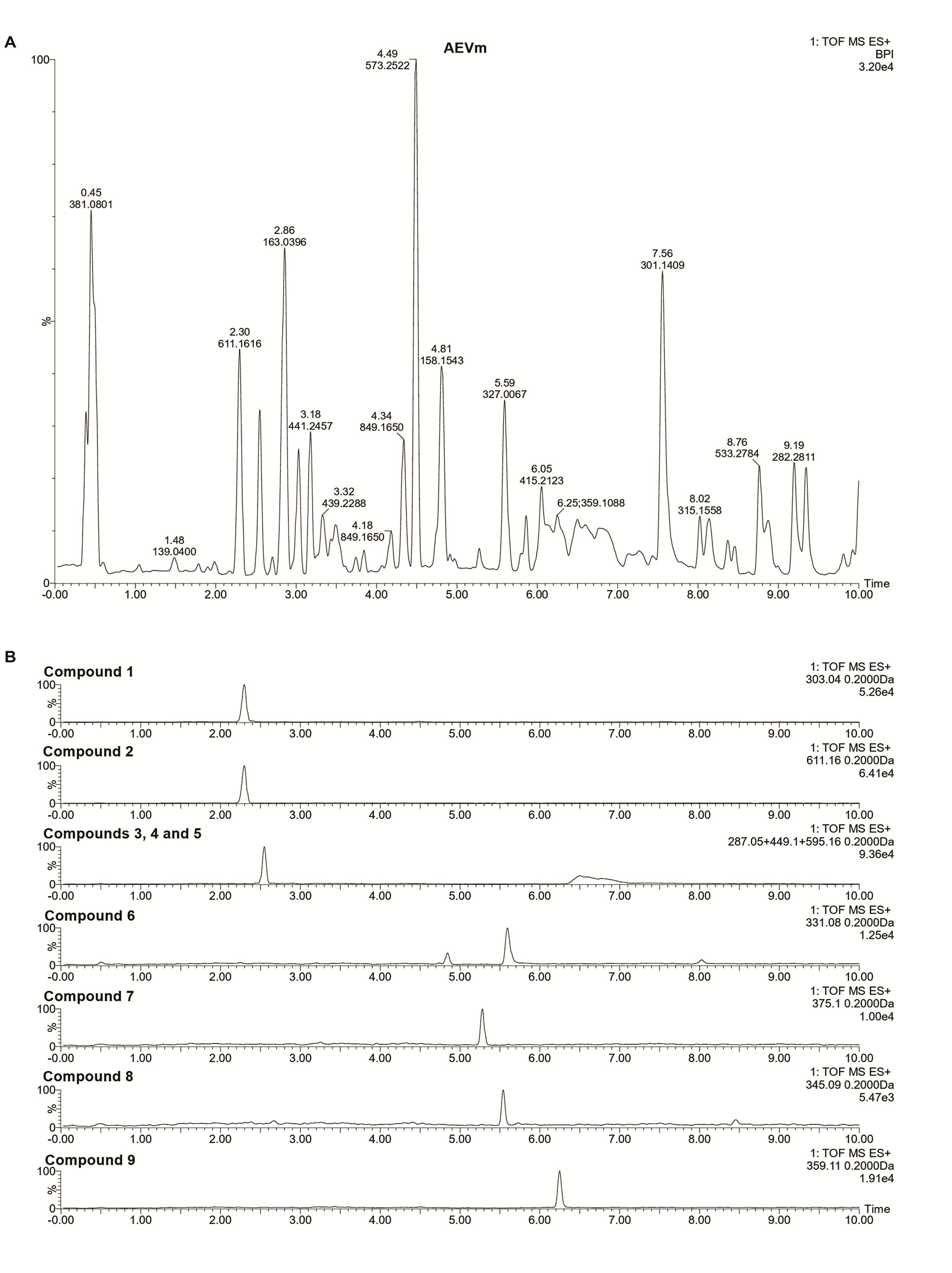
Figure 1 Total ion current chromatograms of aqueous extract of Varronia multispicata (A) and representative chromatograms of isolated flavonoids (B).
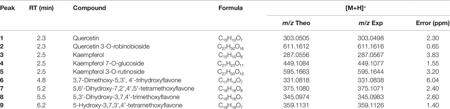
Table 1 Flavonoids identified by liquid chromatography-high-resolution-electrospray ionization-mass spectrometry in aqueous extract of Varronia multispicata.
Aqueous Extract of Varronia multispicata Acute Oral Toxicity
Aqueous Extract of Varronia multispicata Does Not Cause Deaths or Impair General Behavior, Physical, or Histological Aspects
No manifestations of toxicity were observed immediately after acute oral administration of AEVm 2,000 mg/kg and for following 14 days, with no allergic manifestations, behavioral or motor changes. Similarly, there were no deaths.
In addition, there were no prejudice in body weight gain and feed and water intake compared to the control group (Figures 2A–C), nor were there any changes in macroscopic aspects (morphology, color and size) and relative weight of stomach, liver, kidneys, and lungs (Figure 2D). Histopathological evaluation revealed no abnormalities in gastric, pulmonary, hepatic, and renal tissues (data not shown).
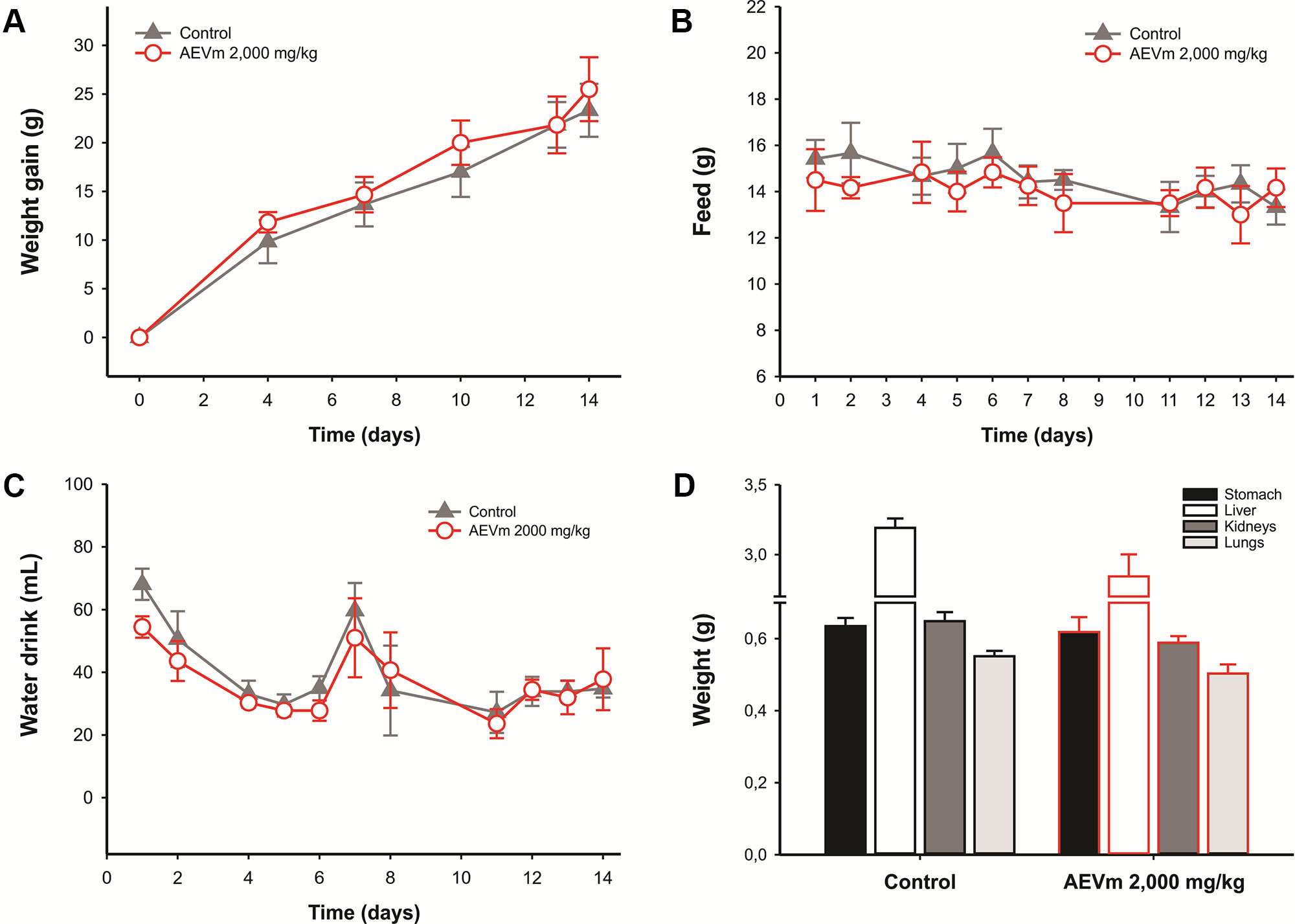
Figure 2 Effect of acute oral administration of aqueous extract of Varronia multispicata (2,000 mg/kg) on (A) body weight gain, (B) water, and (C) food intake over the following 14 days, and relative organ-body weight (D) at the end of the evaluation period. Data expressed as mean ± SEM (n = 6/group).
Aqueous Extract of Varronia multispicata Provides Protection Against Oxidative Damage
When evaluating animal plasma samples at end of observation period, i.e., on day 14 after acute oral extract administration (2,000 mg/kg), no antioxidant activity was detected (Figure 3A), as well as no change in GSH levels (Figure 3B). Despite this, the extract significantly reduced nitrite and MDA plasma levels (p < 0.001 and p < 0.05 respectively versus control; Figures 3C, D).
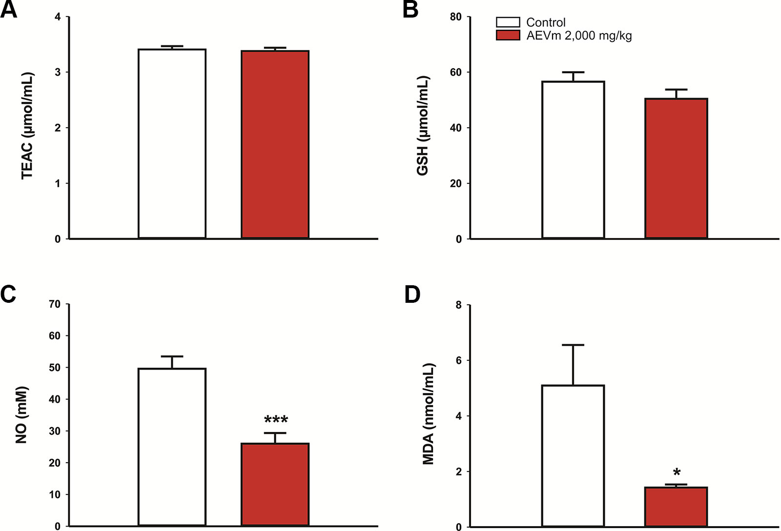
Figure 3 Effect of acute oral administration of aqueous extract of Varronia multispicata (2,000 mg/kg) on oxidative balance after 14 days of follow-up, considering (A) antioxidant activity and plasma levels of (B) GSH, (C) nitrite-NO, and (D) malondialdehyde-MDA. Data expressed as mean ± SEM (n = 6/group). ***p < 0.001; *p < 0.05 versus control. Student’s t-test.
Aqueous Extract of Varronia Multispicata Does Not Interfere With or Down-Regulate Liver Enzymes
In the same context of previous evaluation, animals treated with AEVm maintained serum levels of ALT and AST equivalent to those found in controls (Figures 4A, B). On the other hand, the extract promoted significant reductions in γ-GT and ALP levels (p < 0.05 versus control; Figures 4C, D).
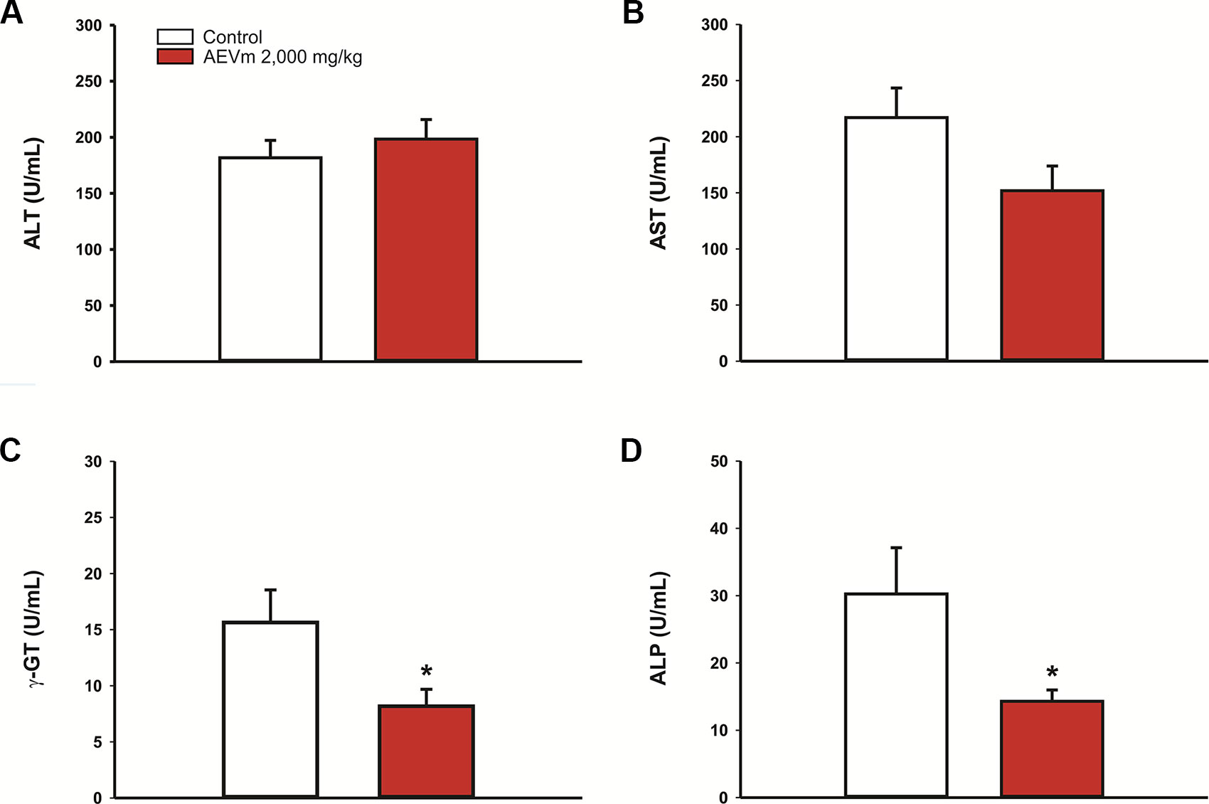
Figure 4 Effect of acute oral administration of aqueous extract of Varronia multispicata (2,000 mg/kg) on the activity of (A) alanine aminotransferase, ALT; (B) aspartate aminotransferase, AST; (C) gamma glutamyl transpeptidase, γ-GT; and (D) alkaline phosphatase, ALP. Data expressed as mean ± SEM (n = 6/group). *p < 0.05 versus control (Student’s t-test).
Antinociceptive Activity
Aqueous Extract of Varronia multispicata Inhibits Nociception in a Dose-Dependent Pattern
Oral pretreatment with AEVm (25, 75, 200, and 400 mg/kg) 1 h before WT induced a dose-dependent inhibition (p < 0.05, 0.001, 0.001, and 0.001 versus control) of acetic acid-induced abdominal writhes in mice when compared to control group (Figure 5A), with a median effective dose (ED50) of 146.89 mg/kg (Figure 5B). In addition, group treated with AEVm 400 mg/kg showed a mean reduction in nociception manifestation of 70%, reaching levels similar to animals treated with indomethacin 10 mg/kg (standard drug; 73%).
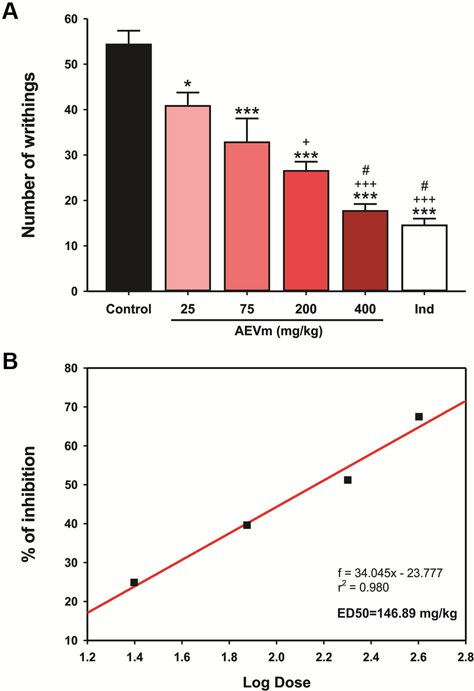
Figure 5 Effect of aqueous extract of Varronia multispicata (AEVm) (25, 75, 200, and 400 mg/kg; o.r.) on (A) acetic acid (0.6%)-induced abdominal writhing and (B) determination of median effective dose-ED50. Data expressed as mean ± SEM (n = 6/group). ***p< 0.001, *p < 0.05 versus control; +p < 0,05, +++p < 0.001 versus AEVm 25 mg/kg; #p < 0.05 versus 75 mg/kg (ANOVA, Holm-Sidak’s test).
Aqueous Extract of Varronia multispicata Does Not Compromise Locomotion and Exploration in Open Field
Applied to verify possible impairment of mobility or sedation-like effect, OF revealed that treatment with ED50 of AEVm does not compromise animals’ locomotion or exploration (Figure 6C), excluding the prejudice of such effects on antinociceptive evaluation.
Antinociceptive Effect of Aqueous Extract of Varronia multispicata Is Due to Modulation of Inflammatory Components of Pain
When AEVm ED50 was previously applied to animals undergoing FT, a significant reduction in paw licking time (P < 0.001 versus control group) was observed in second (inflammatory) phase (Figure 6A, at right). In first (neuropathic) phase of FT (Figure 6A, at left), as in HP (Figure 6B), treatment with AEVm did not interfere with animals’ nociception, which showed behavior equivalent to respective controls. Additionally, in FT, co-treatment with naloxone (0.4 mg/kg; s.c.) inhibited (p < 0.001) the morphine (4 mg/kg; s.c.) effect but not the extract effect (Figure 6A; white bars). This finding suggests that antinociceptive activity of AEVm does not involve opioid receptors activation.
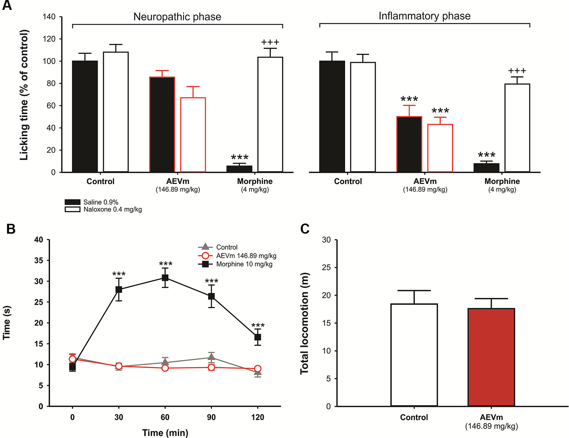
Figure 6 Effect of aqueous extract of Varronia multispicata (AEVm) (146.89 mg/kg; o.r.) on neuropathic and inflammatory nociception in formalin test with evaluation of opioid pathway participation (A), thermal nociception in hot plate test (B), and total locomotion in open field (C). Data expressed as mean ± SEM of six animals/group (n = 10/group for open field). ***p< 0.001 versus control; +++p < 0.001 versus morphine. Formalin test (ANOVA, Tukey´s test); hot plate (RM ANOVA, Holm-Sidak’s test).
Anti-Inflammatory Activity
Aqueous Extract of Varronia Multispicata Inhibits Carrageenan-Induced But Not Dextran-Induced Paw Edema
Pretreatment with 146.89 mg/kg of AEVm significantly inhibited (p < 0.001 versus control) edema formation throughout the evaluation period, promoting similar effect level to that generated by standard drug (Indomethacin 10 mg/kg) (Figure 7A). Same was not observed for dextran-induced edema. In this test, animals treated with AEVm presented edema formation equivalent to control group (Figure 7B).
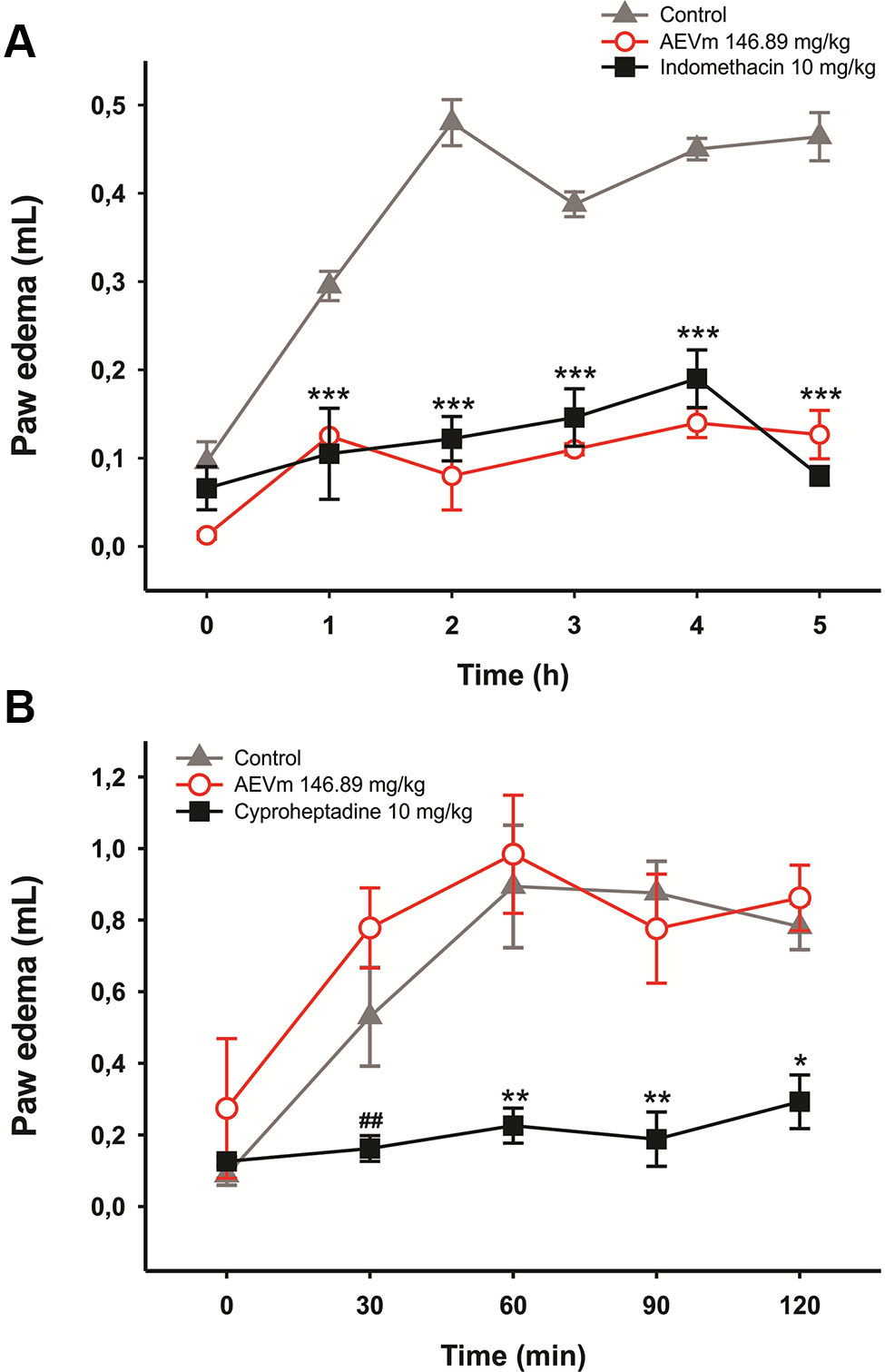
Figure 7 Effect of aqueous extract of Varronia multispicata (AEVm) (146.89 mg/kg; o.r.) on carrageenan (A) or dextran-induced (B) paw edema. Data expressed as mean ± SEM (n = 6/group). *p < 0.05, **p < 0.01, ***p < 0.001 versus control; ##p < 0.01 versus AEVm. RM ANOVA, Holm-Sidaks test.
Aqueous Extract of Varronia multispicata Inhibits Cell Migration and Nitric Oxide Production in Peritonitis Model
In peritonitis model, as intended, carrageenan significantly increased (p < 0.01) nitrite levels in peritoneal fluid compared to white group. The treatment with 146.89 mg/kg of AEVm, in turn, inhibited (p < 0.01 versus carrageenan) this elevation, promoting levels of nitrite equivalent to animals that did not undergo phlogistic induction (Figure 8A). Similarly, the extract also inhibited (p < 0.05 versus carrageenan) carrageenan-induced leukocyte migration. In both cases AEVm effect was like that promoted by Dexamethasone (Figure 8B).
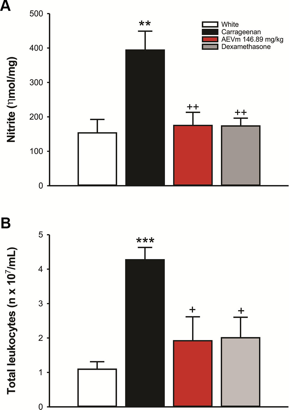
Figure 8 Effect of aqueous extract of Varronia multispicata (AEVm) (146.89 mg/kg; o.r.) on (A) nitrite levels and (B) number of leukocytes in peritoneal fluid in the carrageenan-induced peritonitis model. Data expressed as mean ± SEM (n = 6/group). **p < 0.01, ***p < 0.001 versus White; +p < 0.05, ++p < 0.01 versus Carrageenan. ANOVA, Holm-Sidaks test.
Discussion
This study is the first to evaluate the safety of acute oral administration of AEVm and its effectiveness in treating nociceptive and inflammatory processes in murine models. This initiative came from the insertion of this plant species in Amazonian medicinal culture, where it is used to treat conditions associated with inflammation and pain (Kuroyanagi et al., 2001). However, little is known about its chemical constitution, toxicity, and pharmacological properties, evidencing the relevance of elucidating its therapeutic potential. Our findings demonstrated that AEVm has low toxicity when administered acutely orally, in addition to evidence of hepatoprotection and antioxidant effect. It also demonstrated important antinociceptive and anti-inflammatory activity.
Flavonoids: quercetin (1); quercetin-3-O-rutinoside (2); kaempferol (3); kaempferol-7-O-glycoside (4); kaempferol-3-O-rutinoside (5); 3,7-dimethoxy-5,3’,4’-trihydroxyflavone (6); 5,6’-dihydroxy-7,2’,4’,5’-tetramethoxyflavone (7); 5,3’-dihydroxy-3,7,4’-trimethoxyflavone (8); and 5-hydroxy-3,7,3’,4’-tetramethoxyflavone (9) were identified based on exact masses compared to those described in the literature and compared with patterns isolated by the group (compounds 6 to 9). Flavonoids, quercetin, and kaempferol, as well as their glycosides are widespread in the plant kingdom. Nevertheless, they have a variety of biological activities such as hepatoprotective, antioxidant, and anti-inflammatory (Mohamed et al., 2010; Costa et al., 2012; Sokkar et al., 2016). Flavones are part of another subgroup of flavonoids that are widely studied because they have various pharmacological activities, suggesting that the presence of these flavonoids may contribute to the antinociceptive and anti-inflammatory properties of AEVm found in this study.
The low toxicity was evidenced by absence of deaths following the 2,000 mg/kg dose limit administration in female rats. According to the parameters proposed by OECD (2001), any substance with LD50 between 2,000 and 5,000 mg/kg, as occurred with AEVm, is extruded as a low toxicity xenobiotic. In addition, treatment of animals at limit dose did not impair the general behavior and mobility immediately after administration or over the 14 days of follow-up. Weight gain was also not altered by treatment with extract, indicating that AEVm does not cause damage to nutrition. These data are reinforced by maintenance of feed and water intake at levels equivalent to controls. In evaluation of limit dose systemic repercussions, macroscopic aspects and relative weight of the stomach, liver, kidneys, and lungs were conserved, as well as the cellular architecture and morphology of their tissues. These data are supported by the biochemical evaluation, which showed an absence of interference with ALT and AST activity. γ-GT and ALP, on the other hand, had their activity reduced among animals treated with AEVm. Hepatocellular enzymes are important markers for diagnosis of liver lesions. ALT is considered the most specific marker of liver injury, as AST may also have its levels influenced by changes in other organs, such as the heart. γ-GT is also related to renal function, oxidative stress, and chronic inflammation. Elevation of serum ALP levels is related to hepatobiliary dysfunctions and cholestatic injuries (Olatosin et al., 2014; Wang et al., 2016). Its evaluation, therefore, acts as a predictor of possible drug hepatotoxicity, as occurs with acetaminophen, whose hepatic aggression promotes a characteristic elevation of hepatocellular enzymes (Olatosin et al., 2014). In AEVm case, our biochemical results indicate the absence of tissue damage, especially to liver (Liu et al., 2014b).
The imbalance between oxidant species production and the systemic ability to neutralize them, called oxidative stress, is an important source of xenobiotic-induced cell damage. Our findings showed that AEVm does not impair plasma total antioxidant capacity, nor does it interfere with GSH content. Despite this, the extract exhibited significant evidence of antioxidant potential, as it significantly reduced levels of MDA, a product of lipid peroxidation cell damage, compared with controls. It also reduced NO plasma content, an oxidant radical, assessed indirectly through nitrite concentrations. These properties may be related to flavonoids present in the species (Mohamed et al., 2010; Costa et al., 2012; Sokkar et al., 2016). In addition, antioxidant effect of AEVm may be involved in preserving the histological and functional normality of the liver, an organ with intense oxidative metabolism (Sokkar et al., 2016). Results presented highlight not only the safety of acute oral use of the extract, but also its possible protective effects. We consider, however, the importance of further studies on extract tolerability in subchronic and chronic models.
Based on measured safety, we selected the doses to be applied in evaluation of pharmacological properties. We started at doses equivalent to 20 and 10% of the dose tested for toxicity assessment, i.e., 400 and 200 mg/kg. The other doses were adopted according to drug effect pattern. Antinociceptive and anti-inflammatory properties of AEVm, as well as the possible related mechanisms, were verified by established murine models, starting with WT, a sensitive visceral pain model for evaluation of substances with analgesic potential (Popoola et al., 2016). Acetic acid-induced nociception combines the induction of nociceptive mediators’ expression and/or release (arachidonic acid, cyclooxygenase-2, prostaglandins E2 and F2α, bradykinin, serotonin, and histamine) and non-selective cation channels activation (Hasan et al., 2014). Therefore, it is a model of inflammatory pain and is efficiently modulated by drugs capable of inhibiting prostaglandin biosynthesis, despite the diversity of mechanisms that involve it. Our results demonstrated that AEVm significantly inhibited the writhing generated by acetic acid-induced inflammation at all doses (25, 75, 200, and 400 mg/kg) tested, showing a dose-dependent pattern with an ED50 of 146.89 mg/kg. These findings allow us to infer that AEVm may exert its effect by inhibiting the synthesis/release of inflammatory mediators, especially prostaglandins.
To verify the performance of AEVm on peripheral and central components of nociception was performed the FT. For this and other tests we adopted the WT ED50 as standard dose of AEVm, aiming to reduce the number of animals used. FT is a model of clinical pain, which triggers in two phases from the sc administration of formalin. The first (neurogenic) phase, which begins immediately after formalin administration, is characterized by direct activation of peripheral nociceptive fibers, especially C fibers. This neuropathic nociception inhibited by drugs capable of modulating neural pain mechanisms, such as opioids, that also control inflammatory nociception. The second (inflammatory) phase begins after 15 min of noxious stimulation, involving the activation of inflammatory pathways related to prostaglandins, serotonin, histamine, and NO. In this case, anti-inflammatory drugs can effectively control the nociception (Hunskaar and Hole, 1987; De Oliveira et al., 2015; Islam et al., 2019). AEVm (146.89 mg/kg) significantly (p < 0.001) reduces the nociception in FT second phase. However, this effect was not observed in the first phase. Similarly, in HP test, a thermogenic pain model that evokes supraspinatus nociception, therefore modulated by centrally acting analgesics, the extract did not increase latency for nociceptive reflex, i.e., paw lick or jump (Le Bars et al., 2001). In addition, opioid antagonist naloxone reversed the standard drug (morphine) effect in FT but did not modify the AEVm activity. These results allow us to deduce that the antinociceptive activity of AEVm involves peripheral mechanisms, especially inflammatory ones. It also reveals that the extract does not interfere with neurogenic nociceptive mechanisms and that opioid system is not involved in its effects.
Ultimately, to rule out the possibility of these effects being a product of locomotor capacity inhibition, we evaluated the ambulation of mice in open field for 5 min. The animals treated with the extract (146.89 mg/kg) presented locomotion, i. e., the total distance traveled, equivalent to the control group, allowing to conclude that it did not interfere with the locomotor behavior during nociception experiments. This finding is reinforced by the normality of tactile sensitivity and reflexes and absence of lethargy observed in the toxicity assessment, where animals were treated with 2,000 mg/kg of AEVm.
The anti-inflammatory activity of AEVm was evaluated in carrageenan-induced and dextran-induced paw edema and carrageenan-induced peritonitis in rats. Initial moments after phlogistic stimulation by intraplantar injection of carrageenan (0–2 h) are marked by the rise of inflammatory mediators such as histamine, serotonin and bradykinin, with increasing in local blood flow and capillary permeability, resulting in edema initiation (Sengar et al., 2015). In advanced stage (2–6 h) of inflammation, prostaglandin production, due to cyclooxygenase-2 (COX2) induction, and leukocyte migration are considered the key elements for its maintenance (Marzouk et al., 2010; Paiva et al., 2013). Our results revealed that AEVm (146.89 mg/kg) significantly inhibits edema formation in both stages with indomethacin-like effectiveness. Its efficacy in early stage is suggestive of histamine or bradykinin pathways inhibition, as well as possible stabilization of mast cells (Shashidhara et al., 2008). The persistence of the effect on late stage (2–5 h), with an average inhibition of about 74%, however, suggests possible interference with COX induction or activity. In dextran-induced model, whose edema formation occurs by induction of mast cell degranulation and release of histamine and serotonin (Ankier and Neat, 1972; Van Wauwe and Goossens, 1989), the involvement of these pathways in antiedematogenic effect of AEVm was evaluated. Our results demonstrated that the extract does not interfere with dextran-induced inflammation, indicating that histaminergic pathway is not involved in the antiedematogenic effect observed in the carrageenan-induced paw edema.
In the carrageenan-induced peritonitis test AEVm promoted potent inhibition of leukocyte migration and NO production. This test is an acute inflammation model, marked by intense leukocyte migration to the peritoneal cavity, induction of NO synthesis by induced nitric oxide synthase (iNOS) and COX2 expression, resulting in increased prostaglandin concentration. Indeed, several studies have shown the strong influence of NO production in prostaglandin levels, so that inhibition of NO synthesis reduces COX2 expression and prostaglandin production (Aquino et al., 2017). The ability of the extract to inhibit NO overproduction partly clarifies its mechanism of action and reinforces the hypothesis of the involvement of prostaglandin synthesis modulation in the antinociceptive and anti-inflammatory activities of AEVm.
In summary, our results demonstrate that the AEVm is rich in flavonoids, identified by UPLC-ESI-HRMS analysis, which potentially form the basis of its biological properties. They also demonstrated the safety of AEVm, that presented low oral acute toxicity, with antioxidant and hepatoprotective potential. Additionally, the extract has important peripheral antinociceptive and anti-inflammatory activity, which seems to be related to its constituent’s ability to inhibit NO and prostaglandin production, as well as inhibit leukocyte migration. Thus, the extract has shown promise for the development of new natural product aimed at treating pain and inflammation.
Data Availability Statement
The datasets generated for this study are available on request to the corresponding author.
Ethics Statement
The animal study was reviewed and approved by Ethic Committee on Animal Use of the Federal University of Para (CEUA/UFPA).
Author Contributions
The study was conceived and designed by EF-J, CS, and MS. Experiments and data collection were performed by KL, FS-J, JO, TS, DA, SA, and PG. Data were analyzed by EF-J, KL, WP, MC, CM, CS, PG and MS. Drafting of the manuscript: KL and EF-J. Critical revision of the manuscript: EF-J and MS. All authors revised and approved the final version of the manuscript.
Conflict of Interest
The authors declare that the research was conducted in the absence of any commercial or financial relationships that could be construed as a potential conflict of interest.
Acknowledgments
We thank Dr. Silvane Tavares Rodrigues, Brazilian Agricultural Research Corporation (Embrapa) Eastern Amazon (Pará-Brazil) for the identification of plant. We are also grateful to Universidade Federal do Pará (UFPA) and Coordenação de Aperfeiçoamento de Pessoal de Nível Superior (CAPES) for the financial support and to Evandro Chagas Institute for supplying the animals for this project.
References
Alencar De Menezes, J. E. S., Lemos, T. L. G., Pessoa, O. D. L., Braz-Filho, R., Montenegro, R. C., Wilke, D. V. (2005). A cytotoxic meroterpenoid benzoquinone from roots of Cordia globosa. Planta Med. 71 (1), 54–58. doi: 10.1055/s-2005-837751
Ankier, S. I., Neat, M. L. (1972). Some studies on acute inflammation induced by dextran in the mouse. Int. Arch. Allergy Appl. Immunol. 42, 264–277. doi: 10.1159/000230611
Aquino, P. E. A., Magalhães, T. R., Nicolau, L. A. D., Leal, L. K. A. M., Aquino, N. C., Santos, S. M., et al. (2017). The anti-inflammatory effects of N-methyl-(2S,4R)-trans-4-hydroxyl-proline from Syderoxylon obtusifolium are related to its inhibition of TNF-alpha and inflammatory enzymes. Phytomedicine 24, 14–23. doi: 10.1016/j.phymed.2016.11.010
Carvalho, J. C. T., Sertié, J. A. A., Barbosa, M. V. J., Patrício, K. C. M., Caputo, L. R. G., Sarti, S. J. (1999). Anti-inflammatory activity of the crude extract from the fruits of Pterodon emarginatus Vog. J. Ethnopharmacol. 64 (2), 127–133. doi: 10.1016/S0378-8741(98)00116-0
Conforti, F., Sosa, S., Marrelli, M., Menichini, F., Statti, G. A., Uzunov, D. (2009). The protective ability of Mediterranean dietary plants against the oxidative damage: The role of radical oxygen species in inflammation and the polyphenol, flavonoid and sterol contents. Food Chem. 112 (3), 587–594. doi: 10.1016/j.foodchem.2008.06.013
Costa, G., Francisco, V., Lopes, M. C., Cruz, M. T., Batista, M. T. (2012). Intracellular Signaling Pathways Modulated by Phenolic Compounds: Application for New Anti-Inflammatory Drugs Discovery. Curr. Med. Chem. 19 (18), 2876–2900. doi: 10.2174/092986712800672049
De Oliveira, A. M., De Araújo, A. F., Lemos, R. P. L., Conserva, L. M., De Souza Ferro, J. N., Barreto, E. (2015). Antinociceptive and anti-inflammatory activity of the siaresinolic acid, a triterpene isolated from the leaves of Sabicea grisea Cham. & Schltdl. var. grisea. J. Nat. Med. 69 (2), 232–240. doi: 10.1007/s11418-014-0883-3
Dharmasiri, M. G., Jayakody, J. R. A. C., Galhena, G., Liyanage, S. S. P., Ratnasooriya, W. D. (2003). Anti-inflammatory and analgesic activities of mature fresh leaves of Vitex negundo. J. Ethnopharmacol. 87 (2–3), 199–206. doi: 10.1016/S0378-8741(03)00159-4
Diniz, J. C., Viana, F. A., Oliveira, O. F., Silveira, E. R., Pessoa, O. D. L. (2008). Chemical composition of the leaf essential oil of Cordia leucocephala Moric from northeast of Brazil. J. Essent Oil Res. 20 (6), 495–496. doi: 10.1080/10412905.2008.9700068
Dutra, R. C., Campos, M. M., Santos, A. R. S., Calixto, J. B. (2016). Medicinal plants in Brazil: Pharmacological studies, drug discovery, challenges and perspectives. Pharmacol. Res. 112, 4–29. doi: 10.1016/j.phrs.2016.01.021
Ellman, G. L. (1959). Tissue sulfhydryl groups. Arch. Biochem. Biophys. 82, 70–77. doi: 10.1016/0003-9861(59)90090-6
Forzza, R. C., Zappi, D., Souza, V. C. (2016-continuously updated). Flora do Brasil 2020. Jardim Botânico do Rio de Janeiro. Available in: http://reflora.jbrj.gov.br/reflora/floradobrasil/FB105429. (Accessed in: September 30, 2019)
Fun, C. E., Svendsen, A. B. (1990). The essential oil of Cordia cylindrostachya Roem. & Schult. grown on Aruba. J. Essential Oil Res. 2 (4), 209–210. doi: 10.1080/10412905.1990.9697864
Gottschling, M., Miller, J. S., Weigend, M., Hilger, H. H. (2005). Congruence of a phylogeny of Cordiaceae (Boraginales) inferred from ITS1 sequence data with morphology, ecology, and biogeography. Ann. Mo. Bot. Gard. 92 (3), 425–437.
Granger, D. L., Anstey, N. M., Miller, W. C., Weinberg, J. B. (1999). Measuring nitric oxide production in human clinical studies. Methods Enzymol. 301, 49–61. doi: 10.1016/S0076-6879(99)01068-X
Gupta, M., Mazumder, U. K., Gomathi, P., Selvan, V. T. (2006). Antiinflammatory evaluation of leaves of Plumeria acuminata. BMC Complement. Altern. Med. 6, 1–6. doi: 10.1186/1472-6882-6-36
Gurke, M. (1893). Boraginaceae, in Die naturlichen Pflanzenfamilien, vol. 15 . Eds. Engler, K., Prantl, A. (Leipzig: V.W. Engelmann), 49–96.
Hamada, H., Ohkura, Y. (1976). A new photometric method for the determination of serum glutamate pyruvate transaminase activity using pyruvate and glutamate as substrates. Chem. Pharm. Bull. 24, 1865–1869. doi:10.1248/cpb.24.1865
Hasan, M. M., Uddin, N., Hasan, M. R. (2014). Analgesic and antiinflammatory activities of leaf extract of Mallotus repandus (Willd.). Muell. Arg. BioMed. Res. Int. 2014, 1–7. doi: 10.1155/2014/539807
Hegde, K. S., Chandrashekhar, S., Rao, S. N., Manikkoth, S. (2014). Preclinical screening of phyllanthus amarus ethanolic extract for its analgesic and antimicrobial activity. Pharmacognosy Res. 7 (4), 378–384. doi: 10.4103/0974-8490.159577
Henschke, N., Kamper, S. J., Maher, C. G. (2015). The epidemiology and economic consequences of pain. Mayo Clin. Proc. 90 (1), 139–147. doi: 10.1016/j.mayocp.2014.09.010
Henzen, C. (2003). Traitement aux glucocorticoids; risques et effets secondaires. Forum Med. Suisse 19, 442–446. doi: 10.4414/fms.2003.04866
Hormaza, I. M., Amador, M. C. V., Álvarez, G. B., Rodríguez, F. M., Barreiro, M. L., Hernández, E. (2014). Preclinical validation of antinociceptive, anti-inflammatory, and antipyretic activities of Cordia martinicensis leave decoction. Rev. Cubana Plantas Med. 19 (1), 29–39.
Hunskaar, S., Hole, K. (1987). The formalin test in mice: dissociation between inflammatory and non-inflammatory pain. Pain 30 (1), 103114. doi: 10.1016/0304-3959(87)90088-1
Hunskaar, S., Fasmer, O. B., Hole, K. (1985). Formalin test in mice, a useful technique for evaluating mild analgesics. J. Neurosci. Methods 14 (1), 69–76. doi: 10.1016/0165-0270(85)90116-5
Ioset, J. R., Marston, A., Gupta, M. P., Hostettmann, K. (1998). Antifungal and larvicidal meroterpenoid naphthoquinones and a naphthoxirene from the roots of Cordia linnaei. Phytochemistry 47 (5), 729–734. doi: 10.1016/S0031-9422(97)00695-X
Ioset, J. R., Marston, A., Gupta, M. P., Hostettmann, K. (2000). Antifungal and larvicidal cordiaquinones from the roots of Cordia curassavica. Phytochemistry 53 (5), 613–617. doi: 10.1016/S0031-9422(99)00604-4
Islam, S., Shajib, M. S., Rashid, R. B., Khan, M.F., Al-Mansur, M. A., Datta, B. K., et al. (2019). Antinociceptive activities of Artocarpus lacucha Buch-ham (Moraceae) and its isolated phenolic compound, catechin, in mice. BMC Complement Altern Med. 19 (1), 214. doi: 10.1186/s12906-019-2565-x
Koster, R., Anderson, M., De Beer, E. J. (1959). Acetic acid for analgesic screening. Fed. Proc. 18, 412–417.
Kumara, N. K. V. M. R. (2001). Identification of strategies to improve research on medicinal plants used in Sri Lanka, In: Proceedings of the WHO Symposium; Galle, Sri Lanka: University of Ruhuna. pp. 12–14.
Kunanusorn, P., Teekachunhatean, S., Sangdee, C., Panthong, A. (2009). Antinociceptive and anti-inflammatory activities of a chinese herbal recipe (DJW) in animal models. Int. J. Appl. Res. Nat. Prod. 2, 1–8.
Kuroyanagi, M., Kawahara, N., Sekita, S., Satake, M., Hayashi, T., Takase, Y. (2003). Dammarane-type triterpenes from the Brazilian medicinal plant Cordia multispicata. J. Nat. Prod. 66, 1307–1312. doi:10.1021/np020483f
Kuroyanagi, M., Seki, T., Hayashi, T., Nagashima, Y., Kawahara, N., Sekita, S. (2001). Anti-androgenic triterpenoids from the Brazilian medicinal plants Cordia multispicata. Chem. Pharm. Bull. 49, 954–957. doi:10.1248/cpb.49.954
Le Bars, D., Gozariu, M., Cadden, S. W. (2001). Acute pain measurement in animals. Part 1. Ann. Fr. Anesth. Reanim. 20 (4), 347–365. doi: 10.1016/s0750-7658(01)00381-1
Liu, W., Xu, Z., Deng, Y., Xu, B., Yang, H., Wei, Y. (2014). Excitotoxicity and oxidative damages induced by methylmercury in rat cerebral cortex and the protective effects of tea polyphenols. Environ. Toxicol. 29, 269–283. doi: 10.1002/tox.21755
Liu, X., Hamnvik, O. P. R., Chamberland, J. P., Petrou, M., Gong, H., Christophi, C. A. (2014). Circulating alanine transaminase (ALT) and γ-glutamyl transferase (GGT), but not fetuin-A, is associated with metabolic risk factors, at baseline and at two-year follow-up: the prospective Cyprus Metabolism Study. Metabolism 63 (6), 773–782. doi: 10.1038/jid.2014.371
Macdonald, A. D., Woolfe, G. (1946). Analgesic action of pethidine derivatives and related compounds. Br. J. Pharmacol. Chemother. 1, 4–14. doi: 10.1111/j.1476-5381.1946.tb00022.x
Malone, M. H. (1977) Pharmacological approaches to natural product screening and evaluation. In: Wagner, H., Wolff, P. (eds) New Natural Products and Plant Drugs with Pharmacological, Biological or Therapeutical Activity. Proceedings in Life Sciences. Berlin, Heidelberg: Springer. doi: 10.1007/978-3-642-66682-7_2
Martínez, I., Amador, M., Álvarez, G., Morón, F., López, M., García, A. (2014). Preclinical validation of antinociceptive, antiinflammatory, and antipyretic activities of Cordia martinicensis leave decoction. Rev. Cubana Plantas Med. 19 (1), 29–39.
Marzouk, B., Marzouk, Z., Haloui, E., Fenina, N., Bouraoui, A., Aouni, M. (2010). Screening of analgesic and anti-inflammatory activities of Citrullus colocynthis from southern Tunisia. J. Ethnopharmacol. 128, 15–19. doi: 10.1016/j.jep.2009.11.027
Miller, J. S., Gottschling, M. (2007). Generic classification in the Cordiaceae (Boraginales): resurrection of the genus Varronia P. Br. Taxon 56, 163–169. doi: 10.2307/25065747
Miller, J. S., Wood, J. R. I. (2008). New boraginaceae from tropical america 6: a new species of varronia from bolivia. Novon 18, 86–89. doi: 10.3417/2006004
Miller, N. J., Rice-Evans, C., Davies, M. J., Gopinathan, V., Milner, A. (1993). A novel method for measuring antioxidant capacity and its application to monitoring the antioxidant status in premature neonates. Clin. Sci. 84 (4), 407–412. doi: 10.1042/cs0840407
Mohamed, A. A., Khalil, A. A., El-Beltagi, H. E. S. (2010). Antioxidant and antimicrobial properties of kaff maryam (Anastatica hierochuntica) and doum palm (Hyphaene thebaica). Grasas y Aceites 61 (1), 67–75. doi: 10.3989/gya.064509
Nathan, C., Ding, A. (2010). Nonresolving Inflammation. Cell 140 (6), 871–882. doi: 10.1016/j.cell.2010.02.029
Novais, T., Costa, J. F. O., David, J. P. L., David, J. M., Queiroz, L. P., França, F. (2003). Atividade antibacteriana em alguns extratos de vegetais do semi-árido brasileiro. Rev. Bras. Farmacognosia 13 (Tabela 1), 5–8. doi: 10.1590/s0102-695x2003000400003
OECD (2001), Test No. 420: Acute Oral Toxicity - Fixed Dose Procedure, OECD Guidelines for the Testing of Chemicals, Section 4, OECD Publishing, Paris. doi: 10.1787/9789264070943-en
Olatosin, T. M., Akinduko, D. S., Uche, C. Z., Bardi, J. (2014). Effects of Moringa oleifera Seed Oil on Acetaminophen-Induced Oxidative Stress and Liver Damage in Wistar Albino Rats. IOSR J. Pharm. Biolo. Scis. 9, 53–59. doi: 10.9790/3008-09215359
Oliveira, J. C. S., Camara, C. A. G., Schwartz, M. O. E. (2007). Volatile constituents of the stem and leaves of cordia species from mountain forests of pernambuco (north-eastern Brazil). J. Essen. Oil Res. 19 (5), 444–448. doi: 10.1080/10412905.2007.9699947
Paiva, D. C. C., Santos, C. A., Diniz, J. C., Viana, F. A., Thomazzi, S. M., Falcão, D. A. (2013). Anti-inflammatory and antinociceptive effects of hydroalcoholic extract from Pseudobombax marginatum inner bark from caatinga potiguar. J. Ethnopharmacol. 149 (2), 416–421. doi: 10.1016/j.jep.2013.06.012
Passos, G. F., Fernandes, E. S., da Cunha, F. M., Ferreira, J., Pianowski, L. F., Campos, M. M. (2007). Anti-inflammatory and anti-allergic properties of the essential oil and active compounds from Cordia verbenacea. J. Ethnopharmacol. 110 (2), 323–333. doi: 10.1016/j.jep.2006.09.032
Phanse, M. A., Patil, M. J., Abbulu, K., Chaudhari, P. D., Patel, B. (2012). In-vivo and in-vitro screening of medicinal plants for their anti-inflammatory activity: An overview. J. Appl. Pharma. Sci. 2 (7), 19–33. doi: 10.7324/JAPS.2012.2704
Phillip, M. A., Graham, R. P. (1995). Designing a coupled assay system for aspartate aminotransferase. Biochem. Edu. 23, 40–41. doi: 10.1016/0307-4412(94)00116-7
Popoola, T. D., Awodele, O., Omisanya, A., Obi, N., Umezinwa, C., Fatokun, A. A. (2016). Three indigenous plants used in anti-cancer remedies, Garcinia kola Heckel (stem bark), Uvaria chamae P. Beauv. (root) and Olax subscorpioidea Oliv. (root) show analgesic and anti-inflammatory activities in animal models. J. Ethnopharmacol. 194, 440–449. doi: 10.1016/j.jep.2016.09.046
Rapisarda, A., Lauk, L., Ragusa, S. (1997). Micromorphological study on leaves of Some Cordia (Boraginaceae) espécies used in tradicional medicine. Economic Bot. 51 (4), 385–391. doi:10.1007/bf02861049
Rauf, A., Farooq, U., Khan, A., Hadda, T. B., Naz, S., Ibrar, A. (2017). Sedative and muscle relaxant activities of diterpenoids from Phlomidoschema parviflorum. Braz. J. Pharmacognosy 27 (5), 636–640. doi: 10.1016/j.bjp.2017.07.003
Re, R., Pellegrini, N., Proteggente, A., Pannala, A., Yang, M., Rice-Evans, C. (1999). Antioxidant activity applying an improved ABTS radical cation decolorization assay. Free Radic. Biol. Med. 26 (9–10), 12317. doi: 10.1016/s0891-5849(98)00315-3
Rodrigues, F. F. G., Oliveira, L. G. S., Rodrigues, F. F. G., Saraiva, M. E., Almeida, S. C. X., Cabral, M. E. S., et al. (2012). Chemical composition, antibacterial and antifungal activities of essential oil from Cordia verbenacea DC leaves. Pharmacognosy Res. 4 (3), 161–165. doi: doi./10.4103/0974-8490.99080
Rosenblum, A., Marsch, L. A., Joseph, H., Portenoy, R. K. (2008). Opioids and the Treatment of Chronic Pain: Controversies, Current Status, and Future Directions. Exp. Clin. Psychopharmacol. 16 (5), 405–416. doi: 10.1037/a0013628
Samy, R. P., Pushparaj, P. N., Gopalakrishnakone, P. (2008). A compilation of bioactive compounds from Ayurveda. Bioinformation 3 (3), 100–110. doi: 10.6026/97320630003100
Santos, R. P., Nunes, E. P., Nascimento, R. F., Santiago, G. M. P., Menezes, G. H. A., Silveira, E. R. (2006). Chemical composition and larvicidal activity of the essential oils of Cordia leucomalloides and Cordia curassavica from the Northeast of Brazil. J. Braz. Chem. Soc. 17 (5), 1027–1030. doi: 10.1590/S0103-50532006000500030
Schalcher, T. R., Borges, R. S., Coleman, M. D., Batista Júnior, J., Salgado, C. G., Vieira, J. L. F. (2014). Clinical Oxidative Stress during Leprosy Multidrug Therapy: Impact of Dapsone Oxidation. PloS One 9, e85712. doi: 10.1371/journal.pone.0085712
Schlechtendal, D., Chamisso, A. (1830). Plantarum mexicanarum a cel. viris Schiede et Deppe collectarum recensio brevis. Linnaea 5, 72–174.
Sengar, N., Joshi, A., Prasad, S. K., Hemalatha, S. (2015). Anti-inflammatory, analgesic and anti-pyretic activities of standardized root extract of Jasminum sambac. J. Ethnopharmacol. 160, 140–148. doi: 10.1016/j.jep.2014.11.039
Shahed-Al-Mahmud, M., Lina, S. M. M. (2017). Evaluation of sedative and anxiolytic activities of methanol extract of leaves of Persicaria hydropiper in mice. Clin. Phytosci. 3, 20. doi:10.1186/s40816-017-0056-5
Shashidhara, S., Bhandarkar, A. V., Deepak, M. (2008). Comparative evaluation of successive extracts of leaf and stem bark of Albizzia lebbeck for mast cell stabilization activity. Fitoterapia 79 (4), 301–302. doi: 10.1016/j.fitote.2008.01.006
Sokkar, N. M., El-Hawary, S. M., Slem, A. M., Talaat, Z. (2016). The phenolic composition of the hepatoprotective and antioxidant fractions of Albizia lebbeck L. Quim. Nova 39 (8), 973–978. doi: 10.5935/0100-4042.20160112
Stapf, M. N. S. (2015) Varronia in Lista de Espécies da Flora do Brasil. Jardim Botânico do Rio de Janeiro. Available in: http://floradobrasil.jbrj.gov.br/jabot/floradobrasil/FB105279.
Szasz, G. (1969). A kinetic photometric method for serum gamma-glutamyl transpeptidase. Clin. Chem. 15 (2), 124–136.
Van Wauwe, J. P., Goossens, J. G. (1989). Arabinogalactan- and dextran-induced ear inflammation in mice: Differential inhibition by H1-antihistamines, 5-HT-serotonin antagonists and lipoxygenase blockers. Agents Act. 28 (1–2), 78–82. doi: 10.1007/BF02022984
Vieira, I. C. G., Silva, J. M. C. (1997). Phenology, fruit set and dispersal of Cordia multispicata Cham., an important weed shrub of abandoned pastures in eastern Amazonia. Rev. Bras. Botânica 20 (1), 51–56. doi: 10.1590/s0100-84041997000100005
Wang, Y. L., Koh, W. P., Yuan, J. M., Pan, A. (2016). Association between liver enzymes and incident type 2 diabetes in Singapore Chinese men and women. BMJ Open Diabetes Res. Care 4 (1), 2–9. doi: 10.1136/bmjdrc-2016-000296
Winter, C. A., Risley, E. A., Nuss, G. W. (1962). Carrageenin-induced edema in hind paw. Exp. Biol. Med. 3 (111), 544–547. doi: 10.3181/00379727-111-27849
Wirth, H., Hürlimann, R., Flückiger, T. (2006). Les AINS et les inhibiteurs de la COX-2: principaux effets indésirables. Forum Méd. Suisse Swiss Med. Forum 6 (12), 284–290. doi: 10.4414/fms.2006.05816
Yagi, T., Kagamiyama, H., Ohtawara, S., Soda, K., Nozaki, M. (1979). A new assay for l-aspartate: 2-oxoglutarate aminotransferase. Analytical Biochem. 100 (1), 20–24. doi: 10.1016/0003-2697(79)90104-0
Yang, T., Xu, Z., Liu, W., Xu, B., Deng, Y., Li, Y., et al. (2015). Alpha-lipoic acid protects against methylmercury-induced neurotoxic effects via inhibition of oxidative stress in rat cerebral cortex. Environ. Toxicol. Pharmacol. 39, 157–166. doi: 10.1016/j.etap.2014.11.020
Keywords: Varronia multispicata (Cham.) Borhidi, antinociceptive, anti-inflammatory, toxicity, folk medicine, flavonoids
Citation: Lopes K, Oliveira J, Sousa-Junior FJC, Santos TdF, Andrade D, Andrade SL, Pereira WL, Gomes PWP, Monteiro MC, e Silva CYY, da Silva MN, Maia CF and Fontes-Júnior EA (2019) Chemical Composition, Toxicity, Antinociceptive, and Anti-Inflammatory Activity of Dry Aqueous Extract of Varronia multispicata (Cham.) Borhidi (Cordiaceae) Leaves. Front. Pharmacol. 10:1376. doi: 10.3389/fphar.2019.01376
Received: 20 September 2019; Accepted: 29 October 2019;
Published: 27 November 2019.
Edited by:
Gokhan Zengin, Selçuk University, TurkeyReviewed by:
Simone Carradori, University "G. d'Annunzio" of Chieti-Pescara, ItalySakina Mohamed Yagi, University of Khartoum, Sudan
Copyright © 2019 Lopes, Oliveira, Sousa-Junior, Santos, Andrade, Andrade, Pereira, Gomes, Monteiro, e Silva, da Silva, Maia and Fontes-Júnior. This is an open-access article distributed under the terms of the Creative Commons Attribution License (CC BY). The use, distribution or reproduction in other forums is permitted, provided the original author(s) and the copyright owner(s) are credited and that the original publication in this journal is cited, in accordance with accepted academic practice. No use, distribution or reproduction is permitted which does not comply with these terms.
*Correspondence: Enéas A. Fontes-Júnior, ZWZvbnRlc0B1ZnBhLmJy