 Lianjun Ao1
 Zhengtao Chen2
 Jiacheng Yin1Yulin Leng1Yue Luo1Xiaoxu Fu1,3,4
 Hanyu Liu1
 Xiaoke Liu1,4Hong Gao1,3,4
 Chunguang Xie1,3,4*
- 1Hospital of Chengdu University of Traditional Chinese Medicine, Chengdu, Sichuan, China
- 2Affiliated Hospital of Jiangxi University of Chinese Medicine, Nanchang, Jiangxi, China
- 3TCM Regulating Metabolic Diseases Key Laboratory of Sichuan Province, Chengdu, Sichuan, China
- 4Department of Endocrinology, Hospital of Chengdu University of Traditional Chinese Medicine, Chengdu, China
Abstract Background: Diabetic cardiomyopathy (DCM) is one of the serious microvascular complications of diabetes mellitus. It is often associated with clinical manifestations such as arrhythmias and heart failure, and significantly reduces the quality of life and years of survival of patients. Endoplasmic reticulum stress (ERS) is the removal of unfolded and misfolded proteins and is an important mechanism for the maintenance of cellular homeostasis. ERS plays an important role in the pathogenesis of DCM by causing cardiomyocyte apoptosis, insulin resistance, calcium imbalance, myocardial hypertrophy and fibrosis. Targeting ERS is a new direction in the treatment of DCM. A large number of studies have shown that Chinese herbal medicine and active ingredients can significantly improve the clinical outcome of DCM patients through intervention in ERS and effects on myocardial structure and function, which has become one of the hot research directions.
Purpose: The aim of this review is to elucidate and summarize the roles and mechanisms of Chinese herbal medicine and active ingredients that have the potential to modulate endoplasmic reticulum stress, thereby contributing to better management of DCM.
Methods: Databases such as PubMed, Web of Science, China National Knowledge Internet, and Wanfang Data Knowledge Service Platform were used to search, analyze, and collect literature, in order to review the mechanisms by which phytochemicals inhibit the progression of DCM by targeting the ERS and its key signaling pathways. Keywords used included “diabetic cardiomyopathy” and “endoplasmic reticulum stress.”
Results: This review found that Chinese herbs and their active ingredients can regulate ERS through IRE1, ATF6, and PERK pathways to reduce cardiomyocyte apoptosis, ameliorate myocardial fibrosis, and attenuate myocardial hypertrophy for the treatment of DCM.
Conclusion: A comprehensive source of information on potential ERS inhibitors is provided in this review. The analysis of the literature suggests that Chinese herbal medicine and its active ingredients can be used as potential drug candidates for the treatment of DCM. In short, we cannot ignore the role of traditional Chinese medicine in regulating ERS and treating DCM, and look forward to more research and new drugs to come.
1 Introduction
Diabetic cardiomyopathy (DCM) is a serious diabetic microvascular complication in the absence of other cardiac risk factors (e.g., coronary artery disease, hypertension, significant valvular disease, etc.) in which there are structural and functional abnormalities of the heart muscle (Jia et al., 2018). The name of the disease was first proposed by S Rubler in 1972 (Rub et al., 1972). There are 2 phenotypes of DCM, restrictive and dilated, both of which are manifestations of heart failure, with the difference being whether the ejection fraction is reduced or not (Macvanin et al., 2023). The European Society of Cardiology (Seferovic et al., 2018) and the American Heart Association (Dunlay et al., 2019) state that type 2 diabetes mellitus (T2DM) often coincides with heart failure, and that the prevalence of heart failure is higher in patients with type 2 diabetes mellitus compared to the general population, at approximately 10%–30%.
At present, Western medical treatment mainly includes lifestyle intervention, blood sugar control, lipid regulation, and anti-heart failure, but long-term drug treatment may lead to drug dependence, impaired liver and kidney function, and other adverse effects, and there are no specific drugs for DCM in Western medicine. And the risk of heart failure may be increased with traditional hypoglycemic drugs such as sulfonylureas and thiazolidinediones (Nakamura et al., 2022). Therefore, exploring traditional chinese medicine (TCM) treatment options for DCM is of positive significance. Several clinical studies have shown that TCM may be effective in the prevention and treatment of DCM in terms of clinical efficacy and symptom improvement (Lu and Zhou, 2010; Du et al., 2018; Shi et al., 2020; Zhang KW. et al., 2022; Niu et al., 2022; Tan et al., 2022). In a randomized controlled study of 82 patients with DCM, the experimental group was treated with Tongluo Nourishing Yin Formula + basic treatment, and the control group was treated with basic treatment. The results showed that the traditional Chinese medicine formula Tongluo Nourishing Yin Formula could improve clinical efficacy and left ventricular ejection fraction (LVEF), left ventricular end diastolic diameter (LVEDD), left ventricular end systolic diameter (LVESD), and left ventricular end systolic diameter (LVEDD) of the patients with DCM. Left ventricular ejection fraction (LVEF), left ventricular end diastolic diameter (LVEDD), left ventricular end systolic diameter (LVESD) cardiac function indexes in DCM patients (Zhang KW. et al., 2022). Benefiting qi-nourishing yin-activating blood circulation formula improved the left ventricular diastolic function, regulated blood glucose, improved the antioxidant capacity of the body, improved the clinical symptoms of the patients, and had a certain preventive effect on the cardiomyopathy caused by diabetes mellitus in the elderly (Lu and Zhou, 2010). A total of 33 RCT studies were included in a meta-analysis of the effects of anthocyanins on cardiometabolic health, which showed that the natural compound anthocyanins can lower blood glucose, blood lipids, and have cardiovascular protective effects (Yang et al., 2017). Many researchers have been involved in order to elucidate the mechanism of action of TCM in the treatment of DCM. It has been demonstrated that the active ingredients of Chinese medicines (Resveratrol (Fang et al., 2022), Daidzein (Laddha and Kulkarni, 2021), Salvianolic acid A (Gong et al., 2023), Rhynchophylline (Liu et al., 2022), Lycium barbarum polysaccharide (Liu et al., 2019), Salidroside (Li et al., 2021), Piperine (Wang et al., 2020), Quercetin (Jiang et al., 2022), Scutellarin (Xu et al., 2021), Luteolin (Li et al., 2019), etc.), herbal monomers (pomegranate peel (Abo-Saif et al., 2023), cinnamon (Kumar et al., 2022), galangin (Abukhalil et al., 2021), etc.), and herbal compound formulas (Fufang Xueshuantong (Peng et al., 2022), Fufang Zhenzhu Tiao Zhi formula (Yan et al., 2022; Yan et al., 2023), Si-Miao-Yong-An decoction (Li et al., 2020), Yunpi-Huoxue-Sanjie formula (Zhang et al., 2022), etc.) can treat DCM through anti-oxidative stress, anti-inflammation, anti-apoptosis, anti-fibrosis, regulation of autophagy, regulation of calcium homeostasis, regulation of cardiac lipotoxicity, and restoration of mitochondrial function.In further mechanistic studies, endoplasmic reticulum stress (ERS) was found to be a key target for treating DCM in TCM.
ERS is crucial in the development of DCM, inducing apoptosis, affecting myocardial autophagy and aggravating DCM, which is one of the important molecular mechanisms of DCM(1). Research has demonstrated that blocking the ERS pathway can decrease ERS, alleviate apoptosis, inflammation, and cardiac dysfunction in DCM myocytes (Nam et al., 2015), which is a potential target for the treatment of DCM. In recent years, the regulation of ERS has become a research hotspot for the prevention and treatment of DCM. A large number of studies have confirmed that Chinese medicine can prevent and treat DCM through the regulation of ERS, but there is still no review study in this direction. Therefore, this paper focuses on the regulation of ERS by TCM to improve DCM. It also provides an overview for further research, clinical application, and development of innovative TCM drugs for the prevention and treatment of DCM. In the study of ERS modulation by TCM, it was found that the active ingredients of TCM [Paeonol (Choy et al., 2016), Oridonin (Zhou et al., 2023), Lycopene (Sun et al., 2022), Salidroside (Wang et al., 2017), Coptisine (Zhou et al., 2021), Mangiferin (Li et al., 2020)] and TCM compound formulas [Compound Tongluo Decoction (Hui et al., 2022), Dahuang Danshen Decoction (Lian et al., 2021), Fuzheng Nizeng Decoction (Chu et al., 2022), Jiedutongluotiaogan formula (Luo et al., 2022), Qishen Granule (Zhang et al., 2020), Tong-Xie-Yao-Fang (Zhang et al., 2022)] can from different pathways regulation of ERS.
We searched Pubmed, Web of science and Chinese National Knowledge Infrastructure (CNKI) for domestic and foreign studies. The search key words are “Endoplasmic Reticulum Stresses”, “diabetic cardiomyopathy.”
2 Endoplasmic reticulum stress
The endoplasmic reticulum (ER) is an organelle in the cytoplasm of eukaryotic cells, whose functions are mainly related to protein synthesis, including post-translational modification, folding, sorting (Sanvictores and Davis, 2022). Many factors can interfere with ER function, causing unfolded or misfolded proteins to accumulate in the ER, called endoplasmic reticulum stress. When unfolded or misfolded proteins accumulate excessively in the ER lumen, an ER-specific adaptive program--the unfolded protein response (UPR), is activated to increase the degradation of these misfolded proteins to maintain protein homeostasis (Guzel et al., 2017). There are three sensors that activate the UPR (as shown in Figure 1), including inositol-requiring factor 1 (IRE1), activated transcription factor 6 (ATF6), and protein kinase R-like endoplasmic reticulum kinase (PERK). Which are located on the endoplasmic reticulum membrane. Under basal conditions, the intraluminal structures of IRE1, ATF6 and PERK are inactive when bound to the ERS chaperone molecule glucose regulatory protein 78 (GRP78), also known as binding immunoglobulin (BIP).When the levels of unfolded or misfolded proteins are elevated, the three proteins mentioned above dissociate from GRP78, which then becomes activated and activates downstream signaling molecules (Frakes and Dillin, 2017).
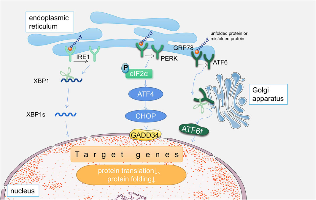
FIGURE 1. Three pathways of unfolded protein response in endoplasmic reticulum stress.
In the IRE1 pathway, its heterodimer IRE1α cleavages X-box binding protein 1 (XBP1) mRNA to generate XBP1S, which degrades the misfolded protein; in addition, IRE1 activates the apoptosis pathway to promotes apoptosis (Yang et al., 2015). In the PERK pathway, activated PERK phosphorylates the eukaryotic translation initiation factor 2 alpha (eIF2α), which prevents the translation of most proteins and reduces the burden of protein folding on the ER (He et al., 2018); It also activates activating transcription factor 4 (ATF4), induces the production of C/EBP homologous protein/growth arrest and DNA damage (CHOP/GADD), and regulates protein translation (Lakshmanan et al., 2013; Younce et al., 2013; Yang et al., 2015). In addition, it can phosphorylate nuclear factor E2-related factor 2 (NRF2), which affects oxidative stress (Yang et al., 2015). In the ATF6 pathway, activation of ERS leads to translocation of ATF6 to the Golgi, cleavage, activation, and release as a 50 kDa amino-terminal cytoplasmic fragment (ATF6f) (Hillary and FitzGerald, 2018). ATF6f is translocated to the nucleus and induces ERS factors (GRP78, XBP1), which are involved in cell death (Lakshmanan et al., 2013; Yang et al., 2015).
When the UPR occurs, the ER first tries to return to homeostasis by dampening the ERS, and if it is able to attenuate the UPR signal by reducing the number of misfolded proteins, the cell will survive; On the other hand, when homeostasis cannot be restored, the UPR signaling persists, activating apoptotic pathways and eventually leading to cell death (Oakes and Papa, 2015).
3 Endoplasmic reticulum stress and diabetic cardiomyopathy
The pathogenesis of DCM is complex, and ERS is one of the important targets. In this study, databases (Pubmed, Web of Science and CNKI) were searched and a total of about 80 articles on research involving ERS mechanisms in DCM were found and finally summarized: ERS may be involved in the pathogenesis of DCM by affecting myocardial fibrosis, myocardial hypertrophy, cardiomyocyte apoptosis, myocardial insulin resistance, and disturbance of intracellular calcium regulation (as shown in Figure 2).
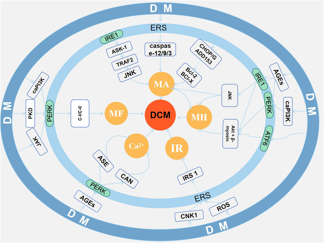
FIGURE 2. Endoplasmic reticulum stress and diabetic cardiomyopathy.
3.1 ERS and myocardial fibrosis
Myocardial fibrosis is one of the important pathological changes in DCM, including the accumulation of extracellular matrix proteins, particularly collagen, which associated with matrix metalloproteinase (MMPs) and MMP inhibitor function, involving sorbitol signaling pathway, diacylglycerol signaling pathway, etc (Asbun and Villarreal, 2006). In both diabetic patients and diabetic animal models, the manifestation of myocardial fibrosis has been observed in the diabetic heart.
It was shown that the T1DM rat model had reduced ejection fraction, elevated type III procollagen and laminin, increased expression of type I collagen, type III collagen and MMP-2/-9, wavy myocardial fibers and significantly increased collagen deposition, suggesting myocardial fibrosis and consequent cardiac dysfunction in this rat; further in vitro experiments showed that this phenomenon may be associated with activated ERS (increased expression of GRP78, GRP94, CHOP and ATF-4) (Hu et al., 2020). In addition, activation of ERS (increased expression of EIF2α, GRP94, and caspase-3) by Janus kinase and transcriptional activator signaling pathways resulted in disorganized cardiomyocyte arrangement and increased extracellular matrix collagen deposition in diabetic rats, which are involved in the process of myocardial fibrosis (Liu et al., 2018). Darnel Prakoso (Prakoso et al., 2020) et al. found that constitutively active phosphatidylinositol 3-kinase (caPI3K) is an important protein of ERS involved in myocardial fibrosis, and they found that decreased expression of caPI3K in DCM rats led to an increase in the expression of ERS-associated proteins GRP94 and CHOP, and an increase in the myocardial content of collagen types I and III and in the expression of connective tissue growth factor (CTGF), transforming growth factor-β (TGF-β), and tissue inhibitor metalloproteinase-2 (TIMP-2) expression, resulting in a significant increase in left ventricular interstitial and perivascular fibrosis. Gene therapy with a recombinant adeno-associated viral vector of the phosphatidylinositol 3 kinase construct (rAAV6-caPI3K) reduced ERS, decreased myocardial fibrosis, and improved myocardial remodeling. In addition, protein kinase D (PKD) is also an important target of ERS involved in cardiac remodeling, and PKD expression was elevated in the DCM rat model and the expression of the ERS chaperone molecule GRP78 was increased, and by correlation analysis, GRP78 was significantly correlated with PKD expression (r = 0.914) (Liu et al., 2015).
3.2 ERS and myocardial hypertrophy
Myocardial hypertrophy is another of the distinct pathological changes seen in DCM. Oxidative stress, insulin resistance and hyperglycemia are the reasons for the promotion of hypertrophic gene expression in cardiomyocytes (Grubic Rotkvic et al., 2021). In the case of cardiac hypertrophy, studies (Prakoso et al., 2020) have shown that caPI3K is also an important signaling pathway involved in ERS. After rAAV6-caPI3K treatment, cardiac natriuretic gene expression can be decreased, and cardiomyocyte width and cross-sectional area can be reduced, and the mechanism may be related to the reduction of ERS by rAAV6-caPI3K. The involvement of advanced glycosylation end products (AGEs) in the progression of ERS-induced DCM was demonstrated by Pei et al. (2018a). In STZ-induced DCM mice, cardiomyocyte cross-sectional area was increased and cardiac function was impaired; their increased phosphorylation of pro-hypertrophic signaling molecules Akt and mTOR correlated with increased phosphorylation of ERS signaling molecules GADD153, EIF2α, and IRE1α, which were ameliorated by aminoguanidine, an AGE inhibitor. It was shown that in HFD-induced DCM mice, the increase in heart weight to tibia length ratio indicates cardiac enlargement, and the increase in cardiomyocyte surface area and cross-sectional area indicates cardiac hypertrophy. The probable mechanism is that ERS initiation activates IRE1, and activation of IRE1 endonuclease activity shears XBP1 to s-XBP1 and activates the JNK pathway, inducing apoptosis, which in turn leads to pathological cardiac hypertrophy (Barr et al., 2015). Jia-hui Tian (Tian et al., 2021) et al. demonstrated that increased cardiomyocyte cross-sectional area, HG-induced increase in cardiomyocyte diameter, and markedly elevated levels of the hypertrophic markers cardiac natriuretic peptide and β-myosin heavy chain mRNA were associated with ERS-activated apoptotic pathways induced by ERS activation (GRP78, XBP-1S, ATF6, P-PERK, ATF4, CHOP) in DCM mice.
3.3 ERS and cardiomyocyte apoptosis
Apoptosis is a form of cardiomyocyte death, also known as programmed cell death, which is manifested by cytoplasmic shrinkage, nuclear enrichment, nuclear overflow, DNA fragmentation, plasma membrane blistering, and finally the formation of apoptotic bodies, and it is the fastest form of cell death; Among them, the protein cascade reaction induced by cysteine-induced aspartate protein hydrolase is a key feature, which amplifies the apoptotic pathway and leads to irreversible cell death (Chen et al., 2020). There are three pathways of ERS-induced apoptosis ERS (Xu et al., 2012). One of them is the c-Jun N-terminal kinase (JNK) signaling pathway, during ERS, activated IRE-1 recruits tumor necrosis factor-related factor 2 (TRAF2) and apoptosis signal-regulated kinase 1 (ASK-1) to activate JNK and downstream signals, in which c-Jun N-terminal inhibitory kinase (JIK) may be involved in regulating this pathway. The second pathway is the caspase-12 pathway, which is activated by multiple pathways during ERS, followed by cleavage of caspase-9 and activation of caspase-3, leading to apoptosis. The third pathway is mediated by transcriptional activation of CHOP/GADD153, which may be associated with enhanced apoptosis by inhibiting the expression of anti-apoptotic Bcl-2 and Bcl-X. In DCM rat hearts, TUNEL staining showed an increase in apoptotic cells in DCM hearts, decreased expression of anti-apoptotic protein Bcl-2, and significantly increased ERS-related proteins GRP78, caspase-9, and caspase-12; In addition, in vitro experiments significantly increased the expression of GRP78, IRE-1α, and CHOP in H9c2 cells cultured in HG; all suggesting that ERS may contribute to DCM development through the apoptotic pathway in DCM (Park et al., 2021). In addition, ERS is also involved in apoptosis in DCM through the CHOP and caspase-12 pathways, and this effect can be ameliorated by exercise (Chengji and Xianjin, 2019). Mitochondrial fusion protein-2 (MFN-2), a key protein linking mitochondria and endoplasmic reticulum, can induce cardiomyocyte apoptosis by mediating oxidative stress through ERS and mitochondrial apoptotic pathways (Yang et al., 2017).
3.4 ERS and myocardial insulin resistance
In addition to the common insulin receptor organs such as liver, fat, and muscle, the insulin signaling pathway is also present in the myocardium. Similar to the three tissues mentioned above, insulin signaling in the heart stimulates GLUT4 translocation to the cardiac cell membrane for glucose uptake via the insulin-insulin receptor substrate (IRS)-PI3K-protein kinase B (AKt) pathway (Jia et al., 2016). Myocardial insulin resistance impairs PI3K/Akt signaling, reduces glucose oxidation, increases intracellular Ca2+, and additionally activates endothelial NOS and reduces NO, leading to the development of DCM (Salvatore et al., 2021). Cannabinoid receptor gene (CNR1) is one of the important targets, Pei et al. (2018b) demonstrated that CNR1 expression was increased in HFD-induced mice with myocardial insulin resistance (decreased expression of IRS1/AMPKα/ACCA) and ERS, and further studies showed that knockdown of CNK1 ameliorated cardiac dysfunction and myocardial apoptosis in mice with DCM, and importantly, ameliorated insulin resistance and decreased ERS, and the in vitro studies further demonstrated that knockdown of CNK and activation of AMPKα ameliorated insulin resistance, suppressed ERS, and relieved DCM. Guanghong Jia and others concluded that in diabetic cardiomyopathy, in the presence of insulin resistance in the heart, nutrient overload leads to the production of ROS, and the excess of ROS disrupts protein folding in the rough endoplasmic reticulum, triggering ERS(68).
3.5 ERS and disturbance of intracellular calcium regulation
There is a special endoplasmic reticulum in heart muscle cells called the sarcoplasmic reticulum (SR) that is involved in the contractile function of the heart muscle. Impaired contractile function in DCM is associated with impaired SR function, which leads to a dysregulation of intracellular Ca2+ regulation, and the main mechanisms include the endoplasmic reticulum Ca2+-ATPase (SERCA2A) pathway (Trost et al., 2002). Furthermore, arrhythmic manifestations are common in DCM patients, and their mechanism is related to ERS. Zhongwei Liu (Liu et al., 2014) et al. showed that in DCM rats treated with ERS inhibitor 4-phenylacetic acid, the expression of ERS chaperone GRP78 was reduced, which inhibited ERS and decreased the number of premature beats, again proving that ERS was associated with arrhythmias in DCM, and further studies showed that PERK was a mediator; Using RNAi technology, the team confirmed that PERK in ERS could increase the activity of calmodulin neuralphosphatase, which induced increased Ca2+ in cardiomyocytes and led to arrhythmias. In addition, in vivo experiments in one study showed disturbed intracellular Ca2+ regulation in cardiomyocytes from DCM mice, while in vitro experiments showed that methylglyoxal-derived AGE (Mg-AGE) promoted oxidation of the calcium-regulated protein SERCA2a and disturbed intracellular Ca2+ homeostasis in isolated cardiomyocyte cultures, and that the above changes could be reversed by use of the ERS chaperone TUDCA(60).
4 TCM modulates ERS for DCM
4.1 Active ingredient in Chinese medicine
4.1.1 Resveratrol (RES)
RES is a natural polyphenolic compound found in herbs such as thuja and cassia (Hou et al., 2019), with a variety of pharmacological effects including anti-inflammatory (Wang et al., 2018),anti-oxidative stress (Xu et al., 2016), nerve protection (Zhang et al., 2020), cardiovascular protection and more (Kaya et al., 2019; Zhang et al., 2022). Studies have shown that RES reduces the heart/body weight ratio, ameliorates myocardial hypertrophy, and improves cardiac function in DCM rats induced by a high-fat diet (HFD) combined with low-dose streptozotocin (STZ); in addition, RES, through PERK/eIF2α, ATF6/CHOP, and IRE1α/JNK-regulated ERS, reduced AGEs-induced myocardial apoptosis in H9C2 cells in vitro (Guo et al., 2015).
4.1.2 Puerarin (PUE)
PUE, a member of the isoflavonoid group of compounds, is the main active ingredient extracted from Lobed Kudzuvine Root. Pharmacological studies have shown its pharmacological effects such as anti-oxidative stress (Zhou et al., 2022), anti-inflammation (Zhou et al., 2022), improvement of insulin resistance (Xu et al., 2021), cardioprotection (Xu et al., 2016), neuroprotection (Zhou et al., 2014). Ma Weibin et al. used high-fat and high-sugar feeding combined with intraperitoneal injection of STZ (35 mg/kg) to prepare diabetic rats with geranylgeranyl intervention, and the results showed that PUE can lower blood glucose, reduce insulin resistance index, inhibit cardiomyocyte apoptosis, ameliorate myocardial fibrosis, and have a protective effect on the myocardium of DCM rats, as shown by Ma Weibin et al. (Ma et al., 2012a; Ma et al., 2012b; Ma et al., 2013). The mechanism may be to attenuate the ERS by reducing the expression of GRP78, CHOP, caspase-3, and caspase−12.)
4.1.3 Astragaloside IV(AS-IV)
AS-IV is one of the active constituents of the traditional Chinese medicine Membranous Milkvetch Root, which has a variety of pharmacological effects, including anti-oxidant stress (Xing et al., 2021),anti-inflammation (Feng et al., 2021),anti-ischemic brain damage (Xiao et al., 2021), and alleviation of fibrosis (Yu et al., 2022).Zhang Xiaofeng et al. (Zhan et al., 2022) used high glucose to induce a myocardial injury model of rat H9c2 cardiomyocytes and observed the effects of astragaloside in combination with geranylgeranyl on cardiomyocyte damage. Compared with the model group, the expression of GRP78, IRE1α, CHOP, XBP1u, XBP1s, and p53 upregulate modulator of apoptosis (PUMA) was decreased and the Bax/Bcl-2 ratio was decreased in the AS-IV combined with PUE group, which showed that the treatment of AS-IV combined with PUE could inhibit the high glucose-induced ERS and the related apoptotic effect on the ER in H9c2 cells, and thus alleviate the high glucose-induced damage to the cardiomyocytes.
4.1.4 Mangiferin (MAN)
MAN (1,3,6,7-tetrahydroxyxanthone-C2-β-d glucoside) is a polyphenolic compound that is widely found in plants of the Anacardiaceae and Gentianaceae families. These plants include the herbs Mango, Anemarrhenae Asphodeloides, Chinese Gentian and so on (Gold-Smith et al., 2016). It has anti-oxidant, anti-virus, anti-tumor, anti-inflammatory, gene regulation, and hepatoprotective effects (Yang et al., 2020). Myocardial enzymes and inflammatory factors were reduced in DCM rats after MAN treatment (Hou et al., 2016), and cumulative reduction of myocardial collagen and improvement of myocardial fibrosis in DCM rats indicated that MAN had myocardial protective effect in DCM rats (Hou et al., 2013). A diabetic model was established by a single intraperitoneal injection of 45 mg/kg STZ in 230 g–280 g SSD rats, and different doses of mangiferin were administered by gavage for 12 weeks after successful modeling, which showed that MAN could ameliorate hyperglycemia, lower left ventricular systolic pressure, and reduce apoptosis rate in DM group rats; the mechanism may be related to MAN reducing cardiomyocyte IRE1, ASK1, and JNK protein expression and attenuating ERS(96).
4.1.5 Ginseng fruit saponins (GFS)
GFS is a ginsenoside analog isolated from the ripe fruit of the traditional Chinese medicine ginseng, which improves insulin resistance (Luo et al., 2005) and protects the nervous and cardiovascular systems (Liu et al., 2016). GFS is also used in the treatment of stroke, Alzheimer’s disease, Parkinson’s disease, hypertension and myocardial ischemia due to its vasodilating, antioxidant and anti-inflammatory properties (Liu et al., 2019). Zhao Liang-chen et al. (Zhao et al., 2014) applied GFS to treat high-fat-high-glucose (HFHG) combined with STZ-induced DCM rats, and the results showed that GFS improved blood sugar, cardiac enzyme profiles, cardiac myocyte abnormalities, and cardiac myocyte apoptosis in DCM rats, which may be related to the fact that GFS decreased the expression of caspase-12 protein in cardiac myocytes. This experiment suggests that GFS can reduce ERS-induced apoptosis and protect cardiomyocytes.
4.1.6 Ginsenoside Rg1(G-RG1)
Ginsenoside, a triterpenoid saponin compound, is the main active ingredient of traditional Chinese medicine ginseng, with anti-tumor, antioxidant and anti-inflammatory effects, widely used in cardiovascular, endocrine and immune system diseases (Yu et al., 2021). G-Rg1 is one of them. Yu Hai-tao et al. (Yu et al., 2016) used high-fat and high-sugar-fed rats for 1 month, induced T2DM by intraperitoneal injection of STZ, and treated with G-Rg1 at high, medium, and low doses for 3 months to observe its effect on the hearts of T2DM rats; G-Rg1 can reduce cardiac troponin-I., reduce the rate of apoptosis of cardiomyocytes, and reduce fibrosis; and it can reduce GRP78, CHOP, cleaved caspase-12, cleaved caspase-3, and elevated Bcl-cl. This suggests that G-Rg1 is indeed able to protect the heart of DCM rats, and its mechanism may be related to the attenuation of ERS in cardiomyocytes by ginsenoside Rg1.
4.1.7 Naringin (Nar)
NAR is a flavanone glycoside, a natural flavonoid compound widely found in grapes and citrus fruits (Ravetti et al., 2023). NAR is the main active ingredient of the Chinese herbal medicines Citri Grandis Exocarpium, Aurantii Fructus Immaturus and Aurantii Fructus. Modern pharmacological studies have shown anti-inflammatory, antioxidant, anticancer, antibacterial and cholesterol-lowering effects (Wu et al., 2021). NAR improves basal glucose transport and lowers blood glucose, attenuates cardiac injury and increases cell viability in T2DM cardiomyocytes, while decreasing ROS production and reducing oxidative stress in the myocardium (Uryash et al., 2021). Further studies showed that Nar could reduce MAD levels and SOD activity in the myocardium of DM rats (modeling by single intraperitoneal injection of STZ at 60 mg/kg), while decreasing the expression of GRP78, CHOP, and caspase-12 proteins, inhibit ERS and oxidative stress, attenuate myocardial cell apoptosis, and reduce myocardial fibrosis in DCM rats (Zhang et al., 2018). However, the crosstalk relationship between oxidative stress and ERS needs further investigation.
4.1.8 Curcumin (CUR)
CUR is a lipophilic polyphenolic compound extracted from turmeric, the main active ingredient of turmeric. It has pharmacological effects such as anti-inflammatory, antioxidant, improvement of glucose homeostasis, regulation of lipid metabolism, improvement of endothelial function and insulin signaling (Kim and Clifton, 2018). In vitro, CUR reduced the expression of ERS proteins (CHOP and GRP78) in palmitic acid (PA) or thapsigargin(THA) induced H9C2 cells, decreased caspase-3 activity, reduced BAX protein expression, and decreased the rate of PA or THA induced apoptosis, demonstrating that CUR protects H9C2 cells from lipotoxicity by ameliorating ERS(108). However, natural curcumin is poorly absorbed, so Yonggang (Wang et al., 2014) synthesized a turmeric analog, C66, from natural turmeric. They treated STZ-induced DCM mice with C66 and demonstrated that C66 could alleviate ERS and improve cardiac fibrosis and apoptosis in DCM mice (C57BL/6J mice, 8–10 weeks old, were selected and modeled by a single intraperitoneal injection of 150 mg/kg STZ. Blood glucose greater than or equal to 250 mg/dL 3 days after injection was used as the modeling criterion) through the IRE-1/ATF4/CHOP/csapase-12 pathway.
4.1.9 Matrine (MAT)
MAT is a tetracyclic quinoline alkaloid, which is the main active ingredient of the traditional Chinese medicine Lightyellow Sophora Root, and also the main ingredient of composite Lightyellow Sophora Root injection; modern research has shown that MAT has the effects of anticancer, anti-fibrosis, anti-osteoporosis, anti-inflammation, anti-apoptosis, and so on (Wu et al., 2021) (Liu et al., 2017) used an STZ-induced DCM rat model (SD rats were injected intraperitoneally with 60 mg/kg STZ twice, and the criterion for modeling was non-fasting blood glucose >16.7 mmol/L for 2 consecutive postinjection sessions) with concomitant intervention with MAT, which was found to attenuate cardiac diastolic function, improve cardiac compliance, and inhibit cardiac fibrosis by attenuating the ATF6/calmodulin/NFAT signaling pathway in the heart in DCM rats; the results suggest that MAT inhibits ATF6 signaling in the ERS as well as its downstream calponin and NFAT pathways, thereby treating DCM. Huijuan and others concluded used MAT intervention in STZ-induced DCM and cardiomyocytes isolated from DCM, and found that MAT could suppress PERK expression and reduce cardiomyocyte apoptosis in DCM rats, indicating that MAT could suppress ERS to achieve therapeutic effects in DCM (Hou et al., 2019).
4.1.10 Astragalus polysaccharide (APS)
APS is one of the main active ingredients extracted from the traditional Chinese medicine Astragalus, and its main pharmacological effects include immune regulation, anti-aging, lowering blood glucose, lowering blood lipids, anti-fibrosis, anti-bacterial, anti-radiation, anti-viral and so on (Zheng et al., 2020). Sun et al. demonstrated that APS improved LVEDD, LVESD, left ventricular end-diastolic volume (LVEDV), left ventricular end-systolic volume (LVESV), fractional shortening (FS), and LVEF indices in DCM rats (after 1 week of acclimation feeding, 200–250 g SD male rats were given a single intraperitoneal injection of 60 mg/kg STZ and those with blood glucose levels >16.7 mmol/L 1 week after injection were included in the study.); and reduced apoptosis in high glucose-cultured H9C2 cells due to inhibition of PERK and ATF6 pathways in ERS by APS (CHOP, p-PERK, p-JNK, p-IRE1α, and caspase-12) (Sun et al., 2019).
4.1.11 Tanshinone IIA (TS-IIA)
TS-IIA (Molecular Formula: C19H18O3, MW: 294.3), is a diterpenoid quinone from S. miltiorrhiza with red needle crystal (Zhong et al., 2021). TS-IIA has pharmacological effects such as anti-tumor, anti-oxidative stress, anti-inflammation, anti-fibrosis, inhibiting apoptosis, inducing autophagy and improving atherosclerosis (Zhang et al., 2019; Wang et al., 2020; Ding et al., 2020; Fang et al., 2020; Bi et al., 2021; Lu et al., 2022). TS-IIA upregulates miroRNA-133 to ameliorate ERS-mediated apoptosis to protect cardiomyocytes (Feng et al., 2016). Further studies showed that TS-IIA alleviated pathological changes in the hearts of diabetic mice (6-week-old SD rats, single intraperitoneal injection of 60 mg/kg STZ, 2 days post-injection, with blood glucose greater than 16 mmol/L as the criterion for modeling) and inhibited the expression of ERS-related proteins and mRNAs (GRP78, CHOP), suggesting that Tan IIA ameliorates DCM by inhibiting ERS (Tao et al., 2019; Wu et al., 2023).
4.2 Chinese herb
4.2.1 White mulberry root-bark (WMR)
WMR has the function of removing heat from lung and relieving asthma, inducing diuresis to alleviate edema. WMR, containing flavonoids, alkaloids and stilbenoids, has antimicrobial, skin-whitening, cytotoxic, anti-inflammatory, and anti-hyperlipidemic properties (Chan et al., 2016). Jiangfang et al. (Lian et al., 2017) used the lyophilized powder of WMR to study its protective effect and mechanism on STZ-induced DCM myocardium(250–300 g SD male rats fed high-fat chow for 8 weeks were added to 25 mg/kg STZ injection, and those with blood glucose >16.7 mmol/L at 1 week post-injection were included in the study), and the results showed that WMR could inhibit the expression of PERK and its phosphorylation, and reduce the expression of eIF2α, ATF4, and CHOP; it was proved that WMR could inhibit ERS and protect the cardiac function of DCM by inhibiting the PERK/eIF2α/ATF4/CHOP pathway.
4.2.2 Ginkgo biloba leaves (GBL)
GBL is widely used to treat diabetic complications, cardiovascular disease, Alzheimer’s, vascular dementia and so on. The components of Ginkgo biloba leaf extract (GBE) contain many constituents, such as terpene trilactones, flavonoids, phenolic acids, amino acids, saccharides, and various organic acids (Horbowicz et al., 2022). GBE has anti-inflammatory, improved innate immunity, anti-apoptotic, antioxidant, anti-fibrotic, and gut microbiota-regulating activities (Kim et al., 2021; Mousavi et al., 2022; Sochocka et al., 2022; Wei et al., 2022; Akanchise and Angelova, 2023). In treating DCM, GBE significantly attenuated cardiomyocyte apoptosis, collagen deposition, and inflammatory responses in HFD(basic diet, 78.85%; fat, 21%; and cholesterol,0.15%) combined with STZ(50 mg/kg/day, 5 days)-induced ApoE−/− mice by inhibiting p-JNK, CHOP, and caspase-12 pathways (Tian et al., 2018). In the treatment of diabetic myocardial injury, GBE may be beneficial.
5 Discussion and conclusion
DCM has a high morbidity and mortality, which seriously affects the quality of life of patients and increases the burden on society. A prospective study showed that after excluding a history of structural heart disease, 100 diabetic patients were included, 48% of whom had DCM, and after a mean follow-up of 48.5 ± 9.0 months, patients with DCM experienced more deaths or cardiovascular events than DM patients without DCM (12.5% vs. 3.9%) (Kiencke et al., 2010). Therefore, it is critical to explore the pathogenesis of DCM. However, the pathogenesis of DCM is complex. Recent studies have identified aldose reductase as a novel target for the development of DCM (Gopal et al., 2023). Phase 3 randomized, placebo-controlled, double-blind, global clinical trial of aldose reductase inhibitors in heart failure due to DCM is now underway, with promising clinical applications (Januzzi et al., 2023). In addition, a potential target for the diagnosis, prognosis and treatment of DCM is expected to be the unique expression profile of microRNA (miR) in the development of DCM (Rhman and Owira, 2022). For example, miR-29b-3 regulates the expression of Sarcolemmal membrane-associated protein (Fang et al., 2023), miR-18a-3p regulates the expression of gasdermin D (Yang et al., 2023), miR-340-5p regulates the expression of myeloid cell leukemia 1 (Zhu et al., 2022), and miR-126 regulates the expression of nuclear Erythroid2-related factor-2 (Asgari et al., 2022), all of which modulate DCM.
ERS is another important mechanism in the development of DCM and affects a wide range of DCM pathologic processes. A large number of studies have shown that ERS is closely related to DCM. By searching the database and summarizing, we concluded: in DCM, hyperglycemia, oxidative stress, and AGEs induce the development of ERS, which leads to myocardial fibrosis, myocardial hypertrophy, myocardial insulin resistance, myocardial cell apoptosis, and disturbances in the intracellular regulation of calcium ions. Ultimately, this leads to myocardial cell injury, which promotes the development of DCM. Current studies have focused on exploring specific targets of ERS-regulated DCM and preliminary exploration of drug therapy, and found that irisin (Li et al., 2022), melatonin (Huang et al., 2022), LCZ696 (Belali et al., 2022), etc. may treat DCM through anti-ERS, but lack of evidence for further clinical studies. Inhibitors of ERS are currently showing promising results in diabetic complications and cardiovascular disease. GSK, a PERK inhibitor, reduces mortality and arrhythmia risk in mice after myocardial infarction (Liu et al., 2021); Proteotoxicity inhibitor 4-PBA attenuates high glucose-induced DCM (Deng et al., 2022). IRE1 inhibitor STF-083010 regulates retinal endothelial inflammatory process under high glucose conditions (Papandreou et al., 2011). IRE1 inhibitor MKC-3946 reduces apoptosis of vascular smooth muscle cells to reduce aortic dissection (Zhang et al., 2022). As can be seen, there are many small molecules that can inhibit endoplasmic reticulum stress, but there is a lack of clinical trial evidence for their safety and efficacy.
In the present study, the beneficial effects of herbal medicines against ERS for the prevention of DCM were also found. Targeting ERS is a potential direction for combating DCM. It was found that the active ingredients of Chinese herbs (RES, PUE, AS-IV, MAN, GFS, G-Rg1, NAR, CUR, MAT, APS, TS-IIA) and Chinese medicines (WMR, GBL) could improve cardiac dysfunction caused by DCM and alleviate the disease progression by modulating endoplasmic reticulum stress (as shown in Table 1). However, there are still some problems in studying the mechanism of DCM modulation by targeted ERS TCM (Jia et al., 2018): Nowadays, the studies on ERS are mostly on a few representative proteins of ERS, involving more myocardial apoptotic pathways and fewer other pathways, which lacks the depth of research, and can be further investigated in depth in the direction of ERS to improve insulin resistance and intracellular calcium homeostasis imbalance (Rub et al., 1972); In the preclinical model of DCM, HFD combined with STZ injection is mostly used to induce (as shown in Table 1), and the drug intervention is mostly to prove the T2DM model before the drug intervention, and there are few descriptions about the DCM model, all of them prove that the DCM model is established after the end of treatment, which cannot make clear whether the role of drug intervention is a therapeutic role or preventive role; In the future, to determine whether it is a model or not, we can randomly sample the indices such as LVSP, LVEDP, and +dp/dtmax before the drug intervention (Macvanin et al., 2023); TCM therapy has multi-pathway and multi-target characteristics, the current study mostly indicates that the therapeutic effect is related to ERS, but cannot fully prove that it is through ERS to play a role in the lack of control group settings, so future studies can add ERS activators or inhibitors as a control group to further validate (Seferovic et al., 2018); The purpose of basic research is to translate it into clinical practice. Currently, most of these studies focus on animal or cellular models, and there is a lack of evidence from clinical trials; follow-up studies require high-quality, randomized, controlled, double-blind, evidence-based research.
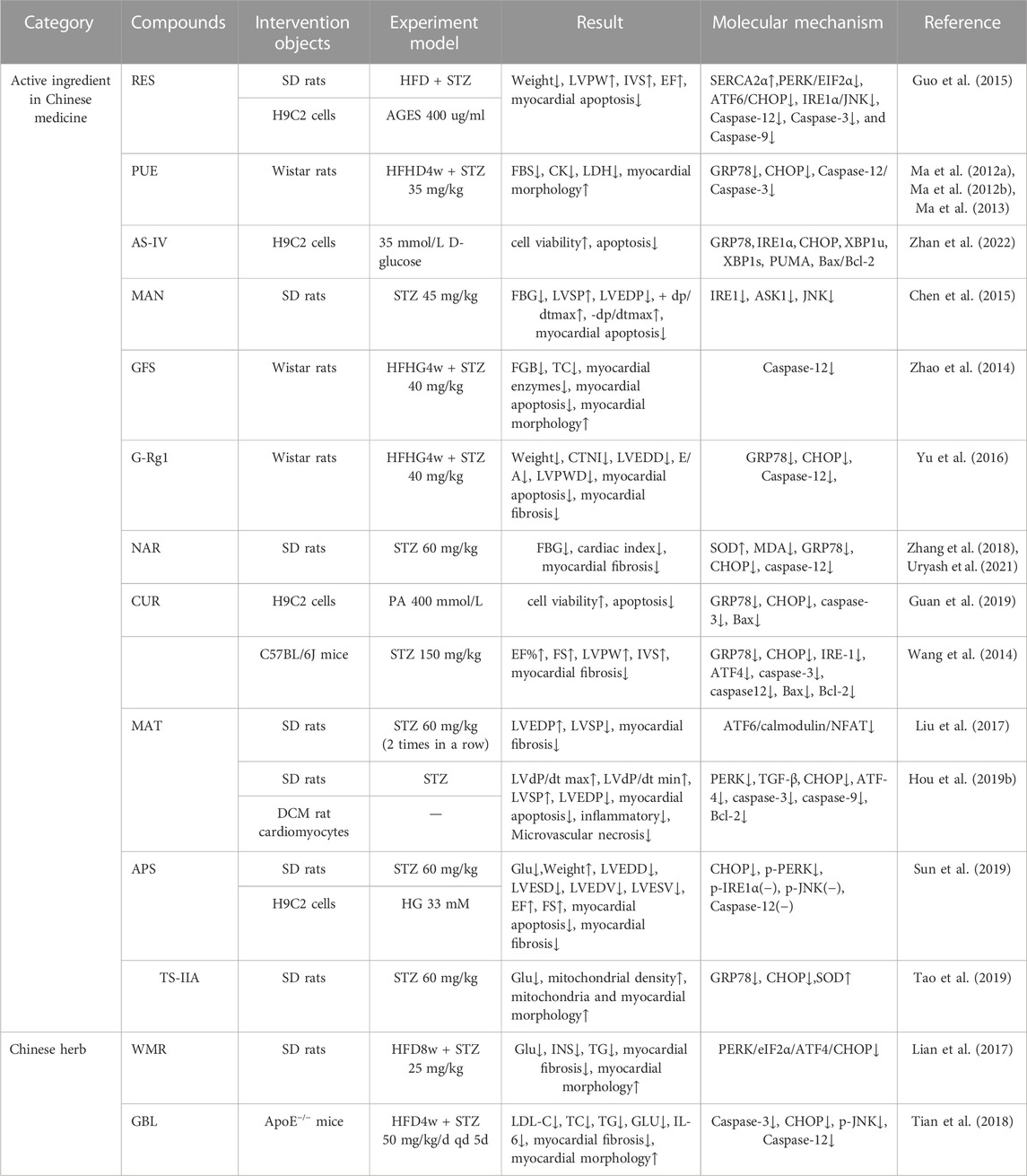
TABLE 1. The effect of traditional Chinese medicine targeting ERS in the treatment of DCM.
Although there are some problems in studying ERS modulation by TCM for treating DCM, with the further development of science, ERS modulation by TCM may provide a new direction for treating DCM. The treatment of diabetes and cardiovascular diseases with Chinese medicines has a history of thousands of years and has achieved good efficacy in Chinese patients, but it is difficult to promote due to the cultural differences between the East and the West, the low quality of medicinal materials, the backwardness of the extraction process, the lack of science in the quality standards and the quality control system, the difficulty in pharmacokinetic studies, the difficulty in evaluating the efficacy of the treatment, the low level of systematization, and the insufficient protection of intellectual property rights (Zhang and Jiang, 2022). It is hoped that more researchers will join in to conduct further in-depth studies on the above issues to accelerate the modernization of Chinese medicine.Under the guidance of Chinese medicine theory, it is hoped that a more precise and personalized treatment plan for patients will be developed through the use of evidence-based Chinese medicine treatment in combination with basic Western medicine treatment. Chinese medicine has unique advantages in regulating ERS to improve DCM, improve the in-depth study of the mechanism of ERS regulation by Chinese medicine compounding, and excavate natural ERS inhibitors from Chinese medicine with complex composition, so as to provide theoretical basis for the research and development of new drugs, and new strategies for preventing and treating DCM by Chinese medicine.
Author contributions
LA: Writing–original draft. ZC: Conceptualization, Project administration, Writing–review and editing. JY: Writing–review and editing. YLe: Conceptualization, Formal Analysis, Writing–review and editing. YLu: Writing–review and editing. XF: Supervision, Writing–review and editing. HL: Writing–review and editing. XL: Writing–review and editing. HG: Writing–review and editing. CX: Funding acquisition, Resources, Supervision, Writing–review and editing.
Funding
The author(s) declare financial support was received for the research, authorship, and/or publication of this article. This work was supported by grants from National Administration of Traditional Chinese Medicine Qi Huang Scholars support project, National Administration of Traditional Chinese Medicine (ZYYCXTD-C-202209), Major Science and Technology Projects of Science and Technology Department of Sichuan Province (2022ZDZX0022).
Conflict of interest
The authors declare that the research was conducted in the absence of any commercial or financial relationships that could be construed as a potential conflict of interest.
Publisher’s note
All claims expressed in this article are solely those of the authors and do not necessarily represent those of their affiliated organizations, or those of the publisher, the editors and the reviewers. Any product that may be evaluated in this article, or claim that may be made by its manufacturer, is not guaranteed or endorsed by the publisher.
Abbreviations
ASE, ASERCA2A; CAN, calmodulin neuralphosphatase; C-I/C-II, type I/II collagen; Ca2+, disturbance of intracellular calcium regulation; IR, insulin resistance; MA, myocardial apoptosis; MH, myocardial hypertrophy; MF, myocardial fibrosis. Straight lines indicate facilitated activation, dashed lines indicate diminished inhibition.
References
Abo-Saif, M. A., Ragab, A. E., Ibrahim, A. O., Abdelzaher, O. F., Mehanyd, A. B. M., Saber-Ayad, M., et al. (2023). Pomegranate peel extract protects against the development of diabetic cardiomyopathy in rats by inhibiting pyroptosis and downregulating LncRNA-MALAT1. Front. Pharmacol. 14, 1166653. doi:10.3389/fphar.2023.1166653
Abukhalil, M. H., Althunibat, O. Y., Aladaileh, S. H., Al-Amarat, W., Obeidat, H. M., Al-Khawalde, A. A. A., et al. (2021). Galangin attenuates diabetic cardiomyopathy through modulating oxidative stress, inflammation and apoptosis in rats. Biomed. Pharmacother. 138, 111410. doi:10.1016/j.biopha.2021.111410
Akanchise, T., and Angelova, A. (2023). Ginkgo biloba and long COVID: in vivo and in vitro models for the evaluation of nanotherapeutic efficacy. Pharmaceutics 15 (5), 1562. doi:10.3390/pharmaceutics15051562
Asbun, J., and Villarreal, F. J. (2006). The pathogenesis of myocardial fibrosis in the setting of diabetic cardiomyopathy. J. Am. Coll. Cardiol. 47 (4), 693–700. doi:10.1016/j.jacc.2005.09.050
Asgari, M., Salehi, I., Ranjbar, K., Khosravi, M., and Zarrinkalam, E. (2022). Interval training and Crataegus persica ameliorate diabetic nephropathy via miR-126/Nrf-2 mediated inhibition of stress oxidative in rats with diabetes after myocardial ischemia-reperfusion injury. Biomed. Pharmacother. 153, 113411. doi:10.1016/j.biopha.2022.113411
Barr, L. A., Shimizu, Y., Lambert, J. P., Nicholson, C. K., and Calvert, J. W. (2015). Hydrogen sulfide attenuates high fat diet-induced cardiac dysfunction via the suppression of endoplasmic reticulum stress. Nitric Oxide 46, 145–156. doi:10.1016/j.niox.2014.12.013
Belali, O. M., Ahmed, M. M., Mohany, M., Belali, T. M., Alotaibi, M. M., Al-Hoshani, A., et al. (2022). LCZ696 protects against diabetic cardiomyopathy-induced myocardial inflammation, ER stress, and apoptosis through inhibiting AGEs/NF-κB and PERK/CHOP signaling pathways. Int. J. Mol. Sci. 23 (3), 1288. doi:10.3390/ijms23031288
Bi, Z., Wang, Y., and Zhang, W. (2021). A comprehensive review of tanshinone IIA and its derivatives in fibrosis treatment. Biomed. Pharmacother. 137, 111404. doi:10.1016/j.biopha.2021.111404
Chan, E. W., Lye, P. Y., and Wong, S. K. (2016). Phytochemistry, pharmacology, and clinical trials of Morus alba. Chin. J. Nat. Med. 14 (1), 17–30. doi:10.3724/SP.J.1009.2016.00017
Chen, Y., Hua, Y., Li, X., Arslan, I. M., Zhang, W., and Meng, G. (2020). Distinct types of cell death and the implication in diabetic cardiomyopathy. Front. Pharmacol. 11, 42. doi:10.3389/fphar.2020.00042
Chen, Y. N., Wang, G. X., and Hu, G. M. (2015). Effect of mangiferin on myocardial apoptosis in diabetic rats. Chin. J. Public Health 31 (04), 453–455. doi:10.11847/zgggws2015-31-04-21
Chengji, W., and Xianjin, F. (2019). Exercise protects against diabetic cardiomyopathy by the inhibition of the endoplasmic reticulum stress pathway in rats. J. Cell Physiol. 234 (2), 1682–1688. doi:10.1002/jcp.27038
Choy, K. W., Mustafa, M. R., Lau, Y. S., Liu, J., Murugan, D., Lau, C. W., et al. (2016). Paeonol protects against endoplasmic reticulum stress-induced endothelial dysfunction via AMPK/PPARδ signaling pathway. Biochem. Pharmacol. 116, 51–62. doi:10.1016/j.bcp.2016.07.013
Chu, Y. M., Wang, T. X., Jia, X. F., Yang, Y., Shi, Z. M., Cui, G. H., et al. (2022). Fuzheng Nizeng Decoction regulated ferroptosis and endoplasmic reticulum stress in the treatment of gastric precancerous lesions: a mechanistic study based on metabolomics coupled with transcriptomics. Front. Pharmacol. 13, 1066244. doi:10.3389/fphar.2022.1066244
Deng, Y. W., Liu, F., Li, Z. T., Gao, J. H., Zhao, Y., Yang, X. L., et al. (2022). Hyperglycemia promotes myocardial dysfunction via the ERS-MAPK10 signaling pathway in db/db mice. Lab. Invest. 102 (11), 1192–1202. doi:10.1038/s41374-022-00819-2
Ding, B., Lin, C., Liu, Q., He, Y., Ruganzu, J. B., Jin, H., et al. (2020). Tanshinone IIA attenuates neuroinflammation via inhibiting RAGE/NF-κB signaling pathway in vivo and in vitro. J. Neuroinflammation 17 (1), 302. doi:10.1186/s12974-020-01981-4
Du, A. Y., Zhang, P. Y., and Zuo, W. B. (2018). Clinical observation and related mechanism study of HuangQi-BaoXin Decoction in the treatment of diabetic cardiomyopathy. J. Chin. Med. Mater. 41 (08), 2007–2010. doi:10.13863/j.issn1001-4454.2018.08.050
Dunlay, S. M., Givertz, M. M., Aguilar, D., Allen, L. A., Chan, M., Desai, A. S., et al. (2019). Type 2 diabetes mellitus and heart failure: a scientific statement from the American heart association and the heart failure society of America: this statement does not represent an update of the 2017 ACC/AHA/HFSA heart failure guideline update. Circulation 140 (7), E294–E324. doi:10.1161/CIR.0000000000000691
Fang, P., Ye, Z. Q., Li, R., She, D. M., Zong, G. N., Zhang, L. Y., et al. (2023). Glucagon-like peptide-1 receptor agonist protects against diabetic cardiomyopathy by modulating microRNA-29b-3p/SLMAP. Drug Des. Dev. Ther. 17, 791–806. doi:10.2147/DDDT.S400249
Fang, W. J., Li, X. M., Zhou, X. K., and Xiong, Y. (2022). Resveratrol improves diabetic cardiomyopathy by preventing asymmetric dimethylarginine-caused peroxisome proliferator-activated receptor-γ coactivator-1α acetylation. Eur. J. Pharmacol. 936, 175342. doi:10.1016/j.ejphar.2022.175342
Fang, Z. Y., Zhang, M., Liu, J. N., Zhao, X., Zhang, Y. Q., and Fang, L. (2020). Tanshinone IIA: a review of its anticancer effects. Front. Pharmacol. 11, 611087. doi:10.3389/fphar.2020.611087
Feng, H., Zhu, X., Tang, Y., Fu, S., Kong, B., and Liu, X. (2021). Astragaloside IV ameliorates diabetic nephropathy in db/db mice by inhibiting NLRP3 inflammasome-mediated inflammation. Int. J. Mol. Med. 48 (2), 164. doi:10.3892/ijmm.2021.4996
Feng, J., Li, S. S., and Chen, H. W. (2016). Tanshinone IIA ameliorates apoptosis of cardiomyocytes induced by endoplasmic reticulum stress. Exp. Biol. Med. 241 (18), 2042–2048. doi:10.1177/1535370216660634
Frakes, A. E., and Dillin, A. (2017). The UPR(ER): sensor and coordinator of organismal homeostasis. Mol. Cell 66 (6), 761–771. doi:10.1016/j.molcel.2017.05.031
Gold-Smith, F., Fernandez, A., and Bishop, K. (2016). Mangiferin and cancer: mechanisms of action. Nutrients 8 (7), 396. doi:10.3390/nu8070396
Gong, D. F., Sun, S. C., Wang, R. R., Dawuti, A., Kong, D. W., Liu, R. Q., et al. (2023). Salvianolic acid A improve mitochondrial respiration and cardiac function via inhibiting apoptosis pathway through CRYAB in diabetic cardiomyopathy. Biomed. Pharmacother. 160, 114382. doi:10.1016/j.biopha.2023.114382
Gopal, K., Karwi, Q. G., Dakhili, S. A. T., Wagg, C. S., Zhang, L. Y., Sun, Q. Y., et al. (2023). Aldose reductase inhibition alleviates diabetic cardiomyopathy and is associated with a decrease in myocardial fatty acid oxidation. Cardiovasc. Diabetol. 22 (1), 73. doi:10.1186/s12933-023-01811-w
Grubic Rotkvic, P., Planinic, Z., Liberati Prso, A. M., Sikic, J., Galic, E., and Rotkvic, L. (2021). The mystery of diabetic cardiomyopathy: from early concepts and underlying mechanisms to novel therapeutic possibilities. Int. J. Mol. Sci. 22 (11), 5973. doi:10.3390/ijms22115973
Guan, G., Lei, L., Lv, Q., Gong, Y., and Yang, L. (2019). Curcumin attenuates palmitic acid-induced cell apoptosis by inhibiting endoplasmic reticulum stress in H9C2 cardiomyocytes. Hum. Exp. Toxicol. 38 (6), 655–664. doi:10.1177/0960327119836222
Guo, R., Liu, W., Liu, B., Zhang, B., Li, W., and Xu, Y. (2015). SIRT1 suppresses cardiomyocyte apoptosis in diabetic cardiomyopathy: an insight into endoplasmic reticulum stress response mechanism. Int. J. Cardiol. 191, 36–45. doi:10.1016/j.ijcard.2015.04.245
Guzel, E., Arlier, S., Guzeloglu-Kayisli, O., Tabak, M. S., Ekiz, T., Semerci, N., et al. (2017). Endoplasmic reticulum stress and homeostasis in reproductive physiology and pathology. Int. J. Mol. Sci. 18 (4), 792. doi:10.3390/ijms18040792
He, Y., Zhou, L., Fan, Z., Liu, S., and Fang, W. (2018). Palmitic acid, but not high-glucose, induced myocardial apoptosis is alleviated by N-acetylcysteine due to attenuated mitochondrial-derived ROS accumulation-induced endoplasmic reticulum stress. Cell Death Dis. 9 (5), 568. doi:10.1038/s41419-018-0593-y
Hillary, R. F., and FitzGerald, U. (2018). A lifetime of stress: ATF6 in development and homeostasis. J. Biomed. Sci. 25 (1), 48. doi:10.1186/s12929-018-0453-1
Horbowicz, M., Szablinska-Piernik, J., Goraj-Koniarska, J., Miyamoto, K., Ueda, J., and Saniewski, M. (2022). Changes in polar metabolites content during natural and methyl-jasmonate-promoted senescence of Ginkgo biloba leaves. Int. J. Mol. Sci. 23 (1), 266. doi:10.3390/ijms23010266
Hou, C. Y., Tain, Y. L., Yu, H. R., and Huang, L. T. (2019a). The effects of resveratrol in the treatment of metabolic syndrome. Int. J. Mol. Sci. 20 (3), 535. doi:10.3390/ijms20030535
Hou, H., Zhang, Q., Dong, H., and Ge, Z. (2019b). Matrine improves diabetic cardiomyopathy through TGF-β-induced protein kinase RNA-like endoplasmic reticulum kinase signaling pathway. J. Cell Biochem. 120 (8), 13573–13582. doi:10.1002/jcb.28632
Hou, J., Zheng, D., Fung, G., Deng, H., Chen, L., Liang, J., et al. (2016). Mangiferin suppressed advanced glycation end products (AGEs) through NF-κB deactivation and displayed anti-inflammatory effects in streptozotocin and high fat diet-diabetic cardiomyopathy rats. Can. J. Physiol. Pharmacol. 94 (3), 332–340. doi:10.1139/cjpp-2015-0073
Hou, J., Zheng, D., Zhong, G., and Hu, Y. (2013). Mangiferin mitigates diabetic cardiomyopathy in streptozotocin-diabetic rats. Can. J. Physiol. Pharmacol. 91 (9), 759–763. doi:10.1139/cjpp-2013-0090
Hu, J., Lu, X., Zhang, X., Shao, X., Wang, Y., Chen, J., et al. (2020). Exogenous spermine attenuates myocardial fibrosis in diabetic cardiomyopathy by inhibiting endoplasmic reticulum stress and the canonical Wnt signaling pathway. Cell Biol. Int. 44 (8), 1660–1670. doi:10.1002/cbin.11360
Huang, K. M., Luo, X. L., Zhong, Y., Deng, L., and Feng, J. (2022). New insights into the role of melatonin in diabetic cardiomyopathy. Pharmacol. Res. Perspe 10 (1), e00904. doi:10.1002/prp2.904
Hui, Z., Wang, S., Li, J., Wang, J., and Zhang, Z. (2022). Compound Tongluo Decoction inhibits endoplasmic reticulum stress-induced ferroptosis and promoted angiogenesis by activating the Sonic Hedgehog pathway in cerebral infarction. J. Ethnopharmacol. 283, 114634. doi:10.1016/j.jep.2021.114634
Januzzi, J. L., Butler, J., Del Prato, S., Ezekowitz, J. A., Ibrahim, N. E., Lam, C. S. P., et al. (2023). Rationale and design of the aldose reductase inhibition for stabilization of exercise capacity in heart failure trial (ARISE-HF) in patients with high-risk diabetic cardiomyopathy. Am. Heart J. 256, 25–36. doi:10.1016/j.ahj.2022.11.003
Jia, G., DeMarco, V. G., and Sowers, J. R. (2016). Insulin resistance and hyperinsulinaemia in diabetic cardiomyopathy. Nat. Rev. Endocrinol. 12 (3), 144–153. doi:10.1038/nrendo.2015.216
Jia, G., Hill, M. A., and Sowers, J. R. (2018). Diabetic cardiomyopathy: an update of mechanisms contributing to this clinical entity. Circ. Res. 122 (4), 624–638. doi:10.1161/CIRCRESAHA.117.311586
Jiang, C., Li, D., Chen, L., Liu, Y., Zhao, Y., Mei, G., et al. (2022). Quercetin ameliorated cardiac injury via reducing inflammatory actions and the glycerophospholipid metabolism dysregulation in a diabetic cardiomyopathy mouse model. Food Funct. 13 (14), 7847–7856. doi:10.1039/d2fo00912a
Kaya, S. T., Bozdogan, O., Ozarslan, T. O., Taskin, E., Eksioglu, D., Erim, F., et al. (2019). The protection of resveratrol and its combination with glibenclamide, but not berberine on the diabetic hearts against reperfusion-induced arrhythmias: the role of myocardial K(ATP) channel. Arch. Physiol. Biochem. 125 (2), 114–121. doi:10.1080/13813455.2018.1440409
Kiencke, S., Handschin, R., von Dahlen, R., Muser, J., Brunner-Larocca, H. P., Schumann, J., et al. (2010). Pre-clinical diabetic cardiomyopathy: prevalence, screening, and outcome. Eur. J. Heart Fail 12 (9), 951–957. doi:10.1093/eurjhf/hfq110
Kim, J. K., Choi, M. S., Kim, J. Y., Yu, J. S., Seo, J. I., Yoo, H. H., et al. (2021). Ginkgo biloba leaf extract suppresses intestinal human breast cancer resistance protein expression in mice: correlation with gut microbiota. Biomed. Pharmacother. 140, 111712. doi:10.1016/j.biopha.2021.111712
Kim, Y., and Clifton, P. (2018). Curcumin, cardiometabolic health and dementia. Int. J. Environ. Res. Public Health 15 (10), 2093. doi:10.3390/ijerph15102093
Kumar, C. U., Reddy, S. S., Suryanarayana, P., Patil, M. A., Chary, P. M., Kumar, P. U., et al. (2022). Protective effect of cinnamon on diabetic cardiomyopathy in nicotinamide-streptozotocin induced diabetic rat model. J. Diabetes Metab. Disord. 21 (1), 141–150. doi:10.1007/s40200-021-00948-3
Laddha, A. P., and Kulkarni, Y. A. (2021). Daidzein mitigates myocardial injury in streptozotocin-induced diabetes in rats. Life Sci. 284, 119664. doi:10.1016/j.lfs.2021.119664
Lakshmanan, A. P., Harima, M., Suzuki, K., Soetikno, V., Nagata, M., Nakamura, T., et al. (2013). The hyperglycemia stimulated myocardial endoplasmic reticulum (ER) stress contributes to diabetic cardiomyopathy in the transgenic non-obese type 2 diabetic rats: a differential role of unfolded protein response (UPR) signaling proteins. Int. J. Biochem. Cell Biol. 45 (2), 438–447. doi:10.1016/j.biocel.2012.09.017
Li, L., Chen, X., Su, C., Wang, Q., Li, R., Jiao, W., et al. (2020a). Si-Miao-Yong-An decoction preserves cardiac function and regulates GLC/AMPK/NF-κB and GLC/PPARα/PGC-1α pathways in diabetic mice. Biomed. Pharmacother. 132, 110817. doi:10.1016/j.biopha.2020.110817
Li, L., Luo, W., Qian, Y., Zhu, W., Qian, J., Li, J., et al. (2019). Luteolin protects against diabetic cardiomyopathy by inhibiting NF-κB-mediated inflammation and activating the Nrf2-mediated antioxidant responses. Phytomedicine 59, 152774. doi:10.1016/j.phymed.2018.11.034
Li, S., Jin, S., Chen, W., Yu, J., Fang, P., Zhou, G., et al. (2020b). Mangiferin alleviates endoplasmic reticulum stress in acute liver injury by regulating the miR-20a/miR-101a-Nrf2 axis. J. Biochem. 168 (4), 365–374. doi:10.1093/jb/mvaa056
Li, X. J., Zhang, D. Q., Wang, X. H., Zhang, Q., Qian, L., Song, R. J., et al. (2022). Irisin alleviates high glucose-induced hypertrophy in H9c2 cardiomyoblasts by inhibiting endoplasmic reticulum stress. Peptides 152, 170774. doi:10.1016/j.peptides.2022.170774
Li, Y., Wei, X., Liu, S. L., Zhao, Y., Jin, S., and Yang, X. Y. (2021). Salidroside protects cardiac function in mice with diabetic cardiomyopathy via activation of mitochondrial biogenesis and SIRT3. Phytother. Res. 35 (8), 4579–4591. doi:10.1002/ptr.7175
Lian, J., Chen, J., Yuan, Y., Chen, J., Daud, M. S. M., Luo, L., et al. (2017). Cortex Mori Radicis extract attenuates myocardial damages in diabetic rats by regulating ERS. Biomed. Pharmacother. 90, 777–785. doi:10.1016/j.biopha.2017.03.097
Liang, X., Han, M., Zhang, X., Sun, X., Yu, K., Liu, C., et al. (2021). Dahuang danshen decoction inhibits pancreatic fibrosis by regulating oxidative stress and endoplasmic reticulum stress. Evid. Based Complement. Altern. Med. 2021, 6629729. doi:10.1155/2021/6629729
Liu, J., Zhao, Y., Zhu, Y., Wang, Y., Liu, X., Nie, X., et al. (2022). Rhynchophylline regulates calcium homeostasis by antagonizing ryanodine receptor 2 phosphorylation to improve diabetic cardiomyopathy. Front. Pharmacol. 13, 882198. doi:10.3389/fphar.2022.882198
Liu, M., Li, Y., Liang, B., Li, Z., Jiang, Z., Chu, C., et al. (2018). Hydrogen sulfide attenuates myocardial fibrosis in diabetic rats through the JAK/STAT signaling pathway. Int. J. Mol. Med. 41 (4), 1867–1876. doi:10.3892/ijmm.2018.3419
Liu, M., Liu, H., Parthiban, P., Kang, G. J., Shi, G. B., Feng, F., et al. (2021). Inhibition of the unfolded protein response reduces arrhythmia risk after myocardial infarction. J. Clin. Investigation 131 (18), e147836. doi:10.1172/JCI147836
Liu, M. Y., Liu, J. Y., Zhang, L. J., Geng, Q. S., and Ge, Y. B. (2019b). Antidepressant-like effects of ginseng fruit saponin in myocardial infarction mice. Biomed. Pharmacother. 115, 108900. doi:10.1016/j.biopha.2019.108900
Liu, M. Y., Ren, Y. P., Zhang, L. J., and Ding, J. Y. (2016). Pretreatment with ginseng fruit saponins affects serotonin expression in an experimental comorbidity model of myocardial infarction and depression. Aging Dis. 7 (6), 680–686. doi:10.14336/AD.2016.0729
Liu, Q., Han, Q., Lu, M., Wang, H., and Tang, F. (2019a). Lycium barbarum polysaccharide attenuates cardiac hypertrophy, inhibits calpain-1 expression and inhibits NF-κB activation in streptozotocin-induced diabetic rats. Exp. Ther. Med. 18 (1), 509–516. doi:10.3892/etm.2019.7612
Liu, X., Xu, Q., Wang, X., Zhao, Z., Zhang, L., Zhong, L., et al. (2015). Irbesartan ameliorates diabetic cardiomyopathy by regulating protein kinase D and ER stress activation in a type 2 diabetes rat model. Pharmacol. Res. 93, 43–51. doi:10.1016/j.phrs.2015.01.001
Liu, Z., Cai, H., Zhu, H., Toque, H., Zhao, N., Qiu, C., et al. (2014). Protein kinase RNA-like endoplasmic reticulum kinase (PERK)/calcineurin signaling is a novel pathway regulating intracellular calcium accumulation which might be involved in ventricular arrhythmias in diabetic cardiomyopathy. Cell Signal 26 (12), 2591–2600. doi:10.1016/j.cellsig.2014.08.015
Liu, Z., Zhang, Y., Tang, Z., Xu, J., Ma, M., Pan, S., et al. (2017). Matrine attenuates cardiac fibrosis by affecting ATF6 signaling pathway in diabetic cardiomyopathy. Eur. J. Pharmacol. 804, 21–30. doi:10.1016/j.ejphar.2017.03.061
Lu, T. C., Wu, Y. H., Chen, W. Y., and Hung, Y. C. (2022). Targeting oxidative stress and endothelial dysfunction using tanshinone IIA for the treatment of tissue inflammation and fibrosis. Oxid. Med. Cell Longev. 2022, 2811789. doi:10.1155/2022/2811789
Lu, Y. P., and Zhou, Y. J. (2010). Observation on the curative effect of 38 cases of diabetic cardiomyopathy treated with Benefiting qi-nourishing yin-activating blood circulation formula combined with western medicine. Benefiting qi-nourishing yin-activating blood Circ. formula 42 (09), 33–34.
Luo, J., Jin, W., Jin, M., Pan, W., Gao, S., Zhao, X., et al. (2022). Jiedutongluotiaogan formula restores pancreatic function by suppressing excessive autophagy and endoplasmic reticulum stress. Pharm. Biol. 60 (1), 1542–1555. doi:10.1080/13880209.2022.2107019
Luo, L., Yin, H. J., Zhang, Y., Jiang, Y. R., Liu, Y., and Shi, D. Z. (2005). Effect of ginseng fruit saponins on insulin sensitivity index in high fat-fed rats. Zhong Xi Yi Jie He Xue Bao 3 (6), 463–465. doi:10.3736/jcim20050611
Ma W. B., Jiang R. L., Lei S., Wang L. C., Xia G. L., Wu J. L.et al. (2013). The expression of endoplasmic reticulum stress protein CHOP in myocardium of diabetic cardiomyopathy rats and the intervention effect of puerarin china tianjin: The Third National Academic Conference on Integrated Traditional Chinese and Western Medicine in Critical Care Medicine.
Ma, W. B., Jiang, R. L., Lei, S., Wang, L. C., Xia, G. L., Wu, J. L., et al. (2012b). Research on the expression of Caspase 12 in rats with diabeticcardiomyopathy and the intervention of puerarin. Chin. J. Crit. Care Med. Ed. 5 (01), 6–12. doi:10.3877/cma.j.issn.1674-6880.2012.01.002
Ma W. B., Jiang R. L., Lei S., Xia G. L., Wang L. C., Wu J. L.et al. (2012a). Effect of puerarin on endoplasmic reticulum stress in diabetic cardiomyopathy rats. Ten-year review and prospect of critical care medicine-2012 zhejiang academic annual meeting of critical care medicine Zhejiang, China: Jiaxing.
Macvanin, M. T., Gluvic, Z., Radovanovic, J., Essack, M., Gao, X., and Isenovic, E. R. (2023). Diabetic cardiomyopathy: the role of microRNAs and long non-coding RNAs. Front. Endocrinol. (Lausanne) 14, 1124613. doi:10.3389/fendo.2023.1124613
Mousavi, S. N., Hosseinikia, M., Yousefi Rad, E., and Saboori, S. (2022). Beneficial effects of Ginkgo biloba leaf extract on inflammatory markers: a systematic review and meta-analysis of the clinical trials. Phytother. Res. 36 (9), 3459–3469. doi:10.1002/ptr.7544
Nakamura, K., Miyoshi, T., Yoshida, M., Akagi, S., Saito, Y., Ejiri, K., et al. (2022). Pathophysiology and treatment of diabetic cardiomyopathy and heart failure in patients with diabetes mellitus. Int. J. Mol. Sci. 23 (7), 3587. doi:10.3390/ijms23073587
Nam, D. H., Han, J. H., Lee, T. J., Shishido, T., Lim, J. H., Kim, G. Y., et al. (2015). CHOP deficiency prevents methylglyoxal-induced myocyte apoptosis and cardiac dysfunction. J. Mol. Cell Cardiol. 85, 168–177. doi:10.1016/j.yjmcc.2015.05.016
Niu, M. M., Zhang, H. F., Tang, Y. G., Wu, J., and Yu, X. H. (2022). Clinical observation on the treatment of early senile diabetic cardiomyopathy with the prescription of yiqiyangyinhuoxuetongluofang. Chin. J. Gerontology 42 (06), 1316–1318. doi:10.3969/j.issn.1005-9202.2022.06.011
Oakes, S. A., and Papa, F. R. (2015). The role of endoplasmic reticulum stress in human pathology. Annu. Rev. Pathol. 10, 173–194. doi:10.1146/annurev-pathol-012513-104649
Papandreou, I., Denko, N. C., Olson, M., Van Melckebeke, H., Lust, S., Tam, A., et al. (2011). Identification of an Ire1alpha endonuclease specific inhibitor with cytotoxic activity against human multiple myeloma. Blood 117 (4), 1311–1314. doi:10.1182/blood-2010-08-303099
Park, I. H., Shen, G. Y., Song, Y. S., Jong Cho, Y., Kim, B. S., Lee, Y., et al. (2021). Granulocyte colony-stimulating factor reduces the endoplasmic reticulum stress in a rat model of diabetic cardiomyopathy. Endocr. J. 68 (11), 1293–1301. doi:10.1507/endocrj.EJ21-0016
Pei, S. J., Zhu, H. Y., Guo, J. H., Zhang, X., and Deng, Z. J. (2018b). Knockout of CNR1 prevents metabolic stress-induced cardiac injury through improving insulin resistance (IR) injury and endoplasmic reticulum (ER) stress by promoting AMPK-alpha activation. Biochem. Biophys. Res. Commun. 503 (2), 744–751. doi:10.1016/j.bbrc.2018.06.070
Pei, Z., Deng, Q., Babcock, S. A., He, E. Y., Ren, J., and Zhang, Y. (2018a). Inhibition of advanced glycation endproduct (AGE) rescues against streptozotocin-induced diabetic cardiomyopathy: role of autophagy and ER stress. Toxicol. Lett. 284, 10–20. doi:10.1016/j.toxlet.2017.11.018
Peng, M., Liu, H., Ji, Q., Ma, P., Niu, Y., Ning, S., et al. (2022). Fufang Xueshuantong improves diabetic cardiomyopathy by regulating the wnt/β-catenin pathway. Int. J. Endocrinol. 2022, 3919161. doi:10.1155/2022/3919161
Prakoso, D., De Blasio, M. J., Tate, M., Kiriazis, H., Donner, D. G., Qian, H., et al. (2020). Gene therapy targeting cardiac phosphoinositide 3-kinase (p110α) attenuates cardiac remodeling in type 2 diabetes. Am. J. Physiol. Heart Circ. Physiol. 318 (4), H840–H852. doi:10.1152/ajpheart.00632.2019
Ravetti, S., Garro, A. G., Gaitan, A., Murature, M., Galiano, M., Brignone, S. G., et al. (2023). Naringin: nanotechnological strategies for potential pharmaceutical applications. Pharmaceutics 15 (3), 863. doi:10.3390/pharmaceutics15030863
Rhman, M. A., and Owira, P. (2022). The role of microRNAs in the pathophysiology, diagnosis, and treatment of diabetic cardiomyopathy. J. Pharm. Pharmacol. 74 (12), 1663–1676. doi:10.1093/jpp/rgac066
Rubler, S., Dlugash, J., Yuceoglu, Y. Z., Kumral, T., Branwood, A. W., and Grishman, A. (1972). New type of cardiomyopathy associated with diabetic glomerulosclerosis. Am. J. Cardiol. 30 (6), 595–602. doi:10.1016/0002-9149(72)90595-4
Salvatore, T., Pafundi, P. C., Galiero, R., Albanese, G., Di Martino, A., Caturano, A., et al. (2021). The diabetic cardiomyopathy: the contributing pathophysiological mechanisms. Front. Med. (Lausanne) 8, 695792. doi:10.3389/fmed.2021.695792
Sanvictores, T., and Davis, D. D. (2022). Histology, rough endoplasmic reticulum. StatPearls. Treasure Isl. (FL).
Seferovic, P. M., Petrie, M. C., Filippatos, G. S., Anker, S. D., Rosano, G., Bauersachs, J., et al. (2018). Type 2 diabetes mellitus and heart failure: a position statement from the Heart Failure Association of the European Society of Cardiology. Eur. J. Heart Fail 20 (5), 853–872. doi:10.1002/ejhf.1170
Shi, R., Chang, L. P., and Deng, Y. (2020). Clinical study on intervention of early diabetic cardiomyopathy by huotan jiedu Tongluo method guided by collateral disease theory. Mod. J. Integr. Traditional Chin. West. Med. 29 (17), 1877–1880.
Sochocka, M., Ochnik, M., Sobczyński, M., Gębura, K., Zambrowicz, A., Naporowski, P., et al. (2022). Ginkgo biloba leaf extract improves an innate immune response of peripheral blood leukocytes of Alzheimer's disease patients. Nutrients 14 (10), 2022. doi:10.3390/nu14102022
Sun, N., Yang, T., Tang, Y., Zhao, Y., Wang, H., Zhao, S., et al. (2022). Lycopene alleviates chronic stress-induced liver injury by inhibiting oxidative stress-mediated endoplasmic reticulum stress pathway apoptosis in rats. J. Agric. Food Chem. 70 (45), 14414–14426. doi:10.1021/acs.jafc.2c06650
Sun, S., Yang, S., An, N., Wang, G., Xu, Q., Liu, J., et al. (2019). Astragalus polysaccharides inhibits cardiomyocyte apoptosis during diabetic cardiomyopathy via the endoplasmic reticulum stress pathway. J. Ethnopharmacol. 238, 111857. doi:10.1016/j.jep.2019.111857
Tan, J., Yi, X. L., Ren, D., Yuan, C. Y., Wu, G. Q., Liu, G. Q., et al. (2022). Clinical study treatment of diabetic cardiomyopathy with Zicui Tongmai recipe. Shaanxi J. Traditional Chin. Med. 43 (06), 715–719. doi:10.3969/j.issn.1000-7369.2022.06.009
Tao, S. L., Chen, L. Y., Song, J. M., Zhu, N. N., Song, X. Y., Shi, R. L., et al. (2019). Tanshinone IIA ameliorates diabetic cardiomyopathy by inhibiting Grp78 and CHOP expression in STZ-induced diabetes rats. Exp. Ther. Med. 18 (1), 729–734. doi:10.3892/etm.2019.7580
Tian, J. F., Liu, Y. F., Liu, Y., Chen, K. J., and Lyu, S. Z. (2018). Ginkgo biloba leaf extract protects against myocardial injury via attenuation of endoplasmic reticulum stress in streptozotocin-induced diabetic ApoE(-/-) mice. Oxidative Med. Cell. Longev. 2018, 2370617. doi:10.1155/2018/2370617
Tian, J. H., Wu, Q., He, Y. X., Shen, Q. Y., Rekep, M., Zhang, G. P., et al. (2021). Zonisamide, an antiepileptic drug, alleviates diabetic cardiomyopathy by inhibiting endoplasmic reticulum stress. Acta Pharmacol. Sin. 42 (3), 393–403. doi:10.1038/s41401-020-0461-z
Trost, S. U., Belke, D. D., Bluhm, W. F., Meyer, M., Swanson, E., and Dillmann, W. H. (2002). Overexpression of the sarcoplasmic reticulum Ca(2+)-ATPase improves myocardial contractility in diabetic cardiomyopathy. Diabetes 51 (4), 1166–1171. doi:10.2337/diabetes.51.4.1166
Uryash, A., Mijares, A., Flores, V., Adams, J. A., and Lopez, J. R. (2021). Effects of naringin on cardiomyocytes from a rodent model of type 2 diabetes. Front. Pharmacol. 12, 719268. doi:10.3389/fphar.2021.719268
Wang, C., Lou, Y., Xu, J., Feng, Z., Chen, Y., Tang, Q., et al. (2017). Endoplasmic reticulum stress and NF-[Formula: see text]B pathway in Salidroside mediated neuroprotection: potential of Salidroside in neurodegenerative diseases. Am. J. Chin. Med. 45 (7), 1459–1475. doi:10.1142/S0192415X17500793
Wang, N., Zhang, X., Ma, Z., Niu, J., Ma, S., Wenjie, W., et al. (2020b). Combination of tanshinone IIA and astragaloside IV attenuate atherosclerotic plaque vulnerability in ApoE(-/-) mice by activating PI3K/AKT signaling and suppressing TRL4/NF-κB signaling. Biomed. Pharmacother. 123, 109729. doi:10.1016/j.biopha.2019.109729
Wang, Y., Feng, F., Liu, M., Xue, J., and Huang, H. (2018). Resveratrol ameliorates sepsis-induced acute kidney injury in a pediatric rat model via Nrf2 signaling pathway. Exp. Ther. Med. 16 (4), 3233–3240. doi:10.3892/etm.2018.6533
Wang, Y., Sun, H., Zhang, J., Xia, Z., and Chen, W. (2020a). Streptozotocin-induced diabetic cardiomyopathy in rats: ameliorative effect of PIPERINE via Bcl2, Bax/Bcl2, and caspase-3 pathways. Biosci. Biotechnol. Biochem. 84 (12), 2533–2544. doi:10.1080/09168451.2020.1815170
Wang, Y., Zhou, S., Sun, W., McClung, K., Pan, Y., Liang, G., et al. (2014). Inhibition of JNK by novel curcumin analog C66 prevents diabetic cardiomyopathy with a preservation of cardiac metallothionein expression. Am. J. Physiol. Endocrinol. Metab. 306 (11), E1239–E1247. doi:10.1152/ajpendo.00629.2013
Wei, C., Zhang, Y., Zhong, X., Lu, S., Zou, X., Yang, Y., et al. (2022). Ginkgo biloba leaf extract mitigates cisplatin-induced chronic renal interstitial fibrosis by inhibiting the epithelial-mesenchymal transition of renal tubular epithelial cells mediated by the Smad3/TGF-β1 and Smad3/p38 MAPK pathways. Chin. Med. 17 (1), 25. doi:10.1186/s13020-022-00574-y
Wu, S., Lu, D. C., Gajendran, B., Hu, Q. L., Zhang, J., Wang, S. Q., et al. (2023). Tanshinone IIA ameliorates experimental diabetic cardiomyopathy by inhibiting endoplasmic reticulum stress in cardiomyocytes via SIRT1. Phytotherapy Res. 37, 3543–3558. doi:10.1002/ptr.7831
Wu, Y., Cai, C., Xiang, Y., Zhao, H., Lv, L., and Zeng, C. (2021). Naringin ameliorates monocrotaline-induced pulmonary arterial hypertension through endothelial-to-mesenchymal transition inhibition. Front. Pharmacol. 12, 696135. doi:10.3389/fphar.2021.696135
Xiao, L., Dai, Z., Tang, W., Liu, C., and Tang, B. (2021). Astragaloside IV alleviates cerebral ischemia-reperfusion injury through NLRP3 inflammasome-mediated pyroptosis inhibition via activating Nrf2. Oxid. Med. Cell Longev. 2021, 9925561. doi:10.1155/2021/9925561
Xing, L., Fang, J., Zhu, B., Wang, L., Chen, J., Wang, Y., et al. (2021). Astragaloside IV protects against podocyte apoptosis by inhibiting oxidative stress via activating PPARγ-Klotho-FoxO1 axis in diabetic nephropathy. Life Sci. 269, 119068. doi:10.1016/j.lfs.2021.119068
Xu, D. X., Guo, X. X., Zeng, Z., Wang, Y., and Qiu, J. (2021b). Puerarin improves hepatic glucose and lipid homeostasis in vitro and in vivo by regulating the AMPK pathway. Food Funct. 12 (6), 2726–2740. doi:10.1039/d0fo02761h
Xu, H., Zhao, M., Liang, S., Huang, Q., Xiao, Y., Ye, L., et al. (2016b). The effects of puerarin on rat ventricular myocytes and the potential mechanism. Sci. Rep. 6, 35475. doi:10.1038/srep35475
Xu, J., Zhou, Q., Xu, W., and Cai, L. (2012). Endoplasmic reticulum stress and diabetic cardiomyopathy. Exp. Diabetes Res. 2012, 827971. doi:10.1155/2012/827971
Xu, L., Chen, R., Zhang, X., Zhu, Y., Ma, X., Sun, G., et al. (2021a). Scutellarin protects against diabetic cardiomyopathy via inhibiting oxidative stress and inflammatory response in mice. Ann. Palliat. Med. 10 (3), 2481–2493. doi:10.21037/apm-19-516
Xu, S., Gao, Y., Zhang, Q., Wei, S., Chen, Z., Dai, X., et al. (2016a). SIRT1/3 activation by resveratrol attenuates acute kidney injury in a septic rat model. Oxid. Med. Cell Longev. 2016, 7296092. doi:10.1155/2016/7296092
Yan, M., Li, L., Wang, Q., Shao, X., Luo, Q., Liu, S., et al. (2022). The Chinese herbal medicine Fufang Zhenzhu Tiaozhi protects against diabetic cardiomyopathy by alleviating cardiac lipotoxicity-induced oxidative stress and NLRP3-dependent inflammasome activation. Biomed. Pharmacother. 148, 112709. doi:10.1016/j.biopha.2022.112709
Yan, M., Liu, S., Zeng, W., Guo, Q., Mei, Y., Shao, X., et al. (2023). The Chinese herbal medicine Fufang Zhenzhu Tiaozhi ameliorates diabetic cardiomyopathy by regulating cardiac abnormal lipid metabolism and mitochondrial dynamics in diabetic mice. Biomed. Pharmacother. 164, 114919. doi:10.1016/j.biopha.2023.114919
Yang, F., Yu, X., Li, T., Wu, J., Zhao, Y., Liu, J., et al. (2017b). Exogenous H(2)S regulates endoplasmic reticulum-mitochondria cross-talk to inhibit apoptotic pathways in STZ-induced type I diabetes. Am. J. Physiol. Endocrinol. Metab. 312 (3), E190–e203. doi:10.1152/ajpendo.00196.2016
Yang, L., Cheng, C. F., Li, Z. F., Huang, X. J., Cai, S. Q., Ye, S. Y., et al. (2023). Berberine blocks inflammasome activation and alleviates diabetic cardiomyopathy via the miR-18a-3p/Gsdmd pathway. Int. J. Mol. Med. 51 (6), 49. doi:10.3892/ijmm.2023.5252
Yang, L., Zhao, D., Ren, J., and Yang, J. (2015). Endoplasmic reticulum stress and protein quality control in diabetic cardiomyopathy. Biochim. Biophys. Acta 1852 (2), 209–218. doi:10.1016/j.bbadis.2014.05.006
Yang, L. P., Ling, W. H., Du, Z. C., Chen, Y. M., Li, D., Deng, S. Z., et al. (2017a). Effects of anthocyanins on cardiometabolic health: a systematic review and meta-analysis of randomized controlled trials. Adv. Nutr. 8 (5), 684–693. doi:10.3945/an.116.014852
Yang, S., Kuang, G., Zhang, L. K., Wu, S. W., Zhao, Z. Z., Wang, B., et al. (2020). Mangiferin attenuates LPS/D-GalN-Induced acute liver injury by promoting HO-1 in kupffer cells. Front. Immunol. 11, 285. doi:10.3389/fimmu.2020.00285
Younce, C. W., Burmeister, M. A., and Ayala, J. E. (2013). Exendin-4 attenuates high glucose-induced cardiomyocyte apoptosis via inhibition of endoplasmic reticulum stress and activation of SERCA2a. Am. J. Physiol. Cell Physiol. 304 (6), C508–C518. doi:10.1152/ajpcell.00248.2012
Yu, H., Zhen, J., Yang, Y., Gu, J., Wu, S., and Liu, Q. (2016). Ginsenoside Rg1 ameliorates diabetic cardiomyopathy by inhibiting endoplasmic reticulum stress-induced apoptosis in a streptozotocin-induced diabetes rat model. J. Cell Mol. Med. 20 (4), 623–631. doi:10.1111/jcmm.12739
Yu, X., Xiao, Q., Yu, X., Cheng, Y., Lin, H., and Xiang, Z. (2022). A network pharmacology-based study on the mechanism of astragaloside IV alleviating renal fibrosis through the AKT1/GSK-3β pathway. J. Ethnopharmacol. 297, 115535. doi:10.1016/j.jep.2022.115535
Yu, X. X., Li, H., Lin, D. F., Guo, W. Z., Xu, Z. H., Wang, L. P., et al. (2021). Ginsenoside prolongs the lifespan of C. elegans via lipid metabolism and activating the stress response signaling pathway. Int. J. Mol. Sci. 22 (18), 9668. doi:10.3390/ijms22189668
Zhang, J., Ren, J., Liu, Y., Huang, D., and Lu, L. (2020b). Resveratrol regulates the recovery of rat sciatic nerve crush injury by promoting the autophagy of Schwann cells. Life Sci. 256, 117959. doi:10.1016/j.lfs.2020.117959
Zhang, K. W., Yu, X. H., and Guo, H. X. (2022a). Effect of Tongluo yangyin recipe and ventricular remodeling in patients with diabetic cardiomyopathy. Res. Integr. Traditional Chin. West. Med. 14 (02), 81–84. doi:10.3969/j.issn.1674-4616.2022.02.003
Zhang, M., Zheng, Y., Li, X., Wu, H., Liu, P., Zhang, K., et al. (2022c). Tong-Xie-Yao-Fang alleviates diarrhea-predominant irritable bowel syndrome in rats via the GCN2/PERK-eIF2α-ATF4 signaling pathway. Phytomedicine 107, 154350. doi:10.1016/j.phymed.2022.154350
Zhang, Q., Shi, J., Guo, D., Wang, Q., Yang, X., Lu, W., et al. (2020a). Qishen Granule alleviates endoplasmic reticulum stress-induced myocardial apoptosis through IRE-1-CRYAB pathway in myocardial ischemia. J. Ethnopharmacol. 252, 112573. doi:10.1016/j.jep.2020.112573
Zhang, Q., Sun, L., He, L. X., Chen, Z. L., Haran, T. E., Wiman, K. G., et al. (2022d). Evolutionary history of the p53 family DNA-binding domain: insights from an Alvinella pompejana homolog. West China J. Pharm. Sci. 37 (02), 214–217. doi:10.1038/s41419-022-04653-8
Zhang, W. J., Wan, M. W., Gao, K., Zhong, X. D., Xie, Y., Dai, L., et al. (2022e). Pharmacologic IRE1α kinase inhibition alleviates aortic dissection by decreasing vascular smooth muscle cells apoptosis. Int. J. Biol. Sci. 18 (3), 1053–1064. doi:10.7150/ijbs.63593
Zhang, X., Wang, Q., Wang, X., Chen, X., Shao, M., Zhang, Q., et al. (2019). Tanshinone IIA protects against heart failure post-myocardial infarction via AMPKs/mTOR-dependent autophagy pathway. Biomed. Pharmacother. 112, 108599. doi:10.1016/j.biopha.2019.108599
Zhang, X., You, L. Y., Zhang, Z. Y., Jiang, D. X., Qiu, Y., Ruan, Y. P., et al. (2022b). Integrating pharmacological evaluation and computational identification for deciphering the action mechanism of Yunpi-Huoxue-Sanjie formula alleviates diabetic cardiomyopathy. Front. Pharmacol. 13, 957829. doi:10.3389/fphar.2022.957829
Zhang, X. F., Li, S. Y., Liu, Y. M., Liu, J. P., Wu, D., Fu, L., et al. (2022). Effect of astragaloside IV combined with puerarin on high glucose-inducedendoplasmic reticulum stress in H9c2 cells. Chin. J. Pathophysiol. 38 (05), 802–808. doi:10.3969/j.issn.1000-4718.2022.05.005
Zhang, Y. F., Meng, N. N., Li, H. Z., Wen, Y. L., Liu, J. T., Zhang, C. L., et al. (2018). Effect of naringin on oxidative stress and endoplasmic reticulum stress indiabetic cardiomyopathy. China J. Chin. Materia Medica 43 (03), 596–602. doi:10.19540/j.cnki.cjcmm.2018.0013
Zhang, Y. J., and Jiang, Y. Y. (2022). Discussion on primary research direction of modernization of traditinal Chinese medicine in the period of the "14th five-year plan. Mod. Traditional Chin. Med. Materia Medica-World Sci. Technol. 24 (03), 1309–1314. doi:10.11842/wst.20201229002
Zhao, L. C., Zhen, J., and Wei, H. T. (2014). Myocardial protection of Ginseng fruit saponins in streptozotocin-induced diabetic rats. Chin. J. Laboratory Diagnosis 18 (11), 1744–1748.
Zheng, Y. J., Ren, W. Y., Zhang, L. N., Zhang, Y. M., Liu, D. L., and Liu, Y. Q. (2020). A review of the pharmacological action of Astragalus polysaccharide. Front. Pharmacol. 11, 349. doi:10.3389/fphar.2020.00349
Zhong, C. H., Lin, Z. A., Ke, L. Y., Shi, P. Y., Li, S. G., Huang, L. Y., et al. (2021). Recent research progress (2015-2021) and perspectives on the pharmacological effects and mechanisms of tanshinone IIA. Front. Pharmacol. 12, 778847. doi:10.3389/fphar.2021.778847
Zhou, F., Gao, H., Shang, L., Li, J., Zhang, M., Wang, S., et al. (2023). Oridonin promotes endoplasmic reticulum stress via TP53-repressed TCF4 transactivation in colorectal cancer. J. Exp. Clin. Cancer Res. 42 (1), 150. doi:10.1186/s13046-023-02702-4
Zhou, J., Zhang, N., Aldhahrani, A., Soliman, M. M., Zhang, L., and Zhou, F. (2022). Puerarin ameliorates nonalcoholic fatty liver in rats by regulating hepatic lipid accumulation, oxidative stress, and inflammation. Front. Immunol. 13, 956688. doi:10.3389/fimmu.2022.956688
Zhou, Y., Zhou, C., Zhang, X., Vong, C. T., Wang, Y., and Cheang, W. S. (2021). Coptisine attenuates diabetes-associated endothelial dysfunction through inhibition of endoplasmic reticulum stress and oxidative stress. Molecules 26 (14), 4210. doi:10.3390/molecules26144210
Zhou, Y. X., Zhang, H., and Peng, C. (2014). Puerarin: a review of pharmacological effects. Phytother. Res. 28 (7), 961–975. doi:10.1002/ptr.5083
Keywords: diabetic cardiomyopathy, endoplasmic reticulum stress, Chinese herbal medicine, active ingredients, mechanism of action
Citation: Ao L, Chen Z, Yin J, Leng Y, Luo Y, Fu X, Liu H, Liu X, Gao H and Xie C (2023) Chinese herbal medicine and active ingredients for diabetic cardiomyopathy: molecular mechanisms regulating endoplasmic reticulum stress. Front. Pharmacol. 14:1290023. doi: 10.3389/fphar.2023.1290023
Received: 06 September 2023; Accepted: 23 October 2023;
Published: 07 November 2023.
Edited by:
Zhihao Liu, University of Maryland, College Park, United StatesReviewed by:
Syed Anees Ahmed, East Carolina University, United StatesPriyanka Choudhury, Medical College of Wisconsin, United States
Copyright © 2023 Ao, Chen, Yin, Leng, Luo, Fu, Liu, Liu, Gao and Xie. This is an open-access article distributed under the terms of the Creative Commons Attribution License (CC BY). The use, distribution or reproduction in other forums is permitted, provided the original author(s) and the copyright owner(s) are credited and that the original publication in this journal is cited, in accordance with accepted academic practice. No use, distribution or reproduction is permitted which does not comply with these terms.
*Correspondence: Chunguang Xie, eGllY2dAY2R1dGNtLmVkdS5jbg==